Original Article
Causes of excessive blinking in children aged 4-12 years
Ru-Lian Zhao, Yang Wang, Min Liu, Yong Li, Lian-Hong Pi
Department of Ophthalmology, Children’s Hospital, Chongqing Medical University, Chongqing 400014, China Received April 4, 2019; Accepted June 10, 2019; Epub August 15, 2019; Published August 30, 2019
Abstract: The current study aimed to investigate causes of excessive blinking in children aged 4 to 12 years, provid-ing evidence for clinical treatment of abnormal blinkprovid-ing in children. A total of 578 children complainprovid-ing of excessive blinking were recruited. Moreover, a total of 370 children without excessive blinking served as controls. Results showed that children with excessive blinking might have a single etiology or multiple etiologies together. Occurrence of excessive blinking was negatively correlated with age (P<0.001) and affected by sex (P=0.008). There were
sig-nificant differences in tear break-up times (P<0.001), allergic conjunctivitis (P<0.001), corneal epithelial injuries
(P=0.013), diopters of children with ametropia (P=0.001), and times of video display terminal (P<0.001) between
three age groups. Allergic conjunctivitis (P<0.001), positive corneal fluorescein staining (P<0.001), conjunctival li -thiasis/trichiasis (P<0.001), and times of video display terminal (P<0.001) had a negative relationship with breakup
times of tear film. Allergic conjunctivitis and conjunctival lithiasis/trichiasis showed positive relationships with cor
-neal fluorescein staining (P<0.001). Times of video display terminal were positively related to diopters of myopia-related ametropia (P=0.001). All subjects had mild ametropia. In addition, there were significant differences in breakup times of tear film between children with excessive blinking and controls (P=0.047). Thus, ocular surface
diseases, refractive problems, times of video exposure, and tic disorders are causes of excessive blinking. Of these
factors, ocular surface diseases and times of video exposure have relationships with breakup times of tear film.
Moreover, these factors may interact with each other, causing deterioration of excessive blinking.
Keywords: Child, blinking, breakup times of tear film, ocular surface disease, times of video display terminal, tic
disorders
Introduction
Blinking occurrence is normally about 10-15 blinks/minute. Each blink lasts for 0.3-0.4 sec-onds, with an interval of 3-4 seconds between
blinks. Blinking is a natural neural reflex of the
eyelid. It plays an important role in adaptive human behaviors [1]. Excessive blinking is de-
fined as a frequency of blinking higher than 15
blinks/minute[2, 3]. Excessive blinking is of- ten accompanied by ocular symptoms (such as itchy eyes, foreign body sensation of the eyes, ocular fatigue, and photophobia) and charac-terized by excessive unilateral or bilateral eyelid closure and opening with or without abnormal eyeball movement. Excessive blinking is often
intermittent and recurrent. It is difficult to con -trol. In addition, it may be attenuated during gazing and disappear after blinking for several weeks or months. It may recur after disappear-ance [4]. Some children with excessive blinking develop other symptoms, such as crowing the
eyebrows, wrinkling the forehead, and sucking the nose. Excessive blinking has become a common disease in the Department of Pediatric Ophthalmology. In some children, excessive blinking is misdiagnosed as infectious kerato-conjunctivitis, causing long term use of eye drops after misdiagnosis. This may increase incidence of ocular discomforts and injuries. It may even cause side effects of some drugs [5, 6]. Some parents do not pay attention. This may contribute to deterioration of the disease and delay correct treatment. In addition, some par-ents become nervous about this disease and misdiagnose it as “tic disorder”. Thus, some inhibitory drugs have been used for treatment of excessive blinking. These may not only ca- use side effects, but also bring adverse somat-ic and psychologsomat-ical effects [7]. It usually
exten-sively studied in various contexts of disease, such as dry eyes and visual tasks, as well as systemic disease [10, 11]. Causes of excessive blinking are complex. They may include ocular disease, systemic disease, and psychological
disease [12]. Excessive blinking has
significan-tly affected the learning abilities of children. Despite a plethora of information concerning blinking disorders in adulthood, relatively little information exists for evaluation and manage-ment of children presenting with blinking disor-ders. Nonstandard diagnosis and treatments
of excessive blinking also cause significant
ad-verse effects in children and their families. The- refore, more attention should be paid to the diagnosis and treatment of children with exces-sive blinking. Thus, it is imperative to investi-gate and identify causes of excessive blinking, taking targeted treatment. This may assist in avoiding misdiagnosis and mistreatment. The current study investigated children with excessive blinking. Possible causes of exces-sive blinking were explored in these children, aiming to provide evidence for clinical treat-ment of abnormal blinking in children.
Materials and methods
General characteristics
This study adhered to the tenets of the De- claration of Helsinki and was approved by the Institutional Review Board of Children’s Hos-
pital, Chongqing Medical University. Consent
for publication was obtained from all partici- pants.
This study was conducted on children present-ing to the Ophthalmology Clinic with excessive blinking. The study was conducted between December 2016 and November 2018. A total of 578 children, including 341 boys and 237 girls, with excessive blinking ages from 4 to 12 years old, served as the observation group. In addition, 370 matched children without exces-sive blinking, including 220 boys and 150 girls, were randomly recruited as controls.
Inclusion criteria: (1) Children with excessive blinking as the chief complaint; (2) Informed consent was obtained from parents or guard-ians; (3) Children cooperated with examina-tions; and (4) Children did not receive prior treatment for excessive blinking.
Exclusion criteria: (1) Children did not cooper-ate with examinations; (2) Parents or guardians refused to participate in this study; (3) Children
were allergic to fluorescein; and (4) Children
received treatment for excessive blinking be- fore the study.
Methods and data processing
The following information was collected by in- terviewing parents or guardians: (1) Gender, age, duration, and frequency of signs and symp -toms; (2) History of allergies, including the fam-ily history of allergies and allergic diseases; (3) Concomitant focal and systemic symptoms dur-ing excessive blinkdur-ing; (4) Diagnosis of tic dis- orders; (5) Prior diagnosis and treatment of excessive blinking and use of eye drops or anti-biotics; and (6) Times of video display terminal (VDT) each day.
Slit-lamp examination: (1) Routine eye examina-tion: Eyelids, eyelashes, conjunctiva, and cor-nea were examined for papilla, follicles, stones, injury, inflammation, foreign bodies, and scars; (2) Tear meniscus height (TMH): Abnormal tear secretion was defined as TMH lower than 0.3 mm; (3) Breakup times of tear film (BUT): BUT was measured thrice and the mean was cal- culated. Decreases in tear film stability were defined as BUT lower than 10 seconds; and (4) Corneal fluorescein staining (FLS): Positive st-aining suggests corneal epithelial injury [13].
Allergen examination: Skin prick tests were employed to examine common inhaled aller-gens. A positive reaction is defined as a light yellow rash with surrounding erythema. History of allergies, family history of allergies, eye ex- aminations, and allergen examinations were employed for diagnosis of allergic conjuncti- vitis (AC) [14].
Vision examination: The International Visual Chart was used for vision examinations. When visual acuity was ≤0.6 for children aged 4 years and ≤0.8 for children aged ≥5 years, abnormal visual acuity was diagnosed [15].
tropia, and tic
disor-ders with BUT were
also explored. Relati- onships between aller-gic conjunctivitis, con-junctival lithiasis/trich-
iasis with positive FLS,
and times of video dis-play terminal with ty- pes of ametropia were also explored. P<0.05 indicates statistical si-
gnificance.
Cycloplegic retinoscopy: 1% Atropine sulfate eye gel was used three times a day for 3 days to the children <7 years; 1% cyclopentolate was used three times with the interval of 10 minutes for children ≥7 years. Pupillary reac -tions were then tested. Cycloplegia is defined as absence of pupillary light reflex. Streak reti -noscope (YZ24; Six Six Vision Corp, Suzhou, China) was used for retinoscopy [16].
Hyperopia is defined as spherical degree ≥ +2.00 D. Myopia is defined as spherical degree ≤-0.50 D. Astigmatism is defined as absolute cylindrical degree ≥1.00 DC. The diopter is rep -resented by Spherical equivalent refraction (SER). Diopters ≤-3.00 D is mild refractive error, -3.00 D~-6.00 D is moderate refractive error, and ≥-6.00 D is high refractive error. The distri -bution of refraction is expressed as _x ± s
[17-19].
Statistical analysis
Children with excessive blinking in the observa-tion group were divided into three age groups, including the 4-6 group, 7-9 group, and 10-12 group. Children without excessive blinking ser- ved as controls. All data were categorically re- corded, listed, and subjected to statistical an- alysis with SPSS version 22.0. Count data are expressed as rates (%). Measurement data are described by _x± s.
Spearman’s rank correlation analysis was ad-
opted to investigate the relationships of BUT,
allergic conjunctivitis, conjunctival lithiasis or
trichiasis, positive FLS, times of video display
terminal, diopters of ametropia, and tic disor-ders between the 3 age groups. Relationships between gender, allergic conjunctivitis,
con-junctival lithiasis or trichiasis, positive FLS,
ti-mes of video display terminal, diopters of
ame-Pearson’s correlation analysis included the
amount of frequent blinking and ages of the children. BUT and times of video display termi -nal were also included, as well as times of video display terminal and diopters of ametropia.
P<0.05 indicates statistical significance.
Paired-t tests were employed for comparisons
of BUT between children with and without
ex-cessive blinking, as well as gender. P<0.05
indi-cates statistical significance.
Results
A total of 578 children with excessive blinking were enrolled as the observation group. This group included 341 boys (59.00%) and 237 girls (41.00%). The mean age was (6.99±2.26) years old (4-12 years). In addition, there were 370 children without excessive blinking serv- ing as the control group. This group included 220 boys (59.45%) and 150 girls (40.55%). The mean age was (7.02±2.30) years old (4-12 years).
Of children with excessive blinking, allergic con-junctivitis was found in 283 children (48.95%; 188 boys and 95 girls) (Table 1). Allergen ex- aminations showed that most children were allergic to dust mites (61.23%) (dermatophagoi-des pteronyssinus and dermatophagoi(dermatophagoi-des fa- rinae). Conjunctival lithiasis/trichiasis was not- ed in 37 children (6.40%; 22 boys and 15 girls). Tic disorders (TD) were found in 23 children (3.99%; 16 boys and 7 girls) (Table 1). Positive
FLS was shown in 247 children (42.73%; 143
boys and 104 girls) (Table 2).
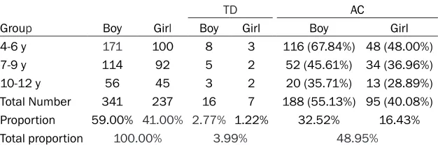
[image:3.612.93.406.84.188.2]Ametropia was observed in 78 children (13.49%; 45 boys and 33 girls). All cases were mild ametropia. Diopters of ametropia in the three groups were, respectively, (1.47±1.61) D, Table 1. Numbers of TD and AC of 3 groups in the observation group
TD AC
Group Boy Girl Boy Girl Boy Girl
4-6 y 171 100 8 3 116 (67.84%) 48 (48.00%)
7-9 y 114 92 5 2 52 (45.61%) 34 (36.96%)
10-12 y 56 45 3 2 20 (35.71%) 13 (28.89%)
Total Number 341 237 16 7 188 (55.13%) 95 (40.08%) Proportion 59.00% 41.00% 2.77% 1.22% 32.52% 16.43%
Total proportion 100.00% 3.99% 48.95%
(-0.49±1.37) D, and (-1.00±1.10) D (Table 3). Times of video display terminal in the three groups were, respectively, (1.27±0.55) h/d, (2.08±0.87) h/d, and (2.82±0.94) h/d (Table 3).
In children with excessive blinking, BUT was,
respectively, (8.57±2.77) s, (8.44±2.49) s, and
(7.87±1.86) s. BUT in children without
frequ-ent blinking was, respectively, (9.34±2.14) s, (8.89±1.65) s, and (8.25±1.93) s (Table 4). TMH of all was higher than 0.3 mm (Table 4). The course of the disease in children with
fre-quent blinking was (1-90) days.
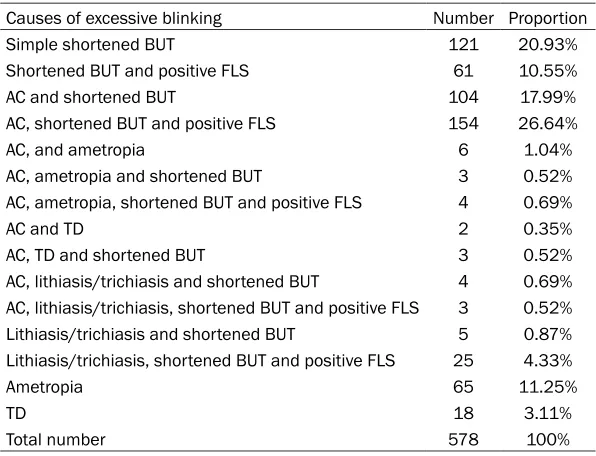
Children with frequent blinking may have a sin -gle cause or multiple causes (Table 5). In chil-dren with excessive blinking, occurrence of
fre-quent blinking was negatively correlated with
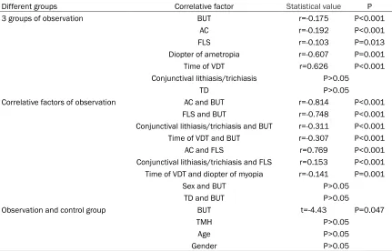
age and affected by sex (Figure 1). Table 6 out-lines correlation analysis statistics of different groups and correlative factors.
Discussion
Reports of excessive blinking in children are
infrequent. Very little guidance exists concern -ing how to evaluate and manage children with excessive blinking. However, excessive blinking has become a common disease in the De- partment of Pediatric Ophthalmology. In this study, it was found that occurrence of exces-sive blinking was negatively correlated with age. Excessive blinking decreases with
increas-es in age. This is mainly due to the blink reflex
and childhood anatomy, as well as physiologi-cal characteristics. The sensor of the blinking
reflex is the cornea. It is controlled by the cen -tral nervous system [20]. In children, the cere-Table 2. FLS and conjunctival lithiasis/trichiasis of 3 groups in the observation group
Group FLS Lithiasis/trichiasis
Boy Girl Boy Girl
4-6 y 79 (46.20%) 52 (52.00%) 11 (6.43%) 7 (7.00%)
7-9 y 45 (39.47%) 38 (41.30%) 8 (7.01%) 5 (5.43%)
10-12 y 19 (33.92%) 14 (31.11%) 3 (5.36%) 3 (6.66%)
Total Number 143 (41.93%) 104 (43.88%) 22 (6.45%) 15 (6.32%)
Proportion 24.74% 17.99% 3.81% 2.59%
Total proportion 42.73% 6.40%
Notes: observation group: children with excessive blinking; FLS: fluorescein staining.
Table 3. Ametropia and VDT times of 3 groups in the observation group
Group Boy Girl Hypermetropic Myopic Diopter (D) VDT (h/d)
4-6 y 20 (11.69%) 12 (12.00%) 26 (9.59%) 6 (2.21%) 1.47±1.61 1.27±0.55 7-9 y 9 (7.89%) 8 (8.69%) 7 (3.40%) 10 (4.85%) -0.49±1.37 2.08±0.87 10-12 y 16 (28.57%) 13 (28.88%) 4 (3.96%) 25 (24.75%) -1.00±1.10 2.82±0.94 Total 45 (13.19%) 33 (13.92%) 37 (6.40%) 41 (7.09%)
Proportion 13.19% 13.92% 6.40% 7.09%
Notes: observation group: children with excessive blinking; VDT: video display terminal.
Table 4. BUT and TMH of 2 groups in the observation group
Age
Total (n) BUT (s) TMH
Observation
(n=578) (n=370)Control Observation (n=578) (n=370)Control Observation (n=578) (n=370)Control
4-6 y 271 173 8.57±2.77 9.34±2.14 0.32±0.040 0.33±0.023
7-9 y 206 129 8.44±2.49 8.89±1.65 0.33±0.043 0.32±0.033
10-12 y 101 68 7.87±1.86 8.25±1.93 0.32±0.041 0.32±0.039
reduces, the friction may increase during eyeball mo- vement. This may cause da- mage to the corneal
epithe-lium. BUT in children with
excessive blinking was lo- wer than controls. However,
TMH showed no significant
differences between two groups. This indicates that tear secretion is normal
and tear film instability is a
major cause of excessive blinking. When tear stability reduces, the cornea beco- mes dry soon after the eye-lid opens. Excessive blink-ing is helpful for the
recon-struction of tear film. Tear
instability may cause exce- ssive blinking. Kashkouli et al. [22] and Nosch et al.
[23] found that tear film
st-ability directly affected the
frequency of blinking, in ac-cord with present findings. Thus, BUT can be used to reflect the frequency of
bl-inking. Correlation levels of
BUT with other factors
sh-ould be further studied.
Results showed that BUT
had no relationship with
gender, consistent with
fin-dings reported by Pauline
et al. [24]. BUT is negatively
correlated with age,
consis-tent with findings reported
by Cho et al. [25].
Allergic conjunctivitis has become a common disease and a major cause of exces-bral cortex is immature. Thus, over-reaction to
a stimulation is very common. The cornea is rich in sensory nerve endings and sensitive to stimulation. Corneal sensitivity is high in child-hood, but decreases with age.
Ocular surface disease: Tear film locates on the
surface of the cornea and provides a moist environment for the epithelium of cornea and conjunctiva. This may reduce friction during blinking and eyeball movement [21]. Positive
FLS is negative with BUT (p<0.05). When BUT
sive blinking in children. Incidence of infectious diseases has decreased, but incidence of al- lergic diseases has increased with improve-ments in living environimprove-ments, development of health care, and increased air pollution [26]. Present results show that allergic conjunctivi-
tis was negatively related to BUT and positively to positive FLS. When mast cells and eosino -phils become activated, they release many in-
[image:5.612.90.388.82.310.2]flammatory factors. This causes damage to cor -neal and conjunctival epithelial cells and goblet cells. This may result in a lack of mucus layer, Table 5. Causes of excessive blinking
Causes of excessive blinking Number Proportion
Simple shortened BUT 121 20.93%
Shortened BUT and positive FLS 61 10.55%
AC and shortened BUT 104 17.99%
AC, shortened BUT and positive FLS 154 26.64%
AC, and ametropia 6 1.04%
AC, ametropia and shortened BUT 3 0.52%
AC, ametropia, shortened BUT and positive FLS 4 0.69%
AC and TD 2 0.35%
AC, TD and shortened BUT 3 0.52%
AC, lithiasis/trichiasis and shortened BUT 4 0.69%
AC, lithiasis/trichiasis, shortened BUT and positive FLS 3 0.52%
Lithiasis/trichiasis and shortened BUT 5 0.87%
Lithiasis/trichiasis, shortened BUT and positive FLS 25 4.33%
Ametropia 65 11.25%
TD 18 3.11%
Total number 578 100%
Note: BUT: breakup time of tear film; FLS: fluorescein staining; AC: allergic conjunctivi-tis; TD: Tic disorder.
Figure 1. Number of frequent blinking increased with age and sex. Of children
with excessive blinking, the occurrence of frequent blinking was negatively cor
[image:5.612.91.387.352.518.2]damage the corneal epithelium, and reduce
tear film stability [27]. Results showed that
all-ergic conjunctivitis had a negative relationship with age. Incidence of allergic conjunctivitis was higher in boys (32.52%) than girls (16.43%). This is consistent with Rhee’s research [28]. Results may be related to the fact that boys have a matrilineal tendency and a wider range of activities. Thus, they are easily exposed to allergens [29].
Conjunctival lithiasis/trichiasis is also a cause of excessive blinking in children. Results sug-gest that conjunctival lithiasis/trichiasis was
negatively related to BUT and positively to po-sitive FLS. Mechanical stimulation induced by
conjunctival lithiasis/trichiasis may cause da- mage to the ocular surface, reduce tear secre-tion, and damage tear stability, resulting in excessive blinking.
Ametropia: Simple ametropia was found in 65 children with excessive blinking. All cases were mild ametropia. Excessive blinking was shown to be related to refractive error. In mild ame- tropia, the visual organs are over-regulated for compensation, which leads to visual fatigue
and frequent blinking. Coats et al. [2] speculat -ed that ametropia without correction was a major cause of excessive blinking in children. After correction, excessive blinking was attenu-ated or even disappeared in about 87% of chil-dren. Abdi et al. found that correction of ame-tropia improved ocular symptoms in 94% of children, attenuated visual fatigue, and im- proved excessive blinking [30]. Thus, refractive problems are also important causes of exces-sive blinking in children.
[image:6.612.91.522.85.361.2]Tic disorders: Tic disorders are causes of exces-sive blinking in children. Tic disorder refers to a Table 6. Correlation analysis statistics
Different groups Correlative factor Statistical value P
3 groups of observation BUT r=-0.175 P<0.001
AC r=-0.192 P<0.001
FLS r=-0.103 P=0.013
Diopter of ametropia r=-0.607 P=0.001
Time of VDT r=0.626 P<0.001
Conjunctival lithiasis/trichiasis P>0.05
TD P>0.05
Correlative factors of observation AC and BUT r=-0.814 P<0.001
FLS and BUT r=-0.748 P<0.001
Conjunctival lithiasis/trichiasis and BUT r=-0.311 P<0.001
Time of VDT and BUT r=-0.307 P<0.001
AC and FLS r=0.769 P<0.001
Conjunctival lithiasis/trichiasis and FLS r=0.153 P<0.001
Time of VDT and diopter of myopia r=-0.141 P=0.001
Sex and BUT P>0.05
TD and BUT P>0.05
Observation and control group BUT t=-4.43 P=0.047
TMH P>0.05
Age P>0.05
Gender P>0.05
Notes: Observation: children with excessive blinking; control group: children without excessive blinking; BUT: breakup time of tear film; FLS: fluorescein staining; AC: allergic conjunctivitis; TD: Tic disorder; TMH: tear meniscus height. There were
sig-nificant differences in BUT, allergic conjunctivitis, corneal epithelial injury, diopter of children with ametropia and time of VDT
among the three groups, but no difference in conjunctival lithiasis/trichiasis and tic disorder. Allergic conjunctivitis, positive
FLS, conjunctival lithiasis/trichiasis, and time of VDT had a negative relationship with BUT, but TD and sex had no relation
mental disease characterized by motor tics and/or phonic tics [31]. Incidence of TD in boys (2.77%) was higher than that in girls (1.22%). Kurlan et al. found that TD mainly occurred at 2-21 years. Incidence in males was higher than that in females [32], consistent with the current study. Some neuroleptics (mainly antipsychotic drugs) are effective in improving tic disorders. However, antipsychotic drugs have some side effects, including cognitive retardation, drug-induced irritability, anxiety, and depression, leading to adverse effects on the health of dren [7]. Thus, treatment of tic disorders in chil-dren should be cautionary. Over-treatment sh- ould be avoided.
Times of VDT (video display terminal): Results showed that times of VDT were negatively
relat-ed to BUT, but positively relatrelat-ed to the diopter
of myopia. With the development of market
economy and the information era, VDT influ -ences lives of people with its ample function. Long time use of VDT may increase exposure of
palpebral fissure and promote the evaporation of tear film, which may cause dry eye and increase the frequency of blinking [33]. In addi -tion, the characteristics of VDT may increase regulatory and convergence movements of the eyes for clear vision. This may cause eye
dis-comfort and subsequent excessive blinking.
You et al. found that myopia was related to the time interval between two rests during comput-er-related work [34]. Jordan et al. found that times of computer-related work in adolescenc-es with myopia were 0.7-1.6 hours longer than in those with emmetropia [35], consistent with
present findings.
Excessive blinking is also affected by sex (p= 0.008). It was hypothesized that this is related to causes of excessive blinking. It was found that allergic conjunctivitis (48.95%) was the cause of excessive blinking. Incidence of this disease was higher in boys than in girls.
In conclusion, correlation analysis showed that allergic conjunctivitis, corneal epithelial injury, conjunctival lithiasis/trichiasis, and times of
video display terminal were related to BUT. The
pathophysiology of these ocular surface dis-eases may affect ocular surface homeostasis
and disrupt the stability of tear film, increasing
the risk for excessive blinking. Causes of exce- ssive blinking are complex in children. They may include ocular diseases, systemic diseases,
and psychological diseases. There could be a single cause for excessive blinking in some chil-dren, but multiple factors are possible. Active
identification of causes of excessive blinking
and appropriate treatment methods are impor-tant. Clinicians should aim to avoid misdiagno-sis, missed diagnomisdiagno-sis, and over-treatment.
Disclosure of conflict of interest
None.
Address correspondence to: Lian-Hong Pi, Depart- ment of Ophthalmology, Children’s Hospital, Chong-
qing Medical University, Chongqing 400014, China.
E-mail: [email protected] References
[1] Hoppe D, Helfmann S and Rothkopf CA. Hu-
mans quickly learn to blink strategically in re -sponse to environmental task demands. Proc
Natl Acad Sci U S A 2018; 115: 2246-2251.
[2] Coats DK, Paysse EA and Kim DS. Excessive blinking in childhood: a prospective evaluation of 99 children. Ophthalmology 2001; 108: 1556-1561.
[3] Conte A, Defazio G, Ferrazzano G, Hallett M, Macerollo A, Fabbrini G and Berardelli A. Is in -creased blinking a form of blepharospasm? Neurology 2013; 80: 2236-2241.
[4] Schicatano EJ. The effects of attention on
the trigeminal blink reflex. Percept Mot Skills
2016; 122: 444-451.
[5] Coroi MC, Bungau S and Tit M. Preservatives from the eye drops and the ocular surface. Rom J Ophthalmol 2015; 59: 2-5.
[6] Liu Z and Huang C. [A correct understanding of preservatives in eye drops]. Zhonghua Yan Ke Za Zhi 2015; 51: 641-644.
[7] Osland ST, Steeves TD and Pringsheim T. Ph-
armacological treatment for attention deficit
hyperactivity disorder (ADHD) in children with comorbid tic disorders. Cochrane Database Syst Rev 2018; 6: Cd007990.
[8] Doane MG. Interactions of eyelids and tears in corneal wetting and the dynamics of the nor-mal human eyeblink. Am J Ophthalmol 1980; 89: 507-516.
[9] Stern JA, Walrath LC and Goldstein R. The en-dogenous eyeblink. Psychophysiology 1984; 21: 22-33.
[10] Collins MJ, Iskander DR, Saunders A, Hook S, Anthony E and Gillon R. Blinking patterns and corneal staining. Eye Contact Lens 2006; 32: 287-293.
Inter-blink interval visual acuity decay test. Clin Ophthalmol 2009; 3: 501-506.
[12] Nomura R, Hino K, Shimazu M, Liang Y and Okada T. Emotionally excited eyeblink-rate vari-ability predicts an experience of transportation
into the narrative world. Front Psychol 2015; 6:
447.
[13] Bandamwar KL, Papas EB and Garrett Q.
Fl-uorescein staining and physiological state of corneal epithelial cells. Cont Lens Anterior Eye 2014; 37: 213-223.
[14] Nevis IF, Binkley K and Kabali C. Diagnostic ac -curacy of skin-prick testing for allergic rhinitis: a systematic review and meta-analysis. Allergy Asthma Clin Immunol 2016; 12: 20.
[15] Bell AL, Rodes ME and Collier Kellar L. Child-
hood eye examination. Am Fam Physician
2013; 88: 241-248.
[16] Davitt BV, Quinn GE, Wallace DK, Dobson V, Hardy RJ, Tung B, Lai D and Good WV. Astig- matism progression in the early treatment for retinopathy of prematurity study to 6 years of age. Ophthalmology 2011; 118: 2326-2329. [17] Chin MP, Siong KH, Chan KH, Do CW, Chan HH
and Cheong AM. Prevalence of visual impair-ment and refractive errors among different ethnic groups in schoolchildren in Turpan, China. Ophthalmic Physiol Opt 2015; 35: 263-270.
[18] Friedburg D and Kloppel KP. [Early correction
of hyperopia and astigmatism in children leads to better development of visual acuity]. Klin Monbl Augenheilkd 1996; 209: 21-24.
[19] Sheeladevi S, Seelam B, Nukella PB, Modi A, Ali R and Keay L. Prevalence of refractive er-rors in children in India: a systematic review. Clin Exp Optom 2018; 101: 495-503.
[20] Doyle PW, Beegun I and Saleh HA. The
Doyle-Saleh blink reflex. J Laryngol Otol 2017; 131:
347-349.
[21] Arita R, Morishige N, Koh S, Shirakawa R, Ka- washima M, Sakimoto T, Suzuki T and Tsubota
K. Increased tear fluid production as a com -pensatory response to meibomian gland loss: a multicenter cross-sectional study. Ophthal- mology 2015; 122: 925-933.
[22] Kashkouli MB, Zolfaghari R, Es’haghi A, Amir- sardari A, Abtahi MB, Karimi N, Alemzadeh A
and Aghamirsalim M. Tear film, lacrimal drain
-age system, and eyelid findings in subjects
with anophthalmic socket discharge. Am J Ophthalmol 2016; 165: 33-38.
[23] Nosch DS, Pult H, Albon J, Purslow C and Murphy PJ. Relationship between corneal
sen-sation, blinking, and tear film quality. Optom
Vis Sci 2016; 93: 471-481.
[24] Boptom PC and Douthwaite W. Tear breakup time and the effect of lifting the eyelid during its measurement. Clini Exp Optom 2010; 75: 231-235.
[25] Cho P and Yap M. Age, gender, and tear break-up time. Optom Vis Sci 1993; 70: 828-831. [26] Shaker M and Salcone E. An update on ocular
allergy. Curr Opin Allergy Clin Immunol 2016; 16: 505-510.
[27] Akil H, Celik F, Ulas F and Kara IS. Dry eye syn -drome and allergic conjunctivitis in the pediat-ric population. Middle East Afr J Ophthalmol 2015; 22: 467-471.
[28] Rhee CS, Wee JH, Ahn JC, Lee WH, Tan KL, Ahn S, Lee JH, Lee CH, Cho YS, Park KH, Lee KH, Kim KS, Lee A and Kim JW. Prevalence, risk factors and comorbidities of allergic rhinitis in
South Korea: the fifth Korea national health
and nutrition examination survey. Am J Rhinol Allergy 2014; 28: e107-114.
[29] Prescott SL, Taylor A, Roper J, Wahdan A, No-
akes P, Thornton C, Dunstan J and Upham JW.
Maternal reactivity to fetal alloantigens is re-lated to newborn immune responses and
sub-sequent allergic disease. Clin Exp Allergy
2005; 35: 417-425.
[30] Abdi S and Rydberg A. Asthenopia in
school-children, orthoptic and ophthalmological find -ings and treatment. Doc Ophthalmol 2005; 111: 65-72.
[31] Silvestri PR, Baglioni V, Cardona F and Cavanna
AE. Self-concept and self-esteem in patients with chronic tic disorders: a systematic litera-ture review. Eur J Paediatr Neurol 2018; 22: 749-756.
[32] Kurlan R. Handbook of Tourette’s syndrome and related tic and behavioral disorders. New York: Maecel Dekker, 2005.
[33] Tauste A, Ronda E, Baste V, Bratveit M, Moen BE and Segui Crespo MD. Ocular surface and
tear film status among contact lens wearers
and non-wearers who use VDT at work: com-paring three different lens types. Int Arch Occup Environ Health 2018; 91: 327-335. [34] You QS, Wu LJ, Duan JL, Luo YX, Liu LJ, Li X,
Gao Q, Wang W, Xu L, Jonas JB and Guo XH.
Factors associated with myopia in school chil -dren in China: the Beijing childhood eye study. PLoS One 2012; 7: e52668.