Exercise has persistent effects on learning and
memory throughout the lifespan: analysis of the
underlying mechanisms
Bibiana Castagna Mota
Thesis submitted for the degree of Doctor of Philosophy at the University of Dublin, Trinity College Dublin.
Submitted April 2018
Department of Physiology and Trinity College Institute of Neuroscience
I. Declaration
I declare that this thesis has not been submitted as an exercise for a degree at this or any other university and it is entirely my own work.
I agree to deposit this thesis in the University’s open access institutional repository or allow the library to do so on my behalf, subject to Irish Copyright Legislation and Trinity College Library conditions of use and acknowledgement.
______________________________
II. Summary
Ageing is a complex process associated with a decline in organism functions. Ageing affects several cellular and molecular brain mechanisms and is commonly associated with decline in brain function, such as cognition and emotion Moreover, age is characterised by the presence of chronic and low-grade inflammation, which is associated with decreased responsiveness of immune cells to stress and exacerbated inflammatory response. However, sometimes, even though morphological and physiological changes are observed in the brain of aged individuals, they may not experience the cognitive decline usually associated with ageing. In an attempt to explain this discrepancy between an individual’s measured level of brain pathology/or age-related changes and the functional and/or cognitive deficits that are expected to result from that pathology or from normal ageing, the cognitive reserve hypothesis was proposed. Evidence suggest that life exposure and experiences can build brain reserve, by increasing brain resources and making the brain more flexible and capable of recruiting theses resources. In this context, exposure to physical exercise is associated with positive inflammatory modulation and increased brain plasticity, thereby contributing to reduce the unfavourable effects of ageing.
months until old age. All mice were tested every 2 months for non-spatial (NOR) and spatial memory (OD) and at old age they were tested in the MWM and underwent analysis of anxiety and depression-like behaviour. Subsequently, structural MRI was performed, tissue was collected, and glial cells were isolated from the brain tissue. Assessment of BrdU labelling and cell phenotype by immunohistochemistry were used to investigate neurogenesis and assay of mRNA and protein expression of different targets of interest, focusing mainly in inflammatory markers, were carried out. To answer our second question, mice underwent 9 consecutive days of treadmill running prior to a single intraperitoneal injection of a sub-septic dose of lipopolysaccharide. Four hours later, mice were tested for spatial learning and memory and brain tissue was removed for analysis of inflammatory response and glia cells activation.
III. Table of Contents
I. Declaration i
II. Summary ii
III. Table of Contents iv
IV. Acknowledgements xiv
V. List of Abbreviations xvi
VI. List of Figures xxii
VII. List of Tables xxix
Chapter 1: Introduction 1
1.1 Ageing 1
1.1.1 World population ageing 1
1.1.2 Age-related diseases 2
1.2 Cognitive function 3
1.2.1 Learning and memory 3
1.2.2 Memory classification 3
1.2.3 Spatial and recognition memory 4
1.3 The hippocampus 5
1.3.1 The hippocampal formation 5
1.3.2 Role of hippocampus 7
1.4 Cognitive function and ageing 9
1.5 Cellular and molecular mechanisms of learning and memory 9
1.5.1 Synaptic plasticity 9
1.5.2 Synaptogenesis 11
1.5.3 Synaptic plasticity, synaptogenesis and ageing 11
1.5.4 Adult Hippocampal Neurogenesis 12
1.5.4.1 Timeline of hippocampal neurogenesis 13 1.5.4.2 Adult hippocampal neurogenesis in humans 15 1.5.4.3 Balance between and neurogenesis and
gliogenesis
1.5.4.4 Adult hippocampal neurogenesis and learning and memory
16
1.5.4.5 Hippocampal neurogenesis and ageing 17
1.6 Neurotrophins and growth factors 18
1.6.1 Brain-derived neurotrophic factor (BDNF) 19
1.6.2 BDNF-TrkB signalling cascade 20
1.6.2.1 Ras-MAPK/ERK pathway 22 1.6.2.2 The PI3K-Akt pathway 22 1.6.2.3 The PLCg-CaMKII pathway 23
1.6.3 Nerve growth factor (NGF) 23
1.6.4 Insulin-like growth factor 1 (IGF-1) 24
1.6.5 Vascular endothelial growth factor (VEGF) 25 1.6.6 Glial cell line-derived neurotrophic factor (GDNF) 26 1.6.7 Neurotrophins, growth factors and ageing 27
1.7 Inflammation 28
1.7.1 The immune response 29
1.7.2 Inflammatory mediators 30
1.7.2.1 Nuclear factor-kappa B (NF-kB) signalling 30
1.7.2.2 Cytokines 31
1.7.2.3 Chemokines 32
1.7.2.4 Inducible nitric oxide synthase (iNOS) 33 1.7.2.5 Cell surface adhesion molecule CD44 34
1.7.3 Apoptosis and inflammation 34
1.7.4 Neuroinflammation 35
1.7.4.1 Microglia 36
1.7.4.2 Astrocytes 38
1.7.5 Peripheral inflammation and neuroinflammation 39
1.7.6 Models of neuroinflammation 40
1.7.7 Inflammation and ageing 41
1.8 Reserve Hypothesis 42
1.8.1 Reserve concept 42
1.9 Exercise 45
1.9.1 Exercise and memory 45
1.9.2 Exercise and synaptic plasticity 46
1.9.3 Exercise and neurogenesis 46
1.9.4 Exercise and neurotrophins 47
1.9.5 Exercise and inflammation 48
1.9.6 Exercise and ageing 50
1.10 Hypothesis 51
1.11 Objectives 52
Chapter 2: Material and Methods 53
2.1 Materials 53
2.1.1 Animals 53
2.1.2 Animal treatments and interventions 53
2.1.3 General laboratory chemicals 53
2.1.4 General laboratory products and plastics 54 2.1.5 Enzyme-linked immunosorbent assay (ELISA) kits 55 2.1.6 Immunohistochemistry reagents and antibodies 55 2.1.7 Polymerase chain-reaction (PCR) reagents 56 2.1.8 Western immunoblotting reagents and antibodies 56
2.2 Animals 57
2.3 Housing conditions 57
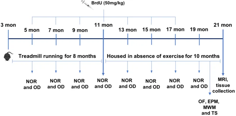
2.4 Experimental design I 57
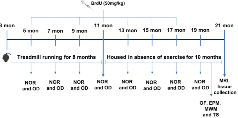
2.5 Experimental design II 59
2.6 Treadmill exercise 59
2.7 BrdU administration 60
2.8 LPS administration 60
2.9 Behavioral assessment 61
2.9.2 Morris water maze (MWM) task 63 2.9.2.1 Morris water maze training 64 2.9.2.2 Morris water maze probe trial 65
2.9.3 Open field task 65
2.9.4 Elevated plus maze (EPM) task 66 2.9.5 Tail suspension test (TST) 66
2.10 Magnetic resonance image (MRI) 67
2.10.1 MRI acquisition 67
2.10.2 MRI data analysis 68
2.11 Tissue collection 69
2.11.1 Tissue preparation 69
2.11.2 Transcardial perfusion and tissue fixation 70 2.11.3 Microglia and astrocyte isolation 70
2.12 Protein quantification 71
2.13 Enzyme linked immunosorbent assay (ELISA) 71
2.14 SDS-PAGE and Western immunoblotting 73
2.14.1 Tissue preparation for SDS polyacrylamaide gel electrophoresis (SDS-PAGE)
73
2.14.2 SDS-PAGE 73
2.14.3 Western immunoblotting 74
2.14.4 Western immunoblotting analysis 77
2.15 Polymerase chain reaction (PCR) analysis 77
2.15.1 Total RNA extraction 77
2.15.2 RNA quantification and reverse transcription 78
2.15.3 RT-PCR 79
2.15.4 RT-PCR analysis 80
2.16 Immunohistochemistry 81
2.16.1 Tissue preparation for immunohistochemistry 81
2.16.2 Fluorescent double staining 82
2.16.3 Microscope imaging and image analysis 83
Chapter 3: The persistent effect of exercise on learning and memory, anxiety and depression-like behaviour throughout the lifespan
86
3.1 Introduction 86
3.2 Methods 88
3.2.1 Animals 88
3.2.2 Experiment design 88
3.2.3 Treadmill exercise protocol 89
3.2.4 Behavioural assessment 90
3.2.4.1 Novel Object Recognition (NOR) task 90 3.2.4.2 Object Displacement (OD) task 91 3.2.4.3 Open Field (OF) task 92 3.2.4.4 Elevated Plus Maze (EPM) test 92 3.2.4.5 Morris Water Maze (MWM) task 93 3.2.4.6 Tail suspension test (TST) 94
3.2.5 Statistical analysis 94
3.3 Results 95
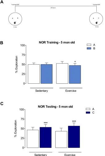
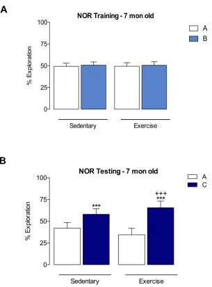
3.3.1 Persistent effects of long-term exercise on non-spatial memory throughout the lifespan: The Novel Object Recognition (NOR) Task
95
3.3.2 Persistent effects of long-term exercise on spatial memory throughout the lifespan: The Object Displacement (OD)
Task
108
3.3.3 Persistent effects of long-term exercise on spatial learning and memory in ageing: The Morris Water Maze (MWM) Task
121
3.3.4 Persistent effects of long-term exercise on anxiety and depression-like behaviour in ageing: The Open Field, the Elevated Plus maze and the Tail Suspension Test
126
3.4 Discussion 132
3.4.3 Anxiety and depression-like behavior in age 135
3.4.4 Summary 137
Chapter 4: Potential mechanisms underlying the persistent effects of exercise on learning and memory throughout the lifespan: the role of inflammation and neurogenesis and the cognitive reserve hypothesis
139
4.1 Introduction 139
4.2 Methods 142
4.2.1 Animals 142
4.2.2 Experimental design 142
4.2.3 Treadmill exercise protocol 143
4.2.4 BrdU administration 144
4.2.5 Magnetic resonance image (MRI) 144
4.2.6 Tissue collection and preparation 144 4.2.7 Analysis of protein expression by ELISA 145 4.2.8 Analysis of mRNA expression by polymerase chain
reaction (RT-PCR)
145
4.2.9 Analysis of protein expression by western immunoblotting 146 4.2.10 Quantification of protein expression by
immunohistochemistry
146
4.2.11 Statistical analysis 147
4.3 Results 148
4.3.1 Effects of age and exercise on mRNA expression of pro- inflammatory cytokines (IL-1b, TNF-a and IL-6) in the hippocampus and cortex
148
4.3.2 Effects of age and exercise on mRNA expression of the anti-inflammatory cytokine IL-10 in the hippocampus and cortex
153
4.3.3 Effects of age and exercise on protein expression of pro- inflammatory cytokines (IL-1b, TNF-a) and anti-
inflammatory cytokine (IL-10) in the hippocampus
4.3.4 Effects of age and exercise on mRNA expression of the glial cell activation markers GFAP, Iba-1 and CD11b in the hippocampus and cortex
157
4.3.5 Effects of age and exercise on mRNA expression of iNOS, Arginase-1 and Mrc-1 in the hippocampus and cortex
161
4.3.6 Effects of age and exercise on mRNA expression of Cx3cl1,Cx3cr1 and CD44, in the hippocampus and cortex
165
4.3.7 Effects of age and exercise on mRNA expression of apoptosis-related proteins, Bax and Bcl-2 and cell
proliferation markers Ki67 in the hippocampus and cortex
168
4.3.8 Effects of age and exercise on mRNA and protein
expression of BDNF, TrkB and p75 in the hippocampus and cortex
171
4.3.9 Effects of age and exercise on mRNA expression of NGF, Igf-1, VEGF and CREB in the hippocampus and cortex
174
4.3.10 Effects of age and exercise on mRNA expression of M1 phenotype markers in an enriched population of microglia
177
4.3.11 Effects of age and exercise on mRNA expression of M2 phenotype markers in an enriched population of microglia
180
4.3.12 Effects of age and exercise on mRNA expression of A1 phenotype markers in an enriched population of astrocytes
182
4.3.13 Effects of age and exercise on mRNA expression of A2 phenotype markers in an enriched population of astrocytes
184
4.3.14 Effects of age and exercise on protein content of
synaptogenesis markers, PSD-95 and Synapsin-1 in the hippocampus
186
4.3.15 Effects of age and exercise on protein expression of TrkB receptor and NF-kB in the hippocampus
188
4.3.16 Effects of age and exercise on astrocyte reactivity and microglia activation in the CA1 and CA3 regions of hippocampus
191
4.3.17 Effects of age and exercise on neurogenesis and gliogenesis in the dentate gyrus of hippocampus
4.3.18 Effects of age and exercise on total brain and hippocampal volumes
199
4.3.19 Effects of age and exercise on mRNA expression of inflammatory markers IL-1b, TNF-a IL-6 and CRP in the
liver
204
4.3.20 Effects of age and exercise on protein expression of inflammatory markers IL-1b and TNF-a in the liver and in
the gastrocnemius muscle 4.4 Table of results
205
207
4.4.1 Cytokines 207
4.4.2 Glial cell markers 208
4.4.3 Inflammatory and apoptotic markers 209 4.4.4 Growth factors, neurotrophins and synaptogenesis
markers
210
4.4.5 Neurogenesis 212
4.4.6 MRI 212
4.5 Discussion 213
4.5.1 Persistent effect of exercise on inflammation in age 213 4.5.2 Lack of effect of exercise on neurotrophin and growth factor
expression in age
221
4.5.3 Persistent effect of exercise on synaptogenesis and neurogenesis in age
222
4.5.4 Summary 225
Chapter 5: The effect of exercise on memory in a mouse model of neuroinflammation: the role of glial cells
227
5.1 Introduction 227
5.2 Methods 229
5.2.1 Animals 229
5.2.2 Experimental design 229
5.2.3 Exercise protocol 230
5.2.5 Object displacement (OD) task 231 5.2.6 Tissue preparation and isolation of microglia and astrocytes 231 5.2.7 Analysis of protein expression by western immunoblotting 232 5.2.8 Analysis of mRNA expression by polymerase chain
reaction (RT-PCR)
232
5.2.9 Statistical Analysis 233
5.3 Results 234
5.3.1 Effects of LPS and exercise on spatial learning and memory
234
5.3.2 Effects of LPS and exercise on mRNA expression of pro- inflammatory cytokines, IL-1b, TNF- α and IL-6 in the hippocampus and cortex
236
5.3.3 Effects of LPS and exercise on mRNA expression of anti- inflammatory cytokines, IL-10 and IL-4 in the hippocampus
and cortex
239
5.3.4 Effects of LPS and exercise on mRNA expression of markers of activation of glial cells in the hippocampus and cortex
242
5.3.5 Effects of LPS and exercise on mRNA expression of fractalkine and cell-surface glycoprotein CD44 in the hippocampus and cortex
247
5.3.6 Effects of LPS and exercise on mRNA expression of neurotrophins, growth factors and markers of apoptosis in
the hippocampus and cortex
250
5.3.7 Effects of LPS and exercise on protein expression of TrkB, Akt, MAPK ERK1/2, CREB and NF- κB in the hippocampus and cortex
256
5.3.8 Effects of LPS and exercise on the expression of M1/M2 markers of microglial polarisation in an enriched population
of microglia
267
5.3.9 Effects of LPS and exercise on the expression of fractalkine receptor Cx3cr1, growth factor IGF-1 and cell-surface
glycoprotein CD44 in an enriched population of microglia
5.3.10 Effects of LPS and exercise on the expression of A1/A2 markers of astrocyte activation in an enriched astrocyte population
274
5.3.11 Effects of LPS and exercise on mRNA expression of growth factors BDNF and GDNF and cell-surface glycoprotein CD44 in an enriched astrocyte population 5.4 Table of Results
279
281
5.4.1 Cytokines 281
5.4.2 Glial cell markers 282
5.4.3 Inflammatory and apoptotic markers 283
5.4.4Growth factors, neurotrophins and signalling proteins 284
5.5 Discussion 286
5.5.1 Behaviour 286
5.5.2 Potential mechanisms underlying altered behaviour 287
5.5.3 Summary 293
Chapter 6: General discussion 294
6.1. Discussion 294
6.2. Limitations and future directions 300
IV. Acknowledgments
I would first and foremost like to thank my supervisor Professor Áine Kelly for all her help, guidance, encouragement and support over these years. It has been an absolute pleasure to work and learn with you in these years and I am sincerely grateful for everything. I wish you all the best in the future Professor Kelly.
I wish to express my gratitude to all the member of Physiology Department, specially Dr. Aedin Minogue for her direct help in the execution of this project and for teaching me so much in these years and Dr Eric Downer for his contribution to my project in the continuation transfer. To the technical and administrative department support for their help over the past few years, specially Noreen, Aidan, Kieran, Alice, Quentin and Christine for the availability and for helping me all the times I needed (even when I was very annoying). I would like to extend my acknowledgments to all the members of Comparative Medicine Unit, for their help with care of the animals over these years.
To all members of TCIN, especially, to Dr Charlotte O´Callaghan, for being so kind and helpful with the behaviour tasks and also to Dr Allison McIntosh and Rustam Rakhmullin for their help with the MRI study. To all MAL and VAC lab members thanks for all the help over these years, specially to Virginia Rivas for the friendship and collaboration in research as well. To members of the Kelly lab and to my officemates, Orla, Steven, Michael, Séan, thanks for everything, for the company and friendship over these years. A special thanks to Dr Sinead Ryan for all the knowledge transmitted and support and to my undergrad and MSc students, thanks for your patience and for teaching me as much as I taught you. And a very big thank you to my best, Ruth Hennessy, thanks for being with me these years, laughing and crying, but always there…your friendship was one of the best things of my PhD, I miss you.
new member of this community Rogério (Roger), thank you for the help with the thesis in these final weeks and for dealing for me with the annoying thesis formatting part. To all my friends in Brazil, specially to the big family BIOEX, thanks for the friendship, even though far away you were always present, I miss you guys.
V. List of Abbreviations
AD Alzheimer disease
ALS Amyotrophic lateral sclerosis
AMPA α-amino-3-hydroxy-5-methyl-4-Isoxazolepropionic acid ANOVA Analysis of variance
APP Amyloid precursor protein APS Ammonium persulfate Arg1 Arginase-1
Aβ Reduced β-amyloid
Bak Pro-apoptotic protein regulator Bax Pro-apoptotic protein regulator BBB Blood-brain barrier
BCA Bicinchoninic acid
Bcl-2 Anti-apoptotic proteins regulator Bcl-w Anti-apoptotic proteins regulator Bcl-Xl Anti-apoptotic proteins regulator BDNF Brain-derived neurotrophic factor
BR Brain reserve
BrdU 5’-bromo-2’-deoxyuridine
BSA Bovine serum albumin
Btg1 Anti-proliferative gene
CA Cornu ammonis
CA1 Cornu ammonis 1
CA2 Cornu ammonis 2
Ca2+ Calcium
CA3 Cornu ammonis 3
CA4 Cornu ammonis 4
CaCl2 Calcium chloride
Calbindin calcium-binding proteins
CD11b Cluster of differentiation molecule 11B CD44 Cell surface adhesion molecule
cDNA Complimentary deoxyribonucleic acid CNS Central nervous system
CR Cognitive reserve
CREB cAMP response element-binding protein CRP C-reactive protein
CSF Cerebrospinal fluid Cx3cl1 Fractalkine
Cx3C1 Fractalkine receptor DA Diacylglycerol
DAMPs Damage-associated molecular patterns DAPI 4',6-diamidino-2-phenylindole
DCX Doublecortin
DG Dentate gyrus
dH2O Distilled water
DNA Deoxyribonucleic acid
dNTPs Deoxyribonucleotide triphosphates EC Entorhinal cortex
EDTA Ethylenediaminetetraacetic acid
ELISA Enzyme-linked immunosorbent assay
E-LTP Early-phase Long-term potentiation eNOS Endotelial nitric oxide synthase enzyme EPM Elevated plus maze
ER Endoplasmic reticulum
ERK Extracellular signal-regulated kinases
EX Exercise group
FOV Field of view FST Forced swim test
GAB1 GRB2 Associated Binding Protein 1 GCL Granular cell layer
Grb2 Growth factor receptor-bound protein 2 H2SO4 Sulfuric Acid
HA Hyaluronan
HBSS Hank’s Balanced Salt Solution HCl Hydrochloric acid
HD Huntington's disease HRP Horseradish peroxidase
IkB Inhibitors of kappa B proteins i.p. Intraperitonial
Iba-1 Ionized calcium-binding adapter molecule 1 IFN-a Interferon alpha
IGF-1 Insulin-like growth factor 1
IGF1R Insulin Like Growth Factor 1 Receptor IGF-2 Insulin-like growth factor 2
IgG Immunoglobulin G
IL-1R Interleukin 1 receptor IL-10 Interleukin-10
IL-13 Interleukin-13 IL-18 Interleukin 18
IL-1ra Interleukin-1 receptor antagonist IL1β Interleukin 1 beta
IL-4 Interleukin-4 IL-6 Interleukin 6
iNOS Inducible nitric oxide synthase IQ Intelligence quotient
KCl Potassium chloride
KH2PO4 Potassium phosphate monobasic
L-LTP Late-phase Long-term potentiation LPP Lateral performant pathways LPS Lipopolysaccharide
LTP Long-term potentiation
MDB Membrane-desalting buffer
MEK Mitogen activated protein kinase kinase
MIPAV Medical Image Processing, Analysis, and Visualization MPP Medial performant pathways
Mrc-1 Mannose receptor C1 MRI Magnetic resonance image MWM Morris water maze
Na2HPO4 Disodium hydrogen phosphate Na3VO4 Sodium Orthovanadate
NaCl Sodium chloride
NaHCO3 Sodium bicarbonate
NeuN Neuronal nuclei
NF-kB Nuclear factor kappa B NGF Nerve growth factor
NLRs Nucleotide oligomerization domain-like receptors NMDA N-methyl D-aspartate
nNOS Neuronal nitric oxide synthase enzyme
NO Nitric oxide
NOR Novel object recognition NOS Nitric oxide synthase enzyme NPCs Neural progenitor cells
NSCs Neural stem cells NT3 Neurotrophin 3 NT4 Neurotrophin 4 NT5 Neurotrophin 5
OCT Optimum cutting temperature compound OD Object displacement
p42 MAPK Isoforms of Erk 2 p44 MAPK Isoforms of Erk 1
p75 Pan-neurotrophin receptor 75
PAGE Polyacrylamide gel electrophoresis
PCR Polymerase chain reaction
PD Parkinson disease
PDK1 Phosphoinositide- dependent kinase-1
PFA Paraformaldehyde
PI3K Phosphatidylinositol 3-kinase
PIP2 Phosphatidylinositol 4,5-bisphosphate PIP3 Phosphatidylinositol (3,4,5)-trisphosphate PKA Protein kinases A
PKC Protein kinases C
PLCg Phospholipase C gamma
proBDNF Pro brain-derived neurotrophic factor PRRs Pattern-recognition receptors
PSD-95 Postsynaptic density protein 95
RNS Reactive nitrogen species ROI Region of interest
ROS Reactive oxygen species Rpm Rotations per minute
RQ Relative quantity
RT-PCR Real time polimerase chain reaction SDS Sodium dodecyl sulphate
SED Sedentary group SGZ Subgranular zone Shc Protein adaptor SOS Sons of sevenless SVZ Subventricular zone TBI Traumatic brain injury TBS Tris buffered saline
TBS-T Tris buffered saline-Tween
TE Echo time
TEMED N,N,N',N'-Tetramethylethylenediamine
TMB 3,3',5,5'-Tetramethylbenzidine TNF-a Tumor necrosis factor alpha TR Repetition time
Tris-HCl Tris hydrochloride
Trk Tyrosine kinase receptor TrkA Tyrosine kinase receptor A
TrkB Tyrosine kinase receptor B TrkC Tyrosine kinase receptor C TST Tail suspension test
VEGF Vascular endothelial growth factor
VEGFR Vascular endothelial growth factor receptor WHO World Health Organization
VI. List of Figures
Figure 1.1 - Ageing population worldwide 2
Figure 1.2 - Types of memory 4
Figure 1.3 - The hippocampus 6
Figure 1.4 - Subfields and neural circuitry of the hippocampus in rodent 7 Figure 1.5 - Adult hippocampal neurogenesis in the subgranular zone
(SGZ) of the dentate gyrus
14
Figure 1.6 - Main BDNF-TrkB signalling cascade 21
Figure 1.7 - Microglia phenotype 38
Figure 1.8 - Reserve hypotheses. 43
Figure 2.1 - Timeline experimental design I 58
Figure 2.2 - Timeline experimental design II 59
Figure 2.3 - Exercise 3/6 animal treadmill 60
Figure 2.4 - Novel object recognition task (NOR) 62
Figure 2.5 - Object displacement task (OD) 63
Figure 2.6 - Morris water maze task (MWM) 64
Figure 2.7 - Open field (OF), tail suspension (TS) and elevated plus maze (EPM) tasks
67
Figure 2.8 - Magnetic resonance imaging (MRI) 68
Figure 2.9 - Coronal brain slice 82
Figure 3.1 - Experimental timeline 89
Figure 3.2 - Novel Object Recognition (NOR) Task 91
Figure 3.3 - Object Displacement (OD) Task 92
Figure 3.4 - Morris Water Maze (MWM) task 93
Figure 3.5 - Performance in NOR task after 2 months of exercise 99 Figure 3.6 - Performance in NOR task after 4 months of exercise 100 Figure 3.7 - Performance in NOR task after 6 months of exercise 101 Figure 3.8 - Performance in NOR task after 8 months of exercise 102 Figure 3.9 - Performance in NOR task 2 months after exercise cessation 103 Figure 3.10 - Performance in NOR task 4 months after exercise
cessation
Figure 3.11 - Performance in NOR task 6 months after exercise cessation
105
Figure 3.12 - Performance in NOR task 8 months after exercise cessation
106
Figure 3.13 - Analysis of NOR task over time 107
Figure 3.14 - Performance in OD task after 2 months of exercise 112 Figure 3.15 - Performance in OD task after 4 months of exercise 113 Figure 3.16 - Performance in OD task after 6 months of exercise 114 Figure 3.17 - Performance in OD task after 8 months of exercise 115 Figure 3.18 - Performance in OD task 2 months after exercise cessation 116 Figure 3.19 - Performance in OD task 4 months after exercise cessation 117 Figure 3.20 - Performance in OD task 6 months after exercise cessation 118 Figure 3.21 - Performance in OD task 8 months after exercise cessation 119
Figure 3.22 - Analysis of OD task over time 120
Figure 3.23 - Performance in the training trial of MWM between 20 and 21 months old
123
Figure 3.24 - Performance in the probe trial of MWM between 20 and 21 months old (time in the quadrants)
124
Figure 3.25 - Performance in the probe trial of MWM between 20 and 21 months old (crossing annlus area)
125
Figure 3.26 - Locomotor and exploratory activity in the OF at 19 months old
128
Figure 3.27 - Anxiety-like behaviour in the EPM at between 19 and 20 months old (entries in the arms)
129
Figure 3.28 - Anxiety-like behaviour in the EPM between 19 and 20 months old (time in the arms)
130
Figure 3.29 - Depression-like behaviour in the TS task at 21 months old 131
Figure 4.1 - Experiment timeline 143
Figure 4.2 - Effects of age and exercise on mRNA expression of IL-1b in the hippocampus and in the cortex
150
Figure 4.3 - Effects of age and exercise on mRNA expression of TNF-a in the hippocampus and in the cortex
Figure 4.4 - Effects of age and exercise on mRNA expression of IL-6 in the hippocampus and in the cortex
152
Figure 4.5 - Effects of age and exercise on mRNA expression of IL-10 in the hippocampus and in the cortex
154
Figure 4.6 - Effects of age and exercise on protein content of IL-1b, TNF-a and IL-10 in the hippocampus
156
Figure 4.7 - Effects of age and exercise on mRNA expression of GFAP in the hippocampus and in the cortex
158
Figure 4.8 - Effects of age and exercise on mRNA expression of Iba-1 in the hippocampus and in the cortex
159
Figure 4.9 - Effects of age and exercise on mRNA expression of CD11b in the hippocampus and in the cortex
160
Figure 4.10 - Effects of age and exercise on mRNA expression of INOS in the hippocampus and in the cortex
162
Figure 4.11 - Effects of age and exercise on mRNA expression of Arg-1 in the hippocampus and in the cortex
162
Figure 4.12 - Effects of age and exercise on mRNA expression of Mrc-1 in the hippocampus and in the cortex
164
Figure 4.13 - Effects of age and exercise on mRNA expression of Cx3cl1 and Cx3cr1 in the hippocampus and in the cortex
166
Figure 4.14 - Effects of age and exercise on mRNA expression of CD44 in the hippocampus and in the cortex
167
Figure 4.15 - Effects of age and exercise on mRNA expression of Bax and Bcl-2 in the hippocampus and in the cortex
169
Figure 4.16 - Effects of age and exercise on mRNA expression of Ki67 in the hippocampus and in the cortex
170
Figure 4.17 - Effects of age and exercise on mRNA expression of BDNF in the hippocampus and in the cortex and in the BDNF protein expression in the hippocampus
172
Figure 4.18 - Effects of age and exercise on mRNA expression of TrkB and p75 receptors in the hippocampus and in the cortex
173
Figure 4.19 - Effects of age and exercise on mRNA expression of NGF and VEGF in the hippocampus and in the cortex
Figure 4.20 - Effects of age and exercise on mRNA expression of Igf-1 and CREB in the hippocampus and in the cortex
176
Figure 4.21 - Effects of age and exercise on mRNA expression of M1 phenotype markers IL-1b and TNF-a in an enriched population of microglia
178
Figure 4.22 - Effects of age and exercise on mRNA expression of M1 phenotype markers iNOS and Iba-1 in an enriched population of microglia
179
Figure 4.23 - Effects of age and exercise on mRNA expression of M2 phenotype markers Arg-1, Mrc-1 and Ym1 in an enriched population of microglia
181
Figure 4.24 - Effects of age and exercise on mRNA expression of A1 phenotype markers TNF-a, iNOS and GFAP in an
enriched population of astrocytes
183
Figure 4.25 - Effects of age and exercise on mRNA expression of A2 phenotype markers IL-10, Arg-1 and Mrc-1 in an enriched population of astrocytes
185
Figure 4.26 - Effects of age and exercise on PSD-95 and Synapsin-1 protein expression in the hippocampus
187
Figure 4.27 - Effects of age and exercise on TrkB receptor protein expression in the hippocampus
189
Figure 4.28 - Effects of age and exercise on NF-kB protein expression in the hippocampus
190
Figure 4.29 - Effects of age and exercise on astrocyte reactivity in CA1 and CA3 subfields of the hippocampus
192
Figure 4.30 - Effects of age and exercise on microglia activation in CA1 and CA3 subfields of the hippocampus
193
Figure 4.31 - Effects of age and exercise on neurogenesis in the dentate gyrus region of the hippocampus
196
Figure 4.32 - Effects of age and exercise on gliogenesis (astrocytes) in the dentate gyrus region of the hippocampus
197
Figure 4.33 - Effects of age and exercise on gliogenesis (microglia) in the dentate gyrus region of the hippocampus
Figure 4.34 - Effects of age and exercise on total brain volume 200 Figure 4.35 - Effects of age and exercise on hippocampal volume 202 Figure 4.36 - Effects of age and exercise on mRNA expression of
peripheral inflammatory markers IL-1β, TNF-a, IL-6 and CRP in the liver
204
Figure 4.37 - Effects of age and exercise on protein expression of inflammatory markers IL-1β and TNF-a in the liver and in
the gastrocnemius muscle
206
Figure 5.1- Experiment timeline 230
Figure 5.2 - Object displacement task (OD) 231
Figure 5.3 - Effect of LPS injection and prior short-term exercise on memory
235
Figure 5.4 - Effect of LPS injection and prior exercise on mRNA expression of IL-1β in the hippocampus and cortex
237
Figure 5.5 - Effect of LPS injection and prior exercise on mRNA expression of TNF-α and IL-6 in the hippocampus and
cortex
238
Figure 5.6 - Effect of LPS injection prior exercise on mRNA expression of IL-10 in the hippocampus and cortex
240
Figure 5.7 - Effect of LPS injection and prior exercise on mRNA expression of IL-4 in the hippocampus and cortex
241
Figure 5.8 - Effect of LPS injection and prior exercise on mRNA expression of Iba-1 and CD11b in the hippocampus and cortex
244
Figure 5.9 - Effect of LPS injection and prior exercise on mRNA expression of GFAP in the hippocampus and cortex
245
Figure 5.10 - Effect of LPS injection and prior exercise on mRNA expression of iNOS, Arg-1 and Mrc-1 in the hippocampus
246
Figure 5.11 - Effect of LPS injection and prior exercise on mRNA expression of Cx3cl1 and Cx3cr1 in the hippocampus and
cortex
248
Figure 5.12 - Effect of LPS injection and prior exercise on mRNA expression of CD44 in the hippocampus and cortex
Figure 5.13 - Effect of LPS injection and prior exercise on mRNA expression of BDNF and TrkB receptor in the
hippocampus and cortex
252
Figure 5.14 - Effect of LPS injection and prior exercise on mRNA
expression of p75 receptor and NGF in the hippocampus and cortex
253
Figure 5.15 - Effect of LPS injection and prior exercise on mRNA expression of Igf-1 and VEGF in the hippocampus and cortex
254
Figure 5.16 - Effect of LPS injection and prior exercise on mRNA expression of Bax and Bcl-2
255
Figure 5.17 - Effect of LPS injection and prior exercise on TrkB receptor protein expression in hippocampus
258
Figure 5.18 - Effect of LPS injection and prior exercise on TrkB receptor protein expression in cortex
259
Figure 5.19 - Effect of LPS injection and prior exercise on Akt protein expression in hippocampus
260
Figure 5.20 - Effect of LPS injection and prior exercise on Akt protein expression in cortex
261
Figure 5.21 - Effect of LPS injection and prior exercise on ERK1/2 protein expression in hippocampus
262
Figure 5.22 - Effect of LPS injection and prior exercise on ERK1/2 protein expression in cortex
263
Figure 5.23 - Effect of LPS injection and prior exercise on CREB protein expression in hippocampus
264
Figure 5.24 - Effect of LPS injection and prior exercise on CREB protein expression in cortex
265
Figure 5.25 - Effect of LPS injection and prior exercise on NF-κB protein expression in hippocampus
266
Figure 5.26 - Effect of LPS injection and prior exercise on mRNA expression of IL-1β and TNF-α in microglia
269
Figure 5.27 - Effect of LPS injection and prior exercise on mRNA expression of iNOS and IL-6 mRNA microglia
Figure 5.28 - Effect of LPS injection and prior exercise on mRNA expression of Arg-1, Ym1 and Mrc-1 in microglia
271
Figure 5.29 - Effect of LPS injection and prior exercise on mRNA expression of Cx3cr1, Igf-1 and CD44 in microglia
273
Figure 5.30 - Effect of LPS injection and prior exercise on mRNA expression of GFAP and TNF-α in astrocytes.
276
Figure 5.31 - Effect of LPS injection and prior exercise on mRNA expression of iNOS and IL-6 in astrocytes.
277
Figure 5.32 - Effect of LPS injection and prior exercise on mRNA expression of IL-10, Arg-1 and Mrc-1 in astrocytes
278
Figure 5.33 - Effect of LPS injection and prior exercise on mRNA expression of BDNF, GDNF and CD44 in astrocytes
VII. List of Tables
Table 2.1 - ELISA kits 73
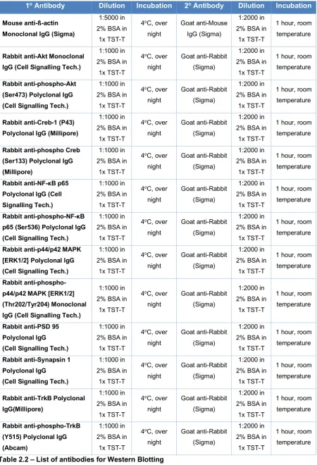
Table 2.2 - List of antibodies for Western Blotting 76
Table 2.3 - Primer used in RT-PCR 80
Table 2.4 - List of antibodies for immunohistochemistry 83 Table 3.1 - Effect of exercise on learning and memory, anxiety and
depression-like behaviour throughout the lifespan
138
Table 4.1 - Effect of exercise on cytokines in age 208 Table 4.2 - Effect of exercise on glial cells marker in age 209 Table 4.3 - Effect of exercise on inflammatory and apoptotic markers
in age
210
Table 4.4 - Effect of exercise on growth factors, neurotrophins, signalling proteins and synaptogenesis markers in age
211
Table 4.5 - Effect of exercise on cell proliferation and neurogenesis in age
212
Table 4.6 - Effect of exercise on brain and hippocampal volumes in age
212
Table 5.1 - Effect of exercise on cytokines 4 hours after LPS challenge
281
Table 5.2 - Effect of exercise on glial cells markers 4 hours after LPS challenge
283
Table 5.3 -Effect of exercise on inflammatory and apoptotic markers 4 hours after LPS challenge
283
Table 5.4 - Effect of exercise on growth factors, neurotrophins and signalling proteins 4 hours after LPS challenge
“Success is not final, failure is not fatal:
it is the courage to continue that counts”
Chapter 1: Introduction
1.1 Ageing
1.1.1 World population ageing
Worldwide, life expectancy has increased due to improvements in longevity and consequently, the world population is rapidly ageing. According to the last United Nations report, around 607 million people were aged 60 or older (~8% of the world’s population) in 2000. Moreover, between 2015 and 2030, the number of people in the world aged 60 years or over is projected to grow from 901 million in to 2015 to 1.4 billion in 2030. By 2050, this number is expected to almost double, reaching nearly 2.1 billion people, representing 16% of the world’s population. The oldest population, people aged 80 years or over, is also growing faster globally. According the United Nations, 125 million people were aged 80 years or over in 2015, and this number is projected to triple in 2050, reaching 434 million of people worldwide.
Figure 1.1 - Ageing population worldwide
World population aged 60–79, and 80 years and over, by development group 2000, 2015, 2030 and 2050 (Source: United Nations. World Population Ageing 2015).
1.1.2 Age-related diseases
Ageing is a multifactorial process associated with a decline in organism functions. The most evident characteristic of ageing is the reduction in the ability of the body to maintain homeostasis (Seidler et al., 2010, Comfort, 1979). Among the alterations related to ageing, one of the most striking is the decline in brain function, bringing increased morbidity, functional limitations, loss of autonomy and consequently reducing the quality of life of this population.
a public health concern and efforts in research, aiming to ameliorate the quality of life of this population, have increased substantially.
1.2 Cognitive function
1.2.1 Learning and memory
One of the most intriguing faculties of the brain is its ability to acquire and store information from an experience and to recall that information at a later point. For this reason, the process of learning and memory is integral and crucial to functional life. We can define learning as the ability to acquire knowledge and change behaviour as a result of an experience (Kolb and Whishaw, 2014) and memory as the processes of encoding, storage and retrieval of learned information (Bunke and Kandel, 2000). Over the past years, studies in animals and humans have significantly enhanced our understanding about how the brain accomplishes these processes (Han and Stevens, 1999).
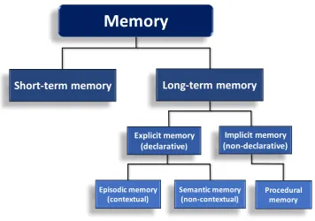
1.2.2 Memory classification
Figure 1.2 - Types of memory
Schematic diagram of the classification of memory (based on Squire, 1986).
Over the years, several studies have contributed new insights to help identify the brain structures and cellular mechanisms involved in memory. In this context, one important difference between these forms of memory seems to be the brain structures involved (Squire, 2004). It has been demonstrated that implicit memory involves areas such as the cerebellum, and basal ganglia, while explicit memory requires areas of the medial temporal lobe, such as entorhinal and perirhinal cortex and, particularly, the hippocampus (Squire, 2004, Squire, 1992).
1.2.3 Spatial and recognition memory
This thesis investigates two types of memory in mice: spatial memory and object recognition (non-spatial) memory. Spatial memory is the ability to encode, store and retrieve information about the spatial arrangement of objects or place (Kessels et al., 2001). This memory is a component of several of the categories in the memory system mentioned previously, involving aspects of both short and long-term memory, as well as procedural (non-declarative), semantic and episodic (declarative) memory (Moscovitch et al., 2006).
Memory
Short-term memory
Explicit memory
(declarative) (non-declarative)Implicit memory
Episodic memory
In rodents, several spatial tasks that are used are based on maze navigation, including the Morris Water Maze (MWM), which is widely employed to test the ability to learn and remember routes and paths using spatial cues (Vorhees and Williams, 2006). The Object Displacement (OD) task is another test used to evaluate spatial memory (object-location memory) and also requires the creation of a spatial layout (Kessels et al., 2001). Studies have demonstrated the hippocampus to be vital for learning, memory, and spatial navigation (Preston and Eichenbaum, 2013)
Recognition memory is the ability to distinguish between a familiar item (previously presented) and a novel item. This type of memory is one of the main components of the declarative memory category (Squire et al., 2007). The most common task used to assess recognition memory, in rodents, is the Novel Object Recognition (NOR) task, which relies in innate preference of the rodent to explore the novel object (Leger et al., 2013). The perirhinal cortex area has been shown to be crucial for remembering and recognition of the features of objects. However, although recognition memory is often described as a hippocampus-independent memory, compelling evidence suggests that the hippocampus plays an important role in processing information related to the familiarity of objects (Buckley, 2005).
1.3 The hippocampus
1.3.1 The hippocampal formation
hippocampal cytoarchitecture. These findings strongly support a functional segmentation of the hippocampus into two main parts, the dorsal (posterior in humans) and the ventral (anterior in humans) portion of the hippocampus (see Figure 1.3) (Fanselow and Dong, 2010, Strange et al., 2014).
Figure 1.3 - The hippocampus
Hippocampal formation (in red) in the rodent (a) and human (b) brain. Hippocampal axis in rodents is ventro-dorsal (a) and in humans is antero-posterior (b). Representative coronal section of the hippocampal formation showing CA1-CA4 and dentate gyrus regions in both the rodent (a) and human (b) brain (adapted from Strange et al., 2014).
The hippocampus exhibits three major excitatory pathways, with the main input running via the entorhinal cortex to the DG, CA3, CA1 and returning to the entorhinal cortex (Anderson et al., 1987, Deng et al., 2010). This is called the tri-synaptic pathway and consists of the perforant pathway, the mossy fibres pathway and the Schaffer collateral pathway (see Figure 1.4). Briefly, the hippocampus receives input from axons in the entorhinal cortex to the granule cells of the DG via the perforant pathway. The granule cells from the DG projects axons to the pyramidal cells in the CA3, via the mossy fibres pathway. Finally, the pyramidal
[image:42.595.108.455.176.460.2]cells from the CA3 region send projections to the CA1 pyramidal cells, via the Schaffer collateral pathway, with subsequent CA1 connection to the subiculum (Deng et al., 2010). This elaborated neural circuitry is the basis to our understanding of the role of hippocampus in learning and memory.
Figure 1.4 – Subfields and neural circuitry of the hippocampus in rodent
The main inputs to the hippocampus from the perirhinal and parahippocampus originate from neurons in the entorhinal cortex (EC), which project their axons to the dentate gyrus through the medial (MPP - II) and lateral (LPP - III) perforant pathways. The mossy fibres connect the cells from the dentate gyrus to CA3, while the Schaffer collaterals connect CA3 pyramidal neurons to CA1 pyramidal neurons. The main output from the hippocampus is from CA1 to the EC via subiculum (Deng et al., 2010).
1.3.2 Role of the hippocampus
region removal, whereas his implicit (non-declarative) memory, such as motor skill abilities and semantic memories was spared (Scoville and Milner, 1957, Corkin, 2002).
Since then, several studies, in humans (Burgess et al., 2002) and animals (Martin and Clark, 2007) provided further evidence of the central role of the hippocampus in the process of learning and memory. In particular, the DG has received special attention for its role in memory formation. It has been demonstrated that lesion of DG blocks memory acquisition, but does not alter retrieval of hippocampal memory (Lee and Kesner, 2004, Madronal et al., 2016). Also, many of the cellular and molecular mechanisms associated with learning and memory, such as long-term potentiation, morphological changes and neurogenesis, are well-established in the DG (Jonas and Lisman, 2014). Furthermore, as the hippocampus is part of the limbic system it plays an important role controlling emotions. Moser and Moser (1998), suggested that dorsal (posterior in humans) and ventral (anterior in humans) portions of hippocampus have different roles (Moser and Moser, 1998). Several lesion studies demonstrated that spatial learning and memory appear to depend on the dorsal portion, while the ventral portion is associated with stress responses and emotional behaviour (Moser et al., 1995, Henke, 1990).
1.4 Cognitive function and ageing
Ageing is characterized by several morphological, chemical and biological changes in brain structures, which are often manifested as deterioration in cognitive function (Bishop et al., 2010). There is an abundance of evidence of age-induced cognitive decline in the literature, in humans (Albert et al., 1995, Atkinson et al., 2007) and in rodents (Means et al., 1993, Latimer et al., 2014). It has been demonstrated that the cognitive decline associated with ageing is due to several changes in specific brain areas and alteration in learning and memory related mechanisms, such as changes in synaptic plasticity and neurogenesis. Specific regions of the brain shrink due ageing, mainly the prefrontal cortex and the hippocampus, both important areas to learning and memory and other complex mental activities (Peters, 2006). Also, age-related changes in behaviour are not limited to cognitive function; studies have also demonstrated increased anxiety in aged mice and mouse models of neurodegenerative disease (Flint et al., 2010). In addition, ageing is associated with inflammation, reduced brain plasticity in the form of either impaired LTP or decreased neurogenesis, and neurodegenerative disease (Grady, 2012).
1.5 Cellular and molecular mechanisms of learning and memory
1.5.1 Synaptic plasticity
Synaptic plasticity can be defined as an activity-dependent modification of the strength or efficacy of synaptic transmission at pre-existing synapses (Bunke and Kandel, 2000). The existence of synaptic plasticity was first described by Cajal (1913) and later by Hebb (1949). Nowadays, these changes in synapse strength and efficacy are believed to represent one of the most important cellular mechanisms associated with the process of learning and memory (Berlucchi and Buchtel, 2009).
resulted in a sustained increase in the synaptic response of the granule cells in the DG and since then, LTP has been widely studied and described in the brain across different species (Artola and Singer, 1993, Bruel-Jungerman et al., 2007). Induction and maintenance of LTP requires the activation of specific receptors and molecular signalling. The most studied form of LTP is N-methyl D-aspartate (NMDA) receptor dependent-LTP and it is divided into three phases: initial, early-phase (E-LTP) and late-early-phase (L-LTP). The initial early-phase, also called short-term potentiation, can last from seconds to minutes and requires the release of glutamate neurotransmitter from the pre synaptic terminal due to an increase in presynaptic levels of Ca2+, in response to an action potential. This initial phase acts
as a prelude to the two subsequent phases, the E-LTP and L-LTP, that are described to be related mainly with post-synaptic signalling (Sweatt, 1999).
The next phase, the E-LTP, can be sustained from minutes to hours and involves the activation of NMDA and α-amino-3-hydroxy-5-methyl-4-isoxazolepropionic acid (AMPA) receptors in the post-synaptic neuron, increasing the influx of Ca2+
into the cell and leading to a subsequently activation of Ca2+-dependent kinases,
such as Ca/calmodulin-dependent kinase (CaMKII) and protein kinases A and C (PKA and PKC) (Mayford, 2007). This cascade is essential to the induction of E-LTP, once activated Ca2+ dependent kinases phosphorylate the post-synaptic
AMPA receptors, making them sensitive to glutamate (Sweatt, 2001, Lynch, 2004). The last phase, known as L-LTP, can last from hours to weeks in vivo (Lynch, 2004) and requires the activation of intracellular signalling and synthesis of new proteins. The kinases activated in E-LTP trigger signalling pathways, which lead to the activation of transcription factors, such as cAMP response element binding protein (CREB) and consequent synthesis of new proteins, including AMPA receptor that are inserted in the post-synaptic membrane and increase the sensitivity of the cell to glutamate (Linden and Routtenberg, 1989, Kelleher et al., 2004).
hippocampus (Ying et al., 2002, Kuipers et al., 2016). Furthermore, a study from our laboratory showed exercise to increase expression of BDNF in the DG together with enhanced LTP and recognition memory (O'Callaghan et al., 2007).
1.5.2 Synaptogenesis
Synaptic organization and remodelling of neural circuits are important mechanism for the acquisition and storage of new memories (Bruel-Jungerman et al., 2007). Synaptogenesis includes the changes in the number of synapses and change in the number, size and density of dendritic spines. Enlargement of existing spines can be observed in minutes (Tanaka et al., 2008), while insertion of AMPA receptors in these spines, important to switch synapses form silent to functional, can take hours (Park et al., 2006b) and it can take days to result in fully functional synapses (Nagerl et al., 2007).
Synaptogenesis has been linked with enhanced cognitive function (Markham and Greenough, 2004, Birch et al., 2013) and it has been shown that exercise can induce the generation of new synapses and up-regulate synaptic proteins in the hippocampus (Shih et al., 2013). Exercise increases the levels of presynaptic proteins, such as synapsin-1 and post-synaptic proteins, such as PSD-95, and it has been linked to increase in BDNF levels and improvements in spatial memory (Vaynman et al., 2003, Siette et al., 2013, Shih et al., 2013). Thus, evidence suggests that remodelling of synapses has a stable and persistent effect on learning and memory.
1.5.3 Synaptic plasticity, synaptogenesis and ageing
which are often related to cognitive impairments and dementia (Loerch et al., 2008, Jouvenceau et al., 1998).
A significant decrease in the expression of synaptic genes with ageing has also been reported, contributing to altered connectivity and integration of brain networks in ageing (Lu et al., 2004). Moreover, a dysfunction in Ca2+ homeostasis can be
observed in the aged brain, which can also contribute to alterations in synaptic plasticity and consequent cognitive impairment (Yankner et al., 2008). In addition, age-related changes in the structure of neuronal synapses have also been reported, characterized by alterations in dendritic branching and reduction in the number of synapses as well as decreased expression of pre-post/synaptic structural proteins (such as synaptophysin, synapsin-1 and PSD-95) and progressive loss of synaptic density (Burke and Barnes, 2006, Ojo et al., 2012). Interestingly, many of these age-related changes in cellular mechanisms and morphology are tightly associated with impairments in learning and memory (Erickson and Barnes, 2003). Taken together, these finds strongly suggest that synaptic plasticity is affected by ageing and linked with cognitive impairments observed in ageing.
1.5.4 Adult Hippocampal Neurogenesis
Nowadays, a wide body of evidence indicates that new neurons are continually being generated in the adult brain. Actively dividing NSCs have been discovered in two specific “neurogenic niches” in the mammalian brain, which are the subventricular zone (SVZ) of the lateral ventricles and the subgranular zone (SGZ) of the DG in the hippocampus (Doetsch and Alvarez-Buylla, 1996, Deng et al., 2010). In the SVZ, adult born neurons differentiate and migrate to become periglomerular and granular neurons of the olfactory bulb (Deng et al., 2010). In contrast, the NSCs originating in the SGZ of DG migrate to the granule cell layer (GCL) and incorporate into local neural circuits as granular cells of the dentate gyrus (van Praag et al., 2002, Duman et al., 2001b).
1.5.4.1 Timeline of hippocampal neurogenesis
The process of adult hippocampal neurogenesis has been divided into a sequence of complex processes, which begins with the proliferation and differentiation of neural progenitor’s cells (NPCs) in the SGZ of DG, followed by migration and maturation of newborn neurons. There are four types of NPCs in the DG, which are the type-1, type-2a, type-2b and type-3 NPCs (Figure 1.5). They can be distinguished from each other based on morphology, proliferation rate and protein expression (Lledo et al., 2006), 2006). Type-1 cells are radial glial-like cells and express the astrocytic protein glial fibrillary acid protein (GFAP), SOX2 and the intermediate filament protein nestin. Type-1 cells have a low proliferation rate and can give rise to Type-2a NPCs (Farioli-Vecchioli et al., 2014). Type-2a cells do not express GFAP, however the nestin expression is maintained. This type of cell is characterized by high proliferation rate and, under favourable conditions, can generate type-2b NPCs (Farioli-Vecchioli et al., 2014, Lledo et al., 2006).
immature neuron to a fully mature neuron transiently expressing the calcium binding protein calretinin, followed by more mature neuronal markers such as neuronal nuclei (NeuN) and calbindin (Steiner et al., 2004).
[image:50.595.91.484.380.621.2]Newborn neurons become functionally integrated into existing DG circuitry within 3 weeks, extending their axons to CA3 region of hippocampus, as indicated by morphological and electrophysiological studies (Hastings and Gould, 1999, Esposito et al., 2005), Once mature, these newborn neurons can be integrated into neuronal network and form synapses with interneurons, mossy cells and CA3 pyramidal neurons (Toni et al., 2008). In this context, these newborn neurons can develop an important role in learning and memory. However, it has been shown that some endogenous and exogenous factors can affect the proliferation, differentiation and survival of the NCSs, including growth factors, neurotransmitters, hormones, transcription factors and inflammation (Klempin and Kempermann, 2007).
Figure 1.5 - Adult hippocampal neurogenesis in the subgranular zone (SGZ) of the dentate gyrus
1.5.4.2 Adult hippocampal neurogenesis in humans
Furthermore, studies have shown that hippocampal neurogenesis in humans can differ from hippocampal neurogenesis in rodents. Spalding and collaborators (2013), demonstrated that in humans, a larger proportion of hippocampal neurons are replaced in adulthood and the age-dependent decline in the rate of hippocampal neurogenesis is less pronounced in humans. Furthermore, hippocampal neurogenesis results in increase in the number of neurons in rodent DG, whereas neurogenesis in the human hippocampus generates a population of neurons with specific functional properties (Spalding et al., 2013). Also, recently, Boldrini and collaborators (2018), reported that healthy older subjects, with no cognitive impairment, show a preserved hippocampal neurogenesis. They demonstrated an age-related decline in the pool of quiescent NSCs in the DG, however, proliferating progenitors and immature neurons pools remain unchanged with ageing (Boldrini et al., 2018).
However, findings about neurogenesis in human are discrepant, while several studies suggested that a large number of new cells are generated in the hippocampus of humans in adulthood, other studies have found only a small number of newborn neurons (Eriksson et al., 1998, Dennis et al., 2016). Most recently, a study analysed post-mortem hippocampal tissue of humans and monkeys and strongly suggested that hippocampal neurogenesis drops from childhood to undetectable levels in adulthood in thesis species (Sorrells et al., 2018). These recent findings about the decline in hippocampal neurogenesis since the early stages of life raises some questions about the possible differences between neurogenesis in humans and other species, where adult neurogenesis has been shown to be well preserved.
1.5.4.3 Balance between and neurogenesis and gliogenesis
the trigger of specific signalling and transcription factors, can drive and determine the NSCs commitment and cell fate (Wen et al., 2009, Rusznak et al., 2016). Accordingly, changes in the balance of neurogenesis and gliogenesis have been reported in a number of pathological conditions of the central nervous system (CNS), including brain injury (Burns et al., 2009, Wang et al., 2016) and neurodegenerative diseases (Winner and Winkler, 2015, Choi et al., 2016).
1.5.4.4 Adult hippocampal neurogenesis and learning and memory
Adult neurogenesis has been linked to neural plasticity and cognition in rodents, as the newborn granule cells have been shown to correlate with modifications in hippocampus-dependent memory (Shors, 2004). For instance, young-adult rats and mice housed in an enriched environment for up to 8 weeks demonstrated increase in hippocampal neurogenesis, which was associated with enhancement in spatial learning and memory performance in the MWM task (Nilsson et al., 1999, Iso et al., 2007). On the other hand, negative modulation of hippocampal neurogenesis has been demonstrated to impair spatial learning and memory. For example, lesion of the cholinergic neurons in the septohippocampal pathway was reported to decrease hippocampal neurogenesis and significantly impair spatial learning and memory performance (Mohapel et al., 2005).
1.5.4.5 Hippocampal neurogenesis and ageing
Adult hippocampal neurogenesis in the dentate gyrus (DG) has been shown to be impaired due to the ageing process in different mammalian species (Seki and Arai, 1995, Cameron and McKay, 1999, Gould et al., 1999, Kempermann et al., 2002, McDonald and Wojtowicz, 2005) and it has also been partially linked to age-related cognitive decline (Lazarov et al., 2010, Mu and Gage, 2011, Galea et al., 2013). In aged rats, the proliferation rate of NSCs in the SGZ of the dentate gyrus can be reduced by approximately 80%, suggesting a link between an age-related decrease in neurogenesis and a decrease in both NSCs proliferation and neural progenitor cell (NPCs) differentiation (Jin et al., 2003, Kuhn et al., 1996).
Several neurological diseases are linked with decreased rates of neurogenesis. For instance, the triple transgenic mouse model of AD (3xTg-AD) is reported to have lower hippocampal neurogenesis when compared to their wild type control, and this decrease is linked to cognitive impairment (Rodriguez et al., 2008). Furthermore, chronic stress and rodent models of anxiety and depression have also been shown to affect hippocampal neurogenesis (Surget et al., 2008). Age-associated decline in neurogenesis may be related to changes in the microenvironment of NSCs, which can interfere with proliferation, differentiation and survival of newborn cells. In this context, inflammation has been shown to play a role in this microenvironment (e.g. microglia activation and pro-inflammatory mediators infiltration from periphery), thereby influencing cell proliferation and survival of newborn neurons (Yirmiya and Goshen, 2011).
hypothesis that age-related decrease in the rate of neurogenesis may be a consequence of changes in the microenvironment of NSCs, such as decreased levels of neurotrophins and growth factors and low-grade inflammation.
1.6 Neurotrophins and growth factors
Neurotrophins belong to the family of growth factors that promote and regulate neuronal development, growth, survival or death as well as neurogenesis and brain plasticity (Blum and Konnerth, 2005) and are widely described to enhance synaptic plasticity and memory, in particular BDNF (Schinder and Poo, 2000, Gooney et al., 2004). The neurotrophin family comprises NGF, BDNF, neurotrophin 3 (NT3) and neurotrophin 4/5 (NT4/5). Neurotrophins are synthetised as precursors proteins, pro-neurotrophins, and later these precursors undergo proteolytic cleavage, to yield the mature-neurotrophins (Mowla et al., 2001).
These proteins act by binding two different membrane receptors, the tyrosine kinase receptor (Trk) family and the pan-neurotrophin (p75) receptor. It has been widely described that NGF binds preferentially to TrkA receptor, while BDNF and NT4/5 are have high affinity for the TrkB receptor and NT3 binds the TrkC receptor (Barbacid, 1995, Huang and Reichardt, 2003). Despite the fact that the p75 receptor was the first neurotrophin receptor to be isolated (Johnson et al., 1986), its ligand affinity and signalling is less understood than the Trk receptors family. It has been shown that the binding affinity of neurotrophins to the p75 receptor is higher for the pro-form of neurotrophins than the mature form (Dechant and Barde, 2002).
to the neurotrophin family but are also widely associated with promotion of cell growth and survival, including the Igf-1, the vascular endothelial growth factor (VEGF) and the glial cell line-derived neurotrophic factor (GDNF).
1.6.1 Brain-derived neurotrophic factor (BDNF)
Among the neurotrophins, BDNF has been most widely studied and shown to play an important role in the mature nervous system. Increased levels of BDNF have been linked to enhanced synaptogenesis, neurogenesis and learning and memory (Carvalho et al., 2008, Nagahara et al., 2009). BDNF was the second member of the neurotrophin family to be discovered, in 1982 (Barde et al., 1982). As mentioned before, BDNF exerts its trophic effects by binding TrkB receptor (Luikart et al., 2003). In the adult brain, BDNF and TrkB receptor can be found in different brain regions and cell types, most abundantly in neurons and astrocytes (Moretto et al., 1994, Barakat-Walter, 1996, Binder and Scharfman, 2004). In the CNS, BDNF is highly expressed in the hippocampus, in association with glutamatergic synapses (Bramham and Messaoudi, 2005), whereas, peripherally, BDNF is found in platelet and endothelial cells (Yamamoto and Gurney, 1990, Fujimura et al., 2002).
Similar to other neurotrophins, BDNF is synthesised as a pro-neurotrophin (proBDNF; ~30kDa), which then is cleaved, generating the mature-BDNF (mBDNF; ~14kDa). Although proBDNF was originally thought to act only intracellularly, evidence has suggested it is secreted and cleaved extracellularly (Barnes and Thomas, 2008). However, studies have shown that most BDNF found and secreted in the CNS is in its mature form (Matsumoto et al., 2008) and the amount of BDNF secreted is regulated in an activity dependent-manner via voltage-gated Ca2+ channels and NMDA receptor (Hartmann et al., 2001, Kolarow
et al., 2007).
1.6.2 BDNF-TrkB signalling cascade
When released into the synaptic cleft, BDNF binds to TrkB receptor in the post synaptic membrane and consequently, a signalling cascade is activated within the cell. Short-term effects of BDNF binding at the TrkB receptor include increase of neurotransmitter release and activation of glutamate receptors in the synapse, whereas the long-term consequences are associated with the activation of intracellular pathways and the modifications in gene expression downstream in the nucleus (Cunha et al., 2010). TrkB receptor is a transmembrane receptor with an extracellular ligand-binding site and several kinase domains.
BDNF and TrkB have a relevant role in several aspects of the hippocampus, including cognitive function and neurogenesis. For instance, higher levels of BDNF in the hippocampus, induced by physical exercise, have been implicated in long-term potentiation and synaptic plasticity (Liu and Nusslock, 2018). Moreover, in mice exposed to enriched environment, BDNF was demonstrated to stimulate hippocampal neurogenesis, as well as BDNF has been reported to have coordinated effects with antidepressants drugs on hippocampal neurogenesis (Rossi et al., 2006, Sairanen et al., 2005).
Figure 1.6 - Main BDNF-TrkB signalling cascade