ORIGINAL RESEARCH
SPINE
Percutaneous Spinal Ablation in a Sheep Model: Protective
Capacity of an Intact Cortex, Correlation of Ablation
Parameters with Ablation Zone Size, and Correlation of
Postablation MRI and Pathologic Findings
XA.N. Wallace,XT.J. Hillen,XM.V. Friedman,XZ.S. Zohny,XB.H. Stephens,XS.C. Greco,XM.R. Talcott, andXJ.W. Jennings
ABSTRACT
BACKGROUND AND PURPOSE: Despite the growing use of percutaneous ablation therapy for the treatment of metastatic spine disease, several issues have yet to be fully addressed. Our aims were to determine whether the vertebral body cortex protects against ablation-induced spinal cord injury; correlate radiofrequency, cryo-, and microwave ablation parameters with resulting spinal ablation zone dimensions and describe normal spinal marrow postablation changes on MR imaging.
MATERIALS AND METHODS:Ten thoracolumbar vertebrae in 3 sheep were treated with radiofrequency ablation, cryoablation, or microwave ablation under fluoroscopic guidance. Technique parameters were chosen to produce ablation zones that exceeded the volume of the vertebral bodies in sheep 1 and were confined to the vertebrae in sheep 2 and 3. Expected ablation zone dimensions were based on data provided by the device manufacturers. Postablation MR imaging was performed at 48 hours (sheep 1) or 7 days (sheep 2 and 3).
RESULTS:In sheep 1, cryoablation and microwave ablations extended into the spinal canal and caused histologically confirmed neurologic injury, but radiofrequency ablation did not. The mean difference between the lengths of the radiofrequency ablation zone dimensions measured on gross pathology compared with those expected was 9.6⫾4.1 mm. The gross pathologic cryo- and microwave ablation zone dimensions were within 1 mm of those expected. All modalities produced a nonenhancing ablation zone with a rim of enhancement, corresponding histologically to marrow necrosis and hemorrhagic congestion.
CONCLUSIONS: An intact cortex appears to protect against radiofrequency ablation-induced spinal cord injury, but not against non-impedance-based modalities. Ablation dimensions produced by microwave and cryoablation are similar to those expected, while radio-frequency ablation dimensions are smaller. Ablation of normal marrow produces a rim of enhancement at the margin of the ablation zone on MR imaging.
ABBREVIATIONS:MWA⫽microwave ablation; RFA⫽radiofrequency ablation
P
ercutaneous, image-guided ablation is a minimally invasive therapy for metastatic spine disease. Several ablation modali-ties are commercially available, including radiofrequency abla-tion (RFA), cryoablaabla-tion, and microwave ablaabla-tion (MWA). RFA and MWA heat the tumor to temperatures above 60°C, at whichpoint protein denaturation, immediate cell death, and coagulative necrosis occur.1Mechanisms of cryoablation-induced cell death
include osmotic bursting, intracellular ice formation, coagulative necrosis, free radical release and apoptosis in response to bio-chemical stress, and stimulated immunologic targeting of tumor cells.2For patients with painful spinal metastases who do not
respond or have a contraindication to radiation therapy, ablation may produce clinically significant pain palliation.3-7Additionally,
Wallace et al8recently reported a 1-year radiologic local control
rate of 70% (21/30) after RFA of spinal metastases, and Tomasian et al5achieved a 97% (30/31) radiologic local control rate of spinal
metastases treated with cryoablation after a median follow-up of 10 months (range, 1–24 months). As a result, recent treatment algorithms for metastatic spine disease have incorporated abla-tion as an alternative opabla-tion for pain palliaabla-tion and local tumor control.9
Received December 28, 2016; accepted after revision March 17, 2017.
From the Mallinckrodt Institute of Radiology (A.N.W., T.J.H., M.V.F., J.W.J.), Depart-ment of Neurosurgery (Z.S.Z., B.H.S.), and Division of Comparative Medicine (S.C.G., M.R.T.), Washington University, St Louis, Missouri.
The authors received ablation probes from DFINE, Galil Medical, and Medtronic for this study.
Please address correspondence to Adam N. Wallace, MD, Mallinckrodt Institute of Radiology, Washington University, 510 South Kingshighway Blvd, St Louis, MO 63141; e-mail: [email protected]
Despite the growing use of percutaneous ablation therapy for the treatment of metastatic spine disease, several important issues have yet to be fully addressed. First, most spinal metas-tases involve the posterior vertebral body where complete tu-mor ablation is essential to prevent localized epidural invasion and spinal cord compression.10,11 However, it is unclear
whether intact cortex acts as a protective barrier for the spinal cord and exiting nerve roots during ablation of the posterior vertebral body.12,13Second, adequate tumor ablation requires
accurate correlations between technique parameters and the resulting ablation zone dimensions. However, existing corre-lations derived from the manufacturers’ preclinical data are based on soft-tissue and artificial models that have different thermal and electrical conductive properties to bone.14Last,
although MR imaging is typically used to assess for local tumor control after ablation of spinal metastases,15diagnosis of
re-sidual tumor is limited by a paucity of literature describing ablation-induced MR imaging findings in normal spinal marrow.
The purposes of this study were to determine whether intact posterior vertebral body cortex protects the spinal cord against radiofrequency-, cryo-, or microwave ablation-induced injury, to correlate the technical parameters of each ablation technique with the resulting dimensions of the ablation zone, and to de-scribe normal spinal marrow postablation changes on MR imaging.
MATERIALS AND METHODS
The protocol of this study was approved by our institutional ani-mal care and use committee.
Ablation Technique
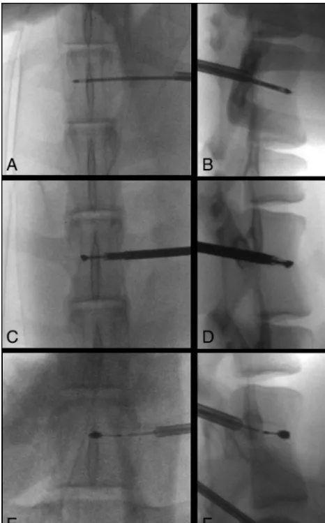
Ablations were performed under fluoroscopic guidance by 4 radiologists (A.N.W., T.J.H., M.V.F., J.W.J.) with 1–9 years of experience performing musculoskeletal tumor ablation. Ten ver-tebral bodies in 3 healthy living adult female sheep (weight range, 55– 60 kg) were ablated. All 3 sheep were sedated by a veterinarian staff member with an intramuscular in-jection of ketamine hydrochloride (2–10 mg/kg) and xylazine (0.2 mg/kg), intu-bated, and maintained under inhaled isoflurane (1%–5%) for general anes-thesia. Mechanical ventilation was used throughout the procedure. The target vertebrae were localized by using a sin-gle-plane fluoroscopy unit, and the planned skin entry sites were sterilized with betadine scrubs and draped. Five-to 10-mm incisions were made at each skin entry site with a No. 11 blade to ac-commodate the percutaneous instru-ments. Unilateral transpedicular osse-ous channels into the vertebral bodies were created for ablation probe place-ment by using the OnControl system (Vidacare, Shavano Park, Texas), which consists of a 10-ga outer can-FIG 1. Fluoroscopic documentation of ablation probe placement
[image:2.594.53.286.47.421.2]in sheep. Anteroposterior (A, C, andE) and lateral (B, D, andF) fluoroscopic images show transpedicular placement of the cryo-ablation probe within the T14 vertebra (Aand B), the radiofre-quency ablation probe within the L3 vertebra (CandD), and the microwave ablation probe within the L6 vertebra (EandF).
Table 1: Technical parameters used for radiofrequency ablation, expected minor-axis diameter of the ablation zones based on nonanimal models, and measured ablation zone diameters on MRI and gross pathology
Watts
Thermocouple
Temperatures (oC) Ablation Time (sec)
Diameter of Ablation Zone Orthogonal to Ablation Probe (mm)
Proximal Distal Expected MRI Gross Pathology
10 72 51 92 20.0 13.4 13.8
5 58 42 107 16.6 9.2 10.0
5 51 40 42 13.0 7.3 8.0
Table 2: Technical parameters used for cryoablation, expected minor-axis diameter of the ablation zones based on nonanimal models, and measured ablation zone diameters on MRI and gross pathology
Probe
Intensity (%)
Freeze Cycles (min)
Thaw (min)
Diameter of Ablation Zone Orthogonal to Ablation Probe (mm) Expected MRI Gross Pathology
IceSphere 60 6.0 7.0 20.0 19.2 20.1
IceSeed 50 4.0 5.0 6.0 6.6 7.4
IceSeed 50 3.5 5.0 6.0 6.4 7.0
[image:2.594.51.378.534.602.2] [image:2.594.53.376.650.727.2]nula and 12-ga inner osteotome. The locations of each probe were documented fluoroscopically (Fig 1).
Ablation Parameters
The technical parameters and expected dimensions of each abla-tion zone based on preclinical data provided by the manufacturers are detailed inTables 1–3. In sheep 1, cryoablation, RFA, and MWA were performed in the T14, L3, and L6 vertebral bodies, respectively. Sheep most commonly have 14 rib-bearing thoracic and 6 lumbar vertebrae, and the spinal cord terminates caudal to the sacrum. Each ablation probe tip was placed 5 mm from the posterior vertebral body cortex on a true lateral fluoroscopic pro-jection. The parameters of radiofrequency, cryo-, and microwave ablation were chosen to produce ablation zones with anteropos-terior, transverse, and craniocaudal dimensions of 30⫻20⫻20 mm, 25⫻20⫻20 mm, and 20⫻20⫻20 mm, respectively. The orientation of each dimension of the ablation zone with respect to the ablation probe was defined as depicted inFig 2. Thus, for each ablation, the radius of the ablation zone extending from the probe tip toward the posterior vertebral body cortex was expected to be 10 mm. Every third vertebra was treated to ensure correct local-ization of histologic ablation-induced spinal cord injury.
In sheep 2 and 3, each ablation probe was placed in the center of the vertebral body, and the parameters of each ablation were chosen to produce an ablation volume that would be confined to the vertebra on the basis of preclinical data provided by the man-ufacturers. In sheep 2, RFA was performed at T14; MWA, at L3; and cryoablation, at L6. In sheep 3, RFA was performed at T14; MWA, at L2; and cryoablation, at L4 and L6.
RFAs were performed with the STAR Tumor Ablation System
(DFINE, San Jose, California). Ablations can be performed at power settings of 5 or 10 W. Two thermocouples located on the shaft of the probe enable real-time monitoring of temperatures 10 and 15 mm from the center of the ablation zone, and the dimen-sions of the ablation zone are estimated from these maximum temperature readings. The expected diameter of each RFA zone orthogonal to the probe is based on the manufacturer’s thermal distribution curves derived from ex vivo porcine liver models.
Cryoablations were performed with the Visual-ICE Cryoabla-tion System (Galil Medical, Arden Hills, Minnesota) with either the IceSeed 1.5 or IceSphere 1.5 cryoprobe. For a given cryoprobe, the volume of the ablation zone is controlled by changing the inten-sity (percentage maximum) and the duration of the freeze cycle. In the present study, 2 freeze cycles were performed and the probe was actively heated to thaw the tissue between cycles. The expected diameter of each ablation zone orthogonal to the cryoprobe was based on the⫺40°C isotherm dimensions pro-duced in a 37°C gel bath.
MWAs were performed by using the Emprint Ablation System (Medtronic, Minneapolis, Minnesota). Ablations were performed at a power setting of 100 W for 10, 15, and 50 seconds. Power settings of 45 and 75 W are also available. The expected diam-eter of each MWA zone was based on an ex vivo porcine kidney model.
Postablation Evaluation
The sheep were clinically evaluated by a veterinarian immediately after each procedure for signs of pain or ablation-induced nerve injury, including lameness or abnormal movement. Subcutane-ous buprenorphine, 0.005– 0.01 mg/kg, was administered every 10 –12 hours, and carprofen, 2– 4 mg/kg, was administered every 12–24 hours for 2 days postoperatively. The sheep were then pe-riodically monitored by veterinary staff before sacrifice for evi-dence of procedure-related complications.
MR Imaging
In sheep 1, MR imaging was performed 48 hours after treatment owing to clinical signs of spinal cord injury and the ethical neces-sity for euthanasia earlier than the planned 7-day time point. MR imaging was performed 7 days after treatment of sheep 2 and 3. Images were obtained on a Magnetom Trio 3T scanner (Siemens, Erlangen, Germany) using the spine coil within the table of the scanner. The protocol included T1-weighted conventional spin-echo images (TR, 783 ms; TE, 10 ms), T2-weighted fast spin-spin-echo images with fat suppression (TR, 3500 ms; TE, 94 ms), and post-contrast conventional spin-echo T1-weighted images with fat suppression obtained after the intravenous bolus administration of 0.1 mmol/kg of gadoversetamide (Optimark; Covidien, Irvine, California). All images were acquired with a 2.5-mm section thickness with 1-mm intersection spacing, 30⫻30 cm FOV, and 288⫻384 matrix. On MR imaging, the ablation zone was defined as the volume of nonenhancement on contrast-enhanced T1-weighted images with fat suppression.15The measurements of
MR imaging ablation zones were performed on high-resolution MR imaging workstation monitors. The anteroposterior, trans-verse, and craniocaudal ablation zone dimensions were measured for sheep 2 and 3. Only the transverse and craniocaudal dimen-FIG 2. Anteroposterior (A) and lateral (B) CT images of a normal
[image:3.594.53.286.78.287.2]lum-bar vertebra. Thethick black linerepresents the unilateral transpe-dicular trajectory of the ablation probe. By definition, the transverse and anteroposterior dimensions of the ablation zone are parallel and perpendicular to the ablation probe in the axial plane, respectively, and the craniocaudal dimension of the ablation zone is perpendicular to the ablation probe in the sagittal plane.
Table 3: Technical parameters used for microwave ablation, expected minor-axis diameter of the ablation zones based on nonanimal models, and measured ablation zone diameters on MRI and gross pathology
Watts
Ablation Time (sec)
Diameter of Ablation Zone Orthogonal to Ablation Probe (mm) Expected MRI
Gross Pathology
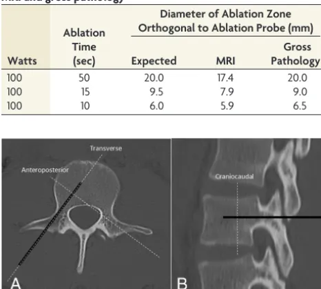
100 50 20.0 17.4 20.0
100 15 9.5 7.9 9.0
sions of each ablation zone were measured in sheep 1, because the anteroposterior dimensions was not confined to the vertebral body marrow. MR signal and enhancement characteristics of the ablation zones of each technique were also subjectively re-viewed by the first and senior authors. No interpretations were discrepant.
Pathology
Following MR imaging, the sheep were euthanized by barbiturate overdose (intravenous pentobarbital, 150 mg/kg) and the verte-bral bodies and spinal cord were harvested by senior neurosurgery residents (B.H.S., Z.S.Z.). Tissues were fixed in 10% neutral buff-ered formalin. After fixation, the vertebral bodies were decalcified in hydrochloric acid– ethylenediaminetetraacetic acid decalcifier (EKI, Joliet, Illinois) to permit cross-sectioning of the bones for gross examination of the ablation sites and trimming of tissues for histopathology. The diameters of the pale tan necrosis orthogonal probe tract were measured once by the first author and a
veteri-nary pathologist with a Westcott 6⬙Clear Plastic Acrylic Ruler (Acme United Corporation, Fairfield, Connecticut). Because the vertebral bodies could be sectioned in only 1 plane, only the trans-verse and craniocaudal diameters of the ablation zones could be measured. Tissues were then paraffin-embedded, sectioned at 5 m, and stained with hematoxylin-eosin for histopathologic evaluation.
RESULTS
Immediately after recovering from anesthesia, sheep 1 demon-strated bilateral lower extremity weakness, which did not improve during 48 hours of observation. MR imaging showed intramed-ullary T2 hyperintensity and enhancement involving the ventral spinal cord at the level of the cryoablated vertebra consistent with ablation-induced injury. Similar findings were seen along the ventrolateral aspect of the spinal cord at the level of the micro-wave-ablated vertebra, likely due to a slightly more oblique angulation of the MWA probe relative to the coronal plane. His-tologically, the ablation zone was char-acterized by white matter necrosis with cavitation and axonal swelling and de-generation (Fig 3). The RFA zone mea-sured 13.8 mm in the craniocaudal di-mension. Because the RFA probe was placed only 5 mm from the posterior vertebral body cortex, the RFA zone should have extended into the spinal ca-nal. Yet, histologic evaluation of the spi-nal cord at this level showed no evidence of ablation-induced injury.
Tables 1–3summarize the correla-tions between the technical parameters of each technique and the resulting di-mensions of the gross pathologic abla-tion zones. The mean differences in mil-limeters among the anteroposterior, transverse, and craniocaudal ablation zone dimensions expected on the basis of the manufacturers’ preclinical data and those measured on MR imaging and gross pathology are presented inTable 4. The dimensions of the RFA zones mea-sured on gross pathology were smaller than those expected on the basis of cor-relations provided by the manufacturer. The mean differences between the lengths of the transverse and craniocau-FIG 3. Sagittal postcontrast T1-weighted image with fat suppression (A) and a T2-weighted image
[image:4.594.53.533.637.696.2](B) of the vertebral level treated with cryoablation show the ablation zone extending beyond the posterior vertebral body into the spinal canal. There is enhancement and intramedullary T2 signal hyperintensity in the ventral aspect of the edematous spinal cord (white arrowheads). There is also inflammation in the soft tissues ventral to the spine where the ablation zone extended beyond the anterior vertebral body cortex (white arrows). Sagittal postcontrast T1-weighted image (C) and axial T2-weighted image (D) of the vertebral level treated with microwave ablation similarly show extension of the ablation zone into the spinal canal (white arrowheads).E, Hema-toxylin-eosin staining of the spinal cord at the level of the cryoablated vertebra at⫻100 total magnification shows axonal necrosis and edema. Similar findings were seen at the microwave-ablated level (not shown).F, Sagittal postcontrast T1-weighted image with fat suppression of the radiofrequency-ablated vertebra shows the posterior margin of the ablation zone confined to the vertebral body.
Table 4: Mean differences among the anteroposterior, transverse, and craniocaudal ablation zone dimensions expected on the basis of manufacturers’ preclinical data and those measured on MRI and gross pathologya
Expected vs MRI (mm)
Expected vs
Pathology (mm) MRI vs Pathology (mm)
AP TV CC TV CC TV CC
Radiofrequency ablation 6.7⫾0.9 11.0⫾2.5 6.6⫾0.7 13.4⫾2.1 5.9⫾0.7 ⫺1.2⫾0.7 ⫺0.8⫾0.1 Cryoablation ⫺0.5⫾0.3 0.1⫾0.7 ⫺0.1⫾0.7 ⫺0.4⫾0.6 ⫺1.0⫾0.5 ⫺3.4⫾0.2 ⫺0.9⫾0.3
Microwave ablation 0.9⫾0.4 0.3⫾0.5 1.4⫾1.0 0.1⫾0.5 0⫾0.4 ⫺0.8⫾0.2 ⫺0.9⫾0.3
Note:—AP indicates anteroposterior; TV, transverse; CC, craniocaudal.
dal RFA zone dimensions measured on gross pathology compared with those expected were 13.4⫾2.1 and 5.9⫾0.7 mm, respec-tively. In contrast, the cryo- and microwave ablation zone dimen-sions measured on gross pathology were within 1 mm of those expected.
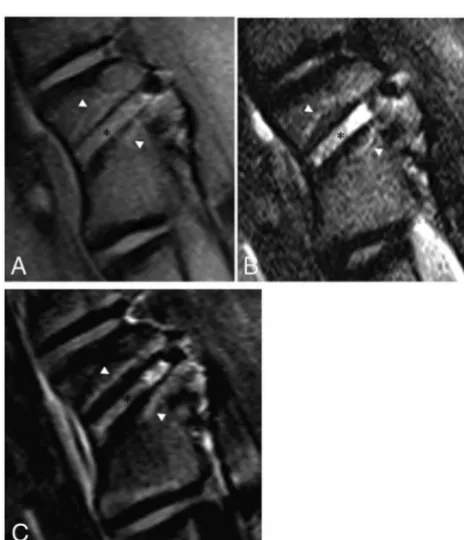
All ablation modalities produced the same subjective signal and enhancement characteristics on MR imaging (Table 5). Rel-ative to normal marrow, the probe tracts demonstrated T1 isoin-tense, T2 hyperinisoin-tense, and enhancing tissue outlined by signal void. The ablation zones were defined by nonenhancing tissue that was slightly T1 hyperintense and T2 isointense and delineated by a thin rim of T1 and T2 hyperintensity and enhancement (Fig 4). The gross and histologic findings seen with all modal-ities were qualitatively similar (Table 5). Gross examination revealed discrete pale tan ablation zones. Histologically, the ablation zones were characterized by widespread marrow ne-crosis with areas of granulation tissue and edematous stroma.
Surrounding the ablation zones were rims of hemorrhagic con-gestion (Fig 5).
DISCUSSION
In the present study, we used a bipolar RFA system in an attempt to produce an ablation zone with a minor-axis diameter greater than the distance from the probe tip to the posterior vertebral body cortex, but caused no histologic evidence of spinal cord in-jury due to the insulating effect of the posterior cortex. Previous animal experiments assessing the safety of RFA in close proximity to the spinal cord have been inconclusive. Dupuy et al12safely
performed monopolar RFA of the posterior vertebral body for 10 minutes, but the maximum temperature generated within the bone was only 48°C, which is below the cytotoxic threshold. In a subsequent porcine study, monopolar RFA performed flush with the posterior vertebral body cortex for 10 minutes at a constant temperature of 90°C at the probe tip caused paraplegia with his-tologically confirmed spinal cord injury.13However, this latter
experiment does not reflect clinical practice because adequate ab-lation of tumor within the posterior vertebral body only requires that the periphery of the ablation zone reach 60°C for a matter of seconds.1
In contrast with these previous studies, the ablation system used in the present experiment has several features that facili-tate its safe use in the spine. First, the distal segment of the probe articulates up to 90°, enabling accurate placement of the tip with respect to the posterior vertebral body cortex. Second, the 2 active thermocouples incorporated into the shaft of the probe allow real-time estimation of the dimensions of the ab-lation zone. Last, monopolar systems require placement of a skin-grounding pad, which, if placed incorrectly, can cause a skin burn. This risk is eliminated with the use of a bipolar probe.16The RFA zones measured on gross pathology were
smaller than those predicted from the manufacturer’s preclin-ical data derived from a soft-tissue model.14Potential reasons
for this discrepancy include the higher impedance and lower thermal conductivity of bone compared with soft tissue and a heat sink effect from blood flow in the vertebrae and epidural space, as well as circulating CSF.1,12
In contrast to RFA, when we produced a cryoablation zone with a diameter greater than the distance from the probe tip to the posterior vertebral body cortex, histologic evaluation of the spinal cord at the treated level showed white matter necrosis with cavi-tation and axonal swelling and degeneration consistent with ab-lation-induced injury. Although there are no prior studies assess-ing the safety of cryoablation within the posterior vertebral body, this result is expected because increased bone density does not limit the propagation of the cryoablation zone.17Several
[image:5.594.54.528.56.113.2]mea-sures may be taken to minimize the risk of this complication clin-ically. First, the distance between the probe tip and the spinal Table 5: MRI, gross, and histopathologic findings produced by all 3 ablation modalities
T1WI T2WI CE-T1WI Histopathology
Probe track Hyperintense Hyperintense Enhancement Hemorrhage, trabecular fragments, serum exudate,
granulation tissue
Center Slightly hyperintense Slightly hypointense No enhancement Marrow necrosis, granulation tissue, intact trabeculae
Rim Hyperintense Hyperintense Enhancement Hemorrhagic congestion
Note:—CE indicates contrast-enhanced.
[image:5.594.53.285.127.397.2]canal must be accurately measured, and the cryoprobe and inten-sity and duration of the freeze cycles can then be chosen to ensure that the ablation zone does not exceed this distance. The dimen-sions of the cryoablation zones measured on gross pathology were within 1 mm of those predicted from the manufacturer’s preclin-ical data derived from a 37°C gel bath model. Second, a coaxial needle with an inner thermocouple can be placed through the neural foramen to monitor the temperature in the epidural space, while the outer cannula is used to inject carbon dioxide for neural insulation and room temperature 5% dextrose in water to warm the epidural space.18Last, the use of
motor-evoked potential monitoring and peripheral motor nerve elec-trostimulation to prevent iatrogenic cryoablation nerve injury have been described.19,20
The present experiment also showed that the posterior verte-bral body cortex is not a barrier to MWA because this ablation zone also extended into the spinal canal and caused histologically evident spinal cord injury. Like cryoablation, this result is ex-pected because MWA is also non-impedance-based. However, the efficiency with which tissue absorbs microwave energy is pro-portional to its water content, and soft tissue is⬎70% water com-pared with the approximately 20% water content of bone.21,22
Therefore, one would expect the MWA zones in the spine to be smaller than those expected from preclinical data derived from an ex vivo soft-tissue model. Yet, such a discrepancy was not ob-served. Accurate sizing of the MWA zone to remain confined to
the vertebral body and injection of car-bon dioxide and room temperature 5% dextrose in water into the epidural space may increase the safety of the procedure. The ablation-induced marrow changes on MR imaging, gross pathology, and histopathology were consistent across all 3 modalities. The necrotic ablation zone was depicted on postcontrast T1-weighted MR imaging with fat suppres-sion as a nonenhancing area surrounded by a thin, enhancing rim of granulation tissue. T2 hyperintense enhancing resid-ual tumor can appear similar to the rim of the ablation cavity. In some cases, FDG PET/CT or biopsy may be needed to differentiate residual tumor from postablation change.15 The ablation
zone dimensions of all 3 modalities mea-sured on gross pathology were slightly larger than those measured on MR im-aging. In the absence of a plausible phys-iologic explanation, the discrepancy is likely related to the resolution limita-tions of MR imaging.
A limitation of this study is that the correlations between technical ablation parameters and ablation zone dimen-sions were derived in normal bone, and these correlations may be altered by the presence of tumor. Also, the derived correlations are specific to the ablation systems used in this experiment and cannot be extrapolated to other devices.
CONCLUSIONS
Intact posterior vertebral body cortex in normal sheep may pro-tect the spinal cord from radiofrequency ablation–induced injury, but not against microwave or cryoablation injury. Ablation di-mensions produced by microwave and cryoablation are similar to those expected, while RFA volumes are smaller. Postablation mar-row changes in normal spinal marmar-row include a rim of enhance-ment at the margin of the ablation zone.
Disclosures: Adam N. Wallace—RELATED:Grant: Medtronic, Galil, DFINE, Com-ments: received donated ablation probes from these companies.* Travis J. Hillen—UNRELATED:Consultancy: Merit Medical,Comments: honorarium for proctoring physician education courses. Jack W. Jennings—UNRELATED: Consul-tancy: Galil, Medtronic, Merit Medical, Bard Medical;Payment for Lectures In-cluding Service on Speakers Bureaus: Merit Medical and Medtronic Speaker Panels;Travel/Accommodations/Meeting Expenses Unrelated to Activities List-ed:Comments: for meetings in which Speaker Panel duties were performed. *Money paid to the institution.
REFERENCES
1. Brace CL.Radiofrequency and microwave ablation of the liver, lung, kidney, and bone: what are the differences?Curr Probl Diagn Radiol2009;38:135– 43CrossRef Medline
2. Rybak LD.Fire and ice: thermal ablation of musculoskeletal tu-mors.Radiol Clin North Am2009;47:455– 69CrossRef Medline FIG 5. A, Gross pathology of the vertebral body shown inFig 2cut along the plane of the
[image:6.594.56.376.46.332.2]3. Wallace AN, Greenwood TJ, Jennings JW.Radiofrequency ablation and vertebral augmentation for palliation of painful spinal metas-tases.J Neurooncol2015;124:111–18CrossRef Medline
4. Anchala PR, Irving WD, Hillen TJ, et al.Treatment of metastatic spinal lesions with a navigational bipolar radiofrequency ablation device: a multicenter retrospective study.Pain Physician2014;17: 317–27Medline
5. Tomasian A, Wallace A, Northrup B, et al.Spine cryoablation: pain palliation and local tumor control for vertebral metastases.AJNR Am J Neuroradiol2016;37:189 –95CrossRef Medline
6. Kastler A, Alnassan H, Aubry S, et al.Microwave thermal ablation of spinal metastatic bone tumors.J Vasc Interv Radiol2014;25:1470 –75
CrossRef Medline
7. Pusceddu C, Sotgia B, Fele RM, et al.Combined microwave ablation and cementoplasty in patients with painful bone metastases at high risk of fracture.Cardiovasc Intervent Radiol2016;39:74 – 80CrossRef Medline
8. Wallace AN, Tomasian A, Vaswani D, et al.Radiographic local con-trol of spinal metastases with percutaneous radiofrequency abla-tion and vertebral augmentaabla-tion.AJNR Am J Neuroradiol2016;37: 759 – 65CrossRef Medline
9. Wallace AN, Robinson CG, Meyer J, et al.The metastatic spine dis-ease multidisciplinary working group algorithms.Oncologist2015; 20:1205–15CrossRef Medline
10. Algra PR, Heimans JJ, Valk J, et al.Do metastases in vertebrae begin in the body or the pedicles? Imaging study in 45 patients.AJR Am J Roentgenol1992;158:1275–79CrossRef Medline
11. Klimo P Jr, Thompson CJ, Kestle JR, et al.A meta-analysis of surgery versus conventional radiotherapy for the treatment of metastatic spi-nal epidural disease.Neuro Oncol2005;7:64 –76CrossRef Medline
12. Dupuy DE, Hong R, Oliver B, et al.Radiofrequency ablation of
spi-nal tumors: temperature distribution in the spispi-nal caspi-nal.AJR Am J Roentgenol2000;175:1263– 66CrossRef Medline
13. Nour SG, Aschoff AJ, Mitchell IC, et al.MR imaging-guided radio-frequency thermal ablation of the lumbar vertebrae in porcine models.Radiology2002;224:452– 62CrossRef Medline
14. Singh S, Saha S.Electrical properties of bone: a review.Clinl Orthop Relat Res1984;186:249 –71
15. Wallace AN, Greenwood TJ, Jennings JW.Use of imaging in the management of metastatic spine disease with percutaneous abla-tion and vertebral augmentaabla-tion.AJR Am J Roentgenol2015;205: 434 – 41CrossRef Medline
16. Wallace AN, Tomasian A, Chang RO, et al.Treatment of osteoid osteomas using a navigational bipolar radiofrequency ablation sys-tem.Cardiovasc Intervent Radiol2016;39:768 –72CrossRef Medline
17. Gangi A, Buy X.Percutaneous bone tumor management.Semin In-tervent Radiol2010;27:124 –36CrossRef Medline
18. Buy X, Tok CH, Szwarc D, et al.Thermal protection during percu-taneous thermal ablation procedures: interest of carbon dioxide dissection and temperature monitoring.Cardiovasc Intervent Radiol 2009;32:529 –34CrossRef Medline
19. Kurup AN, Morris JM, Boon AJ, et al.Motor evoked potential mon-itoring during cryoablation of musculoskeletal tumors.J Vasc In-terv Radiol2014;25:1657– 64CrossRef Medline
20. Tsoumakidou G, Garnon J, Ramamurthy N, et al.Interest of electro-stimulation of peripheral motor nerves during percutaneous ther-mal ablation.Cardiovasc Intervent Radiol2013;36:1624 –28CrossRef Medline
21. Emery JL, Finch E.The fat and water content of the left and right liver before and after birth.Arch Dis Child1954;29:242– 47Medline