A Thesis Submitted for the Degree of PhD at the University of Warwick
Permanent WRAP URL:
http://wrap.warwick.ac.uk/108582
Copyright and reuse:
This thesis is made available online and is protected by original copyright.
Please scroll down to view the document itself.
Please refer to the repository record for this item for information to help you to cite it.
Our policy information is available from the repository home page.
Molecular analysis of chromosome
movement by microtubule
depolymerisation-coupled pulling
Philip Auckland
A thesis submitted for the degree of Doctor of Philosophy
in Interdisciplinary Biomedical Research
Warwick Medical School, The University of Warwick,
Contents
Chapter 1:
Introduction
1.1 Overview of mitosis………..………….12
1.2 Introduction to chromosome congression……….….16
1.3 Kinetochores as the force generator………..…19
1.4 Molecular mechanisms of depolymerisation-coupled pulling……….…23
1.4.1 Physically coupling to depolymerising microtubules………...23
1.4.2 Coordinating microtubule dynamics………...30
1.5 Congression in the absence of end-on pulling……….….35
1.5.1 Lateral sliding……….35
1.5.2 The polar ejection force………..………..38
1.6 Coupling congression with position sensing in the spindle………….…40
1.7 Coupling congression with attachment regulation and signalling……..45
1.7.1 Correcting erroneous kinetochore-microtubule attachments..45
1.7.2 Spindle assembly checkpoint signalling………50
1.8 Integrating current models of chromosome congression………....54
1.9 Thesis aim………...57
Chapter 2:
Methods
2.1 Cell culture and drug treatments……….…582.2 siRNA………...…………...58
2.3 Plasmid construction……….…60
2.3.1 Construction methodology………...60
2.3.2 PCR……….…63
2.3.3 Restriction digests……….…64
2.3.4 DNA Ligation………..64
2.3.5 Bacterial transformation………...………64
2.3.6 Sequencing………...……….65
2.5 siRNA rescue experiments………..….65
2.6 CRISPR Cas9……….66
2.7 Immunofluorescence microscopy………...67
2.8 Live-cell imaging………..………..68
2.9 Electron microscopy………..…70
2.10 Figure preparation………...………72
Chapter 3:
Congressing kinetochores require progressive
loading of Ska complexes to prevent force dependent
detachment
3.1 Introduction……….733.2 The Ska complex is required for the maintenance of bi-orientation during congression………74
3.3 Kinetochore flipping corresponds to lead sister detachment……...…..80
3.4 Force dependent release of the leading sister……….82
3.5 Congression is coupled to an increase in microtubule occupancy at kinetochores………88
3.6 The fate of flipped kinetochore pairs………..91
3.7 Dynamic maturation of the Ska complex during congression…………97
3.8 Ska1 maturation correlates with loss of Bub1 from kinetochores……102
3.9 Summary………...104
Chapter 4:
Distinct contributions of the Ska complex, CENP-F
and CENP-E to depolymerisation-coupled pulling
4.1 Introduction………...…1074.3 Integrating the Ska complex, CENP-F and CENP-E at end-on attached
kinetochores………...….110
4.4 Summary………...114
Chapter 5:
Phospho-dependent force generation by CENP-Q is
essential for the congression of bi-oriented kinetochore pairs
5.1 Introduction………...1165.2 CENP-QS50A rescues kinetochore recruitment of Plk1………...117
5.3 Phosphorylation of CENP-Q S50 is required for chromosome congression………..…119
5.4 Summary……….……..121
Chapter 6:
Discussion
6.1 Summary of findings………..…….1236.2 The role of the Ska complex in congression………...123
6.3 Integrating the Ska complex, CENP-F, CENP-E and MCAK in DCP………...128
6.4 Phosphorylation of CENP-Q serine 50 is essential for chromosome congression………..131
6.5 The contribution of flipping to wild-type congression………...…..133
6.6 Linking Ska complex maturation and SAC signalling……….137
6.7 Conclusions and future directions……….138
References………...…140
A1.1 The Centromere
A1.2 The core constitutive centromere associated network
A1.3 Building the extended CCAN
A1.4 Recruitment of the KMN network by CCAN components
A1.5 Dynamics of the CCAN and KMN
A1.6 KMN dependent loading of outer-kinetochore complexes
A1.7 Kinetochore bound dynein and kinesin
A1.8 The inner centromere
Appendix 2………...192
Bancroft, J., P. Auckland, C.P. Samora, and A.D. McAinsh. 2015.
Chromosome congression is promoted by CENP-Q- and CENP-E-dependent pathways. J Cell Sci. 128:171-184.
Auckland, P., and A.D. McAinsh. 2015. Building an integrated model of chromosome congression. J Cell Sci. 128:3363-3374.
Figures and tables
Figure 1: The stages of mitosis by Whalter Flemming……….….……13
Figure 2: An overview of mitosis………...15
Figure 3: Kinetochore states in prometaphase and metaphase………..…18
Figure 4: Mechanisms of depolymerisation-coupled pulling……….24
Figure 5: Congression of laterally attached kinetochores……….36
Figure 6: Biased congression toward the spindle equator………....42
Figure 7: An integrated model of chromosome congression………55
Figure 8: Congressing chromosomes that are oriented and breathing are bi-oriented………...75
Figure 9: The Ska complex is required for the maintenance of bi-orientation during congression………...77
Figure 10: The Ska complex is not required for bi-orientation……….78
Figure 11: Rescue of the Ska1 depletion phenotype………....79
Figure 12: Validation of Ska complex function using CRISPR Cas9……..81
Figure 13: Kinetochore flipping corresponds to lead sister detachment….83 Figure 14: Testing the contribution of error correction to flipping………....85
Figure 15: Force dependent release of the leading kinetochore………….87
Figure 16: Congression is coupled with an increase in microtubule occupancy at kinetochores – EM analysis………..89
occupancy at kinetochores – IF analysis………..…..90
Figure 18: Flipping in unperturbed HeLa cells………....92
Figure 19: Flipping in an unperturbed RPE1 cell………93
Figure 20: The fates of flipping kinetochore pairs………..…95
Figure 21: Congression timing in Ska1 depleted cells………..96
Figure 22: Ska1 is enriched at aligned bi-oriented kinetochores………….98
Figure 23: Ska1 matures at bi-oriented kinetochore-pairs as they congress………....99
Figure 24: Ska1 may be loaded from the spindle onto the kinetochore...101
Figure 25: Ska1 loading correlates with microtubule occupancy………..103
Figure 26: Ska1 maturation correlates with loss of Bub1 from kinetochores………...…105
Figure 27: CENP-F contributes to the maintenance of bi-orientation during congression……….109
Figure 28: CENP-F recruits the force generating CENP-E to kinetochores………111
Figure 29: CENP-E depletion can rescue flipping………113
Figure 30: The Ska complex and CENP-F independently contribute to DCP………..…115
Figure 31: CENP-QS50A-eGFP can rescue kinetochore Plk1 recruitment………..…118
Figure 33: A molecular model of DCP mediators………....125
Figure 34: A model for bi-oriented kinetochore maturation and attachment regulation during congression………..134
Table 1: Drugs and conditions………...59
Table 2: siRNA oligonucleotides………59
Table 3: PCR primers used for cloning……….61
Table 4: PCR primers used for site-directed mutagenesis………...62
Table 5: Sequencing primers……….…63
Table 6: Primary antibodies for immunofluorescence………....69
Abbreviations
DCP – depolymerisation-coupled pulling
SAC – Spindle assembly checkpoint
Ska – Spindle and kinetochore associated
CENP – Centromere protein
siRNA – Small interfering RNA
IF – Immunofluorescence
SBF-SEM – Serial block face scanning electron microscopy
Declaration
This thesis is submitted to the University of Warwick in support of my
application for the degree of Doctor of Philosophy. It has been composed
by myself and has not been submitted in any previous application for any
degree.
Parts of the introduction and results have been published by the author,
and can be found here:
Auckland, P., and A.D. McAinsh. 2015. Building an integrated model of chromosome congression. J Cell Sci. 128:3363-3374.
Bancroft, J., P. Auckland, C.P. Samora, and A.D. McAinsh. 2015.
Chromosome congression is promoted by CENP-Q- and CENP-E-dependent pathways. J Cell Sci. 128:171-184.
Auckland, P., Clarke, N.I., Royle, S.J., and McAinsh, A.D. 2017.
Inclusion of published work
In appendix 2 I have included three papers to which I have contributed. The
first “chromosome congression is promoted by CENP-Q and CENP-E
dependent pathways” (Bancroft et al., 2015). Here, I conducted all
experiments for figures five and six, and contributed to manuscript
preparation. The second “building an integrated model of chromosome
congression” (Auckland and McAinsh, 2015). Here, I produced all figures
and wrote the manuscript under the supervision of Prof Andrew D.
McAinsh. The third, “congressing kinetochores progressively load Ska
complexes to prevent force-dependent detachment” (Auckland et al, 2017).
Here I conducted all experiments with the exception of the electron
Summary
The alignment of all chromosomes at the spindle equator is a universal
feature of mitosis in metazoans. Kinetochores mediate this migratory event
by either sliding chromosomes along the lattice of spindle microtubules
(lateral sliding) or by coupling them to depolymerising microtubule
plus-ends (depolymerisation-coupled pulling, DCP). While a robust molecular
description of the lateral sliding mechanism has been generated both in
vivo and in vitro over the past decade, similar models of DCP are lacking.
This may represent the comparable complexity of DCP, where multiple
redundant kinetochore factors contribute to specific DCP sub-steps that
together enable congression. Moreover, unlike the lateral sliding motor
CENP-E, many candidate DCP factors have pleiotropic roles in the
kinetochore and convincingly separating these in vivo is complex. Here, we
discuss how combining high-resolution kinetochore tracking with specific
molecular perturbations enables the assignment of distinct DCP functions
to kinetochore components. This enables us to build an integrated model of
chromosome congression in vivo, which acts downstream of the well
established microtubule attachment machinery. We also resolve some of
the previously reported discrepancies associated with depletion of
kinetochore proteins. Finally, this work identifies a previously overlooked
Chapter 1: Introduction
1.1
Overview of mitosis
The field of cell division began with the 17th century discovery of cellula –
small chambers that make up cork (Hooke, 1665). After nearly two
centuries of research, this idea evolved into ‘cell theory’, a proposal by
German biologists Matthias J. Schleden and Theodor Schwaan, which
stated that all living organisms are composed of microscopic units (cells)
that are generated by de novo synthesis (M.J.Schleden, 1838; Schwaan,
1839). Despite being an accepted model for nearly 50 years, several lines
of evidence contradicted this ‘free cell formation’ proposal. Including earlier
findings of cell multiplication by binary fission (Paweletz, 2001; Remak,
1855; von Mohl, 1835), and pioneering work on animal cell division by
Walther Flemming (Flemming, 1882; Paweletz, 2001). Building on the first
observations of metaphase and anaphase by Anton Schneider (Schneider,
1873), Flemming published an extensive description of mitosis called “Cell
substance, nucleus and cell division” in 1882 (Flemming, 1882; Paweletz,
2001) (Fig 1). Here, he described the rearrangement of ‘nuclear threads’
during ‘karyomitosis’ (threadlike metamorphosis of the nucleus). This
process was subdivided into progressive and regressive phases. The
Progressive
[image:14.595.92.503.237.446.2]Regressive
Figure 1: The stages of mitosis by Walther Flemming
threads began to separate, the cell entered the regressive phase, which
encompassed all subsequent mitotic stages until the formation of daughter
nuclei (Flemming, 1882; Paweletz, 2001). In 1888, these threads were
renamed ‘Chromosomen’ (stainable bodies) (Waldeyer, 1888).
Now, mitosis is defined as the equal segregation of replicated DNA from a
single parent into two daughter cells. Importantly, this division creates two
progeny that are genetically identical to one another and the parent cell,
which differentiates this process from meiosis and the production of
gametes (McIntosh, 2016). Mitotic cell division can be observed in all
eukaryotes, and follows a series of highly conserved steps (Fig 2)
(McIntosh, 2016). It begins with prophase, where the long thin chromatin
strands supercoil into compact chromosomes and the nucleolus
disappears. In open mitosis, which is used by somatic cells of higher
eukaryotes, the nuclear envelope then breaks down (NEB) scattering the
condensed chromosomes throughout the cell. This cell is now said to have
entered prometaphase. Simultaneously with NEB, two structures known as
centrosomes, which nucleate dynamic tube-like polymers called
microtubules, move to opposite ends (poles) of the cell. Microtubules grow
from these foci at either pole forming the mitotic spindle, which in turn bind
chromosomes via the kinetochore, a large protein machine that assembles
on the centromere of each sister chromatid (Fig 2). For a detailed structural
description of the kinetochore see appendix 1. The two kinetochores on
1. Prophase
2. Prometaphase
3. Metaphase
4. Anaphase
5. Telophase
6. Cytokinesis
a
b
Centromere Kinetochore Microtubule Chromosome arm (condensed DNA)
[image:16.595.65.518.58.600.2]Centrosome Decondensed DNA
Figure 2: An overview of mitosis
Chromatid Chromosome
each kinetochore making a single attachment to microtubules emanating
from the pole it faces. Once bound to microtubules, the leading kinetochore
generates a directional force that aligns chromosomes at the spindle
equator, an imaginary plane equally distant from either pole. This migratory
event is termed congression, and once complete for all chromosomes the
cell is said to have entered metaphase. Following a short time lag
associated with the silencing of the spindle checkpoint, the cell enters
anaphase. Here, the cohesion between sister-chromatids is broken and the
K-fiber bound to each kinetochore depolymerises, which results in the
segregation of chromatids to opposite poles. Once all chromatids are
positioned at either pole the cell enters telophase, where a nuclear
envelope reforms around each chromatin mass and the DNA
de-condenses. The parental cell is then divided via contraction of a membrane
tethered actomyosin ring prior to being physically cleaved into two daughter
cells, a process termed cytokinesis.
1.2
Introduction to chromosome congression
Congression is the process by which chromosomes are aligned at the
spindle equator during mitosis. This event is highly conserved among
animal cells and has independently evolved in several lineages (Nicklas
and Arana, 1992; Pereira and Maiato, 2012). Suggesting that it is critical for
been the focus of considerable research for almost half a century, and this
has seen what appears to be a simplistic migratory event grow into a
complex multi-modal system of mediators, spatial cues and chromosome
states.
The myriad of challenges presented during congression can be highlighted
by looking at its end-point, metaphase. Here, all chromosomes are aligned
at the spindle equator, an imaginary plane equidistant from either pole, with
sister kinetochores in a bi-oriented state (Fig 3). Also known as an
amphitelic attachment, bi-orientation describes the formation of single,
end-on kinetochore-microtubule attachments to opposite spindle poles (Fig 3).
In contrast, early prometaphase cells appear tumultuous, with
chromosomes distributed throughout the cytoplasm following NEB (Fig 3).
This creates several problems that must be overcome. First, kinetochores
must not only bind microtubules, but also bind in a conformation that
permits migration through the spindle. As such, erroneous attachments
must be destabilised, and the attachment status of all chromosomes must
be communicated. Second, peripheral chromosomes require relocating into
the spindle, enabling interaction with microtubules emanating from opposite
poles. Finally, the kinetochore-microtubule attachment must generate a
directional force that moves chromosomes toward the spindle equator.
Figure 3: Kinetochore states in prometaphase and metaphase 1. Biorientated (amphitelic)
2. Lateral-monoorientated 3. Lateral
6. Syntelic 7. Meroteic
End-on attached kinetochore Laterally attached kinetochore
Unattached kinetochore 4. Monoorientated
5. Unattached
Kinetochores Microtubules DNA
Spindle equator Prometaphase -Aligned Unaligned 5 Polar 1 1 1 1 2 1 7 -spindle equator 4 1 5 Aurora B Aurora B
Polar Unaligned Aligned
1 1 2 2 2 3 3 3 4 4 5 5 6 6 7 7 Aurora B?
*
*
*
*
Metaphase astral-MTs K-Fibre (kinetochore-MT) non-kinetochore-MTTop left, immunofluorescence image of a prometaphase HeLa cell stained for kinetochores (CENP-A, red), microtubules (α-tubulin, green) and DNA (DAPI, blue). Chromosomes are distributed throughout the early spindle, and a subpopulation is already positioned at the spindle equator (dotted line), which is located halfway between the two spindle poles (asterisks). Bottom left, cartoon representation of the various spindle positions and attachment states occupied by prometaphase chromosomes. Chromosomes are categorised as polar if they are located behind the pole within the astral region, unaligned if they are located between the pole and equator, and aligned if they are positioned at the equator. Within these regions, kinetochores attach to spindle microtubules in numerous orientations; (1) bi-orientated (amphitelic), where sister kinetochores are bound to opposite spindle poles, (2) lateral-monoorientated, where one kinetochore in the sister pair is attached end-on and the other is bound to the wall of a pre-existing microtubule bundle, (3) lateral, where both sister
mechanisms that generate force, sense position and regulate attachment to
permit the timely and accurate congression of all chromosomes.
1.3
Kinetochores as the force generator
In principal, two mitotic structures could generate the force that aligns
chromosomes at the spindle equator: (1) kinetochores and (2)
microtubules. Prior to electron microscopy (EM) studies formally identifying
kinetochores as a distinct structure assembled on the centromere (Brinkley
and Stubblefield, 1966), models of congression proposed that microtubule
bundles (termed spindle fibres, now known as K-fibres) mediated
force-generation. The first manifestation of this idea was the ‘polar repulsion’
model published by Darlington in 1937 (Darlington, 1937). Here, it was
proposed that the arrangement of chromosomes at the spindle equator was
“due to repulsion from the poles acting on the centromeres”, and that the
strength of this repulsion was inversely proportional to distance from the
pole. As the repulsive force from either pole would be approximately equal
at the spindle equator, chromosomes would migrate to, and remain in, this
region (Darlington, 1937). This force is now known as polar ejection force
(PEF), which is generated by the walking of chromatin-associated kinesin
along non-kinetochore microtubules (see section 1.6.2) (Ault et al., 1991).
However, while having a role in chromosome position sensing, the principal
Compton, 2001). An alternative idea is that K-fibres pull chromosomes.
This concept formed the basis of the ‘traction fibre’ model suggested by
Östergren in 1951. Here, Östergren proposed that centromeres are pulled
towards their attached pole with a force proportional to K-fibre length
(Östergren, 1951). Congression to the equator would then occur
autonomously as this is where the antagonistic forces are balanced.
Despite gaining some experimental evidence (Hays and Salmon, 1990;
Hays et al., 1982; Östergren, 1945), several key observations discounted
this model. If chromosome position at the spindle equator was maintained
by the balancing of antagonistic forces, the severing of a metaphase K-fibre
should move the sister-pair towards to pole attached to the undamaged
K-fibre. However, UV microbeam studies in Blood Lilly, Newt and Kangaroo
cells demonstrated that the sister-pair only moved a short distance off the
equator in certain cases (Czaban et al., 1993; Spurck et al., 1990). Thus,
the maintenance of chromosomes at the metaphase plate cannot be due to
the balancing of K-fiber pulling forces (Czaban et al., 1993; Spurck et al.,
1990). Moreover, early microtubule labelling studies demonstrated that
K-fibers were relatively stable with the exception of one highly dynamic focus,
which was located proximal to the kinetochore at the microtubule plus-ends.
Within this region, changes in microtubule polymerisation were coupled to
kinetochore movement, with K-fiber depolymerisation associated with
poleward (P, towards the attached pole) motion, and K-fiber polymerisation
motion (Cassimeris and Salmon, 1991; Centonze and Borisy, 1991;
Mitchison et al., 1986; Mitchison and Salmon, 1992; Shelden and
Wadsworth, 1992; Wise et al., 1991). Thus, models of K-fibre force
generation could not account for several experimental observations, and
the kinetochore became the primary force-generating candidate.
A key question remained unanswered; are chromosomes pulled and/or
pushed to the equator? Insight into this problem came from early
computational tracking of kinetochores, which discovered a behaviour
termed ‘directional instability’. Here, both mono-oriented and bi-oriented
sister-pairs would oscillate relative to their associated pole(s), suggesting
that kinetochores existed in different force-generating states (Cassimeris et
al., 1994; Skibbens et al., 1993). To investigate how kinetochore state was
correlated with movement, Skibbens and colleagues used centromere
deformation to infer whether a kinetochore was subject to a pull or push.
They found that during P movement the centromere was frequently
stretched poleward, while during AP movement it was flattened or indented.
This suggested that P motion corresponded to a pull, and AP to a push or
neutral state (Skibbens et al., 1993). In contrast, later studies showed that
both kinetochores were on average equally stretched (Waters et al., 1996).
Direct evidence that a pulling force mediated chromosome congression
came from laser ablation studies. When a congressing bi-oriented
kinetochore-pair was severed between the sisters, the P kinetochore
stopped and switched to P motion (Khodjakov and Rieder, 1996 ).
Moreover, when the P kinetochore was destroyed on a congressing
bi-oriented sister-pair, the chromosome stopped, paused, and the AP sister
switched to P motion (Khodjakov and Rieder, 1996 ). Based on these
observations, a model of congression arose where a P-kinetochore
generated pulling force aligned chromosomes at the spindle equator
(Khodjakov and Rieder, 1996 ; Skibbens et al., 1993; Waters et al., 1996).
Two mechanisms were proposed for how the P-kinetochore produced a
pulling force. The first, termed the ‘pac-man’ model, suggested that
kinetochores catalyse the depolymerisation of K-fiber microtubules, which
allows the pulling of chromosomes through the maintenance of attachment
to the shortening fibre (Gorbsky et al., 1987; Mitchison et al., 1986). The
second, termed the ‘pole-ward flux’ model, suggested that a combination of
microtubule minus-end depolymerisation at the pole and motor driven
microtubule sliding generated a pulling force (Mitchison, 1989). Importantly,
an 80% reduction in flux had no effect on congression in human cells
(Ganem et al., 2005), and chromosome velocity during alignment is faster
than flux in several model organisms (Pereira and Maiato, 2012; Skibbens
et al., 1993). Together, these data suggest that flux does not significantly
contribute to congression. Moreover, work in flies showed that the
‘pac-man’ mechanism was responsible for the poleward movement of
chromosomes in anaphase (Brust-Mascher and Scholey, 2002; Rogers et
microtubules is thought to be the dominant force generating mechanism
that aligns bi-oriented sister-pairs. From here, I use the term
depolymerisation-coupled pulling (DCP) to describe this process.
1.4
Molecular mechanisms of depolymerisation-coupled pulling
Kinetochore movement via DCP can be thought of as a series of sequential
events that are all essential for congression to the spindle equator. First,
the kinetochore needs to form an end-on attachment to the plus-ends of
spindle microtubules, and then remain stably attached as they
depolymerise to generate force. Once migration has initiated, the
kinetochore-pair must ensure that a polymerisation bias is sustained
between sisters, as the K-fibers attached to the P and AP kinetochores
must shorten and grow, respectively (Fig. 4a-c).
1.4.1 Physically coupling to depolymerising microtubules
A microtubule can be thought of as a store of torsional and chemical energy
that can be harnessed to do mechanical work. Microtubule
depolymerisation generates both structural and energetic changes at the
plus-end that are harnessed by kinetochore components to generate force.
In terms of structure, when the stabilising GTP cap of a growing
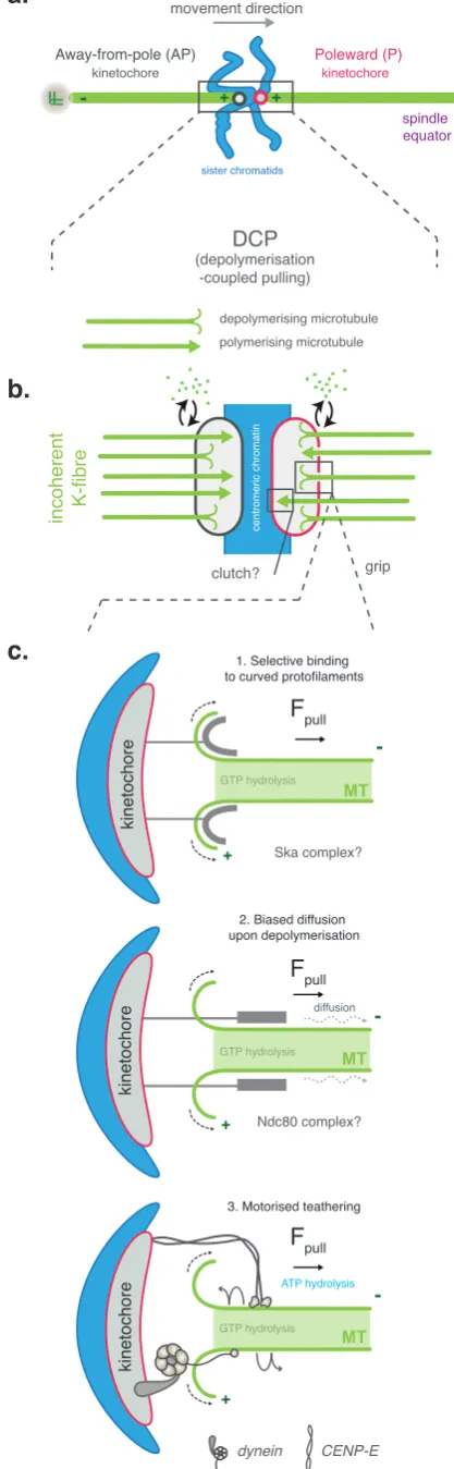
spindle equator -grip incoherent K-fibre movement direction + Fpull MT + Fpull MT kinetochore kinetochore Fpull MT ATP hydrolysis GTP hydrolysis GTP hydrolysis GTP hydrolysis clutch? centromeric chromatin diffusion Away-from-pole (AP) kinetochore sister chromatids Poleward (P) kinetochore DCP (depolymerisation -coupled pulling) Ska complex? Ndc80 complex? + + -depolymerising microtubule polymerising microtubule
a.
b.
c.
1. Selective binding to curved protofilaments2. Biased diffusion upon depolymerisation
[image:25.595.76.285.85.761.2]3. Motorised teathering
Figure 4: Mechanics of
depolymerisation-coupled pulling
(A) Chromosomes that form amphitelic attachments away from the spindle equator utilisedepolymerisation-coupled pulling (DCP) to congress. During this movement,
kinetochores are described as moving poleward (P) if they move towards their attached pole (shown in red), and away-from -the-pole (AP) if moving away from their attached pole (shown in dark grey). (B) The force required for DCP is generated by microtubule depolymerisation at the P kinetochore, which pulls the chromosome towards the equator. This implies that there is a polymerisation or depolymerisation bias between K-fibres that are attached to the P and AP kinetochores, with P K-fibres in a net depolymerising state and AP K-fibres in a net polymerising state. Owing to the incoherent nature of the K-fibre, the P kinetochore might selectively disengage from polymerising microtubules using a clutch-like mechanism. (C) Three potential mechanisms have been proposed for how kinetochores grip
are thought to peel away from the lattice in a ‘rams-horn’ like conformation
(Simon and Salmon, 1990). Early observations from grasshopper
spermatocytes suggested this bending could generate up to 7pN per
kinetochore-bound microtubule, with similar values later being calculated
from centromere stretching in yeast (Nicklas, 1988; Powers et al., 2009). In
contrast, experimentally conjugating glass beads to tubulin polymers in vitro
suggested this force might be significantly higher (65pN) (Grishchuk et al.,
2005). Regardless of its magnitude, this force acts in the P direction, and
could therefore be used to pull chromosomes (Fig 4a-c). Importantly, EM
studies in cells have subsequently identified ‘fibrils’ connecting the
centromere to curving protofilaments, providing the first evidence that
protofilament bending may indeed be linked to poleward kinetochore
movement in mammalian systems (McIntosh et al., 2008). However, the
molecular mediator(s) remains obscure. In contrast, work in yeast has
identified the oligomeric Dam1 complex, which forms a force-coupling ring
around microtubules in vitro (Grishchuk et al., 2008; Miranda et al., 2005;
Umbreit et al., 2014; Westermann et al., 2005), and is required for
microtubule attachment in vivo (Cheeseman et al., 2001; Umbreit et al.,
2014). Despite lacking any true vertebrate homologue, the spindle and
kinetochore associated (Ska) complex is suggested to be functionally
analogous to Dam1, and therefore represents the best
protofilament-coupling candidate in these organisms. This idea has emerged because the
and transduce this force to beads coated in the complex (Fig. 4c) (Schmidt
et al., 2012; Welburn et al., 2009). Moreover, the Ska complex binds
microtubules via the electrostatic interaction of several basic patches in
Ska1 and Ska3 with regions of tubulin that are accessible in straight and
curved lattice configurations (Abad et al., 2014; Abad et al., 2016). This
suggests that the complex can bind both structural states, unlike the end-on
attachment factor Ndc80, whose binding cleft is obscured by filament
curvature (Alushin et al., 2012). In agreement, biochemical analysis
showed that the Ska complex has an equal affinity for curved and straight
microtubules, whereas a truncated monomeric Ndc80 specifically binds the
latter (Schmidt et al., 2012). Nevertheless, the function of the Ska complex
in cells is poorly defined. While some report that small interfering (si) RNA
depletion of any Ska subunit causes a severe congression defect, which
was associated with reduced K-fiber stability (Chan et al., 2012; Gaitanos
et al., 2009; Schmidt et al., 2012; Welburn et al., 2009), others reported a
robust metaphase arrest with few misaligned kinetochore-pairs (Abad et al.,
2014; Hanisch et al., 2006; Jeyaprakash et al., 2012; Theis et al., 2009),
which lead to the downstream failure of sister cohesion and the
asynchronous separation of chromatids (known as cohesion fatigue)
(Sivakumar et al., 2014; Sivakumar et al., 2016). This phenotype was
probably misinterpreted in fixed images as the failure of congression
(Schmidt et al., 2012; Welburn et al., 2009), and may be associated with
anaphase onset (Sivakumar et al., 2014; Sivakumar et al., 2016). It must be
noted however, that more recent studies are far less deterministic in the
reporting of Ska depletion phenotypes (Abad et al., 2016; Redli et al.,
2016), with pleotropic observations being attributed to inter-experimental
variation in siRNA efficiency. Thus, a detailed analysis of kinetochore fates
in cells depleted of Ska via multiple techniques is required to shed more
light on the function of this complex.
In addition to Ska, CENP-F has been proposed to function as another link
between curved protofilaments and the centromere (Volkov et al., 2015).
CENP-F relocates from the nuclear envelope to the kinetochore during
mitosis (Liao H. et al., 1995), where it binds microtubules via two domains
located at opposite ends of the protein (Volkov et al., 2015). Biochemical
analysis suggested that these MTBDs couple the kinetochore to specific
microtubule structures. In this regard, the N-terminal MTBD displays a
preference for curved filaments over straight microtubules, while the
C-terminal MTBD does the opposite (Volkov et al., 2015). Nevertheless, both
regions track depolymerising microtubule plus-ends in vitro and can
transduce this force to beads, suggesting they may both contribute to
force-generation at end-on attached kinetochores (Volkov et al., 2015). In terms
of mechanics, the N-terminal MTBD is likely analogous to Ska, whereas the
C-terminal MTBD may utilize biased diffusion, as described for Ndc80 (see
below). The function of CENP-F at end-on attached kinetochores in cells is,
kinetochore, where it is implicated in the recruitment of dynein and CENP-E
(Bomont et al., 2005; Vergnolle and Taylor, 2007; Yang et al., 2005). These
loading dependencies may entirely account for the observed congression
defect in CENP-F depleted cells. However, by tracking the fate of unaligned
bi-oriented kinetochore-pairs located between the pole and metaphase
plate, another CENP-E loading protein, CENP-Q, was shown to have an
independent role in DCP (Bancroft et al., 2015). As such, the possibility
exists that CENP-F directly contributes to DCP, a hypothesis that can be
tested by following the fates of a specific kinetochore subgroup when
depleted of the protein.
In addition to structural modification, microtubule depolymerisation also
creates an energy gradient away from the plus-end. Proteins that diffuse
along microtubules can utilize this gradient to bias their diffusion along the
lattice during filament disassembly, and therefore generate a P force. This
is because it is more energetically favorable for these molecules to move to
adjacent tubulin dimers, away from the dissembling plus-end, as opposed
to detaching (Hill, 1985; Vladimirou et al., 2011) (Fig. 4c). This idea
underpinned the ‘Hill’s sleeve’ model, a theoretical proposal of how
kinetochores could track with dynamic microtubules (Hill, 1985). One
molecule that may function in a manner reminiscent of a ‘Hill’s sleeve’ is the
KMN component Ndc80/Hec1. Ndc80 binds microtubules via multiple
positively charged regions in both its unstructured N-terminal tail and
Miller et al., 2008; Tooley et al., 2011). Ndc80 forms a cooperative complex
with Nuf2, Spc24 and Spc25 at the kinetochore (the Ndc80 complex), and
loss of this complex in all tested systems leads to the abrogation of end-on
kinetochore-microtubule attachment (Cheeseman and Desai, 2008). This
does not implicate Ndc80 in force generation per se, as it may act solely as
an attachment factor. However, in vitro reconstitution of the entire complex
on dynamic microtubules demonstrated that diffusing ensembles could
track with depolymerising plus-ends, and that single complexes could
‘bounce’ off the tip back to the lattice without dissociating (Powers et al.,
2009). Moreover, beads coated with physiological levels of the complex
could persistently attach to depolymerising plus-ends and transduce force
(Powers et al., 2009). This suggested that the Ndc80 complex has
attachment independent functions, at least in vitro. It must be noted,
however, that a truncated monomeric Ndc80 complex (consisting of Ndc80
and Nuf2 microtubule binding and adjacent regions) does not display
similar behavior (Schmidt et al., 2012). Nevertheless, vertebrate
kinetochores contain at least nine Ndc80 complexes per
kinetochore-microtubule (Johnston et al., 2010), which can self-oligomerize into arrays
(Alushin et al., 2010). Therefore, the possibility exists that the complex
forms a higher order force-coupling structure that operates via biased
diffusion at end-on attached kinetochores
Interestingly, the protofilament binding and biased diffusion pathways are
the Ska complex to kinetochores (Zhang et al., 2012), and the Ska complex
can couple monomeric Ndc80 to depolymerising plus-ends in vitro (Schmidt
et al., 2012). Similarly, Dam1, the proposed Ska functional homologue in
yeast, also acts as a processivity factor for Ndc80 in vitro (Lampert et al.,
2010; Tien et al., 2010). Moreover, EM analysis of purified kinetochores
showed that Dam1 and Ndc80 physically interact, however, this appears to
be mediated by calponin homology (CH) domains and not the loop, as
suggested for the Ska-Ndc80 interaction (Gonen et al., 2012; Zhang et al.,
2012). Therefore, in both vertebrates and yeast, the Ndc80 complex
appears to be upstream in this pathway, and likely forms an initial end-on
attachment to microtubules while recruiting the Ska/Dam1 complex.
Together, these factors then link microtubule depolymerisation to
chromosome migration via protofilament binding and biased diffusion
pathways, respectively. CENP-F potentially contributes to both pathways
via the interaction of distinct MTBDs with specific regions on the tubulin
heterodimer. Nevertheless, analysis of mutants deficient in specific
functions will be required to truly determine if Ndc80 has an attachment
independent role in congression.
1.4.2 Coordinating microtubule dynamics
The persistent migration of a bi-oriented kinetochore-pair towards the
the P and AP K-fibres (Fig 4a,b), as one microtubule bundle must shorten
while the other elongates. Consistent with this notion, work in Xenopus
extracts using end-binding (EB) protein 1 as a marker of microtubule
polymerisation showed high levels of polymerisation at the AP K-fibre
relative to the P K-fibre (Tirnauer et al., 2002). In contrast, EM analysis of
K-fibres in Ptk1 and S2 cells demonstrated that the bundles are incoherent,
containing microtubules in both polymerising and depolymerising states
(VandenBeldt et al., 2006). A finding that was confirmed in recent work that
combined kinetochore tracking with EB3 labelling in HeLa cells (Armond et
al., 2015). As such, the P-kinetochore must prevent polymerising
microtubules from generating an antagonistic force that could impede
chromosome movement. Interestingly, it has been proposed that
kinetochores exert a pushing force during bipolar spindle assembly via the
prometaphase pathway in HeLa cells (Toso et al., 2009).
In this model, the authors suggest that kinetochores push against the
centrosomes via the incorporation of tubulin at K-fiber plus-ends during
microtubule flux (Toso et al., 2009). Therefore, end-on attached
kinetochores may indeed have force-generating states, as suggested by
early tracking experiments (Skibbens et al., 1993). However, both
kinetochore pushing and flux have largely been eliminated as contributors
to the congression of bi-oriented sister-pairs (Ganem et al., 2005;
Khodjakov and Rieder, 1996 ; Levesque and Compton, 2001), which raises
pulling states. Such a state switch could be facilitated by two changes; (1)
the alteration of kinetochore microtubule binding properties such that
depolymerising filaments are specifically engaged with (Fig 4b), and (2)
limiting microtubule polymerisation such that any pushing interactions are
avoided. As the latter mechanism could switch the entire K-fiber into a
depolymerising state, it would require the tight regulation of microtubule
dynamics to account for the observed incoherent nature. In this regard,
several plus-end tracking proteins and molecular motors are known to be
regulators of microtubule dynamics (Cross and McAinsh, 2014; Ferreira et
al., 2014), However, only the kinesin-8 Kif18A and kinesin-13 mitotic
centromere-associated kinesin (MCAK) have been shown to directly affect
the balance of microtubule dynamics within the K-fiber (Armond et al.,
2015).
Kif18A is a highly processive plus-end directed motor that accumulates at
the K-fibre plus-end, forming a comet-like gradient from this focus in a
manner dependent on K-fibre length (Stumpff et al., 2008; Stumpff et al.,
2012). Early studies suggested that Kif18A depletion lead to a severe
congression defect (Mayr et al., 2007). However, subsequent work that
employed high-resolution kinetochore tracking revealed that these
misaligned chromosomes are end-on attached, under tension and
oscillating (Stumpff et al., 2008; Stumpff et al., 2012). Thus, Kif18A
depleted kinetochores are qualitatively indistinguishable from metaphase
equator. This suggests that congression has been successful, and that
these kinetochores instead have a position sensing defect (see section
1.7). Nevertheless, it must be noted that these pseudo-metaphase
kinetochores in Kif18A depleted cells display numerous oscillation
abnormalities. Including an increase in velocity, (Jaqaman et al., 2010;
Stumpff et al., 2011; Stumpff et al., 2008; Stumpff et al., 2012), a decrease
in switching frequency, which, however, is disputed (Jaqaman et al., 2010;
Stumpff et al., 2008), and loss of spatially controlled directional switching
(Stumpff et al., 2012). These defects may explain the reported increase in
polymerisation bias between AP and P kinetochores, which is proposed to
dampen the AP resistive force that would otherwise slow the sister-pair
(Armond et al., 2015). In agreement, Kif18A is suggested to suppress
microtubule dynamics at the AP kinetochore and stimulate direction
switching with in vivo (Du et al., 2010; Stumpff et al., 2008; Stumpff et al.,
2012).
MCAK is a non-motile kinesin that functions as a microtubule depolymerase
(Hunter et al., 2003). In vitro, MCAK can couple to, and generate tension at,
both microtubule ends (Oguchi et al., 2011). However, no such behavior
has been described in vivo. MCAK has been shown to localise to both the
centromere and microtubule plus-ends, and demonstrates a bias to the
P-kinetochore during congression (Honnappa et al., 2009; Kline-Smith et al.,
2004). Abrogation of MCAK has been proposed to cause congression
this effect is inconsistently reported in the literature, particularly in studies
investigating the role of MCAK in metaphase kinetochore dynamics, where
congression appears to have completed (Armond et al., 2015; Jaqaman et
al., 2010). At metaphase, MCAK has been shown to regulate kinetochore
oscillation dynamics, with depleted sister-pairs displaying a decrease in
directional coordination and a reduction in velocity (Jaqaman et al., 2010;
Wordeman et al., 2007). This is consistent with the reported loss of
polymerisation bias between AP and P-sisters in MCAK depleted cells,
which implies that the K-fiber that is bound to the P kinetochore (which is
mediating DCP) has fewer depolymerising microtubules (Armond et al.,
2015). Thus, while MCAK is required for the regulation of kinetochore
movement during metaphase, its role in congression is currently unclear.
Some caution is necessary here as current models of kinetochore dynamics
are derived from the analysis of metaphase chromosomes. While many
concepts are likely conserved, these models may not be immediately
transferrable to congressing kinetochores. Indeed, prometaphase K-fibers
are less stable that those in metaphase cells (Kabeche and Compton,
2013), and the composition of unaligned and aligned kinetochores is known
to differ (Gudimchuk et al., 2013; Kline-Smith et al., 2004; Schmidt et al.,
2010). Moreover, periods of persistent movement are considerably longer
during congression (~2min compared to 10-60 secs in metaphase
(Jaqaman et al., 2010; Khodjakov et al., 1999)). Therefore, a detailed
congressing kinetochores, with emphasis on differentiating AP and P
states, is required to fully decipher the mechanics that underpin DCP.
1.5
Congression in the absence of end-on pulling
1.5.1 Lateral sliding
The formation a bi-oriented attachment was originally thought to be an
essential prerequisite for congression. On the contrary, congression can
also occur before bi-orientation, via the motor dependent sliding of
kinetochores along the microtubule sidewall (Fig 5). Chromosomes that are
located in the spindle periphery following NEB initially migrate to their
proximal pole prior to ejection towards the spindle equator. This movement
is driven by the kinetochore-bound dynein, a minus-end directed motor that
steps towards the pole (Barisic et al., 2014; Li et al., 2007; Savoian et al.,
2000; Yang et al., 2007) (Fig. 5, Step 1). Subsequent equatorially directed
motion is driven CENP-E, a high processive plus-end directed motor that is
enriched at polar chromosomes (Gudimchuk et al., 2013; Kapoor et al.,
2006; Kim et al., 2008; Wood et al., 1997) (Fig. 5, Steps 2 and 3). The
lateral sliding of chromosomes was first demonstrated in a study that used
single kinetochore tracking and correlative EM to analyse the behaviour
and attachment state of congressing sister-pairs in PtK1 cells. Here,
Slide: CENP-E Slide:
Dynein (HSET?)
spindle equator
-3
4
1
5
+
-F
slideMT
Poleward kinetochore (P) lateral attachment
Away-from-pole (AP) kinetochore
kinetochore movement direction
+
+ +
ATP hydrolysis
2
[image:37.595.132.468.122.546.2]dynein CENP-E
Figure 5: Congression of laterall attached kinetochores
alongside the mature K-fibre of an aligned bi-oriented chromosome, a
movement that was facilitated by a force-generating lateral interaction
between the P-kinetochore and K-fibre (Kapoor et al., 2006) (Fig. 5, Chr 2).
This behaviour was abolished by CENP-E depletion, establishing that
CENP-E drives a bi-orientation independent congression mechanism
(Kapoor et al., 2006). Further validation of this model came from studies
that specifically enriched for the lateral sliding pathway. Here, DCP was
prevented by abrogating end-on attachment with depletion of the Ndc80
complex component Nuf2 (Cai et al., 2009). Remarkably, CENP-E
mediated lateral sliding could align all chromosomes in 50% of cells when
the minus-end directed motor HSET was also depleted (Cai et al., 2009).
Similar observations were made when the major end-on attachment factor
Ndc80 was codepleted with MCAK (Iemura and Tanaka, 2015).
Interestingly, these studies highlighted that MCAK and HSET depletion
could suppress an unknown P force, and allow for CENP-E mediated
congression in the absence of end-on attachment, but how? The current
idea is that this effect is indirect, as HSET and MCAK both negatively
regulate the stability of microtubules required for CENP-E sliding toward the
equator (Iemura and Tanaka, 2015).
Somewhat unexpectedly, CENP-E has been implicated in the DCP
mechanism in vivo. In an experiment designed to simulate DCP, where
end-on attached sisters are transported by depolymerising astral
inhibition was found to perturb microtubule tracking (Gudimchuk et al.,
2013). Moreover, CENP-E was shown to track with polymerising and
depolymerising microtubules in vitro, and can transduce the force from
filament disassembly to beads (Gudimchuk et al., 2013). Together, these
data provided compelling evidence that the motor contributes to DCP (Fig
3c). However, 80% of bi-oriented kinetochore pairs in a bipolar spindle
successfully congressed when depleted of CENP-E (Bancroft et al., 2015),
and CENP-E is not required for the oscillation of aligned sister-pairs
(Jaqaman et al., 2010). Nevertheless, microtubule dynamics at aligned
kinetochore-pairs are perturbed when CENP-E is inhibited or depleted
(Maffini et al., 2009). It must be noted that CENP-E is significantly reduced
at aligned sisters when compared to polar kinetochores. As such, the
primary function of CENP-E appears to be as a lateral sliding mediator that
congresses peripheral chromosomes (Barisic et al., 2014).
1.5.2 The polar ejection force
The existence of a polar ejection force (PEF) was proposed by Darlington in
1937. This idea formed the basis of his ‘polar repulsion’ model of
congression (Darlington, 1937), which stated that centromeres are repelled
from the poles with a force inversely proportional to their proximity
(Darlington, 1937). While failing to account for congression (Levesque and
cutting experiments (Rieder et al., 1986). Here, an acentric chromosome
fragment was shown to be actively transported away from its proximal pole
(Rieder et al., 1986). This behaviour was abolished by the addition of
nocodazole, suggesting that microtubules were critical for PEF generation
(Ault et al., 1991). Importantly, these studies also showed that the PEF was
exerted along chromosomes arms, as oppose to the centromere as
originally predicted (Darlington, 1937). Consistent with this, both in vivo and
in vitro suites later showed that plus-end directed chromosome arm
associated kinesins (chromokinesins) Kif4a and Kid create the PEF
(Antonio et al., 2000; Bieling et al., 2010; Brouhard and Hunt, 2005;
Funabiki and Murray, 2000; Levesque and Compton, 2001; Yajima et al.,
2003). Thus, the PEF is an AP force generated by the plus-end directed
walking of chromatin-tethered kinesin along microtubules. As microtubule
concentration is proportional to proximity to the pole, the strength of the
PEF is inversely proportional to distance from the pole (Cane et al., 2013).
In terms of congression, the individual depletion of Kif4a or Kid has little
effect. In contrast, their co-depletion results in significant alignment defects,
suggesting these motors may have independent and/or coordinated
functions (Wandke et al., 2012). In this regard, Kid, the principal PEF
generator, is dispensable for chromosome alignment, but has a role in the
control of aligned kinetochore dynamics and kinetochore position sensing
(Levesque and Compton, 2001; Stumpff et al., 2012; Wandke et al., 2012).
increases the rate of directional switching at, aligned kinetochore-pairs. In
contrast, the function of Kif4a is less well characterised. It has been
suggested to antagonise Kid by reducing microtubule dynamicity in early
mitosis via the suppression of polymerisation, a finding that suggests it may
have a PEF independent function (Wandke et al., 2012). Indeed, this may
involve the regulation of antiparallel microtubule overlaps, a well
documented function of Kif4a in cytokinesis (Nunes Bastos et al., 2013).
Nevertheless, several studies have implicated Kif4a and kinesin-4 motors in
PEF generation, although more work is necessary to tease out the specific
contribution (Antonio et al., 2000; Bieling et al., 2010; Brouhard and Hunt,
2005; Funabiki and Murray, 2000; Levesque and Compton, 2001; Yajima et
al., 2003).
1.6
Coupling congression with position sensing in the spindle
How do kinetochores sense their position in the spindle and feedback this
information to regulate force-generating mechanisms? Early ideas focused
on coordinated kinetochore states in response to tension; however, such a
model failed to consider any integration of spatial information and
kinetochore behaviour during congression could be recapitulated with no
external modulation of behaviour (Khodjakov et al., 1999), i.e. congression
could be achieved with no kinetochore mediated position-sensing.
DCP and lateral sliding mechanisms toward the equator (Fig 6). One such
mechanism is the opposing regulation of microtubule dynamics at
bi-oriented kinetochore pairs by the motor proteins MCAK and Kif18A (see
section 1.5.2). Here, MCAK accumulates at the P-kinetochore (Kline-Smith
et al., 2004), enhancing microtubule depolymerisation at this force
generating interface, which causes chromosomes to move at an increased
velocity toward the equator (Jaqaman et al., 2010) (Fig 6a). Once at the
metaphase plate, MCAK in unloaded from this region and accumulates in
the inner-centromere (Kline-Smith et al., 2004), where it generates a
symmetrical P-force that favours positional equilibrium. Simultaneously, the
length dependent accumulation of Kif18A at the AP attached K-fibre
promotes an AP-to-P state switch at chromosomes moving away from the
equator (Fig 6b) (Stumpff et al., 2012). Mechanistically, the high Kif18A
concentration at the AP K-fibre acts to suppress microtubule dynamics until
the GTP cap is eventually compromised, which leads to microtubule
depolymerisation and directional switching. Once aligned at the equator,
the Kif18A concentration gradient at each sister is roughly identical,
favouring the maintenance of position via quasi-periodic oscillation. Thus,
Kif18A senses K-fiber length whilst MCAK senses kinetochore position.
Recently, the importance of this Kif18A positioning mechanism and the
requirement for chromosome congression have been questioned
(Czechanski et al., 2015). Here, mouse embryonic fibroblasts (MEF)
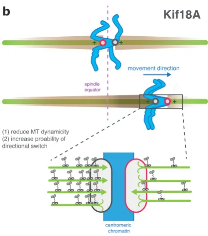
a
Figure 6: Biased congression towrds the spindle equator
b
c
Poleward (P) Away-from-pole (AP) kinetochore
Kif18A accumulation
Polymerising Depolymerising microtubule Kif18A MCAK PEF magnitude Kid + + movement direction 1 2 + + centromeric chromatin
(1) reduce MT dynamicity (2) increase proability of directional switch
Kif18A
+
(1) Increase kinetochore tension (2) Stimulate directional switch
movement direction Arm associated chromatin
PEF (Kid)
F
slide + + movement direction 1 2 + +(1) Increase MT depolymerisation (2) Increase DCP drive force
MCAK
centromeric chromatin spindle equator spindle equator spindle equator(A) Role of MACK in biased congression. At aligned kinetochore pairs (1), the microtubule depolymerase MCAK localises to the inner-centromere. In contrast, at unaligned kinetochore pairs undergoing congression (2) MCAK displays a bias to the P kinetochore, suggesting that it might augment the DCP-mediated drive force and enhance migration towards the spindle equator. (B) Kif18A forms a concentration gradient
along the K-fibre (illustrated by the brown ‘cloud’)
and accumulates at the outer-kinetochore. Here, it acts to suppress microtubule dynamics and
(Kif18Agcd2) successfully progressed through mitosis, entering anaphase
with seemingly unaligned chromosomes (Czechanski et al., 2015).
However, as previously mentioned, kinetochore tracking experiments have
revealed that sister-pairs form bioriented attachments and oscillate with
perturbed dynamics about the spindle equator in Kif18A depleted cells
(Stumpff et al., 2008). This leads to the formation of a broad
‘pseudo-metaphase plate’, and suggests that congression has completed. Further
work is needed to determine the impact of this pseudo-plate affects the
fidelity of mitosis in this cell type, as it is known to be highly detrimental in
HeLa cells (Czechanski et al., 2015).
An alternative idea is that chemical gradients throughout the spindle guide
chromosome movement. In this regard, Plk1 and RanGTP concentration
gradients have been shown to control spindle position (Kiyomitsu and
Cheeseman, 2012), and Aurora A forms a gradient from the pole that is
required for attachment error correction (Ye et al., 2015). Abrogation of the
RanGTP gradient that forms around aligned chromosomes using a
dominant negative RanT24N mutant had no effect on congression (Barisic
et al., 2015). In contrast, polar Aurora A is suggested to bias the lateral
sliding of chromosomes toward the equator via direct regulation of CENP-E
(Kim et al, 2010). Here, the phosphorylation of CENP-E at the pole is
proposed to abrogate its interaction with PP1, a factor that is required for
stable CENP-E-microtubule binding. This is suggested to bias congression
walks on stabilised K-fibre microtubules that are oriented toward the
equator (Kim et al., 2010). It must be noted, however, that while CENP-E
mediated congression requires some form of stabilised microtubules, it is
not dependent upon K-fibres (Cai et al., 2009; Iemura and Tanaka, 2015),
and a new model of directionality based on the ‘tubulin code’ has been
proposed (see below) (Barisic et al., 2015).
As already discussed, the PEF, a decaying AP force field that emanates
from each aster, is symmetrical about the spindle equator and could
therefore bias chromosome migration toward this region (Cane et al.,
2013). The principal PEF generator, Kid, has been shown to induce a
position dependent increase in tension at kinetochores approaching the
pole, which promotes directional switching (Fig. 6c) (Stumpff et al., 2012).
However, its depletion has no effect on congression (Levesque and
Compton, 2001), probably because Kif18A mediates the dominant position
sensing mechanism and can compensate for its loss (Stumpff et al., 2012).
Recently, the ‘tubulin code’ has been suggested to bias CENP-E stepping
toward the equator, and therefore impose directionality on the lateral sliding
mechanism (Barisic et al., 2015). Here, Barisic and colleagues found that
microtubules oriented toward the equator are enriched with detyrosinated
tubulin. Abrogation of this modification resulted in aberrant chromosome
movements that were dependent upon E. Moreover, in vitro,
CENP-E was observed to be more processive and to carry higher loads on
CENP-E transports pole proximal chromosomes preferentially on
detyrosinated microtubule tracks, which are orientated toward the spindle
equator (Barisic et al., 2015).
1.7
Coupling congression with attachment regulation and
signalling
1.7.1 Correcting erroneous kinetochore-microtubule attachments
Despite geometric constraints favouring sister-kinetochores forming
bi-oriented attachments (Cimini et al., 2003; Loncarek et al., 2007), errors in
microtubule attachment are frequenting during prometaphase (Ault and
Rieder, 1992). If left uncorrected, these errors would lead to segregation
defects (Cimini et al., 2001). As such, kinetochores employ a correction
mechanism that selectively destabilises erroneous attachments while
promoting bi-orientation, ensuring that all kinetochores are in an anaphase
compatible configuration prior to segregation. The current model, termed
the “spatial separation” model, dictates that attachment stability is directly
dependent upon tension across the centromere. This is based on classic
observations in grasshopper spermatocytes, which showed that the
normally unstable unipolar attachments could be stabilised by the
application of tension (Nicklas and Koch, 1969). In molecular terms,
yeast, which identified the kinase Ipl1 (Chan and Botstein, 1993). Critically,
it was demonstrated that Ipl1 promoted attachment turnover in the absence
of tension (Tanaka et al., 2002). The vertebrate homologue of Ipl1 is Aurora
B, a ser/thr kinase that is a member of the CPC located at the
inner-centromere (see appendix 1 for a structural description of the CPC) (Adams
et al., 2000). In agreement with models of Ipl1, small molecule inhibition of
Aurora B leads to the stabilisation of incorrect attachments, which are
subsequently corrected upon inhibitor washout (Hauf et al., 2003; Lampson
et al., 2004). Given the centromeric localisation of Aurora B, it was
suggested that substrate proximity to the kinase was critical for attachment
correction, and this was related to the tension. Indeed, the formation of a
bi-oriented attachment is thought to exert enough tension to physically
separate Aurora B from its substrates in the outer-kinetochore. As
erroneous attachments fail to generate sufficient tension, these substrates
are in close proximity to the kinase and can therefore be phosphorylated
(Andrews et al., 2004; Cimini et al., 2006; Tanaka et al., 2002). This idea is
supported by two key lines of experimental evidence; (1) A FRET based
biosensor that reports Aurora B phosphorylation is constitutively
phosphorylated when positioned close to the centromere, irrespectively of
tension, however, if positioned at the outer-kinetochore it is
dephosphorylated when under tension (Welburn et al., 2010). It must be
noted that similar behaviour is observed for endogenous Aurora B
closer to the outer-kinetochore, and therefore forcing constitutive
phosphorylation within this region, microtubule attachments can be
destabilised in a tension independent manner (Liu et al., 2009). A key
prediction of this model is that Aurora B phosphorylation of
outer-kinetochore proteins must alter their microtubule affinity, which in turn
allows for the correction of erroneous attachments. Indeed, experimental
evidence suggests that this is achieved via two mechanisms. First, by
negatively regulating microtubule binding regions within the kinetochore.
Specifically, this involves the phosphorylation of key attachment proteins
Ndc80 and Knl1, which antagonises their electrostatic interaction with
tubulin and therefore decreases microtubule affinity (Cheeseman et al.,
2006; Ciferri et al., 2008; Guimaraes et al., 2008; Miller et al., 2008;
Welburn et al., 2010; Zaytsev et al., 2015). Second, by regulating the
composition of the kinetochore. In this regard, Aurora B phosphorylation of
the microtubule binding Ska and ASTRIN/SKAP complexes antagonises
their recruitment (Chan et al., 2012; Schmidt et al., 2010). Thus, Aurora B
targets a myriad of microtubule binding proteins, which causes a global
reduction in microtubule affinity at low-tension kinetochores.
The spatial separation model is not, however, without caveats. At
low-tension kinetochores, the N-terminus of Ndc80, which contains the principal
error correction target site, is >100nm from Aurora B (Smith et al., 2016).
Therefore it is unclear how the kinase physically interacts with this
centromere. This is based on findings that show the activation of Aurora B
by INCENP occurs in trans, and this concentrates active kinase at the
centromere (Bishop and Schumacher, 2002; Honda et al., 2003; Kelly et
al., 2007; Sessa et al., 2005). This pool then turns over with a t1/2 of ~50s
(Ahonen et al., 2009; Murata-Hori and Wang, 2002), releasing active kinase
that diffuses away from the centromere that is eventually deactivated by
cytoplasmic phosphatases (Kelly et al., 2007). Thus generating a
phospho-gradient. However, it is still unclear if a soluble pool of diffusing kinase can
act on this length scale. Moreover, while syntelically attached kinetochores
(both sisters bound to the same pole) fail to generate tension, merotelic
attachments (one sister in a bi-oriented pair bound to both poles; see Fig 3)
are under tension (Gregan et al., 2011) and are therefore invisible to the
correction machinery. Aurora B is required for the correction of merotelic
attachments, as its inhibition leads to an increase in these errors (Cimini et
al., 2006; Knowlton et al., 2006), however, the mechanism of correction is
less clear. It has been suggested that the merotelically attached sister is
deformed, with the second (incorrect) attachment site projecting towards
the inner-centromere (Cimini et al., 2004). This could bring it in close
proximity to Aurora B, leading to its destabilisation. Moreover, the CPC has
been shown to enrich at merotelically attached sister-pairs (Knowlton et al.,
2006), suggesting there may be an additional detection mechanism that
acts upstream of Aurora B that concentrates it at these errors. Indeed,
mechanism, an idea that is supported by several observations; (1)
metaphase kinetochores display a phenomenon termed ‘breathing’, where
the inter-sister separation oscillates (Jaqaman et al., 2010). During this
oscillation, there are periods of low tension that are comparable to
unattached or erroneously attached kinetochore-pairs. Importantly however,
the bi-oriented sister-pair at low tension does not appear to activate error
correction. (2) Metaphase kinetochores treated with taxol (to induce
centromeric relaxation) do not display a significant increase in
outer-kinetochore phosphorylation by Aurora B (DeLuca et al., 2011), and (3) the
level of Ndc80 dephosphorylation between prometaphase and metaphase
cannot be explained by changes in intra-kinetochore distance. As moving
Ndc80 significantly further from Aurora B via the induction of kinetochore
‘hyper-stretch’ has little effect on its phosphorylation state (Suzuki et al.,
2014). Therefore, once a bi-oriented attachment has formed, the dynamics
of outer-kinetochore phosphorylation, and the tension independent
maintenance of attachment, cannot be explained by changes in subunit
separation at the inter- or intra-kinetochore level. This suggests that error
correction is antagonised or abrogated after bi-orientation by a biochemical
event. This may involve Knl1 mediated recruitment of PP1 (DeLuca et al.,
2011; Liu et al., 2010), which creates a stabilising region of
dephosphorylation within the outer-kinetochore. Therefore, while tension is
contributory to correction, particularly at syntelic attachments, additional