1
Profiles of visuospatial memory dysfunction in opioid exposed and
dependent populations.
Baldacchino, A
1., Tolomeo, S
2., Balfour, D.J
2., Matthews, K
21
Division of Population and Behavioural Science, School of Medicine, St Andrews University, St Andrews, Fife, United Kingdom
2
School of Medicine (Neuroscience), Ninewells Hospital & Medical School, University of Dundee, Dundee, Tayside, United Kingdom
Corresponding Author:
Professor Alex Baldacchino
Division of Population and Behavioural Science School of Medicine
North Haugh
St Andrews University St Andrews, Fife KY16 9TF United Kingdom
2
Abstract
Background:Chronic opioid exposure is common world-wide, but behavioural performance remain
under investigated. This study aimed to investigate visuospatial memory performance in opioid exposed and dependent clinical populations and its associations with measures of intelligence and cognitive impulsivity.
Methods: We recruited 109 participants: i) patients with a history of opioid dependence due to chronic heroin use (n=24), ii) heroin users stabilised on methadone maintenance treatment (n=29), iii) participants with a history of chronic pain and prescribed tramadol and codeine (n=28) and iv) healthy controls (n=28). The neuropsychological tasks from the Cambridge Neuropsychological Test Automated Battery (CANTAB) included the Delayed Matching to Sample (DMS), Pattern Recognition Memory (PRM), Spatial Recognition Memory (SRM), Paired Associate Learning (PAL), Spatial Span Task (SSP), Spatial Working Memory (SWM) and Cambridge Gambling Task (CGT). Pre-morbid general intelligence was assessed using the National Adult Reading Test (NART).
Results: As hypothesised this study identified differential effects of chronic heroin and methadone exposures on neuropsychological measures of visuospatial memory (p<0.01) that were independent of injecting behaviour and dependence status. The study also identified an improvement in DMS performance (specifically at longer delays) when the methadone group was compared to the heroin group and also when the heroin group was stabilised onto methadone.
Results identified differential effects of chronic heroin and methadone exposures on various neuropsychological measures of visuospatial memory independently from addiction severity measures, such as injecting behaviour and dependence status.
4479 Words, 79 References, 5 Tables, 1 Supplementary Table, 2 Figures
3
Working memory is a limited capacity cognitive system that functions to hold information in an active manner to facilitate the performance of complex cognitive tasks (Miyake & Shah, 1999). Such tasks include, for example, language comprehension, learning, abstract thinking (Twamley et al.
2006), problem-solving (Westen, 2006), understanding the meaning of complex texts and planning verbal communications (Zihl et al. 1979). Working memory (WM) is limited in both capacity and duration and is often used synonymously, but inaccurately, with the term “short term memory” (Westen, 2006). Baddeley & Hitch (1974) expanded upon this WM concept and proposed a tripartite working memory model that includes a central executiveand two ‘slave systems’; the phonological loop and the visuospatial store. The visuospatial store is further broken down into (1) visual memory information that includes dimensions such as colour and shape and (2) spatial memory information that includes capacity to understand, reason and to remember
the spatial relations among objects or space. (Baddeley & Logie, 1999; Mammarella et al. 2008). There is accruing evidence that the two components of visuospatial memory are selectively engaged and/or processed by distinct brain regions and neuropsychological functions (Della Sala et al. 1999; Passolunghi et al. 2010, Bormann et al.2015; Erikson et al. 2015).
There are a few brain imaging studies on visuospatial memory impairments among drug users. Kubler and colleagues reported that cocaine dependent individuals were impaired in visuospatial working memory. These were associated with prefrontal, cingulate and striatal regions (Kubler et al.
2005). In another study opiate dependent individuals were impaired in working memory-related
brain areas (Bach et al, 2012).
Hyman and colleagues have conceptualised the behavioural phenomena typically described as ‘addiction’ to a “pathological usurpation of the neural mechanisms of learning and memory that under normal circumstances serve to shape survival behaviours related to the pursuit of rewards and the cues that predict them” (Hyman., 2005; Hyman et al. 2006).
4
impairments in opioid exposed groups are confounded by, for example, comorbid personality disorders (Prosser et al. 2008), anxiety and/or depression (Henry et al. 2012), past and present medical conditions, neurological disorders and history of head trauma and non -fatal overdose (anoxic) episodes(Prosser et al. 2008; Rounsaville et al. 1982; Shmygalev et al. 2011; Specka et al.
2000). Cognitive function may also be influenced by the global sedative effects of opioid drugs, sub-acute responses to the drugs or the presence of untreated withdrawal states (Baldacchino et al.
2016) at time of testing. Table 1 summarises studies that have recorded significant impairments in visuospatial memory in chronic opioid using populations.
INSERT TABLE 1 here
The present study aimed to extend our understanding of neurocognitive performance in dependent and non-dependent opioid users, focusing on visuospatial memory function. Employing an
ambispective cohort design, we tested representative samples of male participants exposed to illicit and therapeutic opioid drugs and matched, non-substance using, healthy controls. Specifically, the study aimed to determine if performance on tasks measuring visuospatial memory, especially delayed memory performance, which is very sensitive for the varying of ‘executive demands’,was affected by (1) the type of the opioid exposure (methadone vs. heroin) at different stages of
treatment seeking, (2) the context (opioids prescribed for pain control compared with illicit opioids) and (3) the presence or absence of syndromal opioid dependence (opioid dependent compared to non–opioid users) and (4) administration route – injectionstatus (opioid dependent and injecting compared with dependent and non-injecting participants). We have previously identified and reported differential effects of heroin, methadone and prescribed analgesic medication on neurocognitive measures of impulsivity (Baldacchino et al. 2014) from the same study cohort.
Method
Participants
Ethical permission for the conduct of this study was provided by the East of Scotland Research Ethics Service (REC reference number: 06/S1401/32). A full description of the participants can be found in
5
and neurodevelopmental disorders, borderline or psychopathic personality disorders, head injuries; individuals with a lifetime history of non-fatal overdose episodes requiring medical attention (e.g. ambulance call out, CPR), co-occurring benzodiazepine, psychostimulant and alcohol dependence. All participants were screened by an experienced clinician (AB) for acute opioid and opioid withdrawal symptoms prior to the neuropsychological testing.
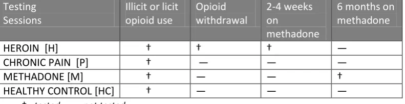
The Heroin group (H) (N=24) were ‘first time’ referrals to a structured Methadone Maintenance Treatment (MMT) programme. The Methadone group (M) (N=29) were established and stable participants in a MMT programme with objective confirmation of absence of illicit drug use for more than six months. Eighteen of the twenty-nine MMT group participants who showed objective continuing clinical stability were retested six months after baseline testing. All recruits making up the H and MMT cohorts presented initially with opioid dependence and reported a history of more than three years of continuous and daily illicit opioid use.
Heroin participants (H) performed repeated neuropsychological testing during a single blinded procedure that permitted the objective observation of participants (a) 3-5 hours after their last illicit heroin administration to minimise the confounding cognitive effects of acute intoxication; (b) 10-15 hours after the last heroin dose in a state of controlled opioid withdrawal and subsequently (c) following more than two weeks on a stable dose of MMT. Clinically this is known as tolerance testing which is a single-blinded procedure that permitted the objective observation of individuals during stages of acute intoxication, withdrawal and subsequent stabilisation on a fixed dose methadone within a period of 7-14 days (Baldacchino, 2011).
The two opioid dependent groups (H and M groups) were matched for lifetime drug use history, morphine equivalent dosages and other drug use (including tobacco smoking) history 30 days prior to baseline testing. The CANTAB neuropsychological tests presented here refer to the standard tests selected from the batteries used at baseline testing. Where available, parallel versions of the tasks were used with the same participant to minimise practice effects.
6
tramadol, codeine, or both, for moderate chronic pain. Both P and HC groups were tested only once (Table 2).
INSERT TABLE 2 here
Instruments
(A) Clinical: All subjects were screened using the MINI Plus v 5.0 (Sheehan et al. 1998), Maudsley Addiction Profile (MAP) (Marsden et al. 1998), and Fagerström Test for Nicotine Dependence (FTND) (Fagerström & Schneider, 1989). Urine samples were collected from all participants to confirm their history of recent opioid intake and to confirm the absence of any other illicit drugs throughout the study period. The Clinical Opiate Withdrawal Scale (COWS) (Wesson & Ling, 2003), quantified the level of opioid withdrawal in the heroin group. A senior research nurse and an experienced clinician conducted the assessments. Both were clinically trained. No participants had HIV or AIDS or other medical comorbidities that could affect cognitive functions.
(B) Cognitive: The neuropsychological tasks from the Cambridge Neuropsychological Test Automated Battery (CANTAB) (Robbins et al. 1994) were selected on the basis of their known sensitivity to detect impairments in (a) visual [Delayed Matching to Sample (DMS), Pattern Recognition Memory (PRM), Spatial Recognition Memory (SRM) and Paired Associate Learning (PAL) and (b) spatial
[Spatial Span Task (SSP), and Spatial Working Memory (SWM) memory performance. Pre-morbid general intelligence was assessed using the National Adult Reading Test (NART) (Nelson, 1982)
(Supplementary Table 1).
Data Analysis
7
Mann Whitney U tests were also performed to examine: (a) sociodemographic characteristics for participants in the H group, comparing those who experienced the lowest (n=8) and highest (n=8) scores on the COWS. Similarly, the same test was used to determine if there were differences between the H group of participants who were tested at baseline and those who were followed up and tested in withdrawal and, subsequently, on methadone. (b) sociodemographic characteristics for participants in the MMT group, comparing those tested at baseline (n=29) and those followed up after six months (n=18). (c) sociodemographic characteristics for participants in the H and M groups comparing those with a lifetime subjective history of injecting illicit opioids (n=41) and those with no history of injecting (n=11). A high COWS score was defined as a score between 18-25; a low COWS score was defined as a score 8-14.
The data were first analysed using an omnibus test to determine if significant differences existed between the groups. If the test revealed significance, appropriate pair wise comparisons were performed. In order to control for family wise error, post hoc Bonferroni corrected pairwise comparisons was used (Fields, 2009). P values <0.01 were considered significant. This minimised the effects of multiple comparisons, subgroup analyses and/or repeated measures as we were considering a family of statistical inferences simultaneously (Sainani, 2009). Those reported as between p<0.05 and p>0.01 are presented as non-significant trends when they are considered relevant to substantiate the interpretation of other significant results.
ANCOVA was used to test for group differences with respect to visuospatial memory performance measures. The PRM and SWM outcomes did not meet assumptions of normality and were square root transformed prior to performing the ANCOVA. However, PAL outcomes were log10 transformed prior to performing the ANCOVA. For incremental levels of difficulty within the testing sessions, the
within‐subject factor DIFFICULTY was introduced, (e.g. SWM (between/within search errors), SSP (span length between 1-9), DMS (0, 4 and 12 second delays), and PAL (1, 2, 3, 6, or 8 shapes)). Homogeneity of variance was assessed using the Mauchly Sphericity Test. Where data sets significantly (p<0.05) violated this requirement, the Greenhouse Geisser Epsilon (^ε) correction parameter for degrees of freedom was used to calculate a more conservative p value for each F ratio.
8
the H and M groups, however we could not draw any particular conclusion about the exposure of the opiate use. In addition, we used DEPENDENCE as a proxy clinical measure of severity without any biological basis.
Similarly, repeated measures ANCOVA was used to evaluate all neuropsychological performance measures between the H group at baseline, in controlled opioid withdrawal and subsequently when stabilised on methadone with presumed opioid receptor occupancy state as a within-subjects factor.
Similarly, repeated measures ANCOVA was performed for the M group at baseline and at six months follow up with duration as a within-subjects factor.
All analyses were conducted using SPSS for Windows (v.18, SPSS Inc. Chicago, Ill.).
Results
Demographic characteristics
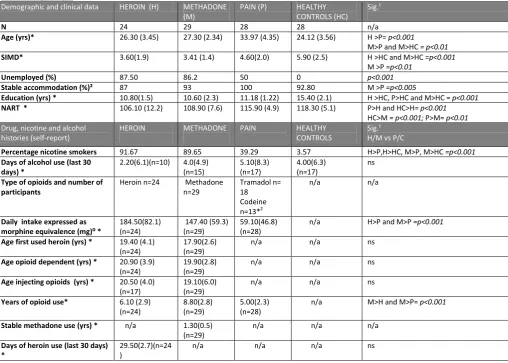
A description of demographics, drug use and smoking variables for the four groups is presented in
Table 3. The H and M groups differed from the P and HC groups with respect to several clinical characteristics. Opioid dependent participants started to drink alcohol approximately two years earlier than the other groups. The mean morphine equivalent daily dose for the P group was significantly lower (59.1 mg) than the H and M groups (165.9mg) (p<0.001).
INSERT TABLE 3 here
When comparing high against low scores for COWS in the H group, there were no differences between age (p=0.88), SIMD score (p=0.75), years in education (p=0.38), years when starting using alcohol (p=0.07), alcohol amount used in last month (p=0.87) or current level of nicotine
dependence (Fagerström scores) (p=0.96)
Similarly, there were no group differences identified on these measures when comparing H group tested at baseline and those retested either through the tolerance testing protocol six months later when taking methadone. There were no significant differences with demographic and drug use characteristics between injecting participants (n=43) and non-injecting participants (n=10). However, NART scores were significantly higher (p<0.01) in the injecting group.
9
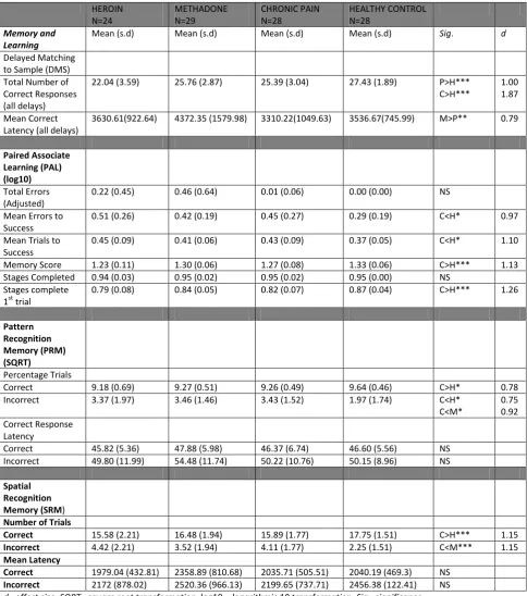
Performance on DMS
There was a significant effect on the percentage of correct responses [GROUP F (3,100) =10.3,
p<0.001]. There were no significant performance differences between groupswith respect to the simultaneous matching condition. Post hoc Bonferroni comparisons, however, showed participants from the H group made significantly more errors than (a) the HC group at the 0 (p<0.005), 4 (p<0.001) and 12 second (p<0.001) delay stages, (b) the P group for the 4 (p<0.01) and 12 (p<0.005) second delay stages and (c) the M group for 0 (p<0.005), 4 (p<0.005) and 12 (p<0.001) second delay stages (Figure 1). In summary the H group exhibited significant delay-dependent memory impairment when compared with the comparison and control groups.
INSERT FIGURE 1 here
Performance on PRM, SRM and PAL
There were no significant GROUP effects on the number of correct trials [F<1] and mean response latencies [F<1] on the PAL and PRM tests. There was a non-significant GROUP trend on the total number of correct trials [F (3,102) = 3.6, p=0.02] on the SRM only.
Spatial memory
Performance on SSP
There was a significant GROUP [F (3,102) =16.8, p<0.001] effect for total errors. Post hoc Bonferroni
comparisons showed that the participants from the H group significantly made more errors compared to the M (p<0.001, d=1.25) and HC (p<0.005, d=1.14) groups (Figure 2)
.
The total error score for the P group lay between those of the H, M and HC groups and did not differ significantly from any of the other three groups (p=1.0).There was also a significant GROUP [F (3,101)=3.7, p<0.01] effect for span length with post hoc Bonferroni comparisons showing the M group was significantly less able to recall successfully the longest sequence compared to HC group (p<0.01, d-1.17). The span length for the H (p=.41) and the P (p=.21) groups lay between those of the M and HC groups and did not differ significantly from any of the other groups (Figure 2).
INSERT FIGURE 2 here
10
There was a non-significant GROUPtrend for: total mean errors [F(3,102)=3.2, p=0.03] and strategy score [F(3,102)=2.9,p=0.04]
INSERT TABLE 4 here
Chronic opioid dependence or injecting status and visuospatial memory performance
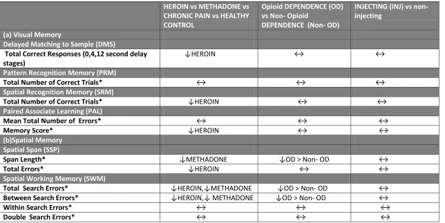
There were no significant effects for either of the factors DEPENDENCE or INJECTING STATUS on any of the DMS, PRM, SRM, and PAL outcome measures.
However, there were significant DEPENDENCE effects for total errors [F (3,104)=6.5, p<0.01] on the SWM, but with no significant DEPENDENCE effects on the strategy score [F(1,104)=4.8,p=0.03]. There was a significant DEPENDENCE status and task difficulty interaction on the SWM test for total errors [F (3,133.75)=6.2, p<0.01]. Analysis using INJECTING status failed to reveal any significant effects or interactions on any SWM outcomes.
There was a significant effect of DEPENDENCE status [F (1,103) = 7.1, p<0.01] for span length on the SSP test, but not for total errors [F (1,104)=1.1, p=0.29]. There was no significant effect on INJECTING status on SSP outcomes.
Type of the opioid exposure at different stages of treatment and visuospatial memory
performance.
When the H group was tested during different states of opioid exposure (tolerance testing) there was a significant effect of on the DMS mean correct latency [F (2, 34.22)=10.5, p<0.001]. Post hoc Bonferroni comparisons showed a significant improvement at the 12 second delay stage (p<0.001) but not the 0 second and 4 second delay conditions. These improvements were noted in comparison with the stable MMT, the ‘withdrawal’ stage (p<0.005) and the illicit heroin stage (p<0.001). There was no effect on PRM, SRM, PAL , SSP and SWM outcomes.
There was a trend (p<0.05) for the M group to improve on DMS and SWM outcomes in selecting the right stimulus following prolonged exposure to a stable dose of methadone. There were no significant additional effects on all PRM, SRM, PAL, and SSP outcomes in the M group following prolonged exposure to a stable dose of methadone.
11
This study identified differential effects of chronic heroin and methadone exposures on neuropsychological measures of visuospatial memory that were independent of estimates of addiction severity (injecting behaviour, dependence status). The study also identified an improvement in DMS performance (specifically at longer delays) when the M group were compared to the H group and also when the H group was tolerance tested and then stabilised on methadone.
INSERT TABLE 5 here
Interpretation
Although there are likely commonalities in the ways in which all opioids can affect cognitive performance, much can be learned from considering the distinctive features of each type of opioid and its effect on visuospatial memory. In this study, we have described significant differences in performance between the heroin, methadone and chronic pain groups. The H but not the M group differentially showed impairment in visual memory whereas both the H and M groups showed impairment in spatial memory. Importantly, the performance of the licit opioid exposed group was broadly similar to that of the HC group. However due to the significantly lower dose equivalence in the licit opioid group one needs to cautiously suggest that the impairments in visuospatial memory reported are evoked by chronic exposure to illicit opioids. Participants with potential confounders, such as impaired mood state (Jollant et al. 2007), non-fatal overdoses, co-morbid personality disorders (Vassileva et al. 2007) or a co-occurrence of polydrug dependence were excluded from the study. Thus, the impairments in visuospatial memory measures, seen in the participants who are opioid dependent, cannot be caused by these potential confounders.
12
However, since DMS outcome impairment did increase significantly as a function of delay in the heroin group, the results are also suggesting that the impairment might also lie in higher order cognitive executive processes rather than solely as impairment in the memory storage process. Additionally, tasks such as Paired Associates Learning (PAL) are associated with hippocampal function and may be highly sensitive to identify those with memory impairments.
Even though this study did not investigate the cognitive impairments observed in response to different opioids using molecular pharmacological techniques one still needs to be aware that heroin, methadone, codeine and tramadol interact with different μ opioid receptor subtypes exhibiting different activation profiles. This results in subtle pharmacological differences in potency, effectiveness, tolerability and neurotoxicity of the drugs (Pasternak, 2012). These opioids also have variable agonist activity at both δ and κ opioid receptors (Pathan & Williams, 2012). Furthermore, the active metabolites of heroin and methadone display multimodal subunit-dependent antagonism of 5-HT3 receptors (Deeb et al. 2009) and methadone, but not heroin, displays N-methyl-
D-aspartate (NMDA) receptor antagonist properties (Davis & Inturrisi, 1999). The licit opioid users were prescribed either tramadol, codeine or both in much lower morphine equivalent doses. Tramadol, like methadone, is an opioid receptor agonist that, in addition to its MOP effects, also have activity at other non-opioid sites through the modulation of serotonin and norepinephrine reuptake (Pathan & Williams, 2012). These cellular and molecular variations might determine different neuropsychological impairments (Baldacchino et al. 2014).
The different neuropsychological impairments observed in the heroin and methadone cohorts might be linked to other factors that could selectively influence visuospatial processing. Human studies found impaired vigilance and slower reaction times in patients receiving high doses of methadone (Hepner et al. 2002). This suggests that there might be a trade-off between the intended effects of opioid agonists and the promotion of cognitive abilities. Current results suggest that spatial working memory capacity is intact in opiate-dependent patients when treated with a moderate opioid dose. However, there may be individual patients (e.g., those treated with high opioid doses, using illicit heroin or using non-opioid drugs frequently) that show deficits in spatial working memory. The strict methodology of our study attempted to minimise such effects.
13
This study recruited treatment-seeking males and, thus, results may not generalise to non-treatment seeking, or female, populations (Ardila et al. 2011; McGivern et al. 2012). It is important to
appreciate the potential impact social deprivation and ageing may have on the neuropsychological performance in opioid dependence (Hackman et al. 2010). Studies indicate that negative and stressful events during the early life period can persistently affect brain development and cognitive function such as learning and memory (Krugers et al. 2017; Hanson et al. 2015). Drug use and risk factor histories of participants were, by necessity, based upon self-report, and no blood, hair or saliva samples taken to validate accuracy of the information. Neuropsychological research has shown that consumption of alcohol, benzodiazepines and psychostimulants are potentially
important confounding variables (Koob & Volkow, 2010). The present study used stringent criteria to exclude regular and dependent users of most psychoactive substances. The exception to this was lack of nicotine use in the healthy controls. We could not control for the effects of this
psychostimulant and this may have influenced our results due to its known effects on visuospatial memory (Richards et al. 2003).This study also conducted urine drug screen analysis to confirm absence of recent amphetamine, opioids, benzodiazepine and cocaine use prior to every session.
Opioid-dependent participants had a mean daily dose of 165 mg morphine equivalent. The P group, however, had a significant lower mean daily dose of 59.1 mg morphine equivalent. Opioids can cause measurable cognitive impairment even at low doses and equi-analgesic doses of different opioids may have nonlinear and non-equivalent adverse cognitive effects (Gagnon et al. 1999). Opioid drug dose is often the only drug treatment variable that is included in the analyses of correlates of performance in visuospatial memory. Grevert et al. (1977) reported a statistically significant correlation (0.37) between methadone doseand trials needed for correct visual
recognition. However, when more rigorous statistical methods have been used (such as covariance or regression analyses), the relationships between methadone (Yin et al. 2012; Prosser et al. 2008; Soyka et al. 2008; Specka et al. 2000) or buprenorphine (Lintzeris et al. 2006; Loeber et al. 2008; Shmygalev et al. 2011) doses and cognitive performance have turned out to be very low and statistically non-significant. In this study we could not repeat cognitive testing in the healthy control and we could not recruit groups with similar socioeconomic status. It would be warranted for future studies as this will give a further confirmation of the cognitive improvement found in this study
Finally we want to highlight that there is no literature to compare, if any, dose related cognitive effects between prescribed methadone, tramadol, codeine and/or combinations.
14
This study identified opioid specific visuospatial memory impairments that need to be considered within the recovery-oriented treatment programmes for opioid dependent populations (Ekthiari et al. 2017). The visuospatial memory impairments will have implications for the successful outcomes of current non-pharmacological approaches, such as relapse prevention techniques and motivational enhancement therapies since all these interventions demand intact sophisticated encoding and retrieval strategies, visual processing and inhibition of irrelevant information. These approaches are reported to improve outcomes in individuals with opioid dependence when they are used to
complement traditional therapeutic interventions (Ruiz-Sánchez de León et al. 2011; Rezapour et al.
2015). The aims of these novel clinical interventions are to improve the general cognitive
functioning, in particular executive and memory functioning, which the results of this study suggest may be compromised in opioid dependent treatment seeking populations.
Acknowledgements
The authors thank NHS Fife Research and Development Department for assistance with the purchase of the CANTAB system. The authors also acknowledge the following services that helped to identify eligible participants. Fife NHS Addiction Services, Tayside Pain Services, Tayside Arrest Referral Team (NCH), Fife Drug Treatment and Testing Order Team, Frontline Fife, Fife Intensive Rehabilitation Support Team, Fife NHS Clinical Psychology Service and a multitude of General Practitioner Practices and NHS Primary Care Health Centres in the Fife and Tayside areas. Finally, to all individuals who gave up so much of their valuable time to participate in this study with the sole aim of helping in the better understanding of the ‘brain’ in addiction.
Declaration of Interest
A.B. has received unrestricted educational grants from Schering Plough, Merck Serono, Lundbeck and Indivior. D.J.B. has received research support from Vifor Pharma and a BBSRC Case award in collaboration with MSD and an honorarium from the Society for Research on Nicotine & Tobacco as Editor-in-Chief the Society’s research journal, Nicotine & Tobacco Research. K.M. has chaired advisory boards for studies of deep brain stimulation for obsessive compulsive disorder sponsored by Medtronic. He has received educational grants from Cyberonics Inc. and Schering Plough, and he has received research project funding from Merck Serono, Lundbeck and Indivior and also from St Jude Medical for a multicentre clinical trial of deep brain stimulation for depression. He has received travel and accommodation support to attend meetings from Medtronic, St Jude Medical, the
15
Ardila M, Rosselli E, Matute O, Inozemtseva (2011). Gender differences in cognitive development. Developmental Psychology47, 984-990
Awh E., Vogel EK, Oh SH (2006). Interactions between attention and working memory. Neuroscience
139(1), 201–208.
Baddeley AD, Hitch, GJ (1974). Working memory. In: Bower, G.A. (ed). Recent Advances in Learning
and Motivation. New York: Academic Press.
Baddeley AD, Logie RH (1999). Working memory: The multiple component model. In Miyake, A & Shah, P (eds.). Models ofWorking Memory. New York: Cambridge University Press.
Bach P, Vollstädt-Klein S, Frischknecht U, Hoerst M, Kiefer F, Mann K, Ende G, Hermann D (2012). Diminished brain Functional Magnetic Resonance imaging activation in patients on opiate
maintenance despite normal spatial working memory task performance. Clinical Neuropharmacology35(4), 153–160
Baldacchino A, Balfour DJ, Matthews K (2014). Impulsivity and opioid drugs: differential effects of heroin, methadone and prescribed analgesic medication. Psychological Medicine 8,1-13.
Baldacchino, A, Balfour D, Passetti F, Humphris G, Matthews K (2012). The neuropsychological
consequences of chronic opioid use: a quantitative review and meta-analysis. Neuroscience and Biobehavioral Review, 36, 2056-2068.
Baldacchino A (2011)Procedure for Tolerance Testing (A1), NHS Fife Addiction Services. Fife Area and Drug Therapeutic Committee (ADTC), Fife. https://www.fifeadtc.scot.nhs.uk/media/7032/a1-procedure-for-tolerance-testing-final-april-2011.pdf. Accessed 23rd September 2018.
Bechara A (2005).Decision making, impulse control and loss of willpower to resist drugs: a neurocognitive perspective. Nature Neuroscience 8(11), 1458-1463.
Beck SJ, Hanson CA, Puffenberger SS, Benninger KL, Benninger WB (2010).A controlled trial of working memory training for children and adolescents with ADHD. Journal of Clinical Child & Adolescent Psychology39(6), 825–836.
Bickel WK, Yi R, Landes RD, Hill PF, Baxter C (2011). Remember the future: Working memory
training decreases delay discounting among stimulant addicts. Biological Psychiatry 69(3), 260–265.
Bormann T, Seyboth M, Umarova R, Weiller C (2015). ‘I know your name, but not your number’–
patients with verbal short-term memory deficits are impaired in learning sequences of digits.
Neuropsychologia72, 80–86.
Cohen J (1992). A power primer. Psychological Bulletin112, 155‐159.
Courtney SM, Undgerleider LG, Keil K, Haxby JV (1996). Object and spatial visual working memory
16
Darke S, Sims J, McDonald S, Wickes W (2000). Cognitive impairment among methadone
maintenance patients. Addiction95(5), 687-695.
Day M, Langston R, Morris RGM (2003). Glutamate-receptor-mediated encoding and retrieval of
paired-associate learning. Nature424, 205–209.
Davis AM, Inturrisi CE (1999). D-Methadone blocks morphine tolerance and
N-methyl-D-aspartate-induced hyperalgesia. Journal of Pharmacology and Experimental Therapeutics289, 1048–1053.
Deeb TZ, Sharp D, Hales TG (2009). Direct subunit-dependent multimodal 5-hydroxytryptamine receptor antagonism by methadone. Molecular Pharmacology75, 908–917.
Della Sala S, Gray C, Baddeley A, Allamano N, Wilson L (1999). Pattern span: a tool for unwelding
visuo-spatial memory. Neuropsychologia37(10), 1189–1199.
EkhtiariH, Rezapour T, Robin RL, Aupperle L, Paulus MP (2017). Neuroscience-informed
psychoeducation for addiction medicine: A neurocognitive perspective. Progress in Brain Research ISSN 0079-6123 https://doi.org/10.1016/bs.pbr.2017.08.013. Accessed 20th January 2018.
Eriksson J, Vogel EK,Lansner A, Bergstrom F, Nyberg L (2015). Neurocognitive Architecture of Working Memory. Neuron88, 33-46.
Ersche KD, Clark L, London M, Robbins TW, Sahakian BJ (2006). Profile of executive and memory
function associated with amphetamine and opiate dependence. Neuropsychopharmacology 31, 1036–1047.
Ersche KD, Fletcher PC, Lewis SJG, Clark L, Stocks-Gee G, London M, Deakin JB, Robbins TW,
Sahakian BJ (2005). Abnormal frontal activations related to decision-making in current and former
amphetamine and opiate dependent individuals. Psychopharmacology180, 612–623.
Ersche KD, Sahakian BJ (2007). The neuropsychology of amphetamine and opiate dependence: Implications for treatment. Neuropsychological Review17, 317-336.
Fagerstrom KO, Schneider NG (1989). Measuring nicotine dependence: A review of the Fagerstrom
Tolerance Questionnaire. Journal of Behavioral Medicine12, 159–182.
Fernandez-Serrano MJ, Perez-Garcia M, Verdejo-Garcia A (2011). What are the specific vs. generalised effects of drugs of abuse on neuropsychological performance? Neuroscience and Biobehavioural Reviews35(3), 377-406.
Field A (2009). Discovering Statistics Using SPSS (3rd edn.). London: SAGE Publications.
Gagnon B, Bruera E (1999) Differences in the ratios of morphine to methadone in patients with neuropathic pain versus non-neuropathic pain. Journal of Pain Symptom Management 18,120-125.
17
Gruber SA, Tzilos GK, Silveri MM, Pollack M, Renshaw PF, Kaufman MJ, Yurgelun-Todd DA (2006).
Methadone maintenance improves cognitive performance after two months of treatment.
Experimental and Clinical Psychopharmacology14(2), 157–164.
Hackman DA, Farah MJ, Meaney MJ (2010). Socioeconomic status and the brain: Mechanistic insights from human and animal research. Nature Reviews Neuroscience11, 651-659.
Hanson JL, Nacewicz BM, Sutterer MJ, Cayo AA, Schaefer SM, Rudolph KD, Shirtcliff EA,Pollak SD,
Davidson RJ (2015). Behavioral problems after early life stress, contributions of the hippocampus and amygdala. Biological Psychiatry 77, 314-323.
Henry PK, Umbricht A, Kleykamp BA, Vandrey R, Strain EC, Bigelow GE, Mintzer MZ(2012). Comparison of cognitive performance in methadone maintenance patients with and without current cocaine dependence. Drug and Alcohol Dependence124, 167–171.
Hepner IJ, Homewood J, Taylor AJ (2002). Methadone disrupts performance on the working memory version of the Morris water task. Physiology and Behavior 76(1), 41-49.
Herath P, Kinomura S, Roland PE (2001). Visual recognition: evidence for two distinctive mechanisms from a PET study. Human Brain Mapping 12(2), 110-119.
Hyman SE, Malenka RC, Nestler EJ (2006). Neural mechanisms of addiction: the role of
reward-related learning and memory. Annual Review of Neuroscience 29, 565-598.
Hyman SE (2005). Addiction: a disease of learning and memory. American Journal of Psychiatry162(8), 1414-1422.
Koob GF, Volkow ND (2010). Neurocircuitry of addiction. Neuropsychopharmacology 35, 217-238.
Krugers HJ, Arpa JM, Xiong H, Kanatsou S, Lesuis SL, Korosi, A, Joels M, Lucassen PJ (2017). Early life adversity: Lasting consequences for emotional learning. Neurobiology of Stress6, 14-21.
Lepsien J, Griffin IC, Devlin JT, Nobre AC (2005). Directing spatial attention in mental representations: interactions between attentional orienting and working-memory load. Neuroimage 26(3), 733-743.
Lintzeris N, Mitchell TB, Bond A, Nestor L, Strang J (2006). Interactions on mixing diazepam with methadone or buprenorphine in maintenance patients. Journal of Clinical Psychopharmacology26, 274–283.
Loeber S, Kniest A, Diehl A, Mann K, Croissant B (2008). Neuropsychological functioning of opiate-dependent patients: A nonrandomized comparison of patients preferring either buprenorphine or methadone maintenance treatment. American Journal of Drug and Alcohol Abuse34, 584–593.
18
Marsden J, Gossop G, Stewart D, Best D, Farrell M, Lehmann P, Edwards C, Strang J (1998). The Maudsley Addiction Profile (MAP): A brief instrument for assessing treatment outcome. Addiction 93(12), 1857-1867.
McGivern RF, Adams A, Handa RJ, Pineda JA (2012). Men and women exhibit a differential bias for
processing movement versus objects PLoS One7 e32238
http://dx.doi.org/10.1371/journal.pone.003223. Accessed 20th August 2018.
Mervis CB, Robinson BF, Pani JR (1999). Cognitive and behavioural genetics: visuospatial construction. American Journal of Human Genetics 65, 1222–1229.
Mintzer MZ, Stitzer ML (2002). Cognitive impairment in methadone maintenance patients. Drug and
Neuropsychological assessment of substance abusers: review and recommendations. Journal of Substance Abuse Treatment2, 5-17.
Miyake A, Shah P (1999). Models of working memory. Mechanisms of active maintenance and executive control. Cambridge: Cambridge University Press.
Nelson HE (1982). National Adult Reading Test(NART): Test manual. Windsor, UK: NFER. Nelson.
Panwar K, Rutherford HJV, Mencl WE, Lacadie CM, Potenza MN, Mayes LC (2014). Differential associations between impulsivity and risk-taking and brain activations underlying working memory in adolescents. Addictive Behaviors 39, 1606–1621.
Ornstein TJ, Iddon JL, Baldacchino AM, Sahakian BJ, London M, Everitt BJ, Robbins TW (2000). Profiles of cognitive dysfunction in chronic amphetamine and heroin abusers.
Neuropsychopharmacology23(2), 113-126.
Passolunghi MC, Mammarella IC (2010). Spatial and visual working memory ability in children with
difficulties in arithmetic word problem solving. European Journal of Cognitive Psychology22(6), 944– 963.
Pasternak GW (2012). Preclinical pharmacology and opioid combinations. Pain Medicine13, S4–S11.
Pathan H, Williams J (2012). Basic opioid pharmacology: an update. British Journal of Pain6, 11–16.
Prosser J, Cohen LJ, Steinfeld M, Eisenberg D, London ED, Galynker II (2006). Neuropsychological functioning in opiate dependent subjects receiving and following methadone maintenance treatment. Drug and Alcohol Dependence84, 240–247.
Prosser J, Eisenberg D, Davey E, Steinfeld M, Cohen L, London E, Galynker I (2008). Character pathology and neuropsychological test performance in remitted opiate dependence. Substance Abuse Treatment, Prevention, and Policy3, 23.
Rezapour T, Hatami J, Farhoudian A, Sofuoglu M, Noroozi A, Daneshmand R, Samiei A, Ekhtiari H
19
Richards M, Jarvis MJ, Thompson N, Wadsworth ME (2003). Cigarette smoking and cognitive
decline in midlife: evidence from a prospective birth cohort study. American Journal in Public Health 93(6), 994-998.
Robbins TW, Everitt BJ (2002). Limbic-striatal memory systems and drug addiction. Neurobiology of Learning and Memory78, 625–636
Robbins TW, James M, Owen AM, Sahakian BJ, McInnes L, Rabbitt P (1994). Cambridge
Neuropsychological Test Automated Battery (CANTAB): a factor analytic study of a large sample of normal elderly volunteers. Dementia5(5), 266‐281.
Rounsaville JB, Jones C, Novelly RA, Kleber A (1982). Neuropsychological functioning in opiate addicts. The Journal of Nervous and Mental Disease170(4), 209-216.
Ruiz-Sánchez de León JM, Pedrero-Pérez EJ, Rojo-Mota G, Llanero-Luque M, Puerta-García C
(2011). A proposal for a protocol of neuropsychological assessment for use in addictions. Revista di Neurologia53(8), 483-493.
Sainani KL (2009). The problem of multiple testing. PM&R, 1(12), 1098-1103.
Shmygalev S, Damm M, Weckbecker K, Berghaus G, Petzke F, Sabatowski R (2011).The impact of
long-term maintenance treatment with buprenorphine on complex psychomotor and cognitive function. Drug and Alcohol Dependence117, 190-197.
Soyka M, Lieb M, Kagerer S, Zingg C, Koller G, Lehnert P, Limmer P, Kuefner H, Henning-Fast K
(2008). Cognitive functioning during methadone and buprenorphine treatment. Journal of Clinical Psychopharmacology28(6), 699-703.
Specka M, Finkbeiner Th, Lodemann E, Leifert K, Kluwig J, Gastpar M (2000). Cognitive-motor performance of methadone maintained patients. European Addiction Research6, 8-19.
Tassain NV, Fletcher AD, Brasseur L, Degieux P, Chauvin M, Bouhassira D (2003). Long term effects
of oral sustained release morphine on neuropsychological performance in patients with chronic non-cancer pain. Pain104, 389-400.
Tolomeo S, Gray S, Matthews K, Steele JD, Baldacchino A (2016). Multifaceted impairments in impulsivity and brain structural abnormalities in opioid dependence and abstinence. Psychological medicine, 46(13), 2841-2853.
Tolomeo S, Matthews K, Steele JD, Baldacchino A (2018). Compulsivity in opioid
dependence. Progress in Neuro-Psychopharmacology and Biological Psychiatry, 81, 333-339.
Twamley EW, Palmer BW, Jeste PV, Taylor MJ, Heaton RK (2006). Transient and executive function
working memory in schizophrenia Schizophrenia Research87(1-3), 185-190.
Westen D (2006). Implications of research in cognitive neuroscience for psychodynamic
20
Wilson FAW, Scalaidhe SPO, Goldman-Rakic PS (1993). Dissociation of object and spatial processing
domains in primate prefrontal cortex. Science260, 1955–1958.
Winer BJ, Brown DR, Michels KM (1991). Statistical Principles in Experimental Design (3rd edition). New York: McGraw Hill.
Yin LS, Li ZZ, Pang LJ, Zhu CY, Wang SM, Zhang L, Tang WC, Dai J (2012). Effects of methadone maintenance treatment on working memory in male heroin dependent patients. Zhonghua Yi Xue Za Zhi Journal92(7), 464-467
21
Tables
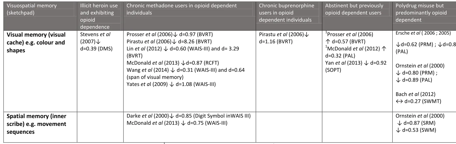
Table 1: Summary of previous research exploring visuospatial memory profiles in opioid-dependent individuals
Visuospatial memory (sketchpad)
Illicit heroin use and exhibiting opioid dependence
Chronic methadone users in opioid dependent individuals
Chronic buprenorphine users in opioid
dependent individuals
Abstinent but previously opioid dependent users
Polydrug misuse but predominantly opioid dependent
Visual memory (visual cache) e.g. colour and shapes
Stevens et al (2007)↓ d=0.39 (DMS)
Prosser et al (2006)↓ d=0.97 (BVRT) Pirastu et al (2006)↓ d=8.26 (BVRT)
Lin et al (2012) ↓ d=0.60 (WAIS-III) and d= 3.29 (BVRT)
McDonald et al (2013) ↓d=0.87 (RCFT)
Wang et al (2014) ↓ d=0.31 (WAIS-III) and d=0.64 (span of visual memory)
Yates et al (2009) ↓ d=1.08 (WAIS-III)
Pirastu et al (2006)↓ d=1.16 (BVRT)
1
Prosser et al (2006) ↑ d=0.57 (BVRT)
1
McDonald et al (2012) ↑ d=0.32 (PAL)
Yan et al (2013) ↓ d=0.92 (SOPT)
Ersche et al ( 2006 ; 2005)
↓d=0.62 (PRM) ; ↓d=0.88 (PAL)
Ornstein et al (2000) ↓ d=0.80 (PRM) ; ↓ d=0.89 (PAL)
Bach et al (2012) ↔ d=0.27 (SWMT)
Spatial memory (inner scribe) e.g. movement sequences
Darke et al (2000)↓ d=0.85 (Digit Symbol inWAIS III) McDonald et al (2013) ↓ d=0.75 (WAIS-III)
Ornstein et al (2000) ↓ d=0.87 (SRM) ↓ d=0.53 (SWM)
*= Opioid group compared with healthy controls unless otherwise stated; 1= Abstinent group compared with methadone and/or buprenorphine group and NOT healthy controls
p<0.05; ↔= no difference in neuropsychological performance; ↓= neuropsychological impairment present; ↑= improvement in neuropsychological performance when compared to healthy controls, d= Cohen’s effect size defined as the difference between two means divided by a standard deviation for the data. Standardised effect sizes are reported regardless of the statistical significance (p-value) of the results reported in the original studies
22
Table 2: Study procedures
Testing Sessions
Illicit or licit opioid use
Opioid withdrawal
2-4 weeks on
methadone
6 months on methadone
HEROIN [H] † † † —
CHRONIC PAIN [P] † — — —
METHADONE [M] † — — †
HEALTHY CONTROL [HC] † — — —
23
Table 3: Comparative demographic, clinical and substance use data for experimental and
control groups
Demographic and clinicaldata HEROIN (H) METHADONE
(M)
PAIN (P) HEALTHY
CONTROLS (HC) Sig.¹
N 24 29 28 28 n/a
Age (yrs)* 26.30 (3.45) 27.30 (2.34) 33.97 (4.35) 24.12 (3.56) H >P= p<0.001
M>P and M>HC = p<0.01
SIMD* 3.60(1.9) 3.41 (1.4) 4.60(2.0) 5.90 (2.5) H >HC and M>HC =p<0.001
M >P =p<0.01
Unemployed (%) 87.50 86.2 50 0 p<0.001
Stable accommodation (%)² 87 93 100 92.80 M >P =p<0.005
Education (yrs) * 10.80(1.5) 10.60 (2.3) 11.18 (1.22) 15.40 (2.1) H >HC, P>HC and M>HC = p<0.001
NART * 106.10 (12.2) 108.90 (7.6) 115.90 (4.9) 118.30 (5.1) P>H and HC>H= p<0.001
HC>M = p<0.001; P>M= p<0.01
Drug, nicotine and alcohol histories (self-report)
HEROIN METHADONE PAIN HEALTHY
CONTROLS
Sig.¹ H/M vs P/C
Percentage nicotine smokers 91.67 89.65 39.29 3.57 H>P,H>HC, M>P, M>HC =p<0.001
Days of alcohol use (last 30 days) *
2.20(6.1)(n=10) 4.0(4.9) (n=15) 5.10(8.3) (n=17) 4.00(6.3) (n=17) ns
Type of opioids and number of participants
Heroin n=24 Methadone n=29
Tramadol n= 18 Codeine n=13*2
n/a n/a
Daily intake expressed as morphine equivalence (mg)⁰ *
184.50(82.1) (n=24) 147.40 (59.3) (n=29) 59.10(46.8) (n=28)
n/a H>P and M>P =p<0.001 Age first used heroin (yrs) * 19.40 (4.1)
(n=24)
17.90(2.6) (n=29)
n/a n/a ns
Age opioid dependent (yrs) * 20.90 (3.9) (n=24)
19.90(2.8) (n=29)
n/a n/a ns
Age injecting opioids (yrs) * 20.50 (4.0) (n=17)
19.10(6.0) (n=29)
n/a n/a ns
Years of opioid use* 6.10 (2.9)
(n=24)
8.80(2.8) (n=29)
5.00(2.3) (n=28)
n/a M>H and M>P= p<0.001
Stable methadone use (yrs) * n/a 1.30(0.5)
(n=29)
n/a n/a n/a
Days of heroin use (last 30 days) *
29.50(2.7)(n=24 )
n/a n/a n/a ns
Sig ¹= significance at p<0.01 two tailed, ²Stable accommodation = own house + rented accommodation + living with parents (excluded hostel, student and homeless),
*=mean total scores (+/- standard deviation), *1= mean,*2= Some participants prescribed Tramadol were also prescribed
Codeine hence total number (31) higher than number recruited (n=28),
24
Table 4: Summary of baseline neuropsychological findings for memory and learning (not
adjusted for covariates).
HEROIN N=24 METHADONE N=29 CHRONIC PAIN N=28 HEALTHY CONTROL N=28 Memory and Learning
Mean (s.d) Mean (s.d) Mean (s.d) Mean (s.d) Sig. d
Delayed Matching to Sample (DMS) Total Number of Correct Responses (all delays)
22.04 (3.59) 25.76 (2.87) 25.39 (3.04) 27.43 (1.89) P>H***
C>H***
1.00 1.87
Mean Correct Latency (all delays)
3630.61(922.64) 4372.35 (1579.98) 3310.22(1049.63) 3536.67(745.99) M>P** 0.79
Paired Associate Learning (PAL) (log10)
Total Errors (Adjusted)
0.22 (0.45) 0.46 (0.64) 0.01 (0.06) 0.00 (0.00) NS
Mean Errors to Success
0.51 (0.26) 0.42 (0.19) 0.45 (0.27) 0.29 (0.19) C<H* 0.97
Mean Trials to Success
0.45 (0.09) 0.41 (0.06) 0.43 (0.09) 0.37 (0.05) C<H* 1.10
Memory Score 1.23 (0.11) 1.30 (0.06) 1.27 (0.08) 1.33 (0.06) C>H*** 1.13
Stages Completed 0.94 (0.03) 0.95 (0.02) 0.95 (0.02) 0.95 (0.00) NS
Stages complete 1st trial
0.79 (0.08) 0.84 (0.05) 0.82 (0.07) 0.87 (0.04) C>H*** 1.26
Pattern Recognition Memory (PRM) (SQRT)
Percentage Trials
Correct 9.18 (0.69) 9.27 (0.51) 9.26 (0.49) 9.64 (0.46) C>H* 0.78
Incorrect 3.37 (1.97) 3.46 (1.46) 3.43 (1.52) 1.97 (1.74) C<H*
C<M*
0.75 0.92 Correct Response
Latency
Correct 45.82 (5.36) 47.88 (5.98) 46.37 (6.74) 46.60 (5.56) NS
Incorrect 49.80 (11.99) 54.48 (11.74) 50.22 (10.76) 50.15 (8.96) NS
Spatial Recognition Memory (SRM)
Number of Trials
Correct 15.58 (2.21) 16.48 (1.94) 15.89 (1.77) 17.75 (1.51) C>H*** 1.15
Incorrect 4.42 (2.21) 3.52 (1.94) 4.11 (1.77) 2.25 (1.51) C<M*** 1.15
Mean Latency
Correct 1979.04 (432.81) 2358.89 (810.68) 2035.71 (505.51) 2040.19 (469.3) NS
Incorrect 2172 (878.02) 2520.36 (966.13) 2199.65 (737.71) 2456.38 (122.41) NS
d= effect size,SQRT= square root transformation; log10 = logarithmic 10 transformation, Sig= significance,
25
Table 5: Summary of results from analysis of visuospatial test outcomes*.Unless specified comparison is with HEALTHY CONTROL and/or
PAIN participants
1HEROIN vs METHADONE vs CHRONIC PAIN vs HEALTHY CONTROL
Opioid DEPENDENCE (OD) vs Non- Opioid
DEPENDENCE (Non- OD)
INJECTING (INJ) vs non- injecting
(a) Visual Memory
Delayed Matching to Sample (DMS)
Total Correct Responses (0,4,12 second delay stages)
↓HEROIN ↔ ↔ Pattern Recognition Memory (PRM)
Total Number of Correct Trials* ↔ ↔ ↔
Spatial Recognition Memory (SRM)
Total Number of Correct Trials* ↓HEROIN ↔ ↔
Paired Associate Learning (PAL)
Mean Total Number of Errors* ↔ ↔ ↔
Memory Score* ↓HEROIN ↔ ↔
(b)Spatial Memory Spatial Span (SSP)
Span Length* ↓METHADONE ↓OD > Non- OD ↔
Total Errors* ↓HEROIN ↔ ↔
Spatial Working Memory (SWM)
Total Search Errors* ↓HEROIN,↓METHADONE ↓OD > Non- OD ↔
Between Search Errors* ↓HEROIN,↓ METHADONE ↓OD > Non- OD ↔
Within Search Errors* ↔ ↔ ↔
Double Search Errors* ↔ ↔ ↔
*
= ANCOVA ‘between subject factor’ of GROUP, DEPENDENCE and INJECTING analysed separately; 1= significant effects with p<0.01, ↓=significant neuropsychological
26
Figures
Legend
Figure 1: DMS-Percentage of correct responses at different delay conditions (Means and Standard Deviation).
Post hoc
Bonferroni comparisons
identified participants from the HEROIN group significantly making more errors than did: the HEALTHY CONTROL group in the 0 (**
p<0.005
), 4
(***
p<0.001
) and 12 second (***
p<0.001)
delay stages, the CHRONIC PAIN group for the 4 (*
p<0.01)
and 12 (**
p<0.005
) second delay stages
and the METHADONE group for 0(**
p< 0.005
), 4 (**
p<0.005
) and 12 (***
p<0.001
) second delay stages.
Sim=
Simultaneous condition
, SD=
Standard Deviation
.
Figure 2:
A-
Total errors in Spatial Span (SSP) task (Means and Standard Deviation). Overall participants significantly made more errors [F (3,102)
=16.8,
p<0.001
].
Post hoc
Bonferroni comparisons identified the HEROIN group participants significantly making more errors compared
to the METHADONE (
p<0.001
) and HEALTHY CONTROL (
p<0.005
) groups. The total errors for the CHRONIC PAIN participants lay
between those of the HEROIN, METHADONE and HEALTHY CONTROL participants and did not differ significantly from these three
groups (p=1.0).
B-