ARTICLE
The Crying Infant: Diagnostic Testing and Frequency
of Serious Underlying Disease
Stephen B. Freedman, MDCM, MSc, FRCPC, Nesrin Al-Harthy, MD, Jennifer Thull-Freedman, MD, MSc
Division of Pediatric Emergency Medicine, Hospital for Sick Children, University of Toronto, Toronto, Ontario, Canada
The authors have indicated they have no financial relationships relevant to this article to disclose.
What’s Known on This Subject
Although the differential diagnosis of crying is extensive, the frequency of severe under-lying disease is unclear. On the basis of very limited data, it has been recommended that corneal fluorescein staining, eyelid eversion, and rectal examination be performed on crying infants.
What This Study Adds
This study confirms that the clinical assessment should guide decision-making in chil-dren presenting with crying. Afebrile infants in the first few months of life should un-dergo urine evaluation. Other investigations should be performed on the basis of clinical findings.
ABSTRACT
OBJECTIVE.To determine the proportion of children evaluated in an emergency
depart-ment because of crying who have a serious underlying etiology. Secondary outcomes included the individual contributions of history, physical examination, and labora-tory investigations in determining a diagnosis.
PATIENTS AND METHODS.We performed a retrospective review of all afebrile patients⬍1
year of age who presented with a chief complaint of crying, irritability, screaming, colic, or fussiness. All children with a serious underlying illness were identified by using a priori defined criteria. Chart review was conducted to determine if history, physical examination, or investigation data contributed to establishing the child’s diagnosis.
RESULTS.Enrollment criteria were met by 237 patients, representing 0.6% of all visits.
A total of 12 (5.1%) children had serious underlying etiologies with urinary tract infections being most prevalent (n⫽3). Two (16.7%) of the serious diagnoses were only made on revisit. Of the 574 tests performed, 81 (14.1%) were positive. How-ever, only 8 (1.4%) diagnoses were assigned on the basis of a positive investigation. History and/or examination suggested an etiology in 66.3% of cases. Unwell appear-ance was associated with serious etiologies. In only 2 (0.8%) children did investiga-tions in the absence of a suggestive clinical picture contribute to the diagnosis. Both of these children were⬍4 months of age and had urinary tract infections. Among children⬍1 month of age, the positive rate of urine cultures performed was 10%. Ocular fluorescein staining and rectal examination with occult blood testing were performed infrequently, and results were negative in all cases. Successful follow-up was completed with 60% of caregivers, and no missed diagnoses were found.
CONCLUSIONS.History and physical examination remains the cornerstone of the evaluation of the crying infant and
should drive investigation selection. Afebrile infants in the first few months of life should undergo urine evaluation. Other investigations should be performed on the basis of clinical findings.Pediatrics2009;123:841–848
I
NFANTS COMMUNICATE ANDexpress discomfort by crying. This can be due to a variety of reasons ranging from hunger or a desire for attention to severe life-threatening illness. Healthy children cry on average nearly 3 hours per day at 6 weeks of age with the peak occurring between 3 PMand 11PM.1By the time parents present to the emergency department (ED) with their crying child, caregivers are often anxious, frustrated, and sleep deprived. These emotions contribute to making the evaluation of the nonverbal, crying infant very difficult.Although the prevalence of excessive crying varies by definition employed,2the peak nevertheless occurs during the second month of life with a prevalence of 1.5% to 11.9%.3Although the differential diagnosis is extensive and involves every organ system, the frequency of severe underlying disease is unclear. Although it has been stated that organic diseases underlie⬍5% of cases of infantile colic,4the only North American ED study of afebrile infants with acute, unexplained, excessive crying reported a “serious” etiology in 61% of such children.5The authors of that study defined a diagnosis as serious if it was deemed “by at least 2 of a panel of 3 pediatricians to require prompt treatment or to have the potential to cause harm if not recognized or left untreated.”5This definition was very inclusive, as otitis media, herpangina, herpes stomatitis, gastroesophageal reflux, and vaccine reactions were among the diagnoses
www.pediatrics.org/cgi/doi/10.1542/ peds.2008-0113
doi:10.1542/peds.2008-0113
This work was presented in part at the 2008 annual meeting of the Pediatric Academic Societies, May 5, 2008, Honolulu, Hawaii; and the 85th annual conference of the Canadian Paediatric Society, June 25, 2008, Victoria, British Columbia, Canada.
Key Words
crying, emergencies, diagnosis
Abbreviation
ED— emergency department
Accepted for publication Jul 2, 2008
Address correspondence to Stephen B. Freedman, MDCM, MSc, FRCPC, Hospital for Sick Children, Division of Pediatric Emergency Medicine, 555 University Ave, Toronto, Ontario, Canada M5G 1X8. E-mail: stephen. [email protected]
represented. Subsequently, this study, which recom-mends routinely performing corneal fluorescein stain-ing, eyelid eversion, and rectal examination, formed the basis for the establishment of guidelines for the evalua-tion of crying infants.
In an attempt to develop evidence-based guidelines for the diagnostic evaluation of afebrile children present-ing with excessive crypresent-ing, we reviewed the clinical find-ings, test results, and diagnoses in a consecutive series of children who presented to the ED with acute, excessive, unexplained crying.
METHODS
Study Site
The Hospital for Sick Children is a tertiary care referral hospital in downtown Toronto. It serves a broad demo-graphic and socioeconomic spectrum of patients and a geographic region that includes urban, suburban, and rural regions. The ED provides care for⬎50 000 patient visits per year and is staffed by attending physicians 24 hours per day.
Patients
This retrospective report includes data from a consecu-tive series of afebrile infants ⬍12 months of age who presented to the ED between January 1, 2005, and Sep-tember 30, 2005, with a chief complaint of crying. Chil-dren were identified by searching our electronic data-base using a chief complaint family word root search for “cry,” “irritable,” “fuss,” “scream,” and “colic.”
Outcomes
Our primary outcome was the proportion of infants who had a potentially serious underlying etiology. To allow us to a priori identify illnesses felt to require prompt diagnosis and therapy, a literature review was per-formed, followed by a focus group discussion with 6 full-time members of our division. The failure to make the diagnosis of any of the disease entities listed in Table 1 was felt to have the potential to result in an adverse outcome. For illnesses identified during data abstraction that had not previously been considered, a panel of 3 pediatric emergency medicine physicians, blinded to his-torical and physical examination features, met to deter-mine if the diagnosis constituted a serious underlying disease.
Secondary outcomes included the proportion of chil-dren with a serious underlying etiology in whom the diagnosis was not suspected on the basis of the initial history or physical examination and whether there ex-isted high-yield investigations that should routinely be performed.
Data Collection
Data were abstracted from our electronic patient chart system. Medical charts were reviewed employing a stan-dardized data collection instrument to record demo-graphic data, history and physical examination findings, diagnostic tests, final diagnosis, and disposition. The study was approved by the hospital’s research ethics
board. Subjects were enrolled after a letter was mailed informing caregivers on how to avoid being contacted. Subsequently, attempts were made to obtain informed consent by telephone.
A follow-up telephone call was performed 9 to 18 months after the index visit to inquire about other phy-sician visits, alternative diagnoses made, and tests or treatments required. Calls were attempted a maximum of 3 times. Caregiver report was confirmed via medical chart review. All alternative diagnoses assigned within 1 week of the index visit were considered as potential etiologies of crying at the index visit.
Definitions
The analysis considered historical variables (review of systems, feeding, perinatal, vaccination, and family his-tory), physical examination (vital signs, general descrip-tion, pertinent findings), tests of blood, urine, and cere-brospinal fluid, imaging studies, stool occult blood testing, eye fluorescein staining, and electrocardiograms. Fever was defined as a documented fever at home or in the ED. Recorded temperatures were adjusted for loca-tion of measurement with 1.1°C and 0.6°C added to axillary and oral temperatures, respectively.6 Children with an adjusted temperature ofⱖ38.0°C were consid-ered to have a fever.6 Children with a tactile fever at home but no evidence of fever in the ED were consid-ered to be afebrile. General appearance was classified for all children by the data abstractor (Dr Al-Harthy) as well, unwell, or unclear.
All tests performed in the ED were recorded, and if positive, the data abstractor indicated whether the test
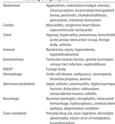
TABLE 1 Serious Underlying Etiologies or Conditions Sorted According to Organ System
Abdominal Appendicitis, malrotation/midgut volvulus,
intussusception, incarcerated/strangulated hernia, peritonitis, choledocholithiasis, pancreatitis, intestinal obstruction
Cardiac Myocarditis, congestive heart failure,
supraventricular tachycardia
Chest Hypoxia, hypercarbia, pneumonia, bronchiolitis,
acute airway obstruction (croup, foreign body, asthma)
General Bacteremia, sepsis, hypovolemia,
hyperbilirubinemia
Genitourinary Testicular/ovarian torsion, genital tourniquet,
urinary tract infection, nephrolithiasis
HEENT Foreign body
Hematologic Sickle cell disease, malignancy, neutropenia,
thrombocytopenia, anemia
Skin/musculoskeletal Septic arthritis, osteomyelitis, digital tourniquet, fracture, dislocation, subluxation,
nonaccidental trauma, cellulitis
Neurologic Bacterial meningitis, encephalitis, intracranial
hemorrhage, hydrocephalus, cerebral edema, epilepsy, degenerative condition
Toxic-metabolic Prenatal drug use, toxic ingestion, electrolyte abnormality, inborn error of metabolism, hyperthyroidism
contributed to the diagnosis. Negative tests were not considered as contributing to a diagnosis, because al-though negative tests can be helpful in ruling out a particular disease, they are generally less diagnostic when evaluating a child with a nonspecific complaint such as crying. All laboratory tests were classified as positive/negative on the basis of age-specific ranges of normal.7 Cultures that grew a clinically significant pathogen were considered positive. Imaging studies were deemed positive on the basis of the interpretation of the responsible specialist. When a test was ordered more than once, we considered only the results of the initial test. Urine dipstick was considered positive if the testing reagent indicated the presence of any leukocyte esterase or nitrites.
For each child, we classified the role of the initial history and physical examination in guiding subsequent investigations. Historical and examination features con-sidered to be indicative of particular diagnoses were identified in advance to guide the abstractor (Dr Al-Harthy) in classifying the contributory role of these vari-ables (Table 2). As has been previously done, all patients were grouped into 1 of 4 categories according to the source(s) of data that contributed to the diagnosis:8
1. Diagnosis was based on the history and/or physical examination alone.
2. Diagnosis was based on positive test results obtained after the history and physical examination failed to suggest a cause.
3. Diagnosis was based on tests ordered to investigate positive findings from the history and/or physical examination that suggested a cause.
4. Neither history, physical examination, nor investiga-tions were diagnostic.
A contributory historical or physical examination vari-able includes any finding that may be connected to the underlying diagnosis. The principle investigator (Dr Freedman) reviewed the classification of all cases that were unclear to the data abstractor. Consistency and reproducibility of the classification scheme was evalu-ated by having a third investigator (Dr Thull-Freedman) review 10% of cases identified at random (SPSS 15.0 for Windows [SPSS Inc, Chicago, IL]), blinded to the assess-ment of the data abstractor. Agreeassess-ment was excellent with avalue of 0.90 (P⬍.001).
Analysis
The required sample size was calculated to yield stable estimates (⫾5%) of the primary outcome measure. We estimated that 10% of our sample would have a serious underlying organic etiology. Thus, a minimum sample of 138 subjects would be required. However, we antici-pated a follow-up telephone call response rate of only 75%. After sample size adjustment, the final size re-quired was 245.
Frequency counts and percentages are given for dis-crete variables, and means, medians, SDs, and interquar-tile ranges are provided for continuous variables. The2
test was used for discrete variables. When the number of observations in any given cell of the contingency table was⬍5, Fisher’s exact test was used. On the basis of the definitions provided above, the diagnostic yield of tests performed was calculated with the number of patients with positive diagnostic tests divided by the number of tested patients. As the underlying etiology may vary by age, we have performed sub-analyses, with children di-vided into groups of 0 to 3 months and ⱖ4 months of age. When the results have been significantly different, we have reported them. All statistical tests were
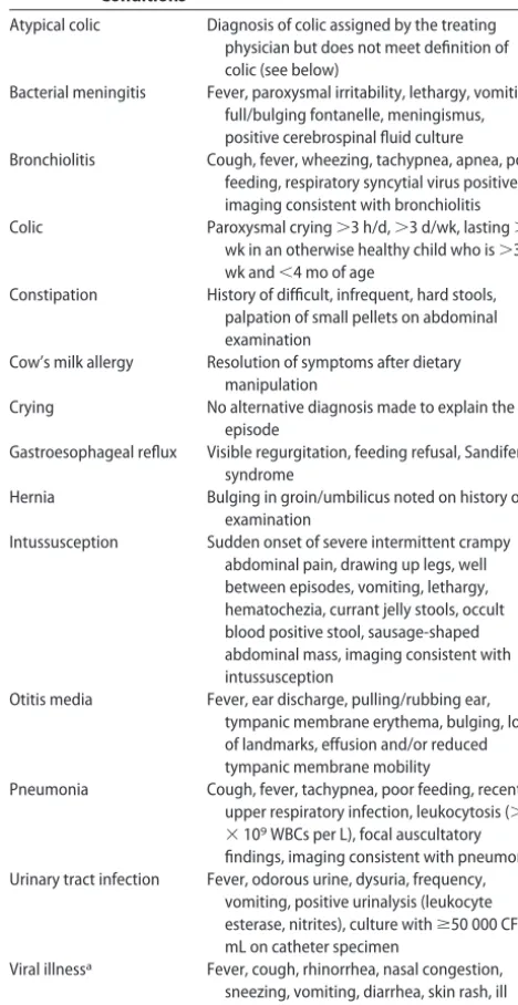
evalu-TABLE 2 Features of History and Physical Examination Considered to Be Helpful in Diagnosing Specific Conditions
Atypical colic Diagnosis of colic assigned by the treating
physician but does not meet definition of colic (see below)
Bacterial meningitis Fever, paroxysmal irritability, lethargy, vomiting, full/bulging fontanelle, meningismus, positive cerebrospinal fluid culture
Bronchiolitis Cough, fever, wheezing, tachypnea, apnea, poor
feeding, respiratory syncytial virus positive, imaging consistent with bronchiolitis
Colic Paroxysmal crying⬎3 h/d,⬎3 d/wk, lasting⬎3
wk in an otherwise healthy child who is⬎3 wk and⬍4 mo of age
Constipation History of difficult, infrequent, hard stools,
palpation of small pellets on abdominal examination
Cow’s milk allergy Resolution of symptoms after dietary manipulation
Crying No alternative diagnosis made to explain the
episode
Gastroesophageal reflux Visible regurgitation, feeding refusal, Sandifer syndrome
Hernia Bulging in groin/umbilicus noted on history or
examination
Intussusception Sudden onset of severe intermittent crampy
abdominal pain, drawing up legs, well between episodes, vomiting, lethargy, hematochezia, currant jelly stools, occult blood positive stool, sausage-shaped abdominal mass, imaging consistent with intussusception
Otitis media Fever, ear discharge, pulling/rubbing ear,
tympanic membrane erythema, bulging, loss of landmarks, effusion and/or reduced tympanic membrane mobility
Pneumonia Cough, fever, tachypnea, poor feeding, recent
upper respiratory infection, leukocytosis (⬎20
⫻109WBCs per L), focal auscultatory
findings, imaging consistent with pneumonia Urinary tract infection Fever, odorous urine, dysuria, frequency,
vomiting, positive urinalysis (leukocyte esterase, nitrites), culture withⱖ50 000 CFU/ mL on catheter specimen
Viral illnessa Fever, cough, rhinorrhea, nasal congestion,
sneezing, vomiting, diarrhea, skin rash, ill contact
WBCs indicates white blood cells; CFU, colony-forming units.
aViral illness includes herpangina, influenza, nasal congestion, roseola, upper respiratory
ated at the 5% level of significance. Statistical analysis was conducted with the use of SPSS.
RESULTS
A total of 37 549 ED visits occurred during the 9-month eligibility period. Two hundred thirty-eight children met the inclusion criteria (0.6% of all visits) (Table 3); 55.0% (131 of 238) were firstborn children. Thirteen percent
(30 of 238) revisited the Hospital for Sick Children’s ED within 1 week of the index visit. Sixty-three percent (150 of 238) were triaged between 6PMand 6AM(Fig 1). Five percent (12 of 238) of our cohort met our pri-mary outcome definition of serious underlying etiology (Table 4). Eighty-three percent (10 of 12) of these chil-dren were correctly diagnosed at the initial visit; 2 (16.7%) of the 12 were diagnosed during the subse-quent 7 days. Overall, the most common serious under-lying disease was urinary tract infection. A total of 14 children were diagnosed with colic (age range: 1.1–3.6 months) and 11 were diagnosed with atypical colic (age range: 0.5– 4.5 months). When comparing patients ⬍4 months of age to those ⱖ4 months of age, there was a tendency for serious illness to be more common among
Triage time
21-24 18-21 15-18
12-15 9-12
6-9 3-6 0-3
C
ount
60
50
40
30
20
10
0
FIGURE 1
Triage time of 238 patients who presented to the ED be-cause of crying. The distribution was nonnormal (P⬍
.001) with the majority of patients presenting between 6
PMand 3AM.
TABLE 3 Baseline Features of Study Patients (Nⴝ238)
Male,n(%) 124 (52)
Age, median (25th–75th percentiles), mo 2.3 (1.0–5.4)
Triage time, mean⫾SD 13:52⫾7:44
Duration of crying, median (25th–75th percentiles), h 27 (3–96) Character of crya
Constant,n(%) 52 (22)
Intermittent,n(%) 93 (39)
Inconsolable,n(%) 45 (19)
Easily consoled,n(%) 44 (18)
Tactile fever at home,n(%), yesb 41 (17)
Taking analgesic/antipyretic,n(%), yesc 61 (26)
Adjusted temperature, mean⫾SD, °Cd 37.2⫾0.4
Heart rate, beats per min⫾SD 136⫾17
Respiratory rate, breaths per min⫾SD 34⫾9
Oxygen saturation, %⫾SD 98⫾3.0
Blood pressure systolic, mean⫾SD 94⫾12
Weight, median (25th–75th percentiles), kg 5.2 (4.2–7.9)
General appearance
Well,n(%) 226 (95)
Unwell,n(%) 7 (3)
Unclear,n(%) 5 (2)
aNot all children had a description of the crying reported. bNo children had a documented fever at home or in the ED. cAnalgesic/antipyretic use included acetaminophen or ibuprofen.
dAdjusted for location of measurement, with 1.1°C and 0.6°C added to axillary and oral
temperatures, respectively.6
TABLE 4 Serious Underlying Diagnoses Sorted According to Time of Diagnosis
Index Visit Within 1 wk of Index Visit
Acute cholecystitis 1 —
Acute lymphoblastic leukemia 1 —
Clavicle fracture 2 —
Epidural hematoma 1 —
Intussusception 1 —
Nephrolithiasis — 1
Pulled elbow 1 —
Spinal muscular atrophy type Ia 1 —
Urinary tract infection 2 1
Total 10 2
aThis 2-month-old infant presented with increased crying and decreased feeding. On
the older cohort (10.4% vs 3.4%); however, this did not achieve statistical significance (P⫽.07).
Investigations
In our cohort of 238 children, anterior eye fluorescein staining was performed and documented in 1 (0.4%) child, and fecal occult blood testing was recorded for 8 (3.4%) children. Both of these tests were negative in all cases (Table 5). Of the 574 tests performed on these children (2.4 tests per child), 81 (14.1%) of 574 were positive, but only 8 (1.4%) diagnoses were assigned on the basis of a positive investigation. Among the 238 children evaluated, the most frequently performed tests were urine analysis (31.1%) and culture (28.6%), both of which contributed to the final diagnosis⬍3% of the times they were performed. All positive urine cultures occurred in the 47 children⬍4 months of age in whom urine specimens were obtained by urinary catheteriza-tion. Of the 60 infants⬍1 month of age, 19 (32%) had urine cultures performed. These infants had the highest
rate of positivity (10% [95% confidence interval: 0.03– 0.30]).
Relative Contributions From History, Physical Examination, and Investigations
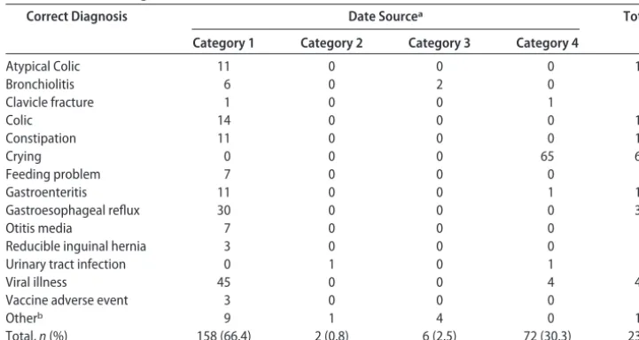
Positive findings on the history and/or physical exami-nation alone suggested the final diagnosis to explain the crying in 66.4% (158 of 238) of children (Table 6). In 6 of 238 (2.5%) other children, the etiology was suggested by the history and/or physical examination with confir-mation by positive investigations. Four of the 7 (57.1%) children described as appearing unwell had a serious underlying illness (P⬍.001). Investigations performed in the absence of a suggestive clinical picture were cru-cial in determining a diagnosis in 2 of 238 (0.8%) chil-dren including (1) a 3-month-old child whose catheter urine sample was positive for nitrites and trace leuko-cytes, whose culture grew 2Escherichia colimorphotypes, both with colony-forming unit counts ofⱖ50⫻106/L (this child’s blood culture additionally grew
Enterococ-TABLE 5 Investigations and Yield of Diagnostic Tests Performed at the Initial Visit (Nⴝ238) Test
Performed
Positive Findings
Finding Contributed to Diagnosis
Diagnoses to Which the Positive Findings
Contributeda
a,n(%) b,n(b/a%) c,n c/b, % c/a, %
Miscellaneous
Fluorescein 1 0 (0) 0 0 0 None
Fecal occult blood 8 1 (12.5) 0 0 0 None
Electrocardiogram 6 1 (16.7) 0 0 0 None
Nasopharyngeal aspirate 6 1 (16.7) 1 100.0 16.7 Bronchiolitis
Blood
White blood cell count 55 12 (21.8) 1 8.3 1.8 Acute lymphoblastic
leukemia
Hemoglobin 55 10 (18.2) 1 10.0 1.8 Acute lymphoblastic
leukemia
Platelet count 50 25 (50.0) 1 2.0 2.0 Acute lymphoblastic
leukemia
Sickle cell screen 4 1 (25.0) 0 0 0 None
Blood gas 31 2 (6.5) 0 0 0 None
Routine electrolytes 44 1 (2.3) 0 0 0 None
Renal function tests 42 1 (2.3) 0 0 0 None
Lactate 2 0 (0) 0 0 0 None
Bilirubin 13 2 (15.4) 0 0 0 None
Liver transaminases 9 1 (11.1) 1 100.0 11.1 Acute cholecystitis
Coagulation profile 8 0 (0) 0 0 0 None
Blood culture 35 1 (2.9) 0 0 0 None
Urine
Urine analysis 74 5 (6.8) 1 20.0 1.4 Urinary tract infection
culture 68 2 (2.9) 2 100.0 2.9 Urinary tract infection (2)
Cerebrospinal fluid
Culture 4 0 (0) 0 0 0 None
Cell count 4 2 (50.0) 0 0 0 None
Chemistry 4 2 (50.0) 0 0 0 None
Radiologic investigations
Chest radiograph 20 8 (40.0)b 1 5.0 12.5 Bronchiolitis
Abdominal radiograph 14 0 (0) 0 0 0 None
Skull radiograph 1 1 (100.0) 1 100.0 100.0 Skull fracture
Abdominal ultrasound 16 2 (12.5) 2 100.0 12.5 Intussusception, acute
cholecystitis
aPatients may have had⬎1 test that contributed to the diagnosis.
bPositive chest radiograph findings as interpreted by the pediatric radiologist included: lower airway inflammation (n⫽4),
cus faecalis, which was felt to be a contaminant); and (2) an 18-day-old male whose catheter urine culture grew E coli with a colony-forming unit count of ⱖ 50 ⫻106/L.
Long-term Follow-up
Three caregivers declined consent for our telephone fol-low-up. Sixty-one percent (143 of 235) of caregivers were successfully contacted after the ED visit. Twenty-nine percent (41 of 143) indicated that they had subse-quently brought their child to see another non–ED-based health care provider; 3.5% (5 of 143) reported seeking additional evaluation in an ED. All serious un-derlying diagnoses reported by the caregivers during follow-up were detected during our chart review.
DISCUSSION
This is the largest sample of children presenting to an ED with crying that has been evaluated. We found that caregivers frequently bring their infants to the ED be-cause of crying, fussiness, screaming, or irritability, and the problem frequently persists after discharge, with 13% of children having a repeat ED visit within 1 week. Our results emphasize the need for a careful evaluation as 5% of children had a serious diagnosis. However, routine investigations in afebrile children with crying seem unwarranted as⬍1% of children received a diag-nosis on the basis of investigations alone. Thus, a selec-tive workup guided by clinical findings seems to be optimal.
Urinary tract infections were the most common seri-ous diagnosis in our cohort, accounting for 25% of all serious etiologies. Crying has previously been reported
as the chief complaint in afebrile children with urinary tract infections.9 The overall yield of urine cultures in our cohort (3%) is similar to that reported in a recent cohort of 200 afebrile crying infants evaluated in Iran.10 The greatest yield occurred among children⬍1 month of age. Although asymptomatic bacteriuria has been re-ported to occur in up to 1% of children ⬍60 days of age,11it is unlikely that this explains all of the positive cultures in our cohort, which includes an 18-day-old infant whose catheter urine specimen was positive for nitrites and leukocytes and whose culture grewE coliⱖ
50⫻106/L. Even among those with normal urine anal-yses, clinicians should not disregard the culture result, as pyuria is not a sensitive marker among neonates, and ⬍50% of febrile infants ⬍8 weeks of age with positive urine cultures have abnormal urinalysis.12Thus, although false-positive urine cultures are possible, even on catheter specimens, we feel that the positive urine cultures in our cohort of patients are important and should not be ignored.
In keeping with other research, we found the most important aspect in the evaluation of the crying infant to be a thorough history and physical examination.5,10 In-vestigations were only helpful in 3% of our cohort. Although some tests, such as nasopharyngeal aspirate, liver function tests, abdominal ultrasound, and skull ra-diographs contributed to the diagnosis⬎10% of the time they were performed, their use was determined on the basis of findings of history or physical examination. Overall, they were infrequently performed in our cohort and thus would not have the same diagnostic properties if used as screening tests.
Although corneal abrasions commonly present with
TABLE 6 Final Diagnoses Categorized According to the Data Sources That Most Likely Contributed to the Diagnoses (Nⴝ238)
Correct Diagnosis Date Sourcea Total
Category 1 Category 2 Category 3 Category 4
Atypical Colic 11 0 0 0 11
Bronchiolitis 6 0 2 0 8
Clavicle fracture 1 0 0 1 2
Colic 14 0 0 0 14
Constipation 11 0 0 0 11
Crying 0 0 0 65 65
Feeding problem 7 0 0 0 7
Gastroenteritis 11 0 0 1 12
Gastroesophageal reflux 30 0 0 0 30
Otitis media 7 0 0 0 7
Reducible inguinal hernia 3 0 0 0 3
Urinary tract infection 0 1 0 1 2
Viral illness 45 0 0 4 49
Vaccine adverse event 3 0 0 0 3
Otherb 9 1 4 0 14
Total,n(%) 158 (66.4) 2 (0.8) 6 (2.5) 72 (30.3) 238
Factors considered to be suggestive or contributory to a diagnosis are listed in Table 2.
aExplanation of data source: category 1, positive history and physical examination only; category 2, positive diagnostic investigations
only; category 3, positive history and physical examination along with positive diagnostic investigations; category 4, history, physical, and diagnostic investigations (if performed) were negative or noncontributory.
bOther includes 1 of each of the following: category 1, cellulitis, dermatitis, gas, infantile spasms, nephrolithiasis, thrush, pulled elbow,
photophobia, excessive tearing, and conjunctival injec-tion in addiinjec-tion to pain, it has been suggested that cor-neal fluorescein staining be performed on all infants with acute, unexplained irritability or excessive crying.13 This recommendation is based primarily on a case series of 20 infants⬍1 year of age who presented with crying or irritability and were diagnosed with corneal abra-sions.13The clinical implications of these findings, how-ever, are unclear, as there was no confirmation by an ophthalmologist, and fluorescein-impregnated papers were employed, which themselves can result in corneal abrasion.14Although, in a previous study, 5% of crying infants were reported to have corneal abrasions,5in the case series,13 no prevalence data were provided. Even once diagnosed, the need for patching and antimicrobial therapy has been debated.14In 2006, a Cochrane review of 11 eligible trials concluded that patching does not improve the rate of healing or reduce pain.15
In our case series, although no children were diag-nosed with a corneal abrasion (prevalence 0% [95% confidence interval: 0%–1.9%]), we cannot conclude that clinicians should forgo searching for abrasions. Al-though it is likely that no child had a significant corneal abrasion as evidenced by our follow-up, less significant abrasions may have been overlooked, as only 1 child had a fluorescein examination documented. Thus, it is pos-sible that a child with an abrasion did not undergo fluorescein examination; it is also possible that many more children had an examination performed but not documented.
Other tests of questionable value are stool occult blood testing and rectal examination. Although consti-pation is not an uncommon condition, constituting 5% (11 of 237; Table 2) of diagnoses in our cohort, the utility of rectal examination is unclear. In a clinical practice guideline, the North American Society for Pediatric Gas-troenterology and Nutrition recommends the routine performance of anorectal examination and occult blood testing.16This expert panel states that the amount, con-sistency, and location of stool within the rectum are all helpful pieces of information. However, a systematic review of the diagnostic value of clinical findings in children with constipation found inconsistent results from rectal examination.17The likelihood ratio of finding stool on rectal examination relative to abdominal radi-ography is close to 1, indicating that stool detected on rectal examination occurs almost as often in children with and without fecal loading on radiography.17Thus, greater evidence is required to support the routine per-formance of rectal examinations in crying children.
A rectal examination may also be performed to obtain stool to test for fecal occult blood. Although of potential importance in children suspected of having intussuscep-tion, in a recent review of 83 cases of intussuscepintussuscep-tion, blood was detected on rectal examination in only 16%, with other findings being significantly more common. These included abdominal pain (81%), vomiting (66%), bloody stools (57%), pallor (47%), lethargy (46%), and abdominal mass (36%).18Given the prevalence of intus-susception of only 0.4% in our cohort, it seems unjus-tified to routinely perform rectal examinations on all
children who present with crying and should be reserved for those with other signs of intussusception.
Missing a serious diagnosis is always a concern when evaluating a crying infant. In our cohort, the most con-cerning diagnosis not initially detected was urinary tract infection in an infant⬍1 month of age. This highlights the need to rule out such infections in very young chil-dren.5The other diagnosis made on revisit, nephrolithi-asis, occurred in an 8-month-old male who represented with a stone that was found at the urethral meatus.
Gastroesophageal reflux disease was diagnosed on the basis of clinical presentation in 13% of our sample. This did not vary significantly by age (14% among those ⬍4 months of age versus 8% among thoseⱖ4 months of age). Previous research employing pH monitoring, however, has shown that clinical findings, such as backarching and sleep disturbance, are poorly associated with pathologic re-flux.19,20Results regarding high-risk groups have been con-tradictory with 1 study finding pathologic reflux to be more common in childrenⱖ3 months of age,20whereas another found it to be more common in infants⬍3 months of age.19 Both studies found that reflux was almost always associ-ated with frequent vomiting or regurgitation and that “si-lent” pathologic reflux was uncommon.19,20
LIMITATIONS
This study was a retrospective chart review. Therefore, the data abstracted may be limited in terms of accuracy and completeness. The absence of a uniform testing protocol, with tests performed instead on the basis of individual physician clinical judgment, means that ab-normalities related to the underlying etiology may have been missed. Because physicians preferentially perform tests on patients with higher pretest probabilities, it is most likely that contributory rates would be lower in untested patients. In addition, because some tests were infrequently performed, we are unable to make firm conclusions regarding their utility in other settings where utilization patterns may be different.
The 90 patients who were discharged with a diagnosis of colic, atypical colic, or crying may have included some patients with serious undetected pathology. However, this is unlikely given that we were able to perform follow-up using our hospital’s database. Data previously collected suggests that 80% of caregivers would bring their child back to our ED should another visit be re-quired.21 In addition to chart review, we were able to contact 61% of caregivers to confirm the diagnosis. Lastly, the presence of a diagnosed condition does not guarantee causality in individual cases. For example, it cannot be proven that the presence of rhinorrhea and a diagnosis of viral illness, or decreased stooling and a diag-nosis of constipation, were the true etiologies of crying. For the purposes of clinical management, in the absence of evidence to the contrary or a more likely explanation, it seems reasonable to accept that such diagnoses are correct and that a causal relationship may exist.
CONCLUSIONS
remains the cornerstone of the evaluation of crying in-fants and should drive the selection of investigations to be performed. Afebrile infants in the first month of life should undergo urine evaluation. Other investigations such as rectal examination and fluorescein staining should be performed on the basis of the findings from the history and physical examination.
ACKNOWLEDGMENTS
We acknowledge the assistance of Kimberly Haladyn and Victoria Paulionis with the data abstraction that was required to complete this research endeavor.
REFERENCES
1. Brazelton TB. Crying in infancy.Pediatrics.1962;29:579 –588 2. Reijneveld SA, Brugman E, Hirasing RA. Excessive infant
crying: definitions determine risk groups.Arch Dis Child.2002; 87(1):43– 44
3. Reijneveld SA, Brugman E, Hirasing RA. Excessive infant crying: the impact of varying definitions. Pediatrics. 2001; 108(4):893– 897
4. Barr RG. Colic and crying syndromes in infants. Pediatrics. 1998;102(5 suppl E):1282–1286
5. Poole SR. The infant with acute, unexplained, excessive crying. Pediatrics.1991;88(3):450 – 455
6. Alpern ER, Henretig FM. Fever. In: Fleisher GR, Ludwig S, Henretig FM, eds.Textbook of Pediatric Emergency Medicine.5th ed. Philadelphia, PA: Lippincott Williams & Wilkins; 2006 7. Cheng A, Williams BA, Sivarajan VB. The Hospital for Sick
Children Handbook of Pediatrics. 10th ed. Toronto, Ontario, Canada: Saunders Canada; 2003
8. Brand DA, Altman RL, Purtill K, Edwards KS. Yield of diag-nostic testing in infants who have had an apparent life-threatening event [published correction appears in Pediatrics. 2005;116(3):802– 803].Pediatrics.2005;115(4):885– 893 9. Du JN. Colic as the sole symptom of urinary tract infection in
infants.CMAJ.1976;115:334 –335
10. Fahimi D, Shamsollahi B, Salamati P, Sotoudeh K. Excessive crying of infancy: a report of 200 cases.Iran J Pediatr.2007; 17(3):222–226
11. Wettergren B, Jodal U, Jonasson G. Epidemiology of bacteri-uria during the first year of life.Acta Paediatr Scand.1985;74(6): 925–933
12. Crain EF, Gershel JC. Urinary tract infections in febrile infants younger than 8 weeks of age.Pediatrics.1990;86(3):363–367 13. Poole SR. Corneal abrasion in infants.Pediatr Emerg Care.1995;
11(1):25–26
14. Levin AV. Eye trauma. In: Fleisher GR, Ludwig S, Henretig FM, eds.Textbook of Pediatric Emergency Medicine. 5th ed. Philadel-phia, PA: Lippincott Williams & Wilkins; 2006
15. Turner A, Rabiu M. Patching for corneal abrasion. Cochrane Database Syst Rev.2006;(2):CD004764
16. Baker SS, Liptak GS, Colletti RB, et al. Constipation in infants and children: evaluation and treatment. A medical position statement of the North American Society for Pediatric Gastro-enterology and Nutrition. J Pediatr Gastroenterol Nutr. 1999; 29(5):612– 626
17. Reuchlin-Vroklage LM, Bierma-Zeinstra S, Benninga MA, Berger MY. Diagnostic value of abdominal radiography in con-stipated children: a systematic review.Arch Pediatr Adolesc Med. 2005;159(7):671– 678
18. Buettcher M, Baer G, Bonhoeffer J, Schaad UB, Heininger U. Three-year surveillance of intussusception in children in Swit-zerland.Pediatrics.2007;120(3):473– 480
19. Heine RG, Jordan B, Lubitz L, Meehan M, Catto-Smith AG. Clinical predictors of pathological gastro-oesophageal reflux in infants with persistent distress.J Paediatr Child Health.2006; 42(3):134 –139
20. Heine RG, Jaquiery A, Lubitz L, Cameron DJ, Catto-Smith AG. Role of gastro-oesophageal reflux in infant irritability.Arch Dis Child.1995;73(2):121–125
DOI: 10.1542/peds.2008-0113
2009;123;841
Pediatrics
Stephen B. Freedman, Nesrin Al-Harthy and Jennifer Thull-Freedman
Disease
The Crying Infant: Diagnostic Testing and Frequency of Serious Underlying
Services
Updated Information &
http://pediatrics.aappublications.org/content/123/3/841 including high resolution figures, can be found at:
References
http://pediatrics.aappublications.org/content/123/3/841#BIBL This article cites 17 articles, 10 of which you can access for free at:
Subspecialty Collections
sub
http://www.aappublications.org/cgi/collection/emergency_medicine_ Emergency Medicine
following collection(s):
This article, along with others on similar topics, appears in the
Permissions & Licensing
http://www.aappublications.org/site/misc/Permissions.xhtml in its entirety can be found online at:
Information about reproducing this article in parts (figures, tables) or
Reprints
DOI: 10.1542/peds.2008-0113
2009;123;841
Pediatrics
Stephen B. Freedman, Nesrin Al-Harthy and Jennifer Thull-Freedman
Disease
The Crying Infant: Diagnostic Testing and Frequency of Serious Underlying
http://pediatrics.aappublications.org/content/123/3/841
located on the World Wide Web at:
The online version of this article, along with updated information and services, is
by the American Academy of Pediatrics. All rights reserved. Print ISSN: 1073-0397.