*Corresponding author:Yavisheva T. M
PHARMACEUTICAL RESEARCH
ISSN: 2395-6429, Impact Factor: 4.656
Available Online at www.journalcmpr.com
Volume 4; Issue 6(A); June 2018; Page No. 3336-3344
DOI: http://dx.doi.org/10.24327/23956429.ijcmpr20180457
Research Article
TO A QUESTION OF STRUCTURAL AND FUNCTIONAL ORGANIZATION OF THE
MORPHOFUNCTIONAL ZONES IN AGE ASPECT. PARTICIPATION OF SRC-KINASE IN
THE WORK OF MORPHOFUNCTIONAL ZONES
Yavisheva T. M and Shcherbakov S.D
Scientific Laboratory of Mechanisms of Stem Cells Regulation, Joint Stock Company “R-Pharm”, Russia
ARTICLE INFO ABSTRACT
In epithelial and stromal tissue there are morphofunctional zones, in which a proliferation and differentiation of cells occur. The zone consists of two subunits with 12 cambial cells in each. Function of cambial cells comes down first of all to an expression of the sufficient amount of inactive Src-kinase in daughter cells, which is necessary for the processes of a cytoskeleton formation and a differentiation. The differentiation is absent when the cambial cells quantity falls to 6. A hypothalamus (the central morphofunctional zone) controls the number of cambial cells. Estrogens, influencing a brain, gradually reduce the amount of inactive Src-kinase in a hypothalamus, which leads to change in a regulation of many hormones and decrease in cambial cells quantity. Thus, the risk of a cancer development and metabolism violation occur with age. Within that, the reduced amount of inactive Src-kinase in a brain leads to shortening of the processes of neurons and deterioration in synoptic transfer, which promotes the development of neurodegenerative diseases. Besides a hypothalamus local factors can also reduce the inactive Src-kinase. Large action of ultraviolet, sharply reducing the amount of inactive Src-kinase in a peripheral morphofunctional zone, can cause the development of a melanoma.
Copyright © 2018 Yavisheva T. M and Shcherbakov S.D. This is an open access article distributed under the Creative Commons Attribution License, which permits unrestricted use, distribution, and reproduction in any medium, provided the original work is properly cited.
INTRODUCTION
In this work we generalized our own material on studying of the space organization of tissues of various organs in norm and with cancer growth in age aspect. The research of this problem is represented very interesting, because the cells function not separately, but in the special communities, providing the tissue specificity and a particular proliferative potential of cells, necessary for functioning of various organs. The changes in the work of the functional cell communities can lead to violation of the major functions of cells and the development of various pathologies.
Now there are data, showing that processes of a proliferation and differentiation take place in the epidermal proliferative units (EPU), which consist of the central (3-4 cells) and peripheral (6-8 cells) group of cells (Booth and Potten, 2000;Potten, 1974;Potten and Booth, 2002 and Savostyanov, 2006).[1, 2, 3, 4].In the EPU center the stem cell is situated, which divides and gives the progeny in the form of peripheral cells. Underlying stromal tissue exerts a great influence on a proliferation and a differentiation of stem cells and forms the so-called niches for them. The idea of niches for the first time was put forward by Schofield (Schofield, 1978),[5] who assumed that hemopoietic stem cells are in a special environment, under the influence of which the stem cells enter
into the proliferation. Only one cell remains the fixed maternal, the other daughter cell migrates and is exposed to a differentiation. But if this cell finds the place in the free niche, then it also becomes maternal.
However a proliferation and differentiation of a stem cell within one EPU cannot be explained. It was necessary to reveal the structures, which are responsible for these processes. As a result, on large clinical and experimental material we found the morphofunctional zones, in which the processes of a proliferation and differentiation of cells proceed. Besides, violation of a proliferation and differentiation of cells within the morphofunctional zones brings to the processes of aging and carcinogenesis. Therefore, it was necessary to attempt to connect the functioning of the morphofunctional zones in norm in age aspect and during malignant tumor development with the work of the central mechanisms, to define a possible role of these mechanisms in age pathology and to reveal some key proteins, participating in this process.
Structural and functional organization of a normal morphofunctional zone
We developed the concept of the morphofunctional zones when studied the space organization of various epithelia in the experimental (mouse skin epidermis, eye cornea anterior epithelium, small intestine epithelium of the mice) and clinical
Article History:
Received 12th March, 2018 Received in revised form 10th April, 2018
Accepted 7th May, 2018 Published online 28th June, 2018
Key words:
material (skin epidermis, pulmonary epithelium, small intestine epithelium, epithelium of esophagus and stomach at people of different ages).
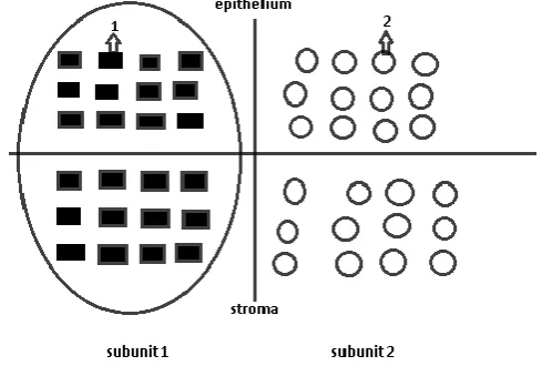
It was revealed that epithelial tissue of peripheral organs and also a basal layer of a skin epidermis are organized in specific structures – morphofunctional zones, in which a division of cambial cells with a further differentiation occur (Yavisheva and Shcherbakov, 2013 and Yavisheva et al., 2004, 2005(a)).[6, 7, 8] The morphofunctional zone consists of two subunits, each of which includes 12 cambial cells. At first the cells of the first subunit divide, therefore 12 pairs of mother and daughter cells are formed, and then of the second. Daughter cells of each subunit stretch in the electric field excited by 12 pairs of these cells and then are exposed to a differentiation (Figure 1).
Figure 1 Normal epithelial morphofunctional zone
1 – the mother cell, 2 – the daughter cell, turned out at cambial cell division, 3 – the cambial cell in inactive state.
The electric field arises because the main axis of the cambial cell is located vertically in relation to a basal membrane. Therefore, one of the cells, which turned out during the division and adjacent to a basal membrane, is not exposed to a differentiation and becomes maternal. It occurs due to the strengthening of the contractile properties of its cortex, which are in turn caused by the action of the growth factors, which are in a basal membrane and have high spastic activity (like the transforming growth factor β). As a result, there is no stretching of a mother cell nucleus, and the density of the chromatin, having the negative charge, increases. The other cell (daughter) is not treated to such action as it is located over it. Therefore, the negative charge in a mother cell in relation to daughter amplifies, which leads to redistribution of the surerficial charges between these two cells and to emergence of some electric field. Stretching of a daughter cell in an electric field occurs parallel to a basal membrane, therefore it changes the direction of the division axis from vertical to horizontal unlike mother cell, in which the main axis corresponds to a cambial cell.
The centrosome, adjacent tightly to a nuclear membrane, is tightened to the leading edge of a daughter cell, therefore the polarity of this cell is established. Because chromosomes are fixed to a nuclear membrane, at nucleus stretching they begin to unwind in particular sites, which opens a way to their transcription and differentiation.
Interestengly, the daughter cells of the skin epidermis, the eye limbus have the tyrosinase activity (Yavisheva et al., 2012). [9] After the daughter cells separate from maternal, they settle down between a basal membrane and the lower poles of the basal cells in the form of oblong melanocytes, and then closer
to a basal layer they asquire more rounded shape. Gradual decrease in melanocytes number correlates with increase in the cell quantity of a basal layer. Therefore, the tyrosinase in epithelial daughter cells, during their stretching in the electric field, becomes more active, and these cells gain features of melanocytes, being the earliest offspring of cambial cells.
It is important to note that in each subunit of a morphofunctional zone not all of 12 cambial cells enter into the proliferation simultaneously: first the first 6, then the other 6. At the entering of the first 6 cambial cells into the mitosis a differentiation of daughter cells is absent, because the field induced by 6 cells is not enough for the stretching of the daughter cells.
So, the process of the epithelial daughter cells differentiation is possible due to their stretching in the electric field. However it occurs only after a preliminary relaxation of their cortex, contracted owing to the own growth factors. This function is performed by a stroma.
We revealed that in the derma papillary layer there are cambial cells which quantity corresponds to epithelial (Yavishevaand Shcherbakov, 2013 and Yavisheva et al., 2007).[6, 10] These cells enter into the mitosis together with the subunits of the epithelial morphofunctional zone and compound with them the uniform epithelial-stromal morphofunctional zone. Each stromal subunit due to its growth factors relaxes the cell cortex of an epithelial subunit, which entered into the mitosis by this time, and provides an opportunity of a further differentiation of epithelial cells (Figure 2).
Figure 2 Normal epithelial-stromal morphofunctional zone. Interaction of
epithelial and stromal components of the first subunit.
1 – the divided cambial cells, 2 – cambial cells in a quiet state
Thus, epithelial tissue of the peripheral organs is constructed by type of the morphofunctional zones in which the differentiation of the epithelial daughter cells, which turned out at the cambial cells division, occurs during their stretching in the electric field, excited by 12 mother and daughter cells pairs of the same subunit. The cortex of the daughter cells has to be relaxed by stromal factors beforehand. 6 cambial cells excite an electric field of threshold level, at which the differentiation of the cells is absent.
the germinal zones of a brain (Mountcastle, 1979; Rakic, 1977, 2003).[11, 12, 13]The formed young neurones gradually migrate along radial fibers and unite in mini-columns. Interestingly, not separate cambial cells enter into the proliferation in the germinal zones of a neocortex and a hippocampus, but groups, consisting of 6-9 cells, then the other 6 proliferate, and only after that they differentiate (Reznikov and Nazarevskaya, 1989).[14]
Thus, the brain germ loci possess the features of the peripheral organs morphofunctional zones. There are data that the germ area of a hippocampus and olfactory bulb of mice is presented not only by subventricular region, but also the loci near the vessels, where cambial cells penetrate from the peripheral blood (Viktorov, 2001).[15]
At the same time in the germ zones of a hippocampus and an olfactory bulb of young mice especially we revealed small cells with the dense cytoplasm, which division axis differed from the other cells (Yavisheva and Shcherbakov, 2017).[16]These cells formed the groups with 3 cells in each, then they entered into the proliferation. Thus, about 6 cambial cells turned out in the group. It explains the fact of the synchronous entering of 6 cambial cells into the proliferation in the germ areas of a brain and also in epithelial and stromal morphofunctional zones of peripheral organs. During the division these cells had the same features, as the cambial cells of peripheral organs tissues. Two cells, which turned out at the division, were in contact with each other, then one of the cells – daughter began to stretch and polarize gradually.
Thus, the germ loci of the brain tissue are constructed by the principle of the the morphofunctional zones, in which the differentiation of neurones occurs only when the number of new cells reaches 12-18. Apparently, the mechanism of the neurones differentiation in the morphofunctional zone is similar to epithelial tissue and occurs due to the influence of the electric field. Among the brain morphofunctional zones the hypothalamus with its hypophysiotropic region is distinguished in a special way and represents the central morphofunctional zone, regulating the trial vegetative functions of an organism (Yavisheva and Shcherbakov, 2012, 2013).[17, 6]
Src-kinase role in the work of the morphofunctional zones
The function of the morphofunctional zones is ensured by many proteins, from which it is necessary to distinguish Src-kinase - one of the key proteins, participating in the most important processes of vital activity of the cells. Src-kinase can be present in the cells in its active and inactive form, and it is important for course of many processes in the cells. In its active form the Src kinase participates in a preliminary relaxation of epithelial daughter cells cortex for the purpose of their further stretching by the forces of the electric field (Yavisheva and Shcherbakov, 2009(a)).[18] So, stromal factors plentifully activate Src-kinase in epithelial cells through the SH3 domain. Further, the active Src-kinase stimulates protein p190RhoGAP, which in turn inactivates RhoA. But the latter participates in the formation of the formins and actin in the cell envelope. Therefore, their quantity decreases in the epithelial cells cortex and the contraction of the cells falls, which gives the possibility for the cells stretching in the electric field.
Interestingly, inactive Src-kinase plays a major role in relation to its active form in the formation of the cell cytoskeleton,
especially the microtubules, necessary for the differentiation. Thus, the daughter cell sizes increase in the process of its stretching in the electric field, which demands the reinforced exocytosis from membranes of Golgi complex for the purpose of the replenishment of the cell membrane. Therefore, there is an increasing receipt of inactive Src-kinase, which is located on these membranes. However, activization of this kinase in the center of the cell happens already due to its SH2 domain. So, the large protein complexes with Src-kinase, PI3K and γ-тубулин are formed on the centrosome (Sulimenko et al., 2006).[19]
Because Src-kinase has low affinity to tubulin, PI3K beforehand phosphorylates tubulin, then Src-kinase bindes to its phosphotyrosine residues by means of the SH2 domain. Therefore, Src-kinase shows not kinase properties, but the the features of the cross-link protein first of all, connecting the dimers of a tubulin and by this participating in microtubules formation. After that the activation of the the Src kinase domain occurs. If this domain was activated before accession to a tubulin, then it would lead to a larger decrease in affinity of Src to a tubulin and falling of microtubules formation. It is observed at a virus carcinogenesis, where the kinase lobe of viruses Src is always activated and interferes with the formation of the host cell cytoskeleton.
Microtubules formation promotes the development of the intermediate microfilaments by means of what the nucleus of a daughter cell stretches in the electric field, excited during 12 cambial cells division, which leads then to the differentiation of daughter cell. Therefore, the function of 12 cambial cells is directed first of all to the expression of certain amount of the inactive Src-kinase (very important protein, which participates in the differentiation) in the daughter cells.
Further, the microenvironment of a daughter cell defines the direction of a differentiation of a daughter cell: towards formation of an epitheliocyte or a fibroblast,which depends on amount of inactive and active Src-kinase in this environment. So, the daughter cell, which turned out at cambial cell division, being in an epithelium, will be exposed to the action of epithelial tissue growth factors, which have spastic properties. Therefore, the expression of inactive Src and RhoA will remain high, which complicates stretching of the top pole, in which centromeres of a daughter cell are located. In this regard the lower pole of a cell with the telomeres, which are located in it, will stretch in an electric field. This will lead to the untwisting of chromosomes near the telomeres and to formation of an epitheliocyte (Figure. 3 a, b).
a b
Figure 3 Formation of epithelial (a) and stromal (b) daughter cells at the
If the division of the cambial cell happened in the stromal tissue, then the top pole of the daughter cell (which is treated to action of the stromal factors, strengthening a portion of the active Src-kinase and reducing a part of inactive Src-kinase and RhoA), will be relaxed. Therefore, the top pole with the centromeres, which are situated in it, will stretch in the electric field more than the lower part of the cell, so this will lead to the untwisting of chromosomes sites closer to the centromeres and to the fibroblast formation.
Thus, in the epithelial cell the untwisting and the further transcription of chromosomes will occur closer to the telomeres, and in fibroblasts – near the centromeres, because in the epithelial cell the amount of inactive Src-kinase increases, and the part of the active Src decreases; in the fibroblast, on the contrary, the amount of the active Src-kinase increases and the portion of inactive Src decreases (Yavisheva and Shcherbakov, 2013).[6]
a b
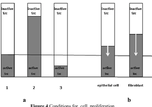
Figure 4 Conditions for cell proliferation.
а). 1 – proliferation happens if inactive Src-kinase more, than moderately exceeds the active form, 2 - proliferation is absent if an expression of inactive Src-kinase weak in relation to the active form, 3 – there is no proliferation if the expression of inactive Src-kinase sharply exceeds an expression of the active Src; b). proliferation of fibroblasts requires larger increase in an expression of inactive Src-kinase, than in epitheliocytes.
The certain ratio of inactive Src-kinase to its active form leads not only to a concrete differentiation of a daughter cell in a morphofunctional zone, but also to the cell proliferation. So, cells enter into mitosis only if inactive Src-kinase more than moderately prevails over the active form, because the active Src inactivates a part of RhoA (Figure. 4 a).Later,Src participates in activization of the transcription factors, and together with the remained portion of RhoA – in formation of a contractile ring. Because in epitheliocytes inactive Src-kinase moderately prevails over the active Src, slight increase of the inactive Src-kinase expression in the microenvironment is enough for beginning of cells proliferation. It explains high proliferative activity of epithelial cells (Figure 4b).
If inactive Src-kinase considerably prevails over the active, then the proliferation will sharply decrease because of strengthening of cell cortex contraction and the decrease of an expression of nucleus transcription factors.
On the contrary, if the active Src-kinase is much more than its inactive form, then a proliferation of cells also is absent as this prevents from the formation of cell cytoskeleton and cytokinesis.
Thus, Src-kinase is directly involved in cells proliferation and differentiation, and both of these processes depend from a ratio of two forms of this protein: the active and inactive. Therefore, it is necessary the sufficient expression of Src-kinase in daughter cells during the electric field action.
The central morphofunctional zone directs processes of aging by means of Src-kinase
So, the daughter cell is exposed to a differentiation after the sufficient expression of inactive Src-kinase in it during its stretching in the electric field, excited at 12 cambial cells division. The weak field of 6 cambial cells does not differentiate the cells. With age there is a decrease in quantity of cambial cells in each subunit of a morphofunctional zone from 12 cells in 20-60 years old people up to 7 over 75 years (Yavisheva and Shcherbakov, 2009(b), 2013).[20, 6] Therefore, the expression of inactive Src-kinase in the morphofunctional zones falls with aging. Systemic falling of cambial cells quantity and an expression of inactive Src-kinase in people after 60 years in the period of a hypoestrogenemia demonstrates the participation of the central structures in this process, among which the main role is played by a hypothalamus with hypophysiotropic region (Yavisheva and Shcherbakov, 2012, 2013).[17, 6]
In the hypothalamus similarly with an epithelial morphofunctional zone the expression of inactive Src-kinase prevails over its active form. This is because the brain develops from the ectoderm in the embryogenesis, in which initially there is a high level of inactive Src-kinase expression. The neural plate during its formation is under the influence of the chord, in which the portion of the active Src-kinase is considerably increased. Therefore in nervous tissue there is only transitional strengthening of the active Src; in this connection inactive Src prevails in neurons (Yavisheva and Shcherbakov, 2012).[17]
Brain neurotransmitters, as well as growth factors of epithelial tissue, promote increase of inactive Src-kinase share and by this interfere with its decrease in nervous tissue. To start the process of synthesis or the exocytosis in a cell-target, it is necessary that the signal, coming to a cell, moderately lowered a part of inactive Src-kinase and the production of neurotransmitters in it. At the same time the portion of the active Src-kinase, which participates in the processes of a transcription, increases. Besides, the active Src-kinase takes part in the formins and actin formation and its moderate quantity is necessary for the contractile ring forming at cell division and also in exercise of an exocytosis.
Srcactive form, which is a kind of "priming" in carrying out the excitation.
Thus, the exciting signal has to reduce the concentration of inactive Src-kinase in a cell to start to pass this signal along the regulatory chain.
The inhibitory signal, on the contrary, carries in itself the information on increase in a portion of inactive Src-kinase in cells-targets. These signals can be transmitted to neurons from environmental tissue and also from peripheral organs.
At childhood the synthesis of many hypothalamic releasing-factors is suppressed owing to high expression of inactive Src-kinase in the hypothalamus and also the influence of other departments of a brain, synthesizing the mediators, which increase a share of inactive Src-kinase in it. Great inhibitory influence on a hypothalamus is exerted by the melatonin, epiphysis hormone, increasing the concentration of inactive Src-kinase in tissues. During the prepubertyits quantity in the organism falls by 40 times in comparison with the childhood.
Decrease in melatonin amount in this period is intimately connected with maturation of ferment systems on digestion of cholesterol (Mazurin, 1984). [24] This inherited programmed mechanism begins to operate only at 5-7-year old children. Cholesterol in turn is a basis for synthesis of steroid hormones. In adrenal cortex in the prepuberty a small synthesis of estrogens occurs, as adrenocorticotropic hormone (AKTH) is not completely blocked by melatonin. In the process of cholesterol digestion, since at 7-years synthesis of estrogens amplify. In a brain, especially a hypothalamus and an epiphysis there are many receptors to estrogens. Estrogens activate Src-kinase by means of G-proteins, which join with its catalytic domain. Therefore, in brain cells the share of inactive Src-kinase decreases, which leads to decrease in quantity of mediators, in particular, of noradrenaline, participating in production and secretion of melatonin, which level falls. It leads to the removal of inhibition from many hypothalamic peptidergic neurons and to development of releasing-factors by them, including gonadotropin-releasing hormone (GnRH). As the result, the period of puberty comes (Yavisheva and Shcherbakov, 2012). [17]
Thus, the concentration of inactive Src-kinase and production of neurotransmitters, rendering the inhibitory effect on peptidergic neurons, decrease in the brain during cholesterol digestion and the increase of estrogens synthesis. Neurotransmitters are synthesized by specialized neurons, which perceive information from the cortex and other parts of the brain and also from the outside. Because inhibitory influence of neurotransmitters weakens, the sexual center of a hypothalamus becomes very sensitive to excitatory stimuli.
Therefore, light quickly activates Src-kinase in the retinamelanopsin cells, then this information is transferred to monoaminergic, further to peptidergic neurons and a pituitary. In this regard, daily fluctuations of gonadotropin secretion during the formation of pubertal phase are noted. So, the portion of the active Src-kinase increases and the share of inactive decreases in the monoaminergic neurons under the influence of light. Therefore, the production of the monoamines, strengthening an expression of inactive Src-kinase, will decrease a little, which will improve the exocytosis. At the same time the conditions for signal transmission in neurocytes of the sexual center will be created and the share of the active Src-kinase will also increase in
them.In the hypothalamic peptidergic neurons the synthesis of GnRH begins and the exocytosis amplifies. GnRH stimulates two other hormones of a pituitary – luteinizing hormone (LH) and follicle-stimulating hormone (FSH). LH is responsible for androgens synthesis in granulosa and ovary theca-cells. FSH strengthens aromatization of androgens, therefore estrogens, which increase a share of the active Src-kinase in tissues-targets, including a brain, are formed. In the process of estrogens formation in ovaries and their action on monoaminergic neurons and neurocytes of sexual center, a further decrease in a share of inactive Src-kinase in these cells occurs, at the same time production of neurotransmitters decreases. Therefore, their inhibitory effect on the hypothalamic peptidergic neurons weakens even more, which leads to strengthening of the exocytosis. Consequently, gradual increase in ovaries estrogens leads to some acceleration of the GnRH secrete rhythm by the peptidergic neurons and to strengthening of the pituitary FSH secretion in the afternoon. At night when light is absent, the rhythm of the GnRH secretion decreases due to the increase in the share of inactive Src-kinase in the cells and deterioration of the exocytosis, which leads to emission of LH. Over time, the considerable decrease in a share of inactive Src-kinase and also in the number of neurotransmitters during the increase of estrogens influence on the neurocytes of the sexual center occurs in these cells.It leads to a larger removal of inhibitory effect from the hypothalamic peptidergic neurons, to increase in a rhythm and amplitude of GnRH secretion. Therefore, at teenagers the further increase in estrogens production will be observed in the afternoon.
Due to the increase in estrogens production there is a further weakening of inactive Src-kinase in the brain cells, which leads to sharp increase of frequency and amplitude of GnRH secretion. This in turn will cause FSH plentiful stimulation and the appearance of the follicular phase of a cycle.
Secretion of LH will be intensive because of the decrease in inhibitory action of neurotransmitters at this time. Therefore, by the end of the pubertal period differences in night and day impulses of these hormones production smooth out and their mature type of secretion is formed.
Thus, estrogens, influencing over the brain tissues since the pubertal period, gradually reduce initially high content of inactive Src-kinase in the brain, especially in the hypothalamus, reducing at the same time the number of neurotransmitters. Hence, their inhibitory effect weakens, and the pass of excitatory signal amplifies.
The above mentioned processes exhaust the resources of inactive Src-kinase in the brain with aging, therefore the period of hyperestrogenemia appears. So, due to the decrease in the inhibitory effect, the excitatory signal not gradually, but very quickly increases a share of the active Src-kinase, necessary for the performance of the exocytosis in the cells, which leads to increase in amplitude of fluctuation of mediators emission from the monoaminergic neurons. This information passes to the hypothalamic peptidergic neurons and then to the pituitary, where the intensive activation of Src-kinase and the increased synthesis of FSH also occur.
especially in a hypothalamus. As a result, the maintenance of FSH will exceed LH, but over time the LH level will also increase in a pituitary, because the reduced number of neurotransmitters does not prevent the production of this hormone.
In the period of a hyperestrogenemia the influence of stroma upon the epithelial tissue increases in the peripheral organs. It is connected with the strengthening of the active Src-kinase expression in the cells, especially in fibroblasts, because they have many receptors to estrogens. Fibroblasts and epithelial cells will enter into mitosis more slowly, because for this process it is necessary more, than at young people, to raise an expression of inactive Src-kinase in the morphofunctional zone. Besides, the increase in the share of the active Src-kinase in relation to its inactive form leads to appearance of a fibroblast-like phenotype of the cells,i.e. the cells begin to stretch closer to centromeres in the process of the differentiation. It causes strengthening of the connective tissue development not only in ovaries, but also in other parenchymatous organs. Further weakening of inactive Src-kinase concentration and neurotransmitters to definitely low level in the central morphofunctional zone leads to change of the hyperestrogenemia for the hypoestrogenemia. In this regard, the excitatory signal at once in the complete measure increases the GnRHsynthesis and the exocytosis, which leads to the large, but not pulsing emission of FSH. From literature data it is known, that if hormone is present constantly and affects in high concentrations on the cell-target, then the insensitivity to this stimulus develops (Tepperman J. and Tepperman H, 1989).[25] Therefore, the ovary cells do not answer to the high signal, passing from a pituitary. Production of LH also amplifies, as the inhibitory effect of various neurotransmitters and brain environmental tissues, which have a weak expression of inactive Src-kinase now, is reduced.
During this period the influence of stroma over the epithelium decreases due to the reduction of estrogens impact and therefore, the active Src-kinase on tissues. The proliferative activity of fibroblasts increases, because their division requires considerably a smaller expression of inactive Src-kinase now, than in the period of a hyperestrogenemia. Unlike fibroblasts the mitotic activity of epithelial cells sharply worsens, because the weak stroma cannot relax a rigidity of epithelial cells cortex. In the period of a hypoestrogenemia the beginning of the decrease in the cambial cells number in the morphofunctional zones is noted. Their quantity at persons of 60 years is 11 cells, and over 75 - sharply falls to 7 cells in one subunit, which is close to the threshold level (6 cells), when the differentiation of the cells is absent (Yavisheva and Shcherbakov, 2009(b), 2012). [20, 17]
Because the influence of estrogens over the brain, especially the central hypothalamic morphofunctional zone leads to weakening of inactive Src-kinase in these structures, therefore a regulation of many pituitary hormones, including AKTH, changes. So, at young people the signal from photoreceptors and the retina melanopsin cells passes to SHN with the reinforced portion of the active Src-kinase in it, because light activates Src-kinase. Further the active Src-kinase weakens the expression of inactive Src-kinase in the hypothalamic peptidergic neurons, therefore the exocytosis and the synthesis of the corticotropin releasing factor, stimulating production of pituitary AKTH, amplify. AKTH intensifies further development of glucocorticoids in adrenal cortex.
At night the signal from the photoreceptors and the retina melanopsin cells decreases, which leads to the falling of the active Src-kinase level. Therefore, the amount of the active Src-kinase in the regulatory chain: photoreceptors - SCN- peptidergic neurons of the hypothalamus, will fall. It leads to the increase in inactive Src-kinase portion in this pathway, which will cause the falling of the releasing-factor synthesis and the deterioration of the exocytosis in the hypothalamus peptidergic neurons. At young people the melatonin and other transmitters, increasing the concentration of the inactive Src-kinase, are available enough in the organism, therefore they suppress metabolic activity of SCN, increasing duration of its circadian cycle. Thus, the AKTH emission is noted at 6-8 a.m.
With aging due to the falling of the inactive Src-kinase level in the brain, the inhibitory influence of the neurotransmitters over SCN decreases, which reduces the duration of SCN own cycle. In this regard AKTH is synthesized at 3 a.m., which leads to the relative AKTH increase in the organism. If the level of the inactive Src-kinase falls considerably in the brain, then it causes activization of the exocytosis in the hypothalamus neurocytes, developing corticotropin releasing factor. In the pituitary the enlarged synthesis of AKTH also begins, the latter stimulates adrenal cortex to the development of the increased quantity of glucocorticoids. Therefore, the absolute quantity of AKTH and glucocorticoids can increase with aging. AKTH participates in a gluconeogenesis and competes with insulin for glucose metabolism. The significant increase in this hormone leads to violation of many types of metabolism, development of the diabetes, the atherosclerosis.
AKTH as well as melanocyte-stimulating hormone (MSH) strengthens a pigmentation of the tissues, because both are included into the common group of the peptides, having this activity. At the same time MSH not so strengthens a melanogenesis, as disseminates a pigment, which is reached by the increase of the active Src-kinase share in the melanosomes. Therefore, the action of AKTH is directed first of all to decrease the inactive kinase and increase the active Src-kinase level in tissues-targets (Yavisheva and Shcherbakov, 2017).[16]
Glucocorticoids unlike AKTH increase the inactive Src-kinase amount, because their insufficient quantity leads to a dispelling of a pigment in tissues-targets. During the accumulation of the synthesized glucocorticoids and the increase of the inactive Src-kinase concentration in tissues, the share of the active Src-kinase, participating in synthesis of AKTH will be suppressed, which will cause the block of its synthesis in the pituitary.
Thus, glucocorticoids as terminative product, block further synthesis of AKTH due to the increase of the inactive Src-kinase in the pituitary and hypothalamic peptidergic neurons. Therefore, glucocorticoids act as a inhibitory signal for AKTH synthesis.
Estrogens unlike glucocorticoids strengthen the FSH function, raising the active Src-kinase share in neurons, which leads to decrease of the inactive Src-kinase expression in the brain. Therefore, estrogens function as a excitatory signal in relation to synthesis of FSH and constantly reduce the reserves of inactive Src-kinase in a brain, especially in a hypothalamus.
content of inactive Src-kinase. However estrogens, influencing over the brain, gradually exhaust the reserves of inactive Src-kinase in the hypothalamus. As a result, a regulation of sexual hormones changes, which leads to the appearance of the puberty, the latter changes for a hyperestrogenemia and then–for hypoestrogenemia. It leads to decrease in the cambial cells quantity in all organism to the level, very close to the critical, at which the malignant tumor can develop. Decrease in the inactiveSrc-kinase share in the central hypothalamic morphofunctional zone affects function of the other hormones, in particular the secretion of AKTH, which can lead to the development of atherosclerosis, metabolic violations, diabetes.
Therefore, maintaining of a steadily high level of inactive Src-kinase in the central morphofunctional zone can lead to a normalization of functionality of peripheral organs tissues.
Tumor process and morphofunctional zones
Thus, aging is more caused by the decrease of inactive Src-kinase expression in the central hypothalamic morphofunctional zone, which leads to the decrease of cambial cells quantity in all organism. Therefore, tumor process is connected with aging very often. Really, over 75 years the cambial cells quantity in each subunit of the morphofunctional zone decreases to 7 and is close to the level (6 cells), when the differentiation is absent. Thus, developing of a malignant tumor in the peripheral morphofunctional zones depends on decrease of cambial cells quantity in them and concentration of inactive Src-kinase.
In this regard the tumor can develop not only at elderly people, but also at young, when there is a decrease in cambial cells number due to the stresses, steady strong stimulating signals (the strong bright light, loud sounds, etc), because they strengthen a share of the active Src-kinase and reduce an expression of inactive Src in the brain. Besides, individual low level of inactive Src-kinase in the brain under adverse conditions can cause critical falling of cambial cells number.
The local reasons can also lead to decrease of cambial cells quantity, including injuries, burns. In other cases the cambial cells number can remain normal, but the expression of the proteins (inactive Src-kinase), responsible for the formation of cell cytoskeleton and a differentiation, at the same time decreases (Yavisheva and Shcherbakov, 2014).[[26] For example, inflammation at which there is an accumulation of the active forms of oxygen or influence of ultraviolet (UV) reduce the level of inactive Src-kinase in the cells, because it quickly becomes more active under the influence of these factors. It is shown above that in epithelial daughter cells at their stretching in an electric field the tyrosinase becomes more active. Large influence of UV can sharply strengthen a tyrosinase expression. Activization of a tyrosinase requires the reduction of copper ions, which are in its active site, without what the oxidation of tyrosine is impossible. This process is carried out due to the thiol groups; the inactive Src-kinase, which occurs in the melanosomes is one of donors of these SH groups (Mallozzi et al., 2001) [27]
Hence, the tyrosinase will compete with inactive Src-kinase in the conditions of the enlarged action of UV, therefore the share of inactive Src, participating in a cytoskeleton formation will decrease considerably. As a result, nuclei stretching of such cells will be very weak, which can lead to the development of a low- differentiated tumor – melanoma.
Thus, the malignant tumor can develop in the case of falling in cambial cells number to 6, when the electric field, excited by such cells quantity cannot express in the daughter cells enough portion of inactive Src-kinase, necessary for the development of their cytoskeleton and a further differentiation; or when the share of inactive Src-kinase for various reasons decreases in daughter cells to critical level though at the same time the cambial cells number can be high.
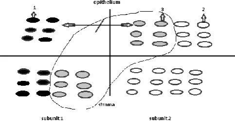
It is simpler to examine the process of a malignant tumor formation in the morphofunctional zones, based on the cambial cells quantity. At the same time it is necessary to consider the influence of the stromaover the epithelial tissue (Figure 5).
Figure 5 The scheme of a malignant tumor development in a
morphofunctional zone. Interaction of electric fields of two epithelial subunits is absent.
1 – activated daughter cells, 2-non-activated daughter cells, 3-interaction of daughter cells of different subunits.
When we studied a dysplasia and cancer of the esophagus, a carcinoma of the stomach, breast cancer, it was revealed that the malignant tumor arises in one subunit as a result of the decrease in cambial cells number in it not less, than twice (Yavisheva and Shcherbakov, 2009(a), 2013 and Yavisheva et al., 2005(b)).[18, 6, 28]It should be noted that during formation of a tumor the underlying stroma is not changed. At the entering of such subunit into a proliferation only 6 pairs of mother and daughter cells are formed. It was shown above that daughter cells cannot be stretched by forces of an electric field without preliminary relaxation of their cortex. This function is performed by growth factors of stromal subunit. However in a stromal component there will be still factors of 6 odd cells. When the other subunit enter into mitosis these 6 odd stromal cells will influence over just formed daughter cells of the second subunit. But the differentiation of daughter cells of the first and second subunits will be absent, because 6 pairs of mother and daughter cells cannot excite the electric field, sufficient for daughter cells stretching. Besides, interaction between the fields of two subunits is also absent, because the fields are very weak In fact, the cells do not differentiate in a small electric field, at the same time the concentric- spherical type of a symmetry is created, where the cancer structures are located in concentric orbits.
The decrease in a share of inactive Src-kinase causes the change in other departments of a brain
inactive Src-kinase participates in formation of a cytoskeleton, a melanogenesis and a differentiation of cells (Yavisheva and Shcherbakov, 2016) [29]. Therefore dendrites, axons and growth cones, where microtubules and actin microfilaments are included, are formed with participation of inactive Src-kinase. Therefore, decrease in a share of inactive Src-kinase leads to shortening of nerve-cell processes and decrease in interrelation between neurons with aging.
Besides, in old mice the proliferative activity of hippocampal cells in comparison with olfactory bulbs is sharply reduced. It is caused by additional impact on hippocampal cells of glucocorticoids, which strengthen inactive Src-kinase. Receptors to glucocorticoids in a hippocampus are distributed not diffusively, but locally and most expressed in the CA1-2
fields. (Gerlach and McEwen, 1972).[30] Interestingly, the field CA1 first of all is exposed to pathological changes at a
senile dementia and Alzheimer's disease (Vinogradova, 1975).[31]
Really, glucocorticoids, which quantity increases with aging, pass a inhibitory signal for nervous cells, reducing an exocytosis and signal transmission in synapses to other neurons. Besides, the sharp local dominance of inactive Src-kinase in an area of function of glucocorticoids leads to strengthening of spastic properties of the nervous cells cortex and their processes. All these factors complicate pathway of nervous signals and promote the development of neurodegenerative violations. If the person has the low individual level of inactive Src-kinase in the brain, then this can accelerate and aggravate the pathological process.
CONCLUSION
1. In epithelial and stromal tissue there are morphofunctional zones, consisting of two subunits (with 12 cambial cells in each), in which the processes of a cell proliferation and differentiation occur. 2. Src-kinase is one of the key proteins in the
morphofunctional zones work.
3. The function of cambial cells of the morphofunctional zones is directed first of all to an expression and maintaining of a certain level of the inactive Src-kinase, necessary for a cytoskeleton formation and a differentiation of daughter cells.
4. 12 cambial cells of one subunit of a morphofunctional zone are necessary for these purposes. 6 cells express a critical number of inactive Src-kinase at which the differentiation of cells is absent.
5. The central hypothalamic morphofunctional zone plays a prime role in a regulation of cambial cells number and degree of inactive Src-kinase expression. Estrogens, influencing over a brain and a hypothalamus, lead to the exhaustion of this protein resource and change of a hormonal regulation of organs and tissues. The periods of hyper-and hypoestrogenemiaare caused, in which the proliferative activity of cambial cells falls and reaches 7 cells in people over 75 years old, that is close to the threshold level (6 cells), at which the differentiation of cells is absent.
6. The local mechanisms also participate in a regulation of the cambial cells quantity and degree of inactive Src-kinase expression. The local factors, reducing the cambial cells number to 6 or an expression of inactive Src-kinase to critical level can lead to the malignant tumor development. Threshold decrease in an
expression of inactive S-kinase at influence of ultraviolet leads to the development of a melanoma. 7. The secretion of many hormones, including AKTH,
changes at decrease of inactive Src-kinase amount in a brain and especially in a hypothalamus. This leads to increase in concentration of glucocorticoids, large number of which is collected in a hippocampus. The raised production of AKTH can lead to a metabolic disorder, development of an atherosclerosis, a diabetes. 8. In a hippocampus the cambial cells quantity and an
expression of inactive Src-kinase also decreases, which worsens a neurons cytoskeleton formation and nerve-cell processes. It is promoted by the low individual level of inactive Src-kinase, which was initiated in an embryogenesis. Besides, local increase in inactive Src-kinase due to the action of the extended quantity of glucocorticoids leads to decrease in the work of synapses and difficulty of signaling, which in total can promote the development of neurodegenerative changes.
References
1. Booth, C. and Potten, C. Gut instincts: thoughts on intestinal epithelial stem cells. Journal of Clinical Investigations.2000.105 :1493-1499.
2. Potten, CS.. The epidermal proliferative unite: the possible role of the central based cell. Cell and Tissue Kinetics.1974.Volume 7(1):77-88.
3. Potten, CS. and Booth, C. Keratinocytes stem cells: a
commentary. Journal of Investigative
Dermatology.2002. Volume 119(4):888-899.
4. Savostyanov, GA. Principles of structural histology. Space organization of epithelia. St. Petersburg: Nauka;2006.
5. Schofield, R. The relationship between the spleen colony forming cell and hemopoietic stem cell. A hypothesis. Blood cells.1978. Volume 4(1-2):7-25. 6. Yavisheva, TM. and Shcherbakov, SD.
Epithelial-stromal morphofunctional zones: structure and functions. Moscow: RAMN; 2013.
7. Yavisheva, TM., Shcherbakov, SD, Golubeva, IS., Sharafutdinov, GZ. and, Savluchinskaya, LA. To a question of the structural organization of a basal layer and the morphofunctional features of mouse skin cambial cells. ByulletenEksperimentalnoiBiologii I Meditsiny, 2004.Volume 137(5):584-588.
8. Yavisheva, TM., Shcherbakov, SD. and Sharafutdinov, GZ. To a question of epithelial cells differentiation in the system of tissue units. Doklady AN.2005(a).Volume 401(6): 833-836.
9. Yavisheva, TM., Shcherbakov, SD., Golubeva, IS, Savluchinskaya, LA. and Rizhova, NI. The relationship between the epidermal melanocytes, Langerhans cells and epidermal cambial cells. Byulleten EksperimentalnoiBiologii I Meditsiny. 2012.153(3):346-349.
10. Yavisheva T.M., Shcherbakov S.D., Golubeva I.S., Sharafutdinov G.Z. Interaction of cambial dermal cells (fibroblasts) and epidermis in morphofunctional zone of mouse skin. Byulleten Eksperimentalnoi Biologii I Meditsiny.2007.144(11):594-599.
Neurosciences.4-th Study Program. Cambridge etc.: The MIT Press: 21-42.1979.
12. Rakic, P. Prenatal development of the nervous system in rhesus monkey. Philosophical Transactions of the Royal Society B: Biological Sciences.1977.Volume 278: 245-260.
13. Rakic P.Developmental and evolutionary adaptations of cortical radial glia. Cerebral Cortex.2003. Volume 13(6): 541-549.
14. Reznikov, KYu, Nazarevskaya, GD. A proliferation and a cytogenesis in the developing hippocampus of a mouse.Moscow:Nauka;1989.
15. Viktorov, IV. Stem cells of mammals brain: biology of stem cells. IzvestiyaAN.Biological series. 2001. No.6: 646-655.
16. Yavisheva, TM. and Shcherbakov, SD. Some aspects of morphofunctional organization of germinal regions of the hippocampus and the olfactory bulb in young and old mice. International Journal of Innovative Studies in Medical Sciences.2017.Volume 1(1): 4-11.
17. Yavisheva, T M. and Shcherbakov, SD. Participation of the morphofunctional zones in aging process. Uspekhigerontologii. 2012. Volume 25(4):604-611. 18. Yavisheva, T M. and Shcherbakov, SD. Features of a
proliferation and a differentiation of cambial and daughter cells of the morphofunctional zones in a normal epithelium and cancer in age aspect. Uspekhi gerontologii. 2009(a).Volume.22(4): 605-613.
19. Sulimenko, V., Draberova, E. and Sulimenko, T., Regulation of microtubule formation in activated mast cells by complexes of gamma-tubuline with Fyn and Syk kinases. The Journal of Immunology. 2006.Volume 176 (12): 7243-7253.
20. Yavisheva, T M. and Shcherbakov, S.D. Morphofunctional changes of cambial cells and their derivatesin human skin in age aspect. Byulleten EksperimentalnoiBiologii I Meditsiny.2009(b). Volume 8(9):326-329.
21. Aikawa, R., Komuro, I., Yamazaki, T., Zou, Y., Kudoh, S., Tanaka, M., Shiojima, I., Hiroi Y. and Yazaki,Y. Oxidative stress activates extracellular signal-regulated kinases through Src and Ras in cultured cardiac myocytes of neonatal rats. Journal of Clinical Investigation.1997.100(7):1813-1821.
22. Burova, EB., Gonchar, IV.and Nikolsky, NN.Stat1 and stat3 activation by oxidative stress in A431 cells involvessrc-dependent EGF receptor tranactivation. Tsitologiya. 2003.45(5):466-477.
23. Devary, Y., Gottlieb, RA., SmealT. and Karin, M. The mammalian ultraviolet response is triggered by activation of Src tyrosine kinases. Cell. 1992. 71(7):1081-1091.
24. Mazurin, AV. Diseases of digestive organs at children.M oscow: Medicine;1984.
25. Tepperman, J. and Tepperman, H. Metabolic and endocrine physiology. Moscow: Mir; 1989.
26. Yavisheva, TM. and Shcherbakov, SD. Development of melanoma and cancer without decreasing of cambial cells number in morphofunctional zones. Advances in Bioscience and Biotechnology. 2014.Volume5(6):493-500.
27. Mallozzi, C.; Di Stasi, M. and Minetti, M. Peroxynitrite-dependent activation of src tyrosine kinases lyn and hck in erithrocytes is under mechanistically different pathways of redox control. Free Radical Biology and Medicine.2001. 30(10):1108-1117.
28. Yavisheva, T M., Shcherbakov, S D. and Savluchinskaya, LA. Features of the morphofunctional zones work in normal epithelium, fibroadenoma and breast cancer. Dokady AN.2005 (b). Volume 404(1):125-128.
29. Yavisheva, TM. and Shcherbakov, SD . Participation of an inactive and active Src-kinase in formation of a cytoskeleton and melanogenesis in Hep2 cells. International Journal of Current Microbiology and Applied Sciences.2016.5(12):583-593
30. Gerlach, JL. And McEwen, BS. Rat brain binds adrenal steroid hormones. Radioautography of Hippocampus with cortecosterone. Science.1972. 175:1133.
31. Vinogradova, OS. Hippocampus and memory. Moscow: Nauka; 1975.
How to cite this article:
Yavisheva T. M and Shcherbakov S.D (2018) 'To a question of structural and functional organization of the morphofunctional zones in age aspect. Participation of src-kinase in the work of morphofunctional zones', International Journal of Current Medical And Pharmaceutical Research, 04(6), pp. 3336-3344.