PR0IEOGLYCANS IN
N
e u r u u k iio n
By
Wai Cheong George Yip
A thesis sutxnSted Ibrthe degree of Doctor of Philosophy
University of London
2001
Developmental Biology Unit
Institute of Child Health
University College London
30 Guilford Street
London
ProQuest Number: U642687
All rights reserved
INFORMATION TO ALL USERS
The quality of this reproduction is dependent upon the quality of the copy submitted.
In the unlikely event that the author did not send a complete manuscript and there are missing pages, these will be noted. Also, if material had to be removed,
a note will indicate the deletion.
uest.
ProQuest U642687
Published by ProQuest LLC(2015). Copyright of the Dissertation is held by the Author.
All rights reserved.
This work is protected against unauthorized copying under Title 17, United States Code. Microform Edition © ProQuest LLC.
ProQuest LLC
789 East Eisenhower Parkway P.O. Box 1346
A
b s tr a c t
Glycosaminoglycans are made up of repeating disaccharide subunits
consisting of a hexosamine and a hexose or hexuronic acid. They
occur freeiy or as carbohydrate side chains in proteogiycans. Although
widely distributed, their importance in embryogenesis is incompletely
understood. This thesis investigates their roles in neurulation and
neural crest migration.
Primary neurulation occurs between E8.5 and E10.5 in the mouse
embryo. Neural tube closure in the spinal region is accomplished by
bending at the median hinge point and at paired dorsolateral hinge
points. Histochemicai and immunohistochemical studies show that
suiphated glycosaminoglycans are present in the basement membrane
of the closing neural tube, as well as elsewhere in the embryo.
Culturing E8.5 CD1 mouse embryos in the presence of chlorate, a
competitive inhibitor of glycosaminoglycan suiphation, suppressed
median hinge point formation. This was associated with increased
bending at the dorsolateral hinge points, resulting in accelerated
posterior neuropore closure. Suppression of median hinge point
formation was prevented by addition of heparan sulphate, but not
chondroitin sulphate, de-N- or de-O-sulphated heparan sulphate, to the
culture medium. This may be due to the requirement for heparan
sulphate in Sonic hedgehog induction of median hinge point formation.
Chondroitin sulphate also influenced posterior neuropore closure.
closure of the neuropore, whereas exogenous chondroitin sulphate
accelerated closure. However, the median and dorsolateral hinge
points were unaffected. The mechanism of action of chondroitin
sulphate is unknown and requires further investigation.
Besides their role in neurulation, chondroitin sulphate proteoglycans are
known to inhibit cell migration. Splotch mouse mutants, where
mutations in Pax-3 result in deficiencies of neural crest derived
structures, have been shown by in situ hybridisation to over-express
versican mRNA. To determine whether the glycosaminoglycan
component was increased, chondroitin sulphate was quantified in E9.5
splotch embryos photospectroscopically using a 1,9-dimethylmethylene
blue binding assay. It was found that mutants contained larger amounts
of chondroitin sulphate than wild type embryos. On the other hand,
there was no difference in the net synthetic rate, as determined by ^^S-
labelling of chondroitin sulphate in cultured splotch embryos. This
suggests that the defect may lie in the mutants' ability to degrade
chondroitin sulphate, resulting in its accumulation and inhibition of
neural crest migration.
These experiments underline the importance of suiphated
glycosaminoglycans in mouse development and raise the possibility that
these molecules may contribute to development of neural tube defects
A
c k n o w le d g e m e n ts
I would like to take this opportunity to express my heartfelt thanks to the
many people who helped in the production of this thesis.
Firstly, I am very grateful to my supervisors, Andy Copp and Patrizia
Ferretti, for their generosity, inspiration and guidance throughout the
course of my studies in London. I would also like to thank my first
supervisor, the late Peter Thorogood, who introduced me to the function
of heparan sulphate in embryonic development. Although this project
has taken a substantial change in direction from the one we planned
initially, I am sure he would have enjoyed reading this thesis.
I would like to show my appreciation for the many past and present
members of the Developmental Biology and Neural Development Units
who have been very helpful and who have made my stay in London so
enjoyable. I would like to specially thank Patricia Cogram for sharing
her tips on embryo culture; Carlos Gaston, Jason Neil-Dwyer, Anita
Petiot, Viccy Reed and Patricia Ybot-Gonzalez for showing me the
various practical aspects of Molecular Biology and for sharing reagents;
Rachel Moore, Paul O’Neill and Sarah Reid for practical protocols and
help with microscopy and immunohistochemistry; Joe Chan for his
computer expertise; and Melanie MacKay and Fang Zhang for
stimulating discussions. I am also grateful to Dave Stanton for his help
with the scintillating counter and Angie Wade for her expert advice on
I would like to thank the National University of Singapore for the award
of an Overseas Graduate Scholarship for my studies, as well as
Professors E A Ling and S K Leong, Associate Professors S S W Tay
and B H Bay, and my other friends and colleagues in the Department of
Anatomy, National University of Singapore for their kind support.
Last, but not least, I would like to express my deepest gratitude to my
parents for their love, kindness and encouragement over the years.
C
o n t e n t s
A b s tra c t__________________________________________________ 2
A ckn o w le d g e m e n ts________________________________________4
Contents__________________________________________________ 6
List o f Figures____________________________________________ 15
List o f T a b le s____________________________________________ 20
A b b re via tio n s____________________________________________ 27
Ch a p t e r 1 Ge n e r a l In t r o d u c t io n________________ 25
1.1 In tro d u ctio n _________________________________________ 26
1.2 G lycosam inoglycans and p ro te o g lyca n s________________26
1.2.1 Glycosaminoglycans___________________________26
1.2.1.1 Differences between heparan sulphate and
h e p a rin ________________________________30
1.2.2 Proteoglycans________________________________32
1.2.2.1 Syndecans_____________________________ 32
1.2.2.2 Glypicans_______________________________33
1.2.2.3 Basement membrane proteoglycans________ 33
1.2.2.4 Hyalectans_____________________________ 34
1.2.2.5 Other proteoglycans_____________________ 34
1.2.3 Biosynthesis_________________________________34
1.2.3.1 Linkage region__________________________ 35
1.2.3.2 Glycosaminoglycan c h a in _________________ 37
1.2.4 Turnover _________________________________ 43
1.2.4.1 Mucopolysaccharidoses__________________ 43
1.2.5 Biological fu n c tio n____________________________44
1.2.6 Mutations involving proteoglycan synthesis______48
1.2.6.1 Heparan sulphate proteoglycans___________48
1.2.6.2 Chondroitin sulphate proteoglycans_________55
1.3 Neurulation_________________________________________ 57
1.3.1 Primary neurulation____________________________ 57
1.3.2 Secondary neurulation_________________________ 61
1.3.3 Hinge points in neurulation_____________________ 61
1.3.4 Mechanisms o f neural tube closure_______________64
1.3.4.1 Intrinsic forces__________________________ 66
1.3.4.2 Extrinsic fo rc e s _________________________68
1.3.4.3 Combination of force s___________________ 70
1.3.5 Developmental events associated with neurulation_ 71
1.3.6 Glycosaminoglycans and neurulation_____________73
1.3.7 Neural tube defects____________________________ 76
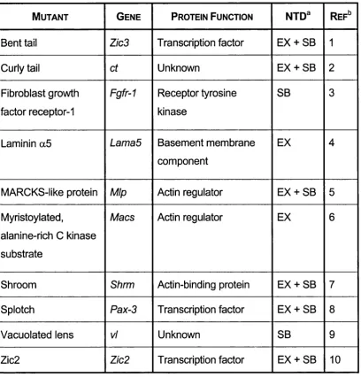
1.3.7.1 Mouse mutants with neural tube defects_____77
1.4 Neural crest_________________________________________ 83
1.4.1 Neural crest ceil m igration_______________________83
1.4.2 Guidance o f neural crest migration_______________ 85
1.4.2.1 Permissive molecules___________________ 86
1.4.2.1.1 Fibronectin, laminin, collagen and
integrin_______________________ 86
1.4.2.1.2 Thrombospondin-1______________88
1.4.2.2 Non-permissive molecules________________ 88
1.4.2.2.1 Chondroitin sulphate proteoglycans 89
1.4.2.2.2 F-spondin_____________________ 93
1.4.2.2A T-cadherin_____________________ 94
1.4.2.2.5 Eph and ephrins________________95
1.4.2.2.6 E rbB 4________________________ 95
1.4.2.3 Multiple directional cues___________________ 96
1.4.3 Mutations affecting the neurai c r e s t______________98
1.5 Aims of the thesis__________________________________ 101
Ch a p t e r 2 Ma t e r ia l s______________________________102
2.1 Générai reagents and equipm ent_____________________103
2.2 Giycosaminogiycans_______________________________ 103
2.3 E n zym es__________________________________________ 103
2.4 Source of mice and rats_____________________________ 104
2.5 Micro-dissection___________________________________ 105
2.6 Embryo cuiture____________________________________ 105
2.7 Polymerase chain reaction___________________________ 105
2.8 Agarose gei electrophoresis_________________________ 106
2.9 Molecular size markers______________________________ 106
2.10 Histological reagents and equipment__________________106
2.11 Microscopy________________________________________107
2.12 Primary antibodies_________________________________ 107
2.12.1 Heparan s u ip h ate_____________________________107
2.12.2 Chondroitin suiphate__________________________ 108
2.13 Control antibodies _________________________________ 108
2.14 Reagents for detection______________________________ 109
2.15 Bacterial p lasm ids_________________________________ 109
2.16 Transformation of bacteria___________________________ 109
2.17 RNA labelling_______________________________________113
2.19 Anion exchange chromatography____________________ 113
2.20 Quantification of giycosaminogiycans________________ 114
2.21 Quantification of proteins____________________________ 114
2.22 Statisticai anaiysis_________________________________ 114
2.23 Moiecuiar modeiiing________________________________ 114
2.24 Other equipment___________________________________ 115
Ch a p t e r 3 Me t h o d s_____________________________ 116
3.1 Cuiture of intact embryos____________________________ 117
3.1.1 Preparation o f rat serum_______________________117
3.1.2 Collection o f embryos_________________________118
3.1.3 Culture and assessment o f the embryos_________118
3.2 Genotyping of splotch (Sp^^) embryos________________ 122
3.2.1 Polymerase chain reaction_____________________125
3.2.2 Agarose gel electrophoresis___________________ 125
3.3 Histochemistry____________________________________ 126
3.3.1 Fixation and wax embedding___________________127
3.3.2 TESPA coating o f slides_______________________127
3.3.3 Sectioning__________________________________ 128
3.3.4 De-waxlng and re-hydratlon____________________128
3.3.5 Haematoxylln and eosin staining_______________ 128
3.3.6 Alclan blue staining___________________________129
3.4 immunohistochemistry______________________________ 129
3.4.1 Pre-treatment________________________________ 130
3.4.2 Binding o f primary antibodies_________________ 132
3.4.3 Signal amplification and detection______________ 132
3.5 Non-radioactive/n S/ÏU hybridisation_________________ 133
3.5.1.1 Preparation of Miller’s LB Agar and LB Broth_ 134
3.5.1.2 Transformation of competent cells__________135
3.5.2 Small scale Isolation of plasmid D N A___________ 135
3.5.3 Preparation o f riboprobes______________________137
3.5.3.1 Restriction digestion _______________ 137
3.5.3.2 Electrophoresis________________________ 139
3.5.3.3 Plasmid linearisation____________________ 139
3.5.3.4 Synthesis of digoxigenin-labelled probes____ 140
3.5.3.5 Agarose/formaldehyde gel electrophoresis 141
3.5.4 Specimen preparation_________________________142
3.5.5 Pre-hybridisation_____________________________142
3.5.6 Hybridisation________________________________ 142
3.5.7 Post-hybridisation washes_____________________143
3.5.8 Detection o f digoxigenin-iabeiied riboprobes_____143
3.5.9 Preparation o f embryo powder__________________144
3.6 Radiolabelling of giycosaminogiycans_________________ 144
3.6.1 Radioactive embryo cuiture____________________145
3.6.2 Purification o f giycosaminogiycans____________ 145
3.6.3 Separation o f giycosaminogiycans______________146
3.6.3.1 Preparation of anion exchange colum n_____ 147
3.G.3.2 Anion exchange chromatography__________147
3.6.4 Scintillation counting_________________________ 148
3.6.5 identification o f giycosaminogiycans___________ 148
3.6.5.1 Degradation by heparitinase______________ 149
3.6.5.2 Degradation by chondroitinase A B C _______ 149
3.7 Quantification of suiphated glycosaminoglycans________150
3.7.1 Pre-treatment o f sam ples______________________150
3.7.2 Dye-binding assay____________________________ 151
Ch a p t e r 4 Dis t r ib u t io n o f Su l p h a t e d
Gl y c o s a m in o g l y c a n s_____________________________ 154
4.1 Introduction_________________________________________155
4.2 Results_____________________________________________ 158
4.2.1 Regional morphological differences during spinal
neurulation___________________________________158
4.2.2 Alclan blue histochemistry_____________________ 160
4.2.3 Immunohistochemistry________________________ 163
4.2.3.1 Antigen recognition_____________________ 163
4.2.3.2 Antibody titration_______________________ 164
4.2.3.3 Expression pattern of heparan su lp h a te ____ 166
4.2.3.4 Expression pattern of chondroitin sulphate__ 169
4.3 Discussion__________________________________________ 173
4.3.1 Suiphated glycosaminoglycans and spinal neurulation
_____________________________________________173
4.3.2 Suiphated glycosaminoglycans and neural crest
m igration____________________________________ 176
4.3.3 Comparison between Alclan blue staining and
Immunostalning_______________________________177
4.3.4 Critique o f fixative u s e d_______________________ 181
Ch a p t e r 5 He p a r a n Su l p h a t e a n d Sp in a l
Ne u r u l a t io n______________________________________ 1S3
5.1 Introduction_________________________________________ 184
5.2 Results_____________________________________________ 187
5.2.1 Titration o f chlorate concentration_______________187
5.2.2 Inhibition o f glycosaminoglycan suiphation results In
5.2.3 Effect o f chlorate on glycosaminoglycan suiphation
____________________________________________ 197
5.2.3.1 Elution profile of ‘normally-sulphated’
glycosaminoglycans____________________ 197
5.2.3.2 Elution profile of glycosaminoglycans from
chlorate-treated embryos_________________ 204
5.2.4 Specific requirement for heparan suiphate in posterior
neuropore ciosure____________________________ 211
5.2.5 importance o f N- and 0-suiphate groups in heparan
suiphate_____________________________________215
5.2.6 Effect o f glycosaminoglycan suiphation on hinge
point form ation_______________________________220
5.2.7 Effect o f giycosaminogiycan suiphation on Sonic
hedgehog signaiiing__________________________ 228
5.3 Discussion_________________________________________ 237
5.3.1 Mechanism o f neurai foid eievation and bending 237
5.3.1.1 Heparan sulphate and actin microfilaments 239
5.3.1.2 Heparan sulphate and the cell c y c le _______ 240
5.3.1.3 Heparan sulphate and Sonic hedgehog signalling
_____________________________________ 242
5.3.1.4 Heparan sulphate and structural support of the
neural p la te ___________________________ 245
5.3.2 Different requirements for heparan suiphate in craniai
and spinal neurulation________________________ 246
5.3.3 Heparan suiphate and other deveiopmentai events 248
5.3.4 Chondroitin suiphate and neuruiation____________249
Ch a p t e r 6 Ch o n d r o it in Su l p h a t e a n d Sp in a l
6.1 Introduction________________________________________ 252
6.2 Results_____________________________________________254
6.2.1 Degradation o f chondroitin suiphate retards posterior
neuropore ciosure____________________________ 254
6.2.2 Chondroitin suiphate acceierates posterior neuropore
ciosure______________________________________262
6.2.3 Chondroitin suiphate and hinge point form ation 262
6.2.4 Chondroitin suiphate does not reguiate posterior
neuropore ciosure by affecting the amount o f heparan
suiphate_____________________________________264
6.3 Discussion__________________________________________268
6.3.1 Ceil-matrix interaction in spinai neuruiation______ 268
6.3.2 Ceii-celi adhesion in spinai neuruiation___________269
6.3.3 Chondroitin suiphate and fibrobiast growth factor-2
____________________________________________ 271
6.3.4 Chondroitin sulphate influences craniai and spinai
neuruiation through different mechanisms_______ 272
C h a p t e r 7 S u lp h a te d G ly c o s a m in o g ly c a n s a n d t h e
Sp l o t c h M u t a n t __________________________________ 2 7 4
7.1 Introduction________________________________________ 275
7.2 Resuits_____________________________________________ 278
7.2.1 Quantification o f suiphated giycosaminogiycans _278
7.2.2 Comparison o f net rate o f synthesis o f sulphated
giycosaminogiycans__________________________ 287
7.3 Discussion__________________________________________295
7.3.1 Chondroitin suiphate proteogiycans and neurai crest
7.3.2 Metabolism o f sulphated glycosaminoglycans In the
splotch mouse m u ta n t________________________ 298
7.3.3 Sulphated glycosaminoglycans and neural tube
defects In the splotch mouse mutant____________ 301
Ch a p t e r 8 Co n c l u s io n____________________________ 305
8.1 Introduction________________________________________ 306
8.2 Heterogeneity of suiphated giycosaminogiycans_______ 306
8.3 Suiphated giycosaminogiycans and human p athology 309
8.4 Future w ork________________________________________ 311
Ap p e n d ix 1 Ar e a u n d e r a Cu r v e___________________3 16
A1.1 Introduction________________________________________ 317
A1.2 Trapezium m etho d __________________________________ 317
A1.3 incompiete resolution of heparan suiphate and chondroitin
suiphate p e a k s _____________________________________ 319
A1.4 Accuracy of estimation of the area under an incompieteiy
resoived p e a k ______________________________________ 321
L
is t
o f
F
ig u r e s
Figure 1.1 Disaccharide subunits of heparan sulphate and chondroitin
sulphate_______________________________________ 28
Figure 1.2 Biosynthesis of the proteoglycan linkage re g io n 36
Figure 1.3 Biosynthesis of heparan sulphate__________________ 38
Figure 1.4 Biosynthesis of chondroitin sulphate________________ 40
Figure 1.5 Neural tube closure______________________________ 60
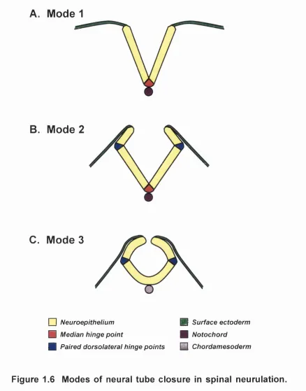
Figure 1.6 Modes of neural tube closure in spinal neuruiation 62
Figure 1.7 Hypothesis on hinge point formation in spinal neuruiation
_____________________________________________ 65
Figure 2.1 Vector map of plasmid pBluescript II K S + ___________ 110
Figure 2.2 Vector map of plasmid pBluescript S K -_____________ 111
Figure 2.3 Restriction map of mouse Patched plasmid__________ 112
Figure 2.4 Restriction map of mouse Sonic hedgehog p lasm id 112
Figure 3.1 Genotyping splotch (Sp^^) embryos using the polymerase
chain rea ction_________________________________ 123
Figure 3.2 Immunohistochemical detection of mouse antigen using
mouse primary a n tibo dy________________________ 131
Figure 3.3 Digoxigenin-labelling of RNA by in vitro transcription 138
Figure 4.1 Haematoxylin and eosin stained transverse sections
through the posterior neuropore region_____________ 159
Figure 4.2 Aldan blue staining of transverse sections through the
posterior neuropore region_______________________ 161
Figure 4.3 Antibody recognition of heparan sulphate and chondroitin
sulphate______________________________________165
Figure 4.5 Immunostalning for heparan sulphate in transverse
sections through the posterior neuropore region 170
Figure 4.6 Immunostalning for chondroitin sulphate in transverse
sections through the posterior neuropore region 171
Figure 5.1 Titration of chlorate concentration_________________188
Figure 5.2 Effect of chlorate on embryonic growth parameters____191
Figure 5.3 Effect of chlorate on Brown and Fabro morphological score
____________________________________________ 192
Figure 5.4 Effect of chlorate on embryonic protein content_______ 193
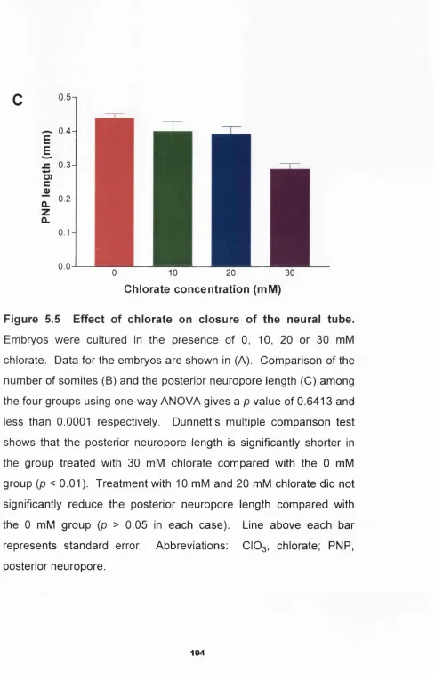
Figure 5.5 Effect of chlorate on closure of the neural tu b e _______ 194
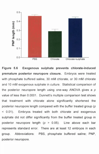
Figure 5.6 Exogenous sulphate prevents chlorate-induced premature
posterior neuropore closure______________________ 196
Figure 5.7 Standard conductivity curve for sodium chloride 198
Figure 5.8 Radioactive decay curve of ^^S-sulphur____________ 199
Figure 5.9 Standard curve for quantification of protein content using
the BCA Protein Assay Kit_______________________ 200
Figure 5.10 Anion exchange chromatography of ^^S-labelled
glycosaminoglycans extracted from cultured CD1 embryos
201
Figure 5.11 Limitation of anion exchange chromatography in peak
resolution_____________________________________ 205
Figure 5.12 Inhibition of suiphation of glycosaminoglycans by chlorate
treatment_____________________________________ 207
Figure 5.13 Identification of the single glycosaminoglycan peak from
chlorate-treated CD1 embryos____________________ 209
Figure 5.14 Effect of chlorate on degree of glycosaminoglycan
suiphation____________________________________ 212
Figure 5.15 Specific requirement for heparan sulphate in spinal
Figure 5.16 Inability of chondroitin sulphate to prevent effect of chlorate
216
Figure 5.17 Effect of pre-digestion of heparan sulphate with
heparitinase___________________________________217
Figure 5.18 Exogenous heparan sulphate does not prevent inhibition of
glycosaminoglycan suiphation by chlorate treatment 218
Figure 5.19 Importance of N-sulphate group in heparan sulphate _221
Figure 5.20 Importance of 0-sulphate group in heparan sulphate _222
Figure 5.21 Heparan sulphate is required for median hinge point
formation during spinal neuruiation________________223
Figure 5.22 Chlorate reduces the suiphation of glycosaminoglycans in
the posterior neuropore region____________________ 227
Figure 5.23 Plasmid linearisation___________________________ 229
Figure 5.24 Digoxigenin-labelling of RNA probes_______________230
Figure 5.25 Whole mount in situ hybridisation _________________ 232
Figure 5.26 Histological localisation of Sonic hedgehog and Patched
transcripts____________________________________ 234
Figure 6.1 Effect of chondroitinase on embryonic growth parameters
____________________________________________ 255
Figure 6.2 Effect of chondroitinase on embryonic gross morphology
____________________________________________ 257
Figure 6.3 Effect of chondroitinase on posterior neuropore closure258
Figure 6.4 Effect of Streptomyces hyaluronidase on posterior
neuropore closure_____________________________ 260
Figure 6.5 Immunohistochemical localisation of chondroitin sulphate
in the posterior neuropore region after chondroitinase
treatment_____________________________________ 261
Figure 6.6 Effect of exogenous chondroitin sulphate on posterior
Figure 6.7 Lack of effect on hinge point formation in the closing neural
tube with changes in chondroitin sulphate content____ 265
Figure 6.8 Effect of chondroitin sulphate on posterior neuropore
closure is not mediated by changes in heparan sulphate
content_______________________________________ 266
Figure 7.1 Standard curve for quantification of sulphated
glycosaminoglycans using the Blyscan Assay K it____ 279
Figure 7.2 The splotch (Sp^^) mouse m utant_________________ 281
Figure 7.3 Genotyping of splotch (Sp^^) embryos using the
polymerase chain reaction_______________________ 282
Figure 7.4 Growth parameters of splotch (Sp^^) mouse mutant
embryos______________________________________ 283
Figure 7.5 Chondroitin sulphate content of splotch (Sp^^) mutant
embryos______________________________________ 285
Figure 7.6 Heparan sulphate content of splotch (Sp^^) mutant
embryos______________________________________ 286
Figure 7.7 Anion exchange chromatography of ^^S-labelled
glycosaminoglycans extracted from cultured splotch (Sp^^)
embryos______________________________________ 288
Figure 7.8 Net rate of chondroitin sulphate synthesis in splotch (Sp^^)
embryos______________________________________ 291
Figure 7.9 Net rate of heparan sulphate synthesis in splotch (Sp^^)
embryos______________________________________ 292
Figure 7.10 Ratio of chondroitin sulphate:total sulphated
glycosaminoglycans synthesised in splotch (Sp^^)
embryos______________________________________ 293
Figure 7.11 Ratio of heparan sulphate:total sulphated
glycosaminoglycans synthesised in splotch (Sp^^)
Figure A1.1 Trapezium method of calculating the area under a curve
____________________________________________ 318
Figure A 1.2 Estimation of area under an incompletely resolved peak
____________________________________________ 320
L
is t
o f
T
a b le s
Table 1.1.
Table 1.2.
Table 1.3.
Table 1.4.
Table 1.5
Table 3.1
Table 5.1
Mutations involving heparan sulphate proteoglycan
synthesis in Drosophila______________________
Mutations involving heparan sulphate proteoglycan
synthesis in the m o u s e ______________________
49
50
Mutations involving chondroitin sulphate proteoglycan
synthesis in the m o u s e __________________________ 51
Mouse mutants with neural tube defects_____________78
Permissive and non-permissive molecules in neural crest
m igration______________________________________ 97
120
Brown and Fabro Morphological Scoring System _
Effect of under-sulphation of heparan sulphate on the
A
b b r e v ia t io n s
Ab antibody
AEC 3-amino-9-ethylcarbazole
ANP anterior neuropore
APS adenosine 5’-phosphosulphate
ATP adenosine 5 -triphosphate
BCA bicinchoninic acid
BCIP 5-bromo-4-chloro-3-indolyl phosphate
BMP bone morphogenetic protein
bp base pair
BSA bovine serum albumin
OHO Chinese hamster ovary
CIO3 chlorate
cm centimetre
cpm counts per minute
OS chondroitin sulphate
OTP cytidine 5’-triphosphate
DAB 3,3'-diaminobenzidine tetrahydrochloride
DEPC diethylpyrocarbonate
De-N-HS de-N-sulphated heparan sulphate
De-O-HS de-O-sulphated heparan sulphate
DePX dextropropoxyphene
DIG digoxigenin
DMEM Dulbecco’s modified Eagle's medium
DNA deoxyribonucleic acid
dNTP deoxyribonucleoside 5’-tri phosphate
dpm disintegrations per minute
DTT dithiothreitol
EDTA ethylenediaminetetraacetic acid
PCS foetal calf serum
FGF fibroblast growth factor
FGFR fibroblast growth factor receptor
GAG glycosaminoglycan
Gal galactose
GalNAc N-acetylgalactosamine
GIcA glucuronate
GlcNAc N-acetylglucosamine
GPI glycosylphosphatidylinositol
GTP guanosine 5'-triphosphate
HEPES N-(2-hydroxyethyl)piperazine-N’-(2-ethanesulphonic acid)
HRP horseradish peroxidase
HS heparan sulphate
htx-HS heparitinase-treated heparan sulphate
IdoA iduronate
ig immunoglobulin
kb kilobase
LB Agar Lennox L Agar
LB Broth Lennox L Broth
LP long PCR products
MARCKS myristoylated, alanine-rich C kinase substrate
mes mesoderm
mg milligram
mm millimetre
MOPS 3-(N-morpholino)propanesuiphonic acid
mRNA messenger ribonucleic acid
nCi microcurie
P9 microgram
pi microlitre
pm micrometre
microsiemen
NBT nitrobiue tétrazolium
ne notochord
N-CAM neurai celi adhesion molecule
NDST N-deacetylase N-sulphotransferase
neb neuroepithelial basement membrane
nm nanometre
NTMT sodium chioride/Tris/magnesium chloride/Tween buffer
NTP ribonucieoside 5'-triphosphate
OST 0 -suiphotransferase
PAPC 3’-phosphoadenosine 5'-phosphochlorate
PAPS 3’-phosphoadenosine 5’-phosphosuiphate
PBS phosphate buffered saline
PBT PBS/Tween buffer
PCR poiymerase chain reaction
PFA paraformaldehyde
PNP posterior neuropore
RNA ribonucieic acid
RNase A ribonuciease A
SA streptavidin
S04 sulphate
STP short target PCR products
SSC sodium chloride/sodium citrate buffer
TAB Tris/acetic acid/EDTA buffer
TBS Tris buffered saline
I E Tris/EDTA buffer
TESPA 3-aminopropyltriethoxysilane
TGF-p transforming growth factor-p
Tris tris(hydroxymethyl)aminomethane
TTP thymidine 5 -triphosphate
UDP uridine 5 -diphosphate
UTP uridine 5’-triphosphate
Chapter i
G
e n e r a l
1.1
Introduction
Glycosaminoglycans are widely distributed molecules in the animal
kingdom, and are found in organisms ranging from Drosophila to
mammals. They are present in many different tissues and organs, and
are involved in cellular functions such as proliferation, differentiation,
adhesion and migration. These molecules play vital roles in many
physiological and pathological processes, including embryonic
development, tumourigenesis and inflammation. The importance of the
sulphated glycosaminoglycans in neurulation and neural crest cell
migration during mouse embryonic development is examined in this
thesis.
1.2 Glycosaminoglycans and proteoglycans
1 Glycosaminoglycans
Glycosaminoglycans are large polyanionic molecules, consisting of
long, unbranched chains of repeating disaccharide subunits. The
disaccharide subunits are made up of a hexosamine, which is either
glucosamine or galactosamine, and a hexuronic acid (Varki et al, 1999).
Glycosaminoglycans are divided into two main groups, based on the
presence of sulphate groups in the disaccharide subunit. The
commonest example of non-sulphated glycosaminoglycans is
hyaluronan, which consists of repeats of N-acetylglucosamine (GlcNAc)
covalently linked to a protein core to form a proteoglycan (Varki et al,
1999).
The sulphated glycosaminoglycans are further divided into three
groups: heparan sulphate/heparin, chondroitin sulphate/dermatan
sulphate, and keratan sulphate. Heparan sulphate/heparin consists of
repeating disaccharide subunits of N-acetylglucosamine and either
iduronate (IdoA) or glucuronate (Figures 1.1 A to D). The glucosamine,
iduronate and glucuronate residues are modified by the addition of
variable numbers of sulphate groups at the C2, C3 and C6 positions
(Figures 1.1 A, C).
The subunit in chondroitin sulphate comprises N-acetylgalactosamine
(GalNAc) and glucuronate (Figures 1.1E, F). The galactosamine
residues are variably modified by the addition of sulphate groups at the
C4 and C6 positions (Figure 1.1E). In dermatan sulphate, some of the
glucuronate residues are epimerised to form iduronate residues.
Keratan sulphate distinguishes itself from the other sulphated
glycosaminoglycans in the lack of a hexuronic acid. The disaccharide
subunit is made up of N-acetylglucosamine and galactose residues.
At the time of neurulation in the rodent embryo, the main sulphated
glycosaminoglycans synthesised are heparan sulphate and chondroitin
sulphate (Solursh and Morriss, 1977; Copp and Bernfield, 1988a).
Hence, these two molecules form the focus of the remainder of this
A. Heparan sulphate: [GlcA->GlcNAc]n
COOH HoC OH
O OH
H HN COCH3
C. Heparan sulphate: [Id o A ^G IcN A c]^
HgC OH
COOH OH
E. Chondroitin sulphate: [G lc A ^ G a lN A c ]„
COOH
HO
O
OH
HN COCH3
H OH
Figure 1.1 Disaccharide subunits of heparan sulphate and chondroitin sulphate. (A) and (0) show
the chemical structures of heparan sulphate disaccharide subunits, and (E) shows the chondroitin sulphate
subunit. Groups that are variably modified by sulphation during glycosaminoglycan biosynthesis are shown
in red. (B), (D) and (F) are space filling models of the structures in (A), (0) and (E) respectively. Note the
three-dimensional conformational change in the heparan sulphate subunit when the glucuronate residue in
(B) is epimerised to form iduronate in (D). Abbreviations: GalNAc, N-acetylgalactosamine; GIcA,
glucuronate; GlcNAc, N-acetylglucosamine; IdoA, iduronate. Colour code for space filling model: blue,
nitrogen; cyan, carbon; red, oxygen; white, hydrogen. (A), (0) and (E) were modified after Prydz and Dalen
1.2.1.1
Différences between heparan sulphate and
heparin
Although both heparan sulphate and heparin are made up of repeating
subunits of glucosamine and glucuronate/iduronate, there are important
physicochemical and biological differences between the two molecules.
Heparin has an average of 2.5 sulphate groups per disaccharide unit,
making it the most negatively charged glycosaminoglycan known. It is
highly modified by deacetylation, sulphation and épimérisation of the
glucuronate residues to iduronate residues (Conrad, 1998; Varki et al,
1999). The 3 -0 sulphate group on the glucosamine residue is critical
for its anticoagulant activity.
In contrast, heparan sulphate only contains about one sulphate group
per disaccharide subunit. The molecule is less extensively modified by
deacetylation, sulphation and épimérisation. There are approximately
equal amounts of N-sulphated and N-acetylated glucosamine residues
present, and the ratio of 0-sulphate to N-sulphate groups is one or less
(Gallagher and Walker, 1985). Short segments, which are highly
modified and sulphated, are dispersed among the less sulphated
sequences in heparan sulphate, resulting in a structure that is much
more complex compared with heparin (Turnbull et al, 2001).
These differences between heparan sulphate and heparin are reflected
in their physicochemical properties. The average molecular weight,
solubility in 2 M potassium acetate, number of bands on isoelectric
focusing in polyacrylamide gels, and susceptibility to degradation by
1998; Varki et al, 1999). Functionally, heparin is capable of binding
bioactive molecules, such as growth factors, with a higher affinity and
avidity than heparan sulphate due to its higher negative charge. This
affects the availability of these signalling molecules to their cellular
receptors.
Heparin is synthesised in mast cells on its core protein, serglycin. It is
stored in secretory granules and released during inflammation
(Matsumota et al, 1995). Recently, heparin is also discovered to be
synthesised by oligodendrocyte-type-2-astrocyte progenitor cells, which
are precursors of oligodendrocytes and type-2 astrocytes (Stringer et al,
1999). Upon differentiation into oligodendrocytes and astrocytes, these
cells switch from the expression of heparin to heparan sulphate.
In contrast, heparan sulphate is synthesised on a distinct series of core
proteins (see below) and secreted continually by most animal cells.
Hence, heparan sulphate, rather than heparin, is the species that is
encountered by most of the cells in an organism.
The biological significance of the differences between heparan sulphate
and heparin is highlighted by comparing mutant mice that are deficient
in heparan sulphate with those that are deficient in heparin. Mice that
lack heparin are viable and fertile (Forsberg et al, 1999; Humphries et
al, 1999). On the other hand, mice that are deficient in heparan
sulphate have a wide range of abnormalities, depending on which part
of the heparan sulphate biosynthetic pathway is affected. These
abnormalities range from gastrulation defects (Lin et al, 2000),
1998), to neonatal death from severe respiratory distress syndrome
(Ringvall et al, 2000; Fan et al, 2000).
1.2.2
Proteoglycans
Proteoglycans are formed by the covalent attachment of
glycosaminoglycan chains to the serine residues of core proteins via a
linkage region that consists of xylose-galactose-galactose-glucuronate.
Proteoglycans are found on cell surfaces, in the basement membranes
and in the extracellular matrix. The expression pattern of proteoglycans
is developmentally regulated. Proteoglycans are classified by either the
core protein or the predominant glycosaminoglycan species attached.
1.2.2.1
Syndecans
Syndecans are transmembrane core proteins. Four members of the
syndecan family are currently known, named syndecan-1 to 4 (Bernfield
et al, 1992; Salmivirta and Jalkanen, 1995). Syndecans contain an
extracellular domain, a transmembrane domain, and a short
cytoplasmic tail. Glycosaminoglycan chains are attached to the
extracellular domain. The transmembrane and cytoplasmic regions are
highly conserved, as are the glycosaminoglycan attachment sites and
the proteolytic cleavage sites in the extracellular domain. Syndecans
are expressed in a developmentally regulated manner in tissues derived
from all three germ layers (David et al, 1993; Kim et al, 1994; Gould et
al, 1995). Although classified as heparan sulphate proteoglycans,
syndecan-1 and 4 are known to carry chondroitin sulphate chains in
heparan sulphate and chondroitin sulphate on each core protein varies
among different tissues (Kokenyesi and Bernfield, 1994).
1.2.2.2
Glypicans
Glypicans are heparan sulphate proteoglycans that are anchored to the
cell surface by a glycosylphosphatidylinositol (GPI) linkage (Conrad,
1998). The family consists of six known members, named glypican-1 to
6. Like syndecans, the presence of glypicans in a wide variety of
tissues is developmentally regulated (Stipp et al, 1994; Watanabe et al,
1995; Saunders et al, 1997; Litwack et al, 1998; Paine-Saunders et al,
2000).
1.2.2.3
Basem ent membrane proteoglycans
The major proteoglycans in the basement membrane are perlecan,
agrin and bamacan (lozzo, 1998). Perlecan and agrin are heparan
sulphate proteoglycans. Both of them contain laminin-like modules.
Perlecan is expressed in the embryo as early as the pre-implantation
stage. The distribution of fibroblast growth factor-2 (FGF-2) in the
basement membrane parallels that of perlecan in the mouse embryo,
suggesting that perlecan might play a role in regulating FGF-2 signalling
(Aviezer et al, 1994). Agrin is important for the development of
neuromuscular function. Bamacan is a chondroitin sulphate
proteoglycan and helps in maintaining the stability of the basement
1.2.2.4
Hyalectans
The chondroitin sulphate proteoglycans aggrecan, versican, neurocan
and brevican are members of the hyalectan family, based on their
common ability to interact with hyaluronan at their N-terminal and with
lectins at their C-terminal (lozzo, 1998). These proteoglycans exhibit
alternative splicing, and several variants of them are found in the
extracellular matrix in distinct spatial and temporal patterns during
development (Oohira et al, 2000). In addition, a short isoform of
brevican is known which attaches to the plasma membrane by a
glycosylphosphatidylinositol anchor.
1.2.2.5
Other proteoglycans
Many other proteoglycans are known which carry heparan sulphate
and/or chondroitin sulphate chains. Examples of these include collagen
XVIII (heparan sulphate), collagen IX (chondroitin sulphate),
phosphacan (chondroitin sulphate) and betaglycan (both heparan
sulphate and chondroitin sulphate). The term ‘part-time proteoglycan’
has been applied to some of them, such as betaglycan, because
variants of the core proteins exist which do not carry any
glycosaminoglycan side chains (David, 1993; Erickson and Couchman,
2000).
1.2.3 Biosynthesis
The biosynthesis of proteoglycans involves several cellular
compartments (Prydz and Dalen, 2000). The proteoglycan core protein
the Golgi apparatus where synthesis of the glycosaminoglycan chains
takes place. Sugar residues are activated by attachment to uridine 5’-
triphosphate (UTP) in the cytoplasm before they are transported to the
Golgi apparatus for elongation of the glycosaminoglycan chains.
Similarly, the sulphate group donor, 3’-phosphoadenosine 5’-
phosphosulphate (PAPS), is first synthesised in the cytoplasm from
inorganic sulphate and adenosine 5’-triphosphate. The reaction is
catalysed by a bifunctional enzyme named 3’-phosphoadenosine 5’-
phosphosulphate synthase. The donor is then transported to the Golgi
apparatus.
1.2.3.1
Linkage region
Synthesis of the linkage region takes place in the endoplasmic
reticulum/c/s-Golgi network. This begins with the addition of xylose to
the serine residue of the core protein (Figure 1.2). Thereafter,
galactose and glucuronate are sequentially added by their respective
transferases. Although different core proteins are found in heparan
sulphate proteoglycans and chondroitin sulphate proteoglycans, the
same set of hexose transferases is used for synthesis of the linkage
region in both groups. Mutant Chinese hamster ovary (OHO) cells that
lack xylosyl transferase and galactosyltransferase I are unable to
synthesise both heparan sulphate and chondroitin sulphate (Esko et al,
\x y i
I
Core protein
transferase
Xyl->Core protein
Gal transferase I
G a l^ X y i^ C o re protein
Gal transferase II
Gai->Gai->Xyi->Core protein
^ GIcA transferase I
G ic A ^ G a i^ G a i^ X y i-> C o re protein
i
Heparan sulphate Chondroitin sulphate
proteoglycan proteoglycan
Figure 1.2 Biosynthesis of the proteoglycan linkage region.
Proteoglycan synthesis begins with formation of the core protein
chain. This is followed by the sequential addition of components of
the linker tetrasaccharide, which consists of xylose, galactose and
glucuronate, by the respective transferases shown above. The same
set of transferase enzymes is used in synthesis of heparan sulphate
and chondroitin sulphate proteoglycans. Once synthesis of the
linkage region is completed, the biochemical pathway diverges to
form either heparan sulphate or chondroitin sulphate proteoglycan.
1.2.3.2
Glycosaminoglycan chain
The first carbohydrate residue that is attached to the linkage region
determines the identity of the glycosaminoglycan chain that is to be
made. In the synthesis of heparan sulphate, GlcNAc-transferase I adds
the first N-acetylglucosamine residue to the glucuronate residue of the
linkage region (Figure 1.3). Chain elongation then proceeds through
the alternate addition of glucuronate and N-acetylglucosamine residues.
This is catalysed by heparan sulphate copolymerase, a hetero-
oligomeric enzyme complex. The enzyme complex is encoded by the
EXT1/EXT2 tumour suppressor genes, which are the mammalian
homologues of the Drosophila tout-velu gene. Synthesis of the heparan
sulphate chain occurs in the c/s-, medial- and trans- Golgi cisternae
(Prydz and Dalen, 2000). A variable amount of sequential modification
of the heparan sulphate chain occurs through deacetylation,
épimérisation, and N- and 0-sulphation (Figure 1.3).
In chondroitin sulphate synthesis, the enzyme GalNAc-transferase I
attaches the first N-acetylgalactosamine residue to the linkage region
(Figure 1.4). The chain is then elongated by alternate additions of
glucuronate and N-acetylgalactosamine residues. Finally, the
chondroitin sulphate chain is variably modified by the action of 4- and 6-
O sulphotransferases. Synthesis of chondroitin sulphate occurs in the
frans-Golgi network (Prydz and Dalen, 2000).
Further processing of the core protein has been described, which
involves the attachment of either N- or 0-linked oligosaccharides to the
arginine and serine residues respectively of the protein backbone
Figure 1.3 Biosynthesis of heparan sulphate. Synthesis of heparan sulphate begins with addition of
the first N-acetylglucosamine residue to the linker tetrasaccharide by GlcNAc transferase I, which is
encoded by the tumour suppressor gene EXTL2. Elongation of the heparan sulphate chain follows, with
alternate addition of glucuronate and N-acetylglucosamine. This reaction is catalysed by a hetero-
oligomeric heparan sulphate copolymerase enzyme complex that possesses both glucuronate transferase
and N-acetylglucosamine transferase activity. The enzyme complex is the gene product of EXT1/EXT2, the
* mammalian homologues of the Drosophila tout-velu gene. The N-acetyl group on N-acetylglucosamine is
then removed and replaced by a sulphate group. Both reactions are carried out by the same bifunctional
enzyme, N-deacetylase N-sulphotransferase. Some glucuronate residues are epimerised to form iduronate
by glucuronate epimerase. Finally, sulphate groups are added to C2, C3 and C6 positions of the
carbohydrate residues by the respective action of 2-0 , 3 -0 and 6 -0 sulphotransferases. The modification
reactions do not proceed to completion, resulting in a complex heparan sulphate structure where highly
modified and sulphated segments are dispersed among less modified sequences. Abbreviations: GIcA,
glucuronate; GlcNAc, N-acetylglucosamine; IdoA, iduronate; NDST, N-deacetylase N-sulphotransferase;
c
Linker^Protein
^ GlcNAc transferase I (EXTL2)
GlcNAc->Linker-^Protein
— Heparan sulphate copolymerase (EXT1IEXT2)
[GlcA-^GIcNAc]n-^Linker->Protein
[ n d s t
▼ [G lcA^G IcNS03-3,6-diS04]n^Linker->Protein
— [G lc A ^ G IcN S0 3]n->Linker->Protein n a
1 g /c A epimerase
I
^▼ ^ [G lcA ^G IcN S0 3-6-S04]n->Linker->Protein [ld o A ^ G IcN S0 3]n->Linker->Protein ^
|2-osr
n ^ -o s r
2-OST [ldoA-2-S0 4^ G lc N S0 3]„ ^ L in k e r ^ P r o te in ^ [IdoA ^G IcN S O a-B -SO Jn^Linker^Protein
^ 6 - o s r
[ldoA-2-S04->GlcNS03-6-S04]n->Linker->Protein
[GlcA-2-S0 4->GlcNS0 3]n->Linker->Protein
\ 6 - 0 S T
Linker-^Protein
I GalNAc transferase I
GalNAc->Linker->Protein
1
1
GIcA transferase II and GalNAc transferase II
[GlcA->GalNAc]n->Linker->Protein
4-OST and 6-OST
Chondroitin sulphate proteoglycan
Figure 1.4 Biosynthesis of chondroitin sulphate. Synthesis of
chondroitin sulphate begins with attachment of N-
acetylgalactosamine to the terminal glucuronate of the linker
tetrasaccharide by GalNAc transferase I. The chondroitin sulphate
chain elongates by the alternate addition of glucuronate and N-
acetylgalactosamine. These reactions are catalysed by GIcA
transferase II and GalNAc transferase II respectively. The
glycosaminoglycan chain is then subjected to variable amounts of
sulphation by the 4 -0 and 6 -0 sulphotransferases. Abbreviations:
GalNAc, N-acetylgalactosamine; GIcA, glucuronate; OST, O-
1.2.3.3
Regulation of proteoglycan biosynthesis
The glycosaminoglycan attachment site on the core protein is
characterised by serine and glycine- containing sequences that are
flanked by acidic amino acids and adjacent tryptophan residues (Zhang
and Esko, 1994; Zhang et al, 1995). These serine-glycine sequences
tend to be clustered together in heparan sulphate proteoglycans. In
chondroitin sulphate proteoglycans, the sequences tend to be isolated.
Other factors, which are not well understood, help to determine the type
of glycosaminoglycan chain that is attached. For example, the
syndecan-1 core protein is identical regardless of the tissue of origin,
but the ratio of attached heparan sulphate to chondroitin sulphate varies
from one tissue to another (Kokenyesi and Bernfield, 1994). The
glycosaminoglycan chains on a core protein influence the location of the
proteoglycan on the cell surface. For example, glypicans are usually
found on the basolateral surface of epithelial cells. Removal of the
heparan sulphate chains results in apical transport of the core protein
(Mertens et al, 1996). On the other hand, the presence of chondroitin
sulphate chains might promote apical secretion of the proteoglycan
(Kolsetet al, 1999).
The fine structure of a glycosaminoglycan chain, including the amount
of sulphation, deacetylation and épimérisation, appears to be cell type
specific rather than core protein-specific (Kato et al, 1991; Kato et al,
1994). Developmentally regulated expression of multiple isoforms of
the glycosaminoglycan biosynthetic enzymes has been postulated to
contribute to the fine structure (Kitagawa et al, 1997; Fan et al, 1999;
which bioactive molecules are able to bind to the proteoglycan and the
biological effect of this interaction (Guimond et al, 1993; Ishihara, 1994;
Bernfield et al, 1999). For example, heparan sulphate is capable of
binding to and potentiating the activity of fibroblast growth factors.
When neural precursor cells switch from a proliferative to a
differentiative state, there is an accompanying change in the sulphation
pattern of heparan sulphate, which switches the potentiating activity of
the glycosaminoglycan from FGF-2 to FGF-1 (Nurcombe et al, 1993;
Brickman et al, 1998).
Growth factors and other biologically active molecules influence both
the amount and type of proteoglycans that are associated with a cell.
Examples of these bioactive molecules include FGF-2, TGF-p, inositol
and retinoic acid (Elenius et al, 1992; Morriss-Kay and Mahmood, 1992;
Grassel et al, 1995; Schmidt et al, 1995). For example, rat aortic
smooth muscle cells synthesise both syndecan-1 and 2, the relative
proportion of which is altered by different growth factors (Cizmeci-Smith
et al, 1993). TGF-p upregulates expression of syndecan-2 and versican
in smooth muscle cells.
Control of the quantity of proteoglycan associated with a cell occurs not
just at the transcriptional level but post-transcriptionally as well
(Bernfield et al, 1999). For example, a ten-fold increase in the amount
of syndecan-1 is seen in kidney mesenchymal cells during development
without an appreciable change in the mRNA level. In contrast,
abundant transcripts of syndecan-3 are found in the developing rat
1,2
m4 Turnover
Proteoglycans can be secreted directly into the extracellular
environment. This is seen in the case of the basement membrane
proteoglycans and the hyalectans. Proteoglycans can also be shed
from the cell surface by enzymatic action. Transmembrane
proteoglycans such as syndecans are shed by proteolytic cleavage,
while glycosylphosphatidylinositol-linked proteoglycans such as
glypicans are shed by the action of phospholipase C. Proteoglycans
removed by endocytosis are degraded by endo- and exo-glycosidases
and sulphatases in the lysosomes (Yanagishita and Hascall, 1992;
David, 1993). However, not all endocytosed proteoglycans are
degraded. Some endocytosed glypicans are modified and then re
secreted by the cell (Edgren et al, 1997). Transport of heparan sulphate
to the cell nuclei has also been reported (Margolis et al, 1976; Fedarko
and Conrad, 1986).
The turnover of proteoglycans is very rapid. In steady state
radiolabelling of heparan sulphate in cell cultures, turnover occurs
between three and eight hours (Bienkowski and Conrad, 1984). Thus,
glycosaminoglycans can rapidly adapt to different requirements during
embryonic development (Nurcombe et al, 1993; Brickman et al, 1998).
1.2.4.1
Mucopolysaccharidoses
Mucopolysaccharidoses are a group of inherited disorders where there
is abnormal accumulation of glycosaminoglycans due to deficiencies in
certain lysosomal enzymes (Varki et al, 1999). The severity of the
Hunter syndrome, where a lack of iduronate-2-sulphatase leads to the
accumulation of heparan sulphate and dermatan sulphate, can have
normal intelligence and survive up to 60 years of age. On the other
hand, in utero or neonatal death from hydrops foetalis is seen in Sly
syndrome, where a p-glucuronidase deficiency results in an inability to
degrade heparan sulphate, chondroitin sulphate and dermatan
sulphate.
Biological function
Proteoglycans are capable of binding to over one hundred molecules
(Conrad, 1998; Bernfield et al, 1999; Oohira et al, 2000). These
molecules include several classes of signalling molecules that are vital
to embryonic development, such as the fibroblast growth factor and
receptor family, the Wnt proteins, and the Hedgehog proteins. Binding
occurs via the glycosaminoglycan chains of the proteoglycan, the core
protein, or both. In the last case, the glycosaminoglycan chains and
core protein may act synergistically to increase the affinity of the
proteoglycan for the signalling molecule (Herndon et al, 1999).
Specific binding is seen between glycosaminoglycans and signalling
molecules. The signalling molecule usually contains one or more
binding sequences that are rich in basic amino acids and hydropathic
amino acids (Cardin and Weintraub, 1989). These bind to specific
sequences on the glycosaminoglycan chain, such as the one found in
heparan sulphate to which FGF-2 binds (Habuchi et al, 1992; Turnbull
et al, 1992). The ligand-binding ability of a glycosaminoglycan is
enhanced by the presence of multiple closely arrayed charged sulphate
positively charged domains on the surface of a specific ligand. For
example, the sulphate groups on heparan sulphate influence the ability
of the glycosaminoglycan chain to bind to different members of the
fibroblast growth factor family (Guimond et al, 1993; Ishihara, 1994).
Rotation at the glycosidic linkages of the glycosaminoglycan chain helps
to present these negatively charged groups to the signalling molecule
(Conrad, 1998). It has been postulated that the proteoglycan core
protein facilitates binding of glycosaminoglycan chains to the signalling
molecules by tethering multiple chains together and positioning them in
an optimal position for ligand attachment. Positively charged metal ions
in the vicinity of this molecular interaction can also affect the binding
affinity and avidity (Kan et al, 1996).
The binding of fibroblast growth factors to heparan sulphate has been
extensively studied. Fibroblast growth factors bind to high affinity
fibroblast growth factor receptors, leading to dimérisation and mutual
tyrosine phosphorylation of these receptors. Heparan sulphate acts as
a low affinity co-receptor, facilitating the binding of the growth factor with
its high affinity receptor (Spivak et al, 1994; Schlessinger et al, 1995).
Binding of FGF-2 to heparan sulphate helps to concentrate the growth
factor at the cell surface, and protects it from being degraded
(Rosengart et al, 1988). The growth factor can also be internalised
while bound to heparan sulphate (Murono et al, 1993). FGF-2 induces
syndecan-1 expression, and this has been postulated as a mechanism
by which FGF-2 regulates its own activity (Bernfield et al, 1999).
Besides directly helping signalling molecules to activate their receptors,
heparan sulphate is able to influence signalling in other ways. In the
the signal from the producing cells to the receiving ceiis, as has been
shown in Drosophila (Beliaiche et al, 1998; The et al, 1999) and in mice
(Lin et al, 2000). In addition, heparan sulphate potentiates the activity of
TGF-pi and p2 by binding to the growth factors and preventing them
from being inactivated by a2-macroglobulin (Lyon et al, 1997).
Changes in the amount of heparan suiphate proteoglycan are
associated with different phases of the ceil cycle. Synthesis of the
glycosaminogiycan chain is reduced four foid during mitosis compared
with interphase (Preston et al, 1985). Smooth muscle cells synthesise
and secrete the proteoglycan in G r and G2-phases, but stop at the
beginning of the S-phase and during mitosis (Breton et ai, 1986).
Addition of exogenous heparin blocks the cell cycle in the late G rphase
(Reilly et al, 1989). In addition, the rate of ceil division is inversely
correlated with the amount of nuclear heparan sulphate (Fedarko and
Conrad, 1986; Ishihara et ai, 1986).
Syndecans are found on the basolateral surface of epithelial cells,
colocalised with and bound to actin and other components of the
cytoskeleton (Carey et ai, 1994; Fernandez-Borja et ai, 1995; Carey et
ai, 1996). This helps to stabilise both interceliular and ceil-extraceliular
matrix adhesions (Bernfield et al, 1999). Syndecans play a role in
maintaining the epitheiiai phenotype and prevent epitheliomesenchymal
transformation from occurring. Syndecans are aiso involved in the
transfer of signais from the extraceiluiar matrix into the ceii and the
coupling of cytosoiic downstream signalling molecules. For example,
the binding of syndecan-4 to the extraceliular matrix enabies its
cytopiasmic domain to bind to and activate the catalytic domain of