Article
1
Evolutionary analysis of Rett Syndrome-causing
2
proteins and their pathogenic missense point
3
mutations: structural order–disorder,
post-4
translational modifications, evolutionary rates, and
5
interacting proteins
6
Muhamad Fahmi1, Gen Yasui1, Kaito Seki1, Syouichi Katayama2, Takako Kaneko-Kawano2,
7
Tetsuya Inazu2, Yukihiko Kubota1,3 and Masahiro Ito1,3,*
8
9
1 Graduate School of Life Sciences, Ritsumeikan University; [email protected] (M.F.),
10
[email protected](G.Y.), [email protected](K.S.), [email protected](Y.K.),
11
[email protected](M.I.)
12
2 College of Pharmaceutical Sciences, Ritsumeikan University; [email protected](S.K.),
13
[email protected](T.K.-K.), [email protected](T.I.)
14
3 College of Life Sciences, Ritsumeikan University
15
16
* Correspondence: [email protected] (M.I.)
17
Abstract: Rett syndrome (RTT) is mainly caused by mutations in methyl CpG-binding protein 2,
18
cyclin-dependent kinase-like 5, or forkhead box protein G1. These RTT-causing proteins harbor an
19
intrinsically disordered region (IDR) whose conformation exhibits spatiotemporal heterogeneity,
20
which not only confer versatility to the protein, but also implicates them in diseases. The IDR
21
generally evolves more rapidly than an ordered structure. In this study, we examined the
22
relationship between pathogenic RTT-associated point mutations in RTT-causing proteins and the
23
evolutionary dynamics of sequence features including structural order–disorder, phosphorylation
24
sites, and evolutionary rates. We also analyzed the molecular properties and evolution of proteins
25
that interact with RTT-causing proteins in terms of phylogenetic profiles, tissue specificity,
26
subcellular localization, expression level, and functions. The results indicate that constrained IDRs
27
may function by forming contacts with other regions in the protein sequence causing pathogenic
28
missense mutations likely to arise in the rapidly evolving IDR and affect molecular networks,
29
leading to disease. The results also provide novel insights into the genetic basis for RTT and the
30
evolution of the neocortex in higher vertebrates.
31
32
Keywords: Rett Syndrome; Intrinsically disordered region; phylogenetic profile analysis;
post-33
transcriptional modification; methyl-CpG-binding protein 2; cyclin-dependent kinase-like 5;
34
forkhead box protein G1
35
36
1. Introduction
37
Rett syndrome (RTT; OMIM entry #312750) is a rare disease that was first described by
38
Andreas Rett in 1966 [1]. It mainly affects girls aged 6 to 18 months and is characterized by severe
39
neurodevelopmental impairment such as intellectual disability, movement disorder, and epilepsy [2].
40
Mutations in methyl CpG-binding protein (MECP)2, an X-linked gene involved in the regulation of RNA
41
2 of 20
splicing and chromatin remodeling, are the predominant cause of classic RTT and are present in >
42
90% of patients [3, 4]. Atypical cases of this syndrome are associated with mutations in either
cyclin-43
dependent kinase-like (CDKL)5, forkhead box protein (FOXG)1, or undefined genes [5, 6]. Collectively,
44
MeCP2, CDKL5, and FOXG1 are known as RTT-causing proteins. MeCP2 has been determined
45
shown to have a disordered structure by using various experimental methods approaches, the only
46
predicted structures of are available for FOXG1 have only been investigated by predictions [7-9]. In
47
case of CDKL5, structure of amino terminal kinase domain is already identified, but long carboxy
48
terminal tail has not been clarified [10]. These proteins contain polypeptide segments that are unable
49
to fold spontaneously into three-dimensional structures; the so-called intrinsically disordered regions
50
(IDRs) exist as dynamic ensembles of conformations that rapidly interconvert from molten globule
51
(collapsed) to coiled or premolten globules (extended) as a result of relatively flat energy landscapes
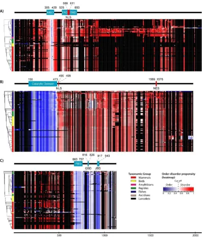
52
[11–14].
53
The different conformations of IDRs and structured regions are dictated by the amino acid
54
sequence; the former generally lack bulky hydrophobic residues [14, 15]. Proteins are composed of
55
either fully structured or fully disordered regions (with the latter referred to as intrinsically
56
disordered proteins [IDPs]) or a combination of the two, which is the case for most eukaryotic
57
proteins [16]. Although protein function has traditionally been elucidated based on a well-defined
58
structure, it is now widely acknowledged that IDRs contribute to diverse functions, which can be
59
classified into six types: entropic chain activity, display site, chaperone, molecular effector, molecular
60
assembler, and molecular scavenger [17–19]. Excluding entropic chain activity, IDRs adopt specific
61
tertiary conformations—at least locally—in order to perform these functions by binding to other
62
proteins, nucleic acids, membranes, and small molecules or respond to changes in their environment
63
that alter their relative free energy landscape [11, 20, 21]. Hence, IDR conformation varies over time—
64
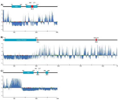
i.e., it exhibits spatiotemporal heterogeneity [22]. Moreover, long IDRs contain more modification
65
sites than fully ordered regions and their conformational flexibility provides more opportunities for
66
displaying these sites [23, 24]. These features explain how proteins with IDRs or IDPs interact with
67
and are tightly regulated by various factors to ensure that appropriate levels of protein are available
68
at the right time to minimize the possibility of inappropriate protein–protein interactions [19]. Thus,
69
altered conformation and availability of proteins with IDRs or IDPs are more likely to be associated
70
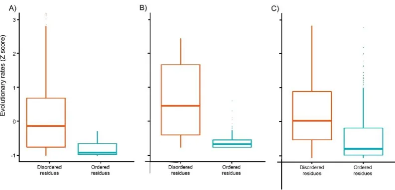
with disease states. However, IDRs generally (but not always) evolve more rapidly than ordered
71
structures owing to different accepted point mutation caused by differences of residue composition,
72
intramolecular contacts, and function [25].
73
Disease-related alleles are introduced into the human population by mutation, are directed in
74
part by random genetic drift, and disappear through purifying selection [26–28]. Restoring MeCP2
75
gene function in an animal model abolished the symptoms of RTT, suggesting that the disorder is
76
treatable [29]. In addition to gene therapy, reactivation of inactivated X chromosome is noted as a
77
new therapeutic method [30, 31]. Moreover, growth factor stimulation (e.g. insulin-like growth factor
78
1) and activation of neurotransmitter pathways (e.g.
2-adrenergic receptor pathway) can partially79
rescue phenotypes of MeCP2 knockout mice (RTT model mice) [32, 33]. Elucidating the molecular
80
basis for RTT can lead to the development of effective treatments.
81
Proteins function as part of interaction networks with other proteins, with human-specific
82
protein–protein interactions established through evolution. RettBASE is a point mutation database
83
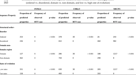
for RTT that provides reliable prevalence data on deleterious mutations [34]. In this study, we used
84
RettBASE to examine the relationship between pathogenic RTT point mutations and the evolution of
85
structural order–disorder, phosphorylation sites, and evolutionary rates of MECP2, CDKL5, and
86
FOXG1 in order to clarify the role of pathogenic mutations in these genes in the development of RTT,
87
and determine how IDRs are involved. In addition, we also analyzed the molecular features and
88
evolution of proteins that interact with RTT-causing proteins based on phylogenetic profile, tissue
89
specificity, subcellular localization, expression level, and function. Our results suggest that
90
Preprints (www.preprints.org) | NOT PEER-REVIEWED | Posted: 1 July 2019 doi:10.20944/preprints201907.0013.v1
pathogenic RTT missense mutations tend to occur in domain regions and affect ordered residues for
91
CDKL5 and FOXG1, and an intrinsically disordered domain (IDD) which can form intramolecular
92
contacts with other disordered regions in MeCP2 could promote the development of pathogenic RTT
93
by missense mutations in the rapidly evolved residues beyond the domain region. The phylogenetic
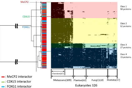
94
cluster analysis revealed that the RTT-causing proteins MeCP2, CDKL5, and FOXG1 and their
95
interaction partners may have been acquired during metazoan evolution and play essential roles in
96
neocortical development.
97
98
2. Results
99
2.1. Structural order–disorder properties of RTT-causing proteins during chordate evolution
100
We retrieved 97, 113, and 108 chordates sequences of MeCP2, CDKL5, and FOXG1, respectively,
101
and constructed a heat map of structural order–disorder propensity for each RTT-causing protein
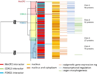
102
according to aligned sequences and taxonomic position in the phylogenetic tree (Supplementary
103
Table S1 and Figure 1). All RTT-causing proteins harbored both ordered and disordered regions; by
104
comparing their distribution to domain and non-domain regions, we found that the catalytic domain
105
and non-domain regions of CDKL5 were ordered and disordered, respectively (Figure 1B). While
106
most regions of MeCP2 were predicted to be disordered, some ordered structures were observed in
107
the methyl-CpG binding domain (MBD) (Figure 1A). Furthermore, FOXG1 showed a varied
108
distribution of ordered–disordered regions corresponding to domain and non-domain regions, with
109
the former predicted to be fully ordered (Figure 1). Despite insertions and deletions were frequently
110
detected in disordered regions, particularly in MeCP2 and FOXG1 (Figure 1A, C), the order-disorder
111
conformation of RTT-causing proteins showed to be stable in chordates, excluding a few
112
conformational transitions of FOXG1 and CDKL5 in mammals and fishes, respectively.
4 of 20
114
Figure 1. Order–disorder propensity of RTT-causing proteins in chordates. Heat maps of order–
115
disorder propensity were generated according to taxonomic position in the phylogenetic tree (rows)
116
and multiple sequence alignment (columns). The heat maps show a color gradient of blue (ordered)
117
to red (disordered), with white as the boundary between the two and black as gaps. Colored boxes
118
between the trees and heat maps indicate the taxonomic group; and bars above the heat maps indicate
119
domain position in the multiple sequence alignment, with light blue and black areas indicating the
120
domain and absence of a domain, respectively. (a–c) Heat maps for MeCP2 (A), CDKL5 (B), and
121
FOXG1 (C) are shown. MBD, TRD, FBD, GBD, JBD, NLS, and NES indicate methyl-CpG binding
122
domain, transcriptional repression domain, forkhead binding domain, Groucho-binding domain,
123
JARID1B binding domain, nuclear localization signal, and nuclear export signal, respectively.
124
125
2.2 Rate of evolution per site in RTT-causing proteins
126
We calculated the evolutionary rate of RTT-causing proteins in chordates to investigate its
127
relationship with structural features and the distribution of pathogenic RTT-causing missense
128
mutations. We used the human sequence as reference and determined standardized evolutionary
129
Preprints (www.preprints.org) | NOT PEER-REVIEWED | Posted: 1 July 2019 doi:10.20944/preprints201907.0013.v1
rate scores (Z scores), with values greater than or less than zero reflecting evolution at a faster and
130
slower than average rate, respectively (Figure 2 and Supplementary Table S2). The results showed
131
that evolutionary rates per site showed similar patterns in all proteins, with low rates of evolution
132
more commonly observed in domains and ordered regions; some exceptional cases such as the
133
transcriptional repression domain (TRD) of MeCP2 showed a higher rate of amino acid substitution.
134
On the other hand, non-domain regions that were also usually disordered—excluding the ordered
135
region surrounding a domain in FOXG1—typically exhibited a higher evolutionary rate, although
136
some regions with low rates of evolution were nonetheless detected (Fig. 2). This was corroborated
137
by the distribution of evolutionary rates for predicted structural order–disorder residues in the three
138
RTT-causing proteins, with disordered residues showing a wide and overlapping distribution that
139
reflected their conservation (P < 2.2e−16 for CDKL5 and FOXG1 and P < 6.409e−08 for MeCP2,
Mann-140
Whitney U test; Fig. 3).
141
142
Figure 2. Rate of evolution per site in human RTT-related proteins. (a–c) Rates of amino acid
143
substitution in MECP2 (A), CDKL5 (B), and FOXG1 (C) are shown as blue areas. The bars above charts
144
indicate the position of the domain in the human sequence, with light blue areas indicating the
145
domain and black lines indicating no domain. Phosphorylated amino acids and pathogenic
146
missense mutation sites are indicated by green and red dots, respectively. The x and y axes
147
represent the sequence length and Z score of evolutionary rate, respectively.
6 of 20
149
Figure 3. Boxplots of evolutionary rates for predicted structural order–disorder residues of human
150
RTT-causing proteins. (a–c) Boxes representing predicted ordered (blue) and disordered (red)
151
structure residues in MECP2 (A), CDKL5 (B), and FOXG1 (C). The x axis and y axes represent
152
predicted conformation and Z score of evolutionary rate, respectively.
153
2.3 Post-translational modifications (PTMs)
154
We predicted PTM (phosphorylation) sites in chordate sequences of RTT-causing proteins and
155
identified conserved PTM sites. We found numerous conserved phosphorylation sites including
156
60/82 in CDKL5, 30/45 in MeCP2, and all 23 sites in FOXG1 in human RTT-causing proteins (Fig. 2
157
green dots and Supplementary Table S3). Structural disorder makes such sites accessible for
158
phosphorylation. PTMs affect the stability, turnover, interaction potential, and localization of
159
proteins within the cell; proteins with disordered regions are more likely to have many PTMs [14,
160
15]. However, half of the phosphorylation sites of FOXG1 were distributed in the ordered region near
161
a domain.
162
2.4 Disease-associated missense mutation distribution in the sequence of RTT-causing proteins
163
Sites of pathogenic RTT-associated missense mutation in human MeCP2, CDKL5, and FOXG1
164
were identified using RettBASE and the features of the corresponding sequence including domain or
165
non-domain regions, predicted ordered or disordered residues, and rapid or slow evolutionary rates
166
were examined. There were 7, 12, and 18 sites in FOXG1, CDKL5, and MeCP2, respectively, that
167
harbored pathogenic missense mutations associated with RTT (Figure 2 and Supplementary Table
168
S4). When the frequencies were combined with that of cases observed for each mutation, MeCP2 had
169
a higher number of cases (1225) than CDKL5 (30) and FOXG1 (eight) (Supplementary Table S4).
170
Pathogenic RTT-associated missense mutations were more frequently detected in domain regions for
171
all proteins, and in ordered and slowly evolving regions for MeCP2 and CDKL5 (Table 1). Based on
172
these results and the tendency for domain regions to have a slower evolutionary rate, we suggest that
173
pathogenic RTT-associated missense mutations in the RTT-causing proteins tend to occur in the
174
domain region irrespective of whether the structure is ordered or disordered. Furthermore, missense
175
mutations in disordered residues have the highest probability of being pathogenic in RTT. On the
176
other hand, many mutation sites in MeCP2 were located close to (or in the case of Ser346Arg and
177
Ser134Cys, overlapped with) phosphorylation sites (Fig. 2), although the frequency of cases
178
harboring these mutation sites was low (only one for each).
179
180
Preprints (www.preprints.org) | NOT PEER-REVIEWED | Posted: 1 July 2019 doi:10.20944/preprints201907.0013.v1
Table 1. Summary of the relationship between frequency of observed cases of pathogenic
RTT-181
associated missense mutation and sequence features at each residue of RTT-causing proteins
182
(ordered vs. disordered, domain vs. non-domain, and low vs. high rate of evolution)
183
Sequence Property
FOXG1 CDKL5 MECP2
Proportion of
predicted
properties
Frequency of
observed
RTT case
p-value
Proportion
of predicted
properties
Frequency of
observed
RTT case
p-value
Proportion of
predicted
properties
Frequency of
observed
RTT case
p-value
Structural
order-disorder
Ordered 253 8 > 0.001 378 30 < 0.001 26 243 < 0.001
disordered 236 0 652 0 460 982
Domain
non-domain region
Domain 127 8 < 0.001 286 30 < 0.001 188 1217 < 0.001
Non-domain 362 0 744 0 298 8
Rates of evolution
Low rates 331 8 > 0.001 630 30 < 0.001 283 1217 < 0.001
Fast rates 158 0 400 0 203 8
184
2.5 Phylogenetic profiling of RTT-causing proteins and their interaction partners
185
We retrieved 240 human proteins interacting with RTT-causing proteins from BioGRID and
186
UniProt databases (Supplementary Table S5) [35, 36]. Phylogenetic profiling and cluster analysis of
187
326 eukaryotes were performed using the retrieved sequences and the sequences of the three
RTT-188
causing proteins as queries (Fig. 4, Supplementary Table S6). The dataset was divided into four
189
clusters, which were defined as class 1 to 4. There were 58 conserved proteins in chordates of class 1,
190
92 in metazoans of class 2, 17 in plants of class 3, and 73 in eukaryotes of class 4. MeCP2 and CDKL5
191
belonged to class 1 whereas FOXG1 belonged to class 2 (Fig. 4).
192
8 of 20
194
Figure 4. Phylogenetic profiling of MeCP2, CDKL5, and FOXG1 proteins and their interaction
195
partners. The horizontal axis shows 326 eukaryotes for which whole genome sequences are available,
196
and the vertical axis shows 240 human proteins related to RTT. Human proteins in each species are
197
shown in black. The phylogenetic tree was divided into four clusters (class 1–4); those conserved
198
across chordates, metazoan, plants, and eukaryotes are shown.
199
2.6 Subcellular localization and Gene Ontology (GO) analysis
200
Based on the evolutionary classification, we determined the subcellular localization of each
201
protein and GO categories in each class (Fig. 5, Supplementary Table S7). Specific GO categories
202
included epigenetic regulation of gene expression, transcriptional regulation, and organ or organ
203
morphogenesis (Fig. 5). We confirmed the evolutionary trends of proteins with specific GO categories
204
and their subcellular localization and found that 129 and 48 proteins in classes 1–4 were expressed in
205
the nucleus only or in the nucleus and cytoplasm, respectively. Proteins in classes 1–4 were
206
represented in the epigenetic regulation of gene expression category, whereas transcriptional
207
regulation was observed only in classes 1 and 2 and organogenesis and organ morphogenesis were
208
mainly observed in class 2 (Fig. 5).
209
Preprints (www.preprints.org) | NOT PEER-REVIEWED | Posted: 1 July 2019 doi:10.20944/preprints201907.0013.v1
210
Figure 5. Subcellular localization and specific GO categories of human RTT-related proteins:
211
Phylogenetic trees show interactors, subcellular localization, and specific GO categories for each
212
protein. The vertical axis shows 240 RTT-related proteins, and each bar shows MeCP2, CDKL5, and
213
FOXG1 interactors; nuclear localization; epigenetic regulation of gene expression; transcriptional
214
regulation; and organogenesis from left to right.
215
2.7 Tissue and organ localization
216
Tissue and organ expression data for 237 proteins were extracted from The Human Protein Atlas
217
as transcripts per million (TPM) values [37]. In addition, four proteins were not expressed in the
218
cerebral cortex. Tissues and organs with specific expression were identified using 195 RTT-related
219
human proteins as queries (Fig. 6, Supplementary Table S8). There were nine proteins that were
220
specifically expressed in the cerebral cortex including apoliprotein E, CDKL5, special AT-rich
221
sequence-binding protein (SATB)2, spalt-like transcription factor (SALL)1, zinc finger protein
222
(ZNF)483, FOXG1, (sex determining region Y)-box (SOX)2, homeodomain-interacting protein kinase
223
(HIPK)2, and histone cluster 2 H3 family member A.
10 of 20
225
Figure 6. Tissue and organ expression analysis of human RTT-related proteins. The vertical and
226
horizontal axes show RTT related proteins and 37 tissue types classified according to the Human
227
Protein Atlas [37]. The tissue expressing each protein satisfying the range determined with Equation
228
3 is shown in black. The lower part of the figure shows the number of specifically expressed proteins.
229
a, adipose tissue; b, adrenal gland; c, appendix; d, bone marrow; e, breast; f, cerebral cortex; g, cervix;
230
uterine; h, colon; i, duodenum; j, endometrium; k, epididymis; l, esophagus; m, fallopian tube; n,
231
gallbladder; o, heart muscle; p, kidney; q, liver; r, lung; s, lymph node; t, ovary; u, pancreas; v,
232
parathyroid gland; x, placenta; y, prostate; z, rectum; aa, salivary gland; ab, seminal vesicle; ac,
233
skeletal muscle; ad, skin; ae, small intestine; af, smooth muscle; ag, spleen; ah, stomach; ai, testis; aj,
234
thyroid gland; ak, tonsil; al, urinary bladder.
235
3. Discussion
236
The spatiotemporal heterogeneity of IDR conformations allows a protein to bind to multiple
237
partners. Proteins with long disordered region are tightly regulated to ensure their availability at the
238
appropriate level and time [19, 22–24]. While mutations in IDR-containing proteins are often
239
associated with diseases, IDR sequences generally evolve more rapidly than sequences of ordered
240
regions. RTT is a progressive postnatal neurodevelopmental disorder in females characterized by
241
intellectual disability, with an incidence of ~1:10,000 [38, 39]. It is mainly caused by mutations in
242
MeCP2 (which contains an IDR) and CDKL5 or FOXG1 (which are only predicted to have this
243
conformation) [7-9]. To obtain insight into the evolution of proteins with IDRs in association with
244
pathogenic associated missense mutation, in this study we analyzed the evolution of the
RTT-245
causing proteins MeCP2, FOXG1, and CDKL5, in the context of structural order–disorder and rate of
246
amino acid substitution per site. We found that all three proteins had a conformation that was
247
conserved across chordates, with IDR residues that evolved rapidly. However, the distributions of
248
evolutionary rates of disordered residues were broad and overlapped with those of ordered residues,
249
indicating that some disordered residues were conserved.
250
Based on these observations, we identified structurally conserved disordered regions with
251
slowly and rapidly evolving residues reflecting constrained and flexible disorder, respectively [19].
252
For the latter, despite rapid evolution of residues, the change from structurally disordered to ordered
253
Preprints (www.preprints.org) | NOT PEER-REVIEWED | Posted: 1 July 2019 doi:10.20944/preprints201907.0013.v1
could affect protein function; hence, amino acid substitutions are constrained to residues that confer
254
structural flexibility. This type of IDR typically functions as an entropic spring, flexible linker, or
255
spacer without becoming structured and is frequently located outside the domain region [19,40-42].
256
In contrast, constrained disorder is associated with protein–protein interaction interfaces that adopt
257
a structured conformation or undergo folding upon binding and are thus constrained in terms of
258
sequence while still requiring flexibility; these modules are usually linear or short linear motifs [19,
259
43, 44]. There are also domains that confer constrained disorder called IDDs [19]. In this study, this
260
was observed in the MBD—which was predicted to be partly disordered—and in the TRD of MeCP2,
261
which is in accordance with previous reports that structured regions are found only in the MBD while
262
other regions including the TRD are extensively disordered [7, 8, 45]. Most domains with conserved
263
disordered regions are involved in DNA, RNA, and protein binding, which has been demonstrated
264
by both domains of MeCP2 [8, 46].
265
Insertions and deletions were frequently detected in disordered regions. This is caused by their
266
flexibility, which makes sequence alignment difficult; a tendency of linear motifs to lie between
267
flexible disordered region; and permutation of functional modules with respect to others during
268
evolution that is possible in disordered region, such as SUMO modification sites in Drosophila and
269
human p53 that are located before and after the oligomerization domain, respectively [19, 47].
270
Phosphorylation is important to modulate the balance of proteins between the bound and unbound
271
state and previous study has reported that kinases target disordered proteins as many as twice on
272
average of structured proteins [48,49]. In this Study, most predicted human PTM (phosphorylation)
273
sites in RTT-causing proteins are conserved across chordates and are located in disordered regions;
274
one exception is FOXG1, in which almost half of the phosphorylation sites are located in predicted
275
ordered regions, indicating that functionally constrained disordered regions beyond the domain can
276
be aligned and that RTT-causing proteins can retain their functional modules from phosphorylation
277
despite harboring numerous insertions and deletions.
278
We also analyzed the distribution of pathogenic RTT-associated missense mutations and found
279
that regardless of the structural order–disorder property, such mutations in RTT-causing proteins
280
tended to occur in the domain region, especially for FOXG1 and CDKL5. Furthermore, although
281
disease-related missense mutations are present in nearly all regions according to RettBase, many
282
were not categorized as pathogenic. For example, R453Q in human MeCP2, which is well
283
documented in UniProt, showed a lower rate of evolution in this study; an in silico analysis of
wild-284
type and mutant proteins using IUPred showed a decrease and shift in IUPred scores at positions
285
335–486 as well as at the C terminus, which initially exhibited a propensity for disorder, although this
286
variant was not categorized as RTT-related [52, 53]. We suggest that pathogenic RTT are
287
predominantly associated with the alteration in domain region. Since this unit is responsible for most
288
of the functions of a protein and harbors residues that evolve more slowly, this can lead to protein
289
aggregation, loss of normal function, and gain of deleterious function if this region fails to adopt its
290
normal conformation [21]. However, this is only relevant for FOXG1 and CDKL5.
291
Missense mutations in ordered residues are likely to be pathogenic according to the proportion
292
of predicted structural order–disorder in the sequence. Moreover, most identified domains are
293
structured and only 14% of Pfam domains have over 50% of residues that are predicted to be
294
disordered [19]. However, missense mutations in disordered residues have the highest probability of
295
being pathogenic in RTT. This is especially true of mutations in the MBD and TRD of MeCP2, which
296
protein is highly expressed in the brain; these two domains cooperatively mediate transcriptional
297
repression of neuronal genes [8, 52]. The MBD is highly conserved, differing by just four residues
298
between human and Xenopus orthologs; the binding properties of this domain can also be modulated
299
by another region outside the domain [8]. Hence, this explains why the presence of missense
300
mutations in other regions such as in rapidly evolving residues that may contribute to flexible
301
disorder could also be deleterious and associated with pathogenic RTT in MeCP2.
302
We investigated the molecular evolution of MeCP2, CDKL5, and FOXG1 and their interacting
303
proteins by phylogenetic cluster analysis and found that 243 RTT-related molecules formed four
304
clusters—i.e., chordates, metazoans, plants, and eukaryotes. Based on these findings, we propose that
12 of 20 acquisition of each RTT-related gene (RTT-causing proteins and their interaction partners) coincides
306
with the emergence of plants, metazoans, and chordates. Among the three RTT-causing proteins,
307
only FOXG1 was a member of class 2, which comprises genes acquired during metazoan evolution.
308
On the other hand, acquisition of MeCP2 and CDKL5 was correlated with chordate evolution. Thus,
309
acquisition of the MeCP2 and CDKL5 genes may have enabled the development of the chordate brain,
310
whereas acquisition of the FOXG1 gene may have been critical for the development of multicellular
311
systems including the metazoan nervous system.
312
Among the 237 class 1 or 2 genes, 233 were detected in the cerebral cortex with nine expressed
313
at a high level (Fig. 6). Seven genes were acquired during metazoan evolution, of which four and
314
three encode MeCP2- and FOXG1-interacting molecules, respectively. Since FOXG1 was also
315
acquired during metazoan evolution, acquisition of FOXG1, SATB2, and SALL1 may have played an
316
important role in development of the neocortex. FOXG1 is transiently expressed in neuronal
317
progenitor cells and regulates their migration to the cortical plate [51]. During this process, FOXG1
318
expression is upregulated, which contributes to cortical plate development [54]. Similarly, the
319
FOXG1-interacting chromatin remodeling factor SATB2 was found to be expressed in the cortical
320
plate and regulate neocortical development [55, 56]. Therefore, it is conceivable that transcriptional
321
co-operation between FOXG1 and SATB2 mediates the laminarization of the neocortex. In support of
322
this possibility, patients with SATB2 mutation exhibit an RTT-like phenotype [57, 58].
323
MeCP2 was acquired during chordate evolution; a prerequisite for this step was the acquisition
324
of MeCP2-interacting molecules such as ZNF483, SOX2, HIPK2, and HIST2H2A. The MeCP2 kinase
325
HIPK2 was shown to be required for induction of apoptotic cell death in neuronal and other cell types
326
via phosphorylation of the MeCP2 N terminus [59]. Given that CDKL5, another MeCP2 kinase, was
327
also acquired during chordate evolution, it is possible that HIPK2 and CDKL5 cooperate to activate
328
MeCP2 during neocortical development. Since, apoptotic cell death increased in CDKL5 knockout
329
mice brain, CDKL5 probably works suppressive in apoptosis process in contrast to HIPK2 [60].
330
Therefore, functional division of their kinases through phosphorylation of MeCP2 are important
331
issue. Indeed, the CDKL5-interacting domain was shown to associate with the C terminus of MeCP2
332
[61]. Hence, CDKL5 may phosphorylates carboxy terminus. Thus, both HIPK2 and CDKL5 may
333
activate MeCP2 by phosphorylating different regions of the protein.
334
It is important to remember that the features of structural order-disorder and phosphorylation
335
sites in this study have been inferred using linear sequence predictors and the sequences and
336
mutation points were retrieved from databases which data have been collected from studies with
337
various methods. However, the results can still be used and considered as idea for further
338
identification of relationship between IDR and diseases.
339
4. Materials and Methods
340
4.1 Sequence retrieval, alignment, and phylogenetic analysis of RTT-causing proteins
341
Orthologous sequences of human RTT-causing proteins (MeCP2, CDKL5, and FOXG1) in
342
chordates were retrieved from the Kyoto Encyclopedia of Genes and Genomes (KEGG) sequence
343
similarity database with a Smith-Waterman similarity score threshold of 100 and the bidirectional best
344
hits (best-best hits) option [62]. The highest similarity score for each species was used for each
RTT-345
causing protein to minimize redundancy. Datasets were created for each RTT-causing protein and then
346
aligned using MAFFT v.7 with the iterative refinement method (FFT-NS-i), with a maximum of 1000
347
iterations [63]. Phylogenetic trees were constructed with the maximum likelihood method using
348
RAxML-HPC2 BlackBox with the RAxML automatic bootstrapping option in the CIPRES Science
349
Gateway [64, 65]. The Jones, Taylor, and Thornton (JTT) amino acid substitution model was used for all
350
datasets.
351
4.2 Structural order–disorder prediction
352
Preprints (www.preprints.org) | NOT PEER-REVIEWED | Posted: 1 July 2019 doi:10.20944/preprints201907.0013.v1
The structural order–disorder propensity of each protein was predicted using IUPred2A [66] using
353
the option for long disordered regions. This prediction had values ranging from 0 (strong propensity
354
for an ordered structure) to 1 (strong propensity for a disordered structure), with 0.5 as the cut-off
355
between propensity for order and disorder. The results for each site of each protein were mapped onto
356
its sequence alignment and taxon position in the phylogenetic tree using iTOL [67].
357
4.3 Rate of evolution per site
358
We calculated the rate of evolution per site of human CDKL5, FOXG1, and MeCP2 relative to their
359
orthologs using Rate4site [68]. The aligned sequences of each protein dataset were calculated using the
360
empirical Bayesian principle with the JTT model and 16 discrete categories of the prior gamma
361
distribution. Gaps were treated as missing data, and outputs were standardized as Z scores. The results
362
of the rate of evolution of each residue were then integrated with the structural order–disorder
363
prediction result and the distribution of the rate of evolution in the structural order and disorder of each
364
protein was evaluated with the Mann-Whitney U test using R software.
365
4.4 PTM prediction
366
We predicted phosphorylation sites using NetPhos 3.1 [69] to infer PTM sites conserved between
367
human CDKL5, FOXG1, and MeCP2 sequences and their orthologs. The predictions had values ranging
368
from 0 (strong propensity for obtaining a negative result) to 1 (strong propensity for obtaining a positive
369
result); we used 0.75 as a cut-off to divide the negative and positive results. The prediction results for
370
each sequence were plotted following multiple sequence alignment of each protein dataset. Predicted
371
PTM sites in each dataset were considered as conserved through evolution if they had a positive value
372
according to the 50% majority rule of the amount of sequence in the alignment.
373
4.5 Point mutations in RTT-causing proteins
374
Point mutations in CDKL5, FOXG1, and MeCP2 were identified from RettBASE [34]. We selected
375
missense mutations that were associated with pathogenic RTT. To investigate the distribution of point
376
mutations with respect to sequence features, we determined the sites of missense point mutations,
377
calculated the frequency of observed cases with the point mutations, and combined the frequency with
378
three different sequence features including domain or non-domain region, predicted ordered or
379
disordered residue, and rapid or slow evolutionary rate (Supplementary Table S2). The frequency
380
counts of observed cases of RTT-associated mutation in two variables of each sequence feature category
381
were compared with the sequence feature proportion based on the prediction using the chi-square test,
382
Fisher’s exact test, and Yates’s chi-square test for sequence feature categories with a score greater than
383
or equal to 5 for all variables, a score less than 5, and a score of 0, respectively.
384
385
4.6 Phylogenetic profiling and cluster analyses of human MeCP2, CDKL5, and FOXG1 and their interacting
386
proteins
387
Sequences of human MeCP2, CDKL5, and FOXG1 and their interaction partners identified with
388
BioGRID (release 2019_03) were obtained from the UniProtKB/Swiss-Prot database (release 2019_04)
389
and used as the dataset [35, 36]. We generated phylogenetic profiles of 326 eukaryotes in the KEGG
390
database using the dataset as query [70]. Phylogenetic profiling is a method for detecting the presence
391
or absence of orthologous proteins in a target organism [71]. The presence or absence of proteins
392
homologous to the query in each species was determined using KEGG Ortholog Cluster (release
393
2019_04) [72]. Profiles were determined based on Manhattan distance and then clustered using Ward’s
394
method [73].
395
4.7 Protein expression in human tissues
14 of 20
Expression levels of human RTT-related proteins in each tissue were extracted from the
397
Human Protein Atlas (release 2019_4) [37] and classified into 37 tissues. Protein expression level was
398
determined using the TPM value, which was corrected for protein expression by gene length.
399
Comparisons of protein expression levels were not shown as a ratio so that proteins with high
400
expression did not skew the results (Equations 1–3). The mean and standard deviation were derived
401
from Equations (1) and (2) and the range was obtained from Equation (3). The range in Equation (3)
402
was taken as the tissue for each of the specifically expressed proteins—i.e., the value was “1” when
403
included in the range of Equation (3) and “0” when it was not included in the expression level of each
404
protein expressed as a percentage. The procedure yielded human protein-specific expression profiles
405
in the context of RTT.
406
𝜇 =
1𝑛
∑
𝑥
𝑖 𝑛𝑖=0
(1)
𝑠 = √
1𝑛
∑
(𝑥
𝑖− 𝜇)
2 𝑛𝑖=0
(2)
𝜇 + 1.65 × 𝑠 < 𝑥
(3)
Here, μ, s, n, and x are the mean, standard deviation, number of samples, and one sample,
407
respectively. The value of 1.65 in Equation (3) is the standard confidence factor for extracting data
408
outside the 90% confidence interval.
409
4. 8 GO analysis
410
Specific GO categories in the target protein group were obtained using the Panther tool [74].
411
Categories with an appearance frequency of P < 0.05 were defined as protein group-specific. In this
412
study, we obtained GO categories specific for human proteins related to RTT that were classified based
413
on defined functions.
414
415
5. Conclusions
416
Evolutionary analyses of RTT-causing proteins provide insight into the distribution of
417
pathogenic RTT missense mutations, which tend to occur in domain regions and affect ordered
418
residues for CDKL5 and FOXG1. However, missense mutations in disordered residues of MeCP2
419
may be more deleterious owing to the numerous binding partners, and can also be found outside the
420
domain region since IDDs could function by forming contacts with other sequences in a protein. Our
421
phylogenetic cluster analysis revealed that the RTT-causing proteins MeCP2, CDKL5, and FOXG1
422
and their interaction partners may have been acquired during metazoan evolution and play essential
423
roles in neocortical development. After the emergence of chordates or vertebrates, interactions
424
between these newly acquired and pre-existing genes may have permitted the evolution of the
425
human neocortex.
426
Author Contributions conceptualization, M.F., Y.K. and M.I. ; methodology, M.F., G.Y., Y.K. and M.I..; software,
427
M.F. and G.Y.; validation, M.F., G.Y. and K.S.; formal analysis, M.F. and G.Y.; investigation, M.F., G.Y., K.S., S.K.,
428
T.K.-K. T.I., Y.K. and M.I.; resources, S.K, T.K.-K. and T.I.; data curation, M.F., Y.K. and M.I. ; writing—original
429
draft preparation, M.F..; writing—review and editing, S.K., T.K.-K. T.I., Y.K. and M.I.; visualization, M.F., G.Y.
430
and K.S.;; supervision, M.I.; project administration, M.I.; funding acquisition, T.I. and M.I.
431
Funding: This study was supported by MEXT-supported program for the strategic research foundation at
432
private universities (2015-2019 to T.I).
433
Acknowledgments: We would like to thank Mr. Takahiro Nakamura for support and helpful comments.
434
Conflicts of Interest: The authors declare no competing interest.
435
Preprints (www.preprints.org) | NOT PEER-REVIEWED | Posted: 1 July 2019 doi:10.20944/preprints201907.0013.v1
Abbreviations
436
APOE Apolipoprotein E
BioGRID Biological General Repository for Interaction Datasets
CDKL5 Cyclin-dependent kinase-like 5
CIPRES Cyberinfrastructure for Phylogenetic Research
DOI digital object identifier
FBD Forkhead box domain
FOXG1 Forkhead box protein G1
GBD Groucho-binding domain
GO Gene Ontology
HIPK2 homeodomain-interacting protein kinase 2
IDD intrinsically disordered domain
IDR intrinsically disordered protein
iTOL Interactive Tree of Life
IUPred Prediction of Intrinsically Unstructured Proteins
JBD JARID1B-binding domain
JTT The Jones, Taylor, and Thornton
KEGG Kyoto Encyclopedia of Genes and Genomes
MAFFT Modified Multiple Alignment Fast Fourier Transform
MBD Methyl-CpG-binding domain
MeCP2 Methyl-CpG-binding protein 2
NES nuclear export signal
NLS nuclear localization signal
OMIM Online Mendelian Inheritance in Man
PTM post-translational modification
RAxML-HPC2 Randomized Axelerated Maximum Likelihood for High
Performance Computing 2
RTT Rett syndrome
RettBASE Rett syndrome Variation Database
SALL1 spalt-like transcription factor 1
SATB2 Special AT-rich sequence-binding protein 2
SOX2 SRY-box transcription factor 2
SSDB Sequence Similarity DataBase
SUMO Small ubiquitin-related modifier
TPM transcripts per million
TRD transcriptional repression domain
Z scores standardized scores
ZNF483 zinc finger protein 483
References
437
1. Rett, A. On a unusual brain atrophy syndrome in hyperammonemia in childhood. Wien Med Wochenschr
438
1966, 116, 723-726.
439
2. Hanefeld, F. The clinical pattern of the Rett syndrome. Brain Dev 1985, 7, 320-325.
16 of 20
3. Amir, R.E.; Van den Veyver, I.B.; Wan, M.; Tran, C.Q.; Francke, U.; Zoghbi, H.Y. Rett syndrome is
441
caused by mutations in X-linked MECP2, encoding methyl-CpG-binding protein 2. Nat Genet 1999, 23,
442
185-188, doi:10.1038/13810.
443
4. Smeets, E.; Schollen, E.; Moog, U.; Matthijs, G.; Herbergs, J.; Smeets, H.; Curfs, L.; Schrander-Stumpel,
444
C.; Fryns, J.P. Rett syndrome in adolescent and adult females: clinical and molecular genetic findings.
445
American journal of medical genetics. Part A 2003, 122a, 227-233, doi:10.1002/ajmg.a.20321.
446
5. Ariani, F.; Hayek, G.; Rondinella, D.; Artuso, R.; Mencarelli, M.A.; Spanhol-Rosseto, A.; Pollazzon, M.;
447
Buoni, S.; Spiga, O.; Ricciardi, S., et al. FOXG1 is responsible for the congenital variant of Rett syndrome.
448
Am J Hum Genet 2008, 83, 89-93, doi:10.1016/j.ajhg.2008.05.015.
449
6. Weaving, L.S.; Christodoulou, J.; Williamson, S.L.; Friend, K.L.; McKenzie, O.L.; Archer, H.; Evans, J.;
450
Clarke, A.; Pelka, G.J.; Tam, P.P., et al. Mutations of CDKL5 cause a severe neurodevelopmental
451
disorder with infantile spasms and mental retardation. Am J Hum Genet 2004, 75, 1079-1093,
452
doi:10.1086/426462.
453
7. Adams, V.H.; McBryant, S.J.; Wade, P.A.; Woodcock, C.L.; Hansen, J.C. Intrinsic disorder and
454
autonomous domain function in the multifunctional nuclear protein, MeCP2. The Journal of biological
455
chemistry 2007, 282, 15057-15064, doi:10.1074/jbc.M700855200.
456
8. Ghosh, R.P.; Nikitina, T.; Horowitz-Scherer, R.A.; Gierasch, L.M.; Uversky, V.N.; Hite, K.; Hansen, J.C.;
457
Woodcock, C.L. Unique physical properties and interactions of the domains of methylated DNA
458
binding protein 2. Biochemistry 2010, 49, 4395-4410, doi:10.1021/bi9019753.
459
9. Toth-Petroczy, A.; Palmedo, P.; Ingraham, J.; Hopf, T.A.; Berger, B.; Sander, C.; Marks, D.S. Structured
460
States of Disordered Proteins from Genomic Sequences. Cell 2016, 167, 158-170.e112,
461
doi:10.1016/j.cell.2016.09.010.
462
10. Canning, P.; Park, K.; Goncalves, J.; Li, C.; Howard, C.J.; Sharpe, T.D.; Holt, L.J.; Pelletier, L.; Bullock,
463
A.N.; Leroux, M.R. CDKL Family Kinases Have Evolved Distinct Structural Features and Ciliary
464
Function. Cell reports 2018, 22, 885-894, doi:10.1016/j.celrep.2017.12.083.
465
11. Dunker, A.K.; Lawson, J.D.; Brown, C.J.; Williams, R.M.; Romero, P.; Oh, J.S.; Oldfield, C.J.; Campen,
466
A.M.; Ratliff, C.M.; Hipps, K.W., et al. Intrinsically disordered protein. Journal of molecular graphics &
467
modelling 2001, 19, 26-59.
468
12. Dyson, H.J.; Wright, P.E. Equilibrium NMR studies of unfolded and partially folded proteins. Nature
469
structural biology 1998, 5 Suppl, 499-503, doi:10.1038/739.
470
13. Uversky, V.N. Protein folding revisited. A polypeptide chain at the folding-misfolding-nonfolding
471
cross-roads: which way to go? Cellular and molecular life sciences : CMLS 2003, 60, 1852-1871,
472
doi:10.1007/s00018-003-3096-6.
473
14. Uversky, V.N.; Gillespie, J.R.; Fink, A.L. Why are "natively unfolded" proteins unstructured under
474
physiologic conditions? Proteins 2000, 41, 415-427.
475
15. Romero, P.; Obradovic, Z.; Li, X.; Garner, E.C.; Brown, C.J.; Dunker, A.K. Sequence complexity of
476
disordered protein. Proteins 2001, 42, 38-48.
477
16. Dunker, A.K.; Babu, M.M.; Barbar, E.; Blackledge, M.; Bondos, S.E.; Dosztanyi, Z.; Dyson, H.J.;
Forman-478
Kay, J.; Fuxreiter, M.; Gsponer, J., et al. What's in a name? Why these proteins are intrinsically
479
disordered: Why these proteins are intrinsically disordered. Intrinsically disordered proteins 2013, 1,
480
e24157, doi:10.4161/idp.24157.
481
17. Tompa, P. Intrinsically unstructured proteins. Trends in biochemical sciences 2002, 27, 527-533.
482
Preprints (www.preprints.org) | NOT PEER-REVIEWED | Posted: 1 July 2019 doi:10.20944/preprints201907.0013.v1
18. Tompa, P. The interplay between structure and function in intrinsically unstructured proteins. FEBS
483
letters 2005, 579, 3346-3354, doi:10.1016/j.febslet.2005.03.072.
484
19. van der Lee, R.; Buljan, M.; Lang, B.; Weatheritt, R.J.; Daughdrill, G.W.; Dunker, A.K.; Fuxreiter, M.;
485
Gough, J.; Gsponer, J.; Jones, D.T., et al. Classification of intrinsically disordered regions and proteins.
486
Chemical reviews 2014, 114, 6589-6631, doi:10.1021/cr400525m.
487
20. Daughdrill, G.W.; Pielak, G.J.; Uversky, V.N.; Cortese, M.S.; Dunker, A.K. Natively disordered proteins.
488
Protein folding handbook 2005, 275-357.
489
21. Uversky, V.N.; Oldfield, C.J.; Dunker, A.K. Intrinsically disordered proteins in human diseases:
490
introducing the D2 concept. Annual review of biophysics 2008, 37, 215-246,
491
doi:10.1146/annurev.biophys.37.032807.125924.
492
22. Uversky, V.N. Intrinsically Disordered Proteins and Their “Mysterious” (Meta)Physics. Frontiers in
493
Physics 2019, 7, doi:10.3389/fphy.2019.00010.
494
23. Diella, F.; Haslam, N.; Chica, C.; Budd, A.; Michael, S.; Brown, N.P.; Trave, G.; Gibson, T.J.
495
Understanding eukaryotic linear motifs and their role in cell signaling and regulation. Frontiers in
496
bioscience : a journal and virtual library 2008, 13, 6580-6603.
497
24. Galea, C.A.; Wang, Y.; Sivakolundu, S.G.; Kriwacki, R.W. Regulation of cell division by intrinsically
498
unstructured proteins: intrinsic flexibility, modularity, and signaling conduits. Biochemistry 2008, 47,
499
7598-7609, doi:10.1021/bi8006803.
500
25. Brown, C.J.; Johnson, A.K.; Dunker, A.K.; Daughdrill, G.W. Evolution and disorder. Current opinion in
501
structural biology 2011, 21, 441-446, doi:10.1016/j.sbi.2011.02.005.
502
26. Blekhman, R.; Man, O.; Herrmann, L.; Boyko, A.R.; Indap, A.; Kosiol, C.; Bustamante, C.D.; Teshima,
503
K.M.; Przeworski, M. Natural selection on genes that underlie human disease susceptibility. Current
504
biology : CB 2008, 18, 883-889, doi:10.1016/j.cub.2008.04.074.
505
27. Kondrashov, F.A.; Ogurtsov, A.Y.; Kondrashov, A.S. Bioinformatical assay of human gene morbidity.
506
Nucleic acids research 2004, 32, 1731-1737, doi:10.1093/nar/gkh330.
507
28. Pritchard, J.K.; Cox, N.J. The allelic architecture of human disease genes: common disease-common
508
variant...or not? Human molecular genetics 2002, 11, 2417-2423, doi:10.1093/hmg/11.20.2417.
509
29. Lyst, M.J.; Bird, A. Rett syndrome: a complex disorder with simple roots. Nat Rev Genet 2015, 16,
261-510
275, doi:10.1038/nrg3897.
511
30. Carrette, L.L.G.; Wang, C.Y.; Wei, C.; Press, W.; Ma, W.; Kelleher, R.J., 3rd; Lee, J.T. A mixed modality
512
approach towards Xi reactivation for Rett syndrome and other X-linked disorders. Proceedings of the
513
National Academy of Sciences of the United States of America 2018, 115, E668-e675,
514
doi:10.1073/pnas.1715124115.
515
31. Shah, R.R.; Bird, A.P. MeCP2 mutations: progress towards understanding and treating Rett syndrome.
516
Genome Med 2017, 9, 17, doi:10.1186/s13073-017-0411-7.
517
32. Mellios, N.; Woodson, J.; Garcia, R.I.; Crawford, B.; Sharma, J.; Sheridan, S.D.; Haggarty, S.J.; Sur, M.
518
beta2-Adrenergic receptor agonist ameliorates phenotypes and corrects microRNA-mediated IGF1
519
deficits in a mouse model of Rett syndrome. Proceedings of the National Academy of Sciences of the United
520
States of America 2014, 111, 9947-9952, doi:10.1073/pnas.1309426111.
521
33. Tropea, D.; Giacometti, E.; Wilson, N.R.; Beard, C.; McCurry, C.; Fu, D.D.; Flannery, R.; Jaenisch, R.;
522
Sur, M. Partial reversal of Rett Syndrome-like symptoms in MeCP2 mutant mice. Proceedings of the
523
National Academy of Sciences of the United States of America 2009, 106, 2029-2034,
524
doi:10.1073/pnas.0812394106.
18 of 20
34. Krishnaraj, R.; Ho, G.; Christodoulou, J. RettBASE: Rett syndrome database update. Human mutation
526
2017, 38, 922-931, doi:10.1002/humu.23263.
527
35. The Universal Protein Resource (UniProt) in 2010. Nucleic acids research 2010, 38, D142-148,
528
doi:10.1093/nar/gkp846.
529
36. Chatr-Aryamontri, A.; Oughtred, R.; Boucher, L.; Rust, J.; Chang, C.; Kolas, N.K.; O'Donnell, L.; Oster,
530
S.; Theesfeld, C.; Sellam, A., et al. The BioGRID interaction database: 2017 update. Nucleic acids research
531
2017, 45, D369-d379, doi:10.1093/nar/gkw1102.
532
37. Uhlen, M.; Fagerberg, L.; Hallstrom, B.M.; Lindskog, C.; Oksvold, P.; Mardinoglu, A.; Sivertsson, A.;
533
Kampf, C.; Sjostedt, E.; Asplund, A., et al. Proteomics. Tissue-based map of the human proteome.
534
Science 2015, 347, 1260419, doi:10.1126/science.1260419.
535
38. Chahrour, M.; Zoghbi, H.Y. The story of Rett syndrome: from clinic to neurobiology. Neuron 2007, 56,
536
422-437, doi:10.1016/j.neuron.2007.10.001.
537
39. Francke, U. Mechanisms of disease: neurogenetics of MeCP2 deficiency. Nature clinical practice.
538
Neurology 2006, 2, 212-221, doi:10.1038/ncpneuro0148.
539
40. Dyson, H.J.; Wright, P.E. Intrinsically unstructured proteins and their functions. Nature reviews.
540
Molecular cell biology 2005, 6, 197-208, doi:10.1038/nrm1589.
541
41. Fahmi, M.; Ito, M. Evolutionary Approach of Intrinsically Disordered CIP/KIP Proteins. Scientific reports
542
2019, 9, 1575, doi:10.1038/s41598-018-37917-5.
543
42. Gsponer, J.; Babu, M.M. The rules of disorder or why disorder rules. Progress in biophysics and molecular
544
biology 2009, 99, 94-103, doi:10.1016/j.pbiomolbio.2009.03.001.
545
43. Bellay, J.; Han, S.; Michaut, M.; Kim, T.; Costanzo, M.; Andrews, B.J.; Boone, C.; Bader, G.D.; Myers,
546
C.L.; Kim, P.M. Bringing order to protein disorder through comparative genomics and genetic
547
interactions. Genome biology 2011, 12, R14, doi:10.1186/gb-2011-12-2-r14.
548
44. Mi, T.; Merlin, J.C.; Deverasetty, S.; Gryk, M.R.; Bill, T.J.; Brooks, A.W.; Lee, L.Y.; Rathnayake, V.; Ross,
549
C.A.; Sargeant, D.P., et al. Minimotif Miner 3.0: database expansion and significantly improved
550
reduction of false-positive predictions from consensus sequences. Nucleic acids research 2012, 40,
D252-551
260, doi:10.1093/nar/gkr1189.
552
45. Wakefield, R.I.; Smith, B.O.; Nan, X.; Free, A.; Soteriou, A.; Uhrin, D.; Bird, A.P.; Barlow, P.N. The
553
solution structure of the domain from MeCP2 that binds to methylated DNA. Journal of molecular biology
554
1999, 291, 1055-1065, doi:10.1006/jmbi.1999.3023.
555
46. Chen, J.W.; Romero, P.; Uversky, V.N.; Dunker, A.K. Conservation of intrinsic disorder in protein
556
domains and families: II. functions of conserved disorder. Journal of proteome research 2006, 5, 888-898,
557
doi:10.1021/pr060049p.
558
47. Mauri, F.; McNamee, L.M.; Lunardi, A.; Chiacchiera, F.; Del Sal, G.; Brodsky, M.H.; Collavin, L.
559
Modification of Drosophila p53 by SUMO modulates its transactivation and pro-apoptotic functions.
560
The Journal of biological chemistry 2008, 283, 20848-20856, doi:10.1074/jbc.M710186200.
561
48. Grimmler, M.; Wang, Y.; Mund, T.; Cilenšek, Z.; Keidel, E.M.; Waddell, M.B.; Jäkel, H.; Kullmann, M.;
562
Kriwacki, R.W.; Hengst, L. Cdk-inhibitory activity and stability of p27Kip1 are directly regulated by
563
oncogenic tyrosine kinases. Cell 2007, 128, pp. 269-280, doi: 10.1016/j.cell.2006.11.047.
564
49. Gsponer, J.; Futschik, M.E.; Teichmann, S.A.; Babu, M.M. Tight regulation of unstructured proteins:
565
from transcript synthesis to protein degradation. Science 2008, 322, pp.1365-1368, doi:
566
10.1126/science.1163581.
567
Preprints (www.preprints.org) | NOT PEER-REVIEWED | Posted: 1 July 2019 doi:10.20944/preprints201907.0013.v1
50. Couvert, P.; Bienvenu, T.; Aquaviva, C.; Poirier, K.; Moraine, C.; Gendrot, C.; Verloes, A.; Andres, C.;
568
Le Fevre, A.C.; Souville, I., et al. MECP2 is highly mutated in X-linked mental retardation. Human
569
molecular genetics 2001, 10, 941-946, doi:10.1093/hmg/10.9.941.
570
51. Vacic, V.; Iakoucheva, L.M. Disease mutations in disordered regions--exception to the rule? Molecular
571
bioSystems 2012, 8, 27-32, doi:10.1039/c1mb05251a.
572
52. Nan, X.; Campoy, F.J.; Bird, A. MeCP2 is a transcriptional repressor with abundant binding sites in
573
genomic chromatin. Cell 1997, 88, 471-481.
574
53. Miyoshi, G.; Fishell, G. Dynamic FoxG1 expression coordinates the integration of multipolar pyramidal
575
neuron precursors into the cortical plate. Neuron 2012, 74, 1045-1058, doi:10.1016/j.neuron.2012.04.025.
576
54. Kumamoto, T.; Toma, K.; Gunadi; McKenna, W.L.; Kasukawa, T.; Katzman, S.; Chen, B.; Hanashima,
577
C. Foxg1 coordinates the switch from nonradially to radially migrating glutamatergic subtypes in the
578
neocortex through spatiotemporal repression. Cell reports 2013, 3, 931-945,
579
doi:10.1016/j.celrep.2013.02.023.
580
55. Alcamo, E.A.; Chirivella, L.; Dautzenberg, M.; Dobreva, G.; Farinas, I.; Grosschedl, R.; McConnell, S.K.
581
Satb2 regulates callosal projection neuron identity in the developing cerebral cortex. Neuron 2008, 57,
582
364-377, doi:10.1016/j.neuron.2007.12.012.
583
56. Britanova, O.; de Juan Romero, C.; Cheung, A.; Kwan, K.Y.; Schwark, M.; Gyorgy, A.; Vogel, T.;
584
Akopov, S.; Mitkovski, M.; Agoston, D., et al. Satb2 is a postmitotic determinant for upper-layer neuron
585
specification in the neocortex. Neuron 2008, 57, 378-392, doi:10.1016/j.neuron.2007.12.028.
586
57. Docker, D.; Schubach, M.; Menzel, M.; Munz, M.; Spaich, C.; Biskup, S.; Bartholdi, D. Further
587
delineation of the SATB2 phenotype. Eur J Hum Genet 2014, 22, 1034-1039, doi:10.1038/ejhg.2013.280.
588
58. Lee, J.S.; Yoo, Y.; Lim, B.C.; Kim, K.J.; Choi, M.; Chae, J.H. SATB2-associated syndrome presenting with
589
Rett-like phenotypes. Clin Genet 2016, 89, 728-732, doi:10.1111/cge.12698.
590
59. Bracaglia, G.; Conca, B.; Bergo, A.; Rusconi, L.; Zhou, Z.; Greenberg, M.E.; Landsberger, N.; Soddu, S.;
591
Kilstrup-Nielsen, C. Methyl-CpG-binding protein 2 is phosphorylated by homeodomain-interacting
592
protein kinase 2 and contributes to apoptosis. EMBO Rep 2009, 10, 1327-1333,
593
doi:10.1038/embor.2009.217.
594
60. Fuchs, C.; Trazzi, S.; Torricella, R.; Viggiano, R.; De Franceschi, M.; Amendola, E.; Gross, C.; Calza, L.;
595
Bartesaghi, R.; Ciani, E. Loss of CDKL5 impairs survival and dendritic growth of newborn neurons by
596
altering AKT/GSK-3beta signaling. Neurobiology of disease 2014, 70, 53-68, doi:10.1016/j.nbd.2014.06.006.
597
61. Mari, F.; Azimonti, S.; Bertani, I.; Bolognese, F.; Colombo, E.; Caselli, R.; Scala, E.; Longo, I.; Grosso, S.;
598
Pescucci, C., et al. CDKL5 belongs to the same molecular pathway of MeCP2 and it is responsible for
599
the early-onset seizure variant of Rett syndrome. Human molecular genetics 2005, 14, 1935-1946,
600
doi:10.1093/hmg/ddi198.
601
62. Sato, Y.; Nakaya, A.; Shiraishi, K.; Kawashima, S.; Goto, S.; Kanehisa, M. Ssdb: Sequence similarity
602
database in kegg. Genome Informatics 2001, 12, 230-231.
603
63. Katoh, K.; Standley, D.M. MAFFT multiple sequence alignment software version 7: improvements in
604
performance and usability. Molecular biology and evolution 2013, 30, 772-780, doi:10.1093/molbev/mst010.
605
64. Miller, M.A.; Pfeiffer, W.; Schwartz, T. Creating the CIPRES Science Gateway for inference of large
606
phylogenetic trees. In Proceedings of 2010 gateway computing environments workshop (GCE); pp.
1-607
8.
20 of 20
65. Stamatakis, A. RAxML-VI-HPC: maximum likelihood-based phylogenetic analyses with thousands of
609
taxa and mixed models. Bioinformatics (Oxford, England) 2006, 22, 2688-2690,
610
doi:10.1093/bioinformatics/btl446.
611
66. Meszaros, B.; Erdos, G.; Dosztanyi, Z. IUPred2A: context-dependent prediction of protein disorder as
612
a function of redox state and protein binding. Nucleic acids research 2018, 46, W329-w337,
613
doi:10.1093/nar/gky384.
614
67. Letunic, I.; Bork, P. Interactive Tree Of Life (iTOL): an online tool for phylogenetic tree display and
615
annotation. Bioinformatics (Oxford, England) 2007, 23, 127-128, doi:10.1093/bioinformatics/btl529.
616
68. Pupko, T.; Bell, R.E.; Mayrose, I.; Glaser, F.; Ben-Tal, N. Rate4Site: an algorithmic tool for the
617
identification of functional regions in proteins by surface mapping of evolutionary determinants within
618
their homologues. Bioinformatics (Oxford, England) 2002, 18 Suppl 1, S71-77,
619
doi:10.1093/bioinformatics/18.suppl_1.s71.
620
69. Blom, N.; Gammeltoft, S.; Brunak, S. Sequence and structure-based prediction of eukaryotic protein
621
phosphorylation sites. Journal of molecular biology 1999, 294, 1351-1362, doi:10.1006/jmbi.1999.3310.
622
70. Kanehisa, M.; Furumichi, M.; Tanabe, M.; Sato, Y.; Morishima, K. KEGG: new perspectives on genomes,
623
pathways, diseases and drugs. Nucleic acids research 2017, 45, D353-d361, doi:10.1093/nar/gkw1092.
624
71. Pellegrini, M.; Marcotte, E.M.; Thompson, M.J.; Eisenberg, D.; Yeates, T.O. Assigning protein funct
![Figure 6. Tissue and organ expression analysis of human RTT-related proteins. The vertical and horizontal axes show RTT related proteins and 37 tissue types classified according to the Human Protein Atlas [37]](https://thumb-us.123doks.com/thumbv2/123dok_us/1087387.1609351/10.595.103.517.79.377/expression-proteins-vertical-horizontal-proteins-classified-according-protein.webp)
