Baghdad Science Journal
Vol.15(4)2018
DOI:http://dx.doi.org/10.21123/bsj.2018.15.4.0381
Application of Randomly Amplified Polymorphic DNA (RAPD) Technique to
Estimate Genetic Distance among Some Methicillin Resistant
Staphylococcus
aureus
Isolated from Different Iraqi Hospitals
Alyaa Mohammed H.
1*Maysaa Adil Ali
2Mohammed Saad Alnasari
3Wedean Al-Hadban
3Received 30/7/2018, Accepted 15/10/2018, Published 9/12/2018
This work is licensed under a Creative Commons Attribution 4.0 International License.
Abstract:
Methicillin resistant Staphylococcus aureus (MRSA) is one of the principal nosocomial causative agents. This bacterium has the capability to resist wide range of antibiotics and it is responsible for many diseases like skin, nose and wounds infection. In this study, randomly amplified polymorphic DNA (RAPD)-PCR was applied with ten random primers to examine the molecular diversity among methicillin resistant
Staphylococcus aureus (MRSA) isolates in the hospitals and to investigate the genetic distance between
them. 90 Isolates were collected from clinical specimens from Iraqi hospitals for a total of 90 isolates. Only 10 strains (11.11%) were found to be MRSA. From these 10 primers, only 9 gave clear amplification products. 91 fragment lines were generated from these primers across all isolates with an average of 10 fragment lines per primer. Of these, 90 (99%) were polymorphic. The size of the amplified bands ranged between 145-2109 bp. The polymorphism percentage for all primers was 100% except OP-X17 primer which gave 86% polymorphism. The genetic distances revealed from Jaccard similarity index was calculated for the 90 RAPD polymorphic fragment lines. The highest genetic distance value 0.959 was between isolate number (1) and (5) and between isolate number (3) and (10), while the lowest genetic distance value 0.218 was between isolate number (6) and (7). This study shows that RAPD-PCR technique assayed with nine primers can be successfully applied to reveal the genetic distances among methicillin resistant Staphylococcus aureus
(MRSA) isolates from different hospitals.
Keywords: Methicillin resistant Staphylococcus aureus (MRSA), randomly amplified polymorphic DNA, Genetic distance, DNA typing.
Introduction:
Staphylococcus aureus is an opportunistic
pathogen that causes nosocomial infections in both developing and developed countries. Some strains are also responsible for human food poisoning as they produce enterotoxins in food stuffs (1, 2, 3). Although immense advances in medical care, Methicillin-resistance S. aureus (MRSA) is still responsible for high mortality rate as a pathogenic agent in both hospital and community environment. MRSA was first reported in 1961 and has become an emerging pathogen in both hospitals and intensive care units as well as in community settings especially in congested places (4, 5). Presently, these bacteria have developed their resistance against
1
Department of Pathological Analyzes, Institute of Medical Technology/ Al- Mansour, Baghdad, Iraq.
2Department of Applied Sciences, University of
Technology, Baghdad, Iraq.
3 Biotechnology Department, College of Sciences,
University of Baghdad, Baghdad, Iraq.
Corresponding author: [email protected]
different types of antibiotics and it considered the main causative agent for hospital acquired infections around the world (6, 7).
The idea of Methicillin resistance for these bacteria depends on SCCmec which is Staphylococcus Cassette-Chromosome (SCCmec). The natural resistance of S.aureus towards methicillin is caused by the expression of mecA gene, a gene produce protein called penicillin binding protein 2a (8, 9). To control the infection of
S. aureus, precise and rapid typing is required and several techniques have been used in other studies (10, 11).
Amplified Polymorphic DNA (RAPD), a simple PCR based method, is used widely for epidemiological studies. RAPD primers can efficiently scan whole chromosomes for the presence of short inverted DNA sequences and amplifies the inverted fragments. The difference in the length of amplification products can be used to differentiate the genetic diversity and to construct genetic fingerprints. This technique needs a low concentration of DNA using short synthetic oligonucleotide primers in length. Depending on their versatility and easy handling, RAPD-PCR technique is extensively used in epidemiological study of MRSA. Moreover, it is very useful for obtaining an accurate microbial database linking genetic marker and their clinical outcomes in order to control their spread (13). The aims of this study are (i) investigate the genetic diversity for MRSA strains obtained from different Iraqi patients using RAPD-PCR, and (ii), the assessment of genetic distance among each isolates using RAPD-PCR technique.
Materials and Methods
:Isolation and Identification of MRSA
All 10 strains of Staphylococcus aureus
were isolated from Iraqi patients attending different hospitals in Baghdad city. Isolates were obtained from nasal, blood and wound samples.
Staphylococcus aureus isolates were identified by
their phenotypic characterizations in different biochemical tests including oxidase, coagulase, and catalase (14). For genotypic identification, the obtained Staphylococcus aureus isolates were detected for the presence of mecA and nuc genes by polymerase chain reaction and all were confirmed to be MRSA isolates.
DNA preparation
The genetic material of the 10 MRSA isolates was isolated via the Promega DNA extraction kit, with 30 μg/ml lysozyme enzyme. Bacterial colonies were grown overnight in brain heart infusion broth at 37 ºC. 1 ml of overnight bacterial growth was centrifuged at 10000 rpm for 5 min. All the extraction steps were followed and the provided solutions were added according to the manufacturer´s recommendations with one additional step which is the 1-hour treatment of bacterial cells with lysozyme prior to extraction steps. Spectrophotometer was used to assess both concentration and purity of the extracted DNA samples, and 0.8% agarose gel was used for checking DNA integrity using gel electrophoresis unit., Subsequently, DNA bands were examined under UV light after staining with ethidium bromide (15).
Primer selection and RAPD- PCR analysis In this study, 10 random primers from (Alpha DNA, USA/Canada) were examined. All primers produced results regarding to amplification and polymorphism including (OP-D18, OP-D20, T07, W02, X12, X17, A03, OP-X06 and OP-Y13) except (OP-A06) which gave no amplified products (Table.1). Amplification was performed using thermal cycler (Labnet international. Inc - USA). A volume of 25 μl was used as a PCR reaction volume mixture including 12.5 μl (1X) Green Master Mix (Promega-USA) consisting of 10mM Tris-HCl (PH8), 50mM KCL, 1.5mM MgCl2, 200μM each deoxynucleotide triphosphate (dNTP) and 1U DNA polymerase. Thermocycling conditions were set at 94°C for 5 min as initial denaturation, and 45 cycles of 1 min at 94°C, 36°C for 1 min and 72°C for 2 min, and a final extension step at 72°C for 10 min, followed by a hold step at 4°C (16, 17). Each DNA sample was amplified twice using the same PCR conditions with the elected primer to ensure the accurate result. Twenty microliters of PCR amplicons were fractionated by electrophoresis in 2% agarose gel at a constant voltage of 5 volt/cm for 2 hour using 0.5X concentration of TBE buffer, which consisted of 10mM Tris-Borate and 1 mM EDTA. It was compared along with 100bp DNA ladder (Promega-USA). The DNA bands were analyzed using UV transilluminator after staining with ethidium bromide.
Table 1. The sequences of the RAPD primers.
No. Primer name Sequense '5---3' 1 Op - D18 GAGAGCCAAC
2 Op - D20 ACCCGGTCAC
3 Op - T07 GGCAGGCTGT
4 Op - W02 ACCCCGCCAA
5 Op - X06 ACGCCAGAGG
6 Op - X17 GACACGGACC
7 Op - X12 TCGCCAGCCA
8 Op - Y13 CACAGCGACA
9 Op - A03 AGT CAG CCAC
10 Op - A06 GGT CCC TGAC
Data analysis:
Molecular weight estimation
Molecular weight for the DNA bands generated from each primer was assessed by using Photo-Capture M.W. program version 1.0 (18). A 100 bp ladder was run together with PCR products as a molecular weight marker.
Primers parameters
studied primer were determined using the following formula:
Polymorphism percentage= No. of polymorphic fragments\ No. of fragment lines amplified by the same primer.
Discrimination power= No. of polymorphic fragments\ total No. of polymorphic fragments obtained.
Efficiency= total No. of bands amplified by primer\ total No. of bands gained.
All scorable bands were transformed into a binary (0-1) matrix ("1" indicate presence and "0" indicate absence) (19).
Genetic distance and Eigenvalue analysis
The polymorphic fragments obtained from RAPD primers in this study were analyzed. The best clean and reproducible amplified bands had been taken into consideration for this study. Genetic distance among the 10 isolates was estimated. Eigenvalue and variance for each isolates were also assessed. All computations were carried out using Palaeontological Statistics (PAST) Software version 1.62 (20).
Results and Discussion:
DNA amplificationGenetic distances among ten MRSA isolates were analyzed using ten different RAPD primers. The ability for each primer for estimation diversity among MRSA isolates was varied. Among these ten decamer primers, there was one primer
(OP-A06) that failed to generate PCR products with all MRSA isolates, therefore, it was eliminated from the analysis. The remaining nine primers showed reliable banding patterns.
RAPD-PCR and Primers parameters
The RAPD-PCR technique is considered one of the most significantly used DNA typing methods. In earlier studies, the distribution of S.
aureus and its root of transmission have been
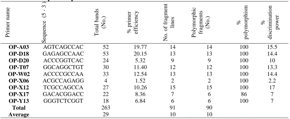
investigated using this technique with several RAPD oligonucleotide (21). The RAPD primers used in this study generated 263 total bands with an average of 29 bands per primer (Table. 2). Each isolate was varied in term of the generated bands, Primer OP-X06 produced 4 bands showing the lowest primer efficiency (1.52%), while primer OP-D18 produced 53 bands showing the highest primer efficiency (20.15%). The molecular weight of the amplified bands was ranged from 145 bp (OP-T07) to 2109 bp (OP-D18). RAPD primers amplified 91 fragment lines across all isolates genomes with average of 10 fragment lines per primer. Among these fragment lines scored, 90 fragment lines (99.0%) were polymorphic with average of 10 polymorphic fragment lines per primer across the 10 MRSA isolates. Primer (OP-X12) amplified 15 fragment lines (100% polymorphism) representing the maximum discrimination power (17%), while primer OP-X06 amplified 2 polymorphic fragment lines (100% polymorphism) representing the minimum discrimination power (2.2 %).
Table 2. RAPD primers parameters.
% d is cr im in atio n p o wer % p o ly m o rp h is m Po ly m o rp h ic fr ag m en ts (No .) No . o f fr ag m en t lin es % p rim er ef ficien cy T o tal b an d s (No .) Seq u en ce (5 3 ) Prim er n am e 15.5 100 14 14 19.77 52 AGTCAGCCAC OP-A03 14.4 100 13 13 20.15 53 GAGAGCCAAC OP-D18 10 100 9 9 5.32 24 ACCCGGTCAC OP-D20 13.3 100 12 12 11.40 30 GGCAGGCTGT OP-T07 14.4 100 13 13 12.54 33 ACCCCGCCAA OP-W02 2.2 100 2 2 1.52 4 ACGCCAGAGG OP-X06 17 100 15 15 10.26 27 TCGCCAGCCA OP-X12 7 86 6 7 8.36 22 GACACGGACC OP-X17 7 100 6 6 6.84 18 GGGTCTCGGT OP-Y13 90 91 263 Total 10 10 29 Average
The arbitrary primers (OP-X12, OP-A03, OP-D18 and OP-T07) were useful for discrimination MRSA isolates of distinct characteristics (Fig. 1 and 2). 100% polymorphism percentage was recognized for all primers except (OP-X17) primer among the
Figure 1. Agarose gel electrophoresis of primer OP-X12 and OP-A03 for DNA samples of the MRSA isolates. M: represent 100 bp ladder. Lanes: from 1-10 represent MRSA isolates.
Figure 2. Agarose gel electrophoresis of primer OP-D18 and OP-T07 for DNA samples of the MRSA isolates. M: represent 100 bp ladder. Lanes: from 1-10 represent MRSA isolates.
Genetic distance and Eigenvalue estimation
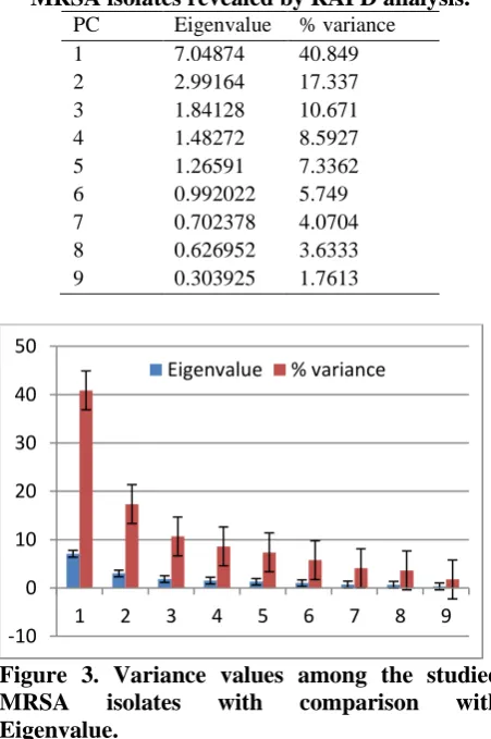
The genetic distances (GD) revealed from Jaccard similarity index was calculated for the 90 RAPD polymorphic fragments according to formula (GD = 1 - similarity) (23), of the 10 MRSA isolates using PAST software version 1.62. Table 3 illustrates the genetic distance values among MRSA isolates. The genetic distance values ranged from (0.218) to (0.959). The isolate number 1 was highly divergent from isolate number 5 as well as isolate number 3 from isolate number 10 with a distance of 0.959. Isolate number 6 was very closely related to isolate number 7 with distance of 0.218. Eigenvalue and the variances for the studied isolates were also estimated using PAST software version 1.62, as
Table 3. Genetic distance’s values among MRSA isolates as revealed by RAPD analysis.
Isolates 1 2 3 4 5 6 7 8 9 10
1 0
2 0.8864 0
3 0.8966 0.8594 0
4 0.8628 0.8449 0.3266 0
5 0.9592 0.9091 0.4694 0.532 0
6 0.8889 0.5953 0.8254 0.8071 0.8704 0
7 0.907 0.4865 0.8549 0.8393 0.9057 0.2188 0
8 0.871 0.625 0.9273 0.9184 0.9303 0.5938 0.5 0
9 0.8948 0.6924 0.8422 0.868 0.8479 0.6667 0.6 0.577 0
10 0.875 0.875 0.9592 0.9025 0.9445 0.8438 0.8667 0.8422 0.8847 0
Table 4. Variance values comparison among MRSA isolates revealed by RAPD analysis.
PC Eigenvalue % variance 1 7.04874 40.849 2 2.99164 17.337 3 1.84128 10.671 4 1.48272 8.5927 5 1.26591 7.3362 6 0.992022 5.749 7 0.702378 4.0704 8 0.626952 3.6333 9 0.303925 1.7613
Figure 3. Variance values among the studied MRSA isolates with comparison with Eigenvalue.
Conclusion:
MRSA, with its virality, distribution and its ability to resist a wide spectrum of antibiotics, make this bacterium one of the most threating agents worldwide. According to the data obtained from this study, RAPD-PCR technique is considered a successful method for MRSA typing and to estimate genetic distances among the studied isolates with its simplicity, rapid and cost-efficient. It was confirmed that there was an extensive genotypic diversity of MRSA from various clinical samples using this technique. Consequently, in light of the data observed in this research and the DNA typing of MRSA isolates, this method can be used to
monitor and understand the pathogenesis and the spread of this bacterium in hospitals, communities and moreover for helping the establishment of a successful surveillance procedures and facilitate global MRSA control.
Conflicts of Interest: None.
References:
1. Graves SF, Kobayashi SD, DeLeo FR. Community-associated methicillin-resistant Staphylococcus aureus immune evasion and virulence. j mol med. 2010;88(2):109-114.
2. Debnath A, Chikkaswamy BK. Randomly Amplified Polymorphic DNA Assay of Methicillin Resistant Staphylococcus aureus Isolated from Clinical Samples from Bengaluru, India. Int J Curr Microbiol Appl Sci. 2015;4(11):342-355.
3. Al-Zahrani NH. PCR-based Random Amplified Polymorphic DNA Fingerprinting of Staphylococcus aureus strains isolated from patients in Jizan Hospital, Saudi Arabia. Life Sci J. 2013;10(12s).
4. Jevons MP, Coe A, Parker M. Methicillin resistance in staphylococci. The Lancet. 1963;281(7287):904-907.
5. Huang H, Flynn NM, King JH, Monchaud C, Morita M, Cohen SH. Comparisons of community-associated methicillin-resistant Staphylococcus aureus (MRSA) and hospital-associated MSRA infections in Sacramento, California. J Clin Microbiol. 2006;44(7):2423-2427.
6. Boyce J. Methicillin-resistantStaphylococcus aureus: A continuing infection control challenge. Eur J Clin Microbiol Infect Dis. 1994;13(1):45.
7. Uzunović S, Ibrahimagić A, Kamberović F, Rijnders MI, Stobberingh EE. Molecular characterization of methicillin-susceptible and methicillin-resistant Staphylococcus aureus in Food handlers in Bosnia and Herzegovina. Open Infect Dis J. 2013;7(1):15-20. 8. Shahkarami F, Rashki A, Ghalehnoo ZR. Microbial susceptibility and plasmid profiles of resistant Staphylococcus aureus and methicillin-susceptible S. aureus. Jundishapur J Microbiol. 2014;7(7).
9. Alipour F, Ahmadi M, Javadi S. Evaluation of different methods to detect methicillin resistance in
-10 0 10 20 30 40 50
1 2 3 4 5 6 7 8 9
Staphylococcus aureus (MRSA). J Infect Public Health. 2014;7(3):186-191.
10.Mostafa S. Molecular typing of methicilin resistant Staphylococcus aureus by spa gene polymorphism. Afr J Microbiol Res. 2013;7(9):755-759.
11.Stranden A, Frei R, Widmer A. Molecular typing of methicillin-resistant Staphylococcus aureus: can PCR replace pulsed-field gel electrophoresis? J Clin Microbiol. 2003;41(7):3181-3186.
12.Singh GK, Bopanna BD, Rindhe G. Molecular characterization of Staphylococcus aureus-human pathogen from clinical samples by RAPD markers. Int J Curr Microbiol App Sci. 2014;3(2):349-354. 13.Idil N, Bilkay IS. Application of RAPD-PCR for
determining the clonality of methicillin resistant Staphylococcus aureus isolated from different hospitals. Brazilian Archives of Biology and Technology. 2014;57(4):548-553.
14.Bergey DH, Holt JG. Bergey’s Manual of Determinative Bacteriology. Baltimore: Maryland; 1994. p. 350.
15.Sambrook J, Fritsch EF, Maniatis T. Molecular cloning: a laboratory manual. Cold spring harbor laboratory press. 1989.
16.Rabbani MA, Pervaiz ZH, Masood MS. Genetic diversity analysis of traditional and improved cultivars of Pakistani rice (Oryza sativa L.) using RAPD markers. Electron j biotechnol. 2008;11(3):52-61.
17.Alansari MS, Al-kazaz AKA, Khierallah HS. Assessment of Genetic Distance Among Some Iraqi
Date Palm Cultivares) Phoenix Dactylifera L.) Using Randomly Amplified Polymorphic DNA. IJS. 2014;55(4B):1833-1843.
18.Muazaz A-h, Jawad Mohammed M, Kanawapee N, Al A-jH, Athiya M, Talib A-k, et al. Genetic Diversity and Relationships among Verbascum Species in Iraq by RAPD-PCR Technique. Global Science Research Journals. 2014; 2(2): 063-074. 19.Khierallah HS, Al-Sammarraie SK, Mohammed HI.
Molecular characterization of some Iraqi date palm cultivars using RAPD and ISSR markers. J Asian Sci Res. 2014;4(9):490.
20. Hammer Ø, Harper D, Ryan P. Paleontological statistics software: package for education and data analysis. Palaeontol Electronica. 2001(4):1-9. 21.Mobasherizadeh S, Shojaei H, Havaei SA,
Mostafavizadeh K, Davoodabadi F, Khorvash F, et al. Application of the Random Amplified Polymorphic DNA (RAPD) Fingerprinting to Analyze Genetic Variation in Community Associated-Methicillin Resistant Staphylococcus Aureus (CA-MRSA) Isolates in Iran. Glob j health sci. 2016; 8(8):185. 22.Mahreen N, Akhtar A, Altaf J. et al. Molecular
genotyping of methicillin-resistant Staphylococcus aureus by employing DNA marker technology. J Biodivers and Environ Sci. 2017; 10(6):330-335. 23.Nei M, Li W-H. Mathematical model for studying
genetic variation in terms of restriction endonucleases. Proceedings of the National Academy of Sciences. 1979;76(10):5269-5273.
ضعب نيب يثارولا دعبلا ريدقتل اندلا ةلسلسل لاكشلاا ددعتملا يئاوشعلا فعاضتلا تارشؤم ةناقت قيبطت
اروكملا تلازع
يفشتسم نم ةلوزعملاو نيليسيثيملل ةمواقملا ةيبهذلا ةيدوقنعلا ت
ةفلتخم ةيقارع تا
يداه دمحم ءايلع
1يلع لداع ءاسيم
2
يراصنلا دعس دمحم
3نابدهلا نايدو
31
,ىطسولا ةينقتلا ةعماجلا ,روصنملا /ينقتلا يبطلا دهعملا ,ةيضرملا تلايلحتلا مسق .قارعلا ,دادغب
.قارعلا ,دادغب ,ةيجولونكتلا ةعماجلا ,ةيئايحلاا تاينقتلا عرف ,ةيقيبطتلا مولعلا مسق
2
3
.قارعلا ,دادغب ,دادغب ةعماج ,مولعلا ةيلك ,ةيئايحلاا تاينقتلا مسق
:ةصلاخلا
تاببسملا نم ةدحاو نيليسيثيملل ةمواقملا ةيبهذلا ةيدوقنعلا تاروكملا تلالاس ربتعتهذه .تايفشتسملا نع ةجتانلا تاباصلإل ةيسيئرلا
فنلاا ,دلجلا تاباصا لثم ضارملاا نم ديدعلا نع ةلوؤسم اهنا امك ةيويحلا تاداضملا نم عساو ىدم ةمواقم ىلع ةردقلا اهل ايريتكبلا ةلسلسل يئاوشعلا فعاضتلا تارشؤم ةناقت مادختسا مت ةساردلا هذه يف .حورجلاو (RAPD) DNA
صحفل ةيئاوشعلا تائدابلا نم ةرشع عم
( عمج مت .مهنيب يثارولا دعبلا نم ققحتللو نيليسيثملل ةمواقملا ةيبهذلا ةيدوقنعلا تاروكملا تلازع نيب يئيزجلا عونتلا 90
تانيع نم ةلزع )
( نأ دجوو , ةفلتخم ةيقارع تايفشتسم نمو ةيريرس 10
( ةبسنبو طقف تلالاس ) 11.11
ل ةمواقم تناك )٪ نيليسيثيمل
. ,ةرشعلا تائدابلا هذه نم
تائدابلا هذه تجتنا .ةحضاو فعاضت جتاون تطعا دق طقف ةعست 91
( لدعمبو تلازعلا عيمجل ةيسيئر ةمزح 10
,ءيداب لكل ةيسيئر مزح )
( اهنمض نم تناك 90
( ةبسنبو ةنيابتم ةمزح ) 99
.)% ( نيب ام ةفعاضملا مزحلا ماجحا تحوارت 145
-2109 يدعاق جوز ) ةيوئملا ةبسنلا .
( ةبسنب تناك تائدابلا لكل يثارولا نيابتلل 100
ءيدابلا ادعام )% OP-X17
( نيابت ةبسن ىطعا يذلا 86
)% ةيثارولا داعبلاا باسح مت .
( رشؤم ىلع دامتعلااب Jaccard similarity
( ل ) 90 يثارو دعب ةميق ىلعا .ةنيابتم ةيسيئر ةمزح ) (0.959)
( مقر ةلزعلا نيب تناك 1
)
( مقر ةلزعلاو 5
( مقر ةلزعلا نيب امو ) 3
( مقر ةلزعلاو ) 10
يثارو دعب ةميق ىندا تناك امنيب ,) (0.218)
( مقر ةلزعلا نيب 6
( مقر ةلزعلاو ) 7
.)
نا رهظت ةساردلا هذه ةلسلسل يئاوشعلا فعاضتلا تارشؤم ةناقت
DNA عم اهرابتخا مت يتلاو 9
نع فشكلل حاجنب اهقيبطت نكمي تائداب
.ةفلتخم تايفشتسم نمو نيليسيثيملل ةمواقملا ةيبهذلا ةيدوقنعلا تاروكملا تلازع نيب ةيثارولا داعبلاا
:ةيحاتفملا تاملكلا تارشؤم ,نيليسيثيملل ةمواقملا ةيبهذلا ةيدوقنعلا تاروكملا
فعاضتلا يئاوشعلا
ددعتملا ةلسلسل لاكشلأا DNA
دعبلا ,
طيمنت ,يثارولا DNA