International Journal of Pharmaceutical Research & Allied Sciences, 2017, 6(1): 145-152
Research Article
ISSN : 2277-3657
CODEN(USA) : IJPRPM
145
MODELING, FORMATION, DESTRUCTION AND SCANNING ELECTRON
MICROSCOPY OF BIOFILMS
Pogorelov A.G.
1, Bakhir V.M.
2, Ipatova L.G.
3, Kuznetsov A.L.
4, Suvorov O.A.
5, Kozlov I.V.
61 Pogorelov Alexander Grigorievich, Doctor of Biological Sciences, e-mail: [email protected], telephone number:+74967739370
2 Bakhir Vitold Mikhailovich, Doctor of Engineering Sciences, e-mail: [email protected], telephone number: +74967739370
3 Ipatova Larisa Grigoryevna, Doctor of Engineering Sciences, e-mail: [email protected], telephone number: +74967739370
4 Kuznetsov Alexander Lvovich, e-mail: [email protected], telephone number: +74967739370 5 Suvorov Oleg Alexandrovich*, Candidate of Engineering Sciences, e-mail: [email protected],
telephone number: +74967739370
6 Kozlov Igor Vladimirovich, Candidate of Engineering Sciences, e-mail: [email protected], telephone number: +74967739370
________________________________________________________________________________________
ABSTRACT
The work was performed to study the cellular population of a biofilm formed on the surface of a tube made of a polymer material, as well as to find a method for its destruction using electrochemically activated solutions. The study was carried out on an experimental stand in the form of a water recirculation system with the possibility of infecting microflora for the cultivation of biofilms and with the possibility of periodically examining the inner surface of the tubes. At the first stage of the study, by means of light and electron scanning microscopy, the biofilm formation was determined by microorganisms spontaneously contaminating the walls of the tubes. The criterion for selecting new means for the destruction of biofilm should be their complex metastable composition with increased reactivity for effectively overcoming the biopolymer matrix and cell destruction At the next stage, the possibility of destroying bacterial biofilms was studied using electrochemically activated solutions obtained by unipolar electrochemical treatment of an aqueous solution of sodium chloride: catholyte - an aqueous solution of sodium hydroxide saturated with hydrogen, and anolyte - an aqueous solution of a metastable mixture of chlorine-containing and hydroperoxide oxidants, obtained by anodic oxidation of sodium chloride solution. As a starting culture, a dry preparation of lactic acid bacteria Lactococcus lactis, Streptococcus thermophilus, Lactobacillus acidophilus, Lactobacillus helveticus, Propionibacterium freudenreichii ssp. Shermani was used. The analysis of microphotographs showed that metastable particles with different electrochemical potentials can have a damaging effect on bacterial biofilm.
Keywords: microbial biofilms, modeling, formation, destruction of biofilms, electrochemically activated solution, scanning electron microscopy, quality, safety and food hygiene
_____________________________________________________________________________________________
146
Currently, almost every enterprise of the agro-food complex associated with the use of liquid media, fouling surfaces of equipment, communications, finished products with microbial biofilms. Biofilms are potential sources of microbiological contamination of products by pathogenic, opportunistic or extraneous, undesirable microorganisms in a certain technological process. Their formation and development takes place at the interface of two phases and consists of the following stages: Primary adhesion; Adaptation of bacteria to the surface of the material: the formation of multilayer cellular structures and clusters as a result of the secretion of extracellular matrix: activation of fission and multiplication [24-26].
Biofilms are one of the most common forms of bacteria in most natural conditions. The term "biofilm" is understood to mean the constantly renewing community of various kinds of microorganisms attached to the phase interface and to each other and surrounded by an extracellular polymer matrix, which is one of the factors of intercellular interaction. Its known that microorganisms, depending on the species, are distinguished by the unique nature of their growth, development and life processes, but the formation of bacterial biofilms is subject to general patterns [15, 19].
The formation of a biofilm is associated with environmental conditions: the quality of the surface, the redox state of the solution, its acidity, the presence of aggressive molecules and other factors. An important problem is the resistance of microflora to antimicrobial agents, its ability to adapt to disinfection and maintain homeostasis for occupying microorganisms after removal of the antimicrobial solution. In particular, when the working solution of the disinfectant dries, its concentration increases, during the local interaction of microorganisms with the active substance, antistress protection cystrons are activated, and the cell adapts to this type of substances. Existence in a fixed state is one of the principles of the habitat of bacteria in different ecotopes, and adhesion is an adaptive reaction that increases the ability to survive in microorganisms under unfavorable growth conditions.
To effectively suppress bacteria in biofilms, a multiple increase in the concentrations of antimicrobial substances (antibiotics, disinfectants) is required in comparison with their values when acting on separate plankton (freely floating) cells. Nevertheless, there are cases of a significant increase in biofilm growth in some microorganisms in the presence of maximum concentrations of biocides. In works [5, 9, 15, 23], it was proved that biofilms, as a rule, exhibit multi-antibiotic resistance, that is resistance to antibiotics of different chemical structure. The same pattern is observed for many disinfectants. Therefore, in order to reduce the survival rate of biofilm bacteria and suppress the formation of biofilms, it is necessary to create new ways of solving this problem.
Promising are compounds whose effect is both in the destruction of pathogenic bacteria in the composition of biofilms due to suppression of biosynthesis, and in the inhibition of their virulence factors (toxins, adhesins, effector proteins), as well as factors that promote the communication of bacteria with each other and the formation of biofilms. Effective practical solutions can be obtained on the basis of studying the processes of formation and destruction of these bacterial agglomerations in laboratory conditions.
MATERIALS AND METHODS
The study was carried out on an experimental stand in the form of a water recirculation system with the possibility of infecting microflora for the cultivation of biofilms and with the possibility of periodically examining the inner surface of the tubes. The working model was a polymer container with a capacity of 2 liters, filled with tap water, through which a PVC tube 5 mm in diameter is passed. At the first stage of the study, the presence of growth of microorganisms on the walls of the tube was determined with the help of the Altamy 105 microscope (Russia) with a magnification of 1-2000, after which the morphological analysis of the biofilm was carried out using a scanning electron microscope JSM-6390A (JEOL, Japan).
147
absolute ethanol. To remove ethyl alcohol, the samples were transferred to hexamethyldisilazane (HMDS) for 30 minutes, and then dried in air.
Prepared samples were mounted on the object holder of the electron microscope by means of a conductive adhesive. Before viewing in the JFC 1600 (JEOL, Japan), a uniform conductive film of platinum 20 nm in thickness was deposited onto the surface of the preparation by ion sputtering of metal in argon plasma medium. The ultrastructure of the biofilm relief on the surface of the tube was studied in a scanning electron microscope JSM-6390A at an accelerating voltage of 10 kV using the secondary electron mode. Scanning electron microscopy was carried out at an amplification from 1000 to 10000.
RESULTS AND DISCUSSION
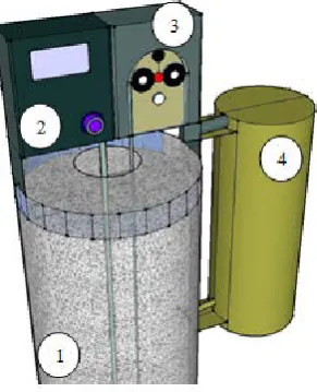
The experiment was carried out in laboratory conditions on a model stand of the original design (working prototype, diagram in Fig. 1). Within 8 weeks, the growth of microorganisms naturally entering the water from the air took place on the inner surface of the tank and on the walls of the tube.
Fig. 1 Scheme of the experimental model stand of the original design for the growth of microorganisms and the formation of a biofilm, where
1 - reservoir volume of 5000 cm3;
2 - compressor and pipeline to saturate the aqueous solution with air oxygen;
3 - peristaltic pump and a set of pipelines for pumping an aqueous solution through a piping system;
4 - system of pipelines, compactly placed in the form of a coiled unit.
The experimental stand was a model of a flow recirculation system with the possibility of spontaneous contamination and additional saturation of the aqueous solution with oxygen. During the work of this system, the formation of biofilms on the walls of pipelines was recorded. An oxygenated aqueous solution through the pump runs the entire length of the pipelines. At the same time, because of the roughness of the PVC tube surface, microorganisms that form a biofilm are retained on it. At the first stage of the study, the growth of microorganisms on the walls of the tube was determined with the Altami 105 microscope.
148
Fig. 2 Microphotographs of the surface obtained with the Altami 105 microscope (x2000)
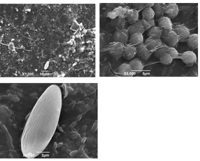
The microphotographs of Figure 2 show the active growth of microbial cells on the surface of the tube. Electron microscopy using the scanning electron microscope JSM-6390A confirmed the presence of formed biofilms both on the outer and inner surfaces of the tube (Figures 3-5).
Fig. 3 Microphotographs of biofilm on the outer surface of the tube (x1000-8500)
149
Fig. 4 Microphotographs of biofilm on the inner surface of the tube (x1000-10000)
Fig. 5 Microphotograph of the inner surface of a clean tube - control (x3000)
Comparison of microphotographs of a clean and contaminated inner surface of the tube confirms the formation of clusters of microorganisms of various shapes represented. Microorganisms have mainly round, oval and rod-like shapes 2-5 microns in size. The bridges connecting the cells to each other, or covering the clusters of cells (an unformed, unstructured substance) obviously represent fragments of the biofilm matrix. The discovery of two obligatory biofilm attributes - bacterial cells and extracellular matrix - served as evidence of the existence of a biofilm. As can be seen from Figures 3-5, under the conditions of the experiment, a biofilm of microorganisms is formed, corresponding to that of existing technological processes in the food industry. Thus, the obtained data indicate that the developed model of the experimental stand of the recirculation system allows one to evaluate the formation of biofilms and conduct their morphological analysis.
The variability and metastability of processes that ensure the life of a community of microorganisms in biofilms requires an adequate approach to the choice of ways to combat such manifestations of life at the micro level. It is important to note that the formation of biofilms is not only one of the pathogenicity factors of microorganisms, but also a process that leads to a significant change in sensitivity to antibacterial drugs. The criterion for new biofilm control products should be their complex changing metastable composition with increased reactivity in order to effectively overcome the biopolymer matrix and destroy cells.
150
electrochemically activated solutions obtained by unipolar electrochemical treatment of an aqueous solution of sodium chloride: a catholyte - an aqueous solution of sodium hydroxide saturated with hydrogen and an anolyte - an aqueous solution of a metastable mixture of chlorine-containing and hydroperoxide oxidants obtained by anodic oxidation of the solution sodium chloride. It is assumed that the most effective way to combat biofilms is the use of metastable substances, such as anolyte (in this experiment Anolyte ANK SUPER was used), manifesting themselves as a "cold flame" [3, 10].
It was established [23] that the biofilm matrix reacts with the biocide only in the case when the biocide molecule has a reactivity with respect to the matrix substance. A metastable mixture of chlorine- and oxygen-containing oxidants possesses a multitude of spontaneously realizing possibilities of irreversible disruption of vital functions of microorganism biofilms at the level of electron transfer reactions [1]. Therefore, this complex is an effective means of destroying microorganisms and their biofilms.
Metastable particles with different electrochemical potentials can have a damaging effect on all large systematic groups of microorganisms - bacteria, mycobacteria, viruses, fungi – and have sporicidal activity. At the same time, they do not harm the cells of human tissues and other higher organisms, i.e. somatic animal cells in the multicellular system, protected by a chemical antioxidant system. The resulting highly active radicals and single atomic oxygen take part in the destruction of microorganisms, interacting with biopolymers capable of oxidation [2].
It is obvious that the result of the action of a particular disinfectant or antibiotic on a biofilm matrix depends not only on the chemical nature of the antimicrobial agent, but also on the chemical composition and architecture of the matrix, which in turn are due to the species and features of the strain of biofilm-forming bacteria [22]. Microphotographs of a biofilm formed on the surface of the inner wall of a tube of a laboratory recirculation system with bifid bacteria and treated with various biocidal agents are shown in Fig. 6. As a starting culture we used a dry preparation of bacteria Lactococcus lactis, Streptococcus thermоphilus, Lactobacillus acidophilus, Lactobacillus helveticus, Propionibacterium freudenreichii ssp. Shermanii with 4х109 CFU / 300 mg of the drug.
A nutrient medium based on distilled water supplemented with sodium hydrogen phosphate and milk was poured into a tube and inoculated with a suspension of a bacterial preparation. Then, growth of microorganisms was observed and biofilm formation was recorded. The control sample was a selected fragment of a clean tube. Disinfection of experimental samples was carried out with biocidal solutions in the flow using a peristaltic pump. The length of each treated tube was 20 cm. The processing time was 5 minutes in the circulation mode. In the case of sequential treatment - first 3 minutes catholyte, then 2 minutes anolyte. The flow rate is 22.93 l / h. After treatment with anolyte, its parameters were determined and compared with the initial ones. Active chlorine content decreased after treatment with anolyte only by 6.7%, and after successive treatment with anolyte and catholyte - by 3%.
(A) control: tap water (B) disinfection with sodium
hydroxide solution 10%
151
(D) Anolyte disinfection421 mg / l active chlorine
(E) two-stage treatment: catholyte 10% and anolyte sequentially
Fig.6. Microphotographs of the inner surface of a tube treated with various biocidal agents (x3000)
As can be seen from the presented data, the results of the experiment on disinfection of the tubes in which the biofilm from lactic acid bacteria was previously grown depended on the method and type of biocide effect.
CONCLUSION
Using a scanning electron microscopy, biofilms formed on the surface of the inner wall of the tube were analyzed. The analysis of microphotographs showed that the result of the action of the electrochemically activated disinfectant on the biofilm matrix depends not only on the chemical nature of the antimicrobial agent, but also on the chemical composition and matrix architecture. It’s in turn, due to the species and features of the strain of biofilm-forming bacteria. It was shown that metastable particles with different electrochemical potential values can have a damaging effect on all large systematic groups of microorganisms - bacteria, mycobacteria, viruses, fungi, spores.
Acknowledgements
This work was supported by the Russian Science Foundation grant 16-16-00020; Institute of Theoretical and Experimental Biophysics, RAS, ul. Institustskaya, 3, Pushchino, 142290, Moscow Region, Russia.
REFERENCES
1. Bakhir, V.M., Electrochemical activation. Inventions, technics, technology. Moscow: VIVA-STAR; 2014, P. 512.
2. Bakhir, V.M., Vtorenko, V.I., Panicheva, S.A., Prilutsky, V.I. et al., On the efficacy and safety of chemical agents for disinfection, pre-sterilization treatment and sterilization: Disinfection, 2003. 1. 29-36.
3. Bakhir, V.M., Ipatova, L.G., Suvorov, O.A., Pogorelov, A.G., Microbial biofilms: mechanisms of formation and interaction with the environment, new approaches to the destruction of biofilms: In the collection: Food. Ecology. Quality Proceedings of the 13th International Scientific and Practical Conference, 2016, 125-130.
4. Golub, A.V., Bacterial biofilms - a new goal of therapy: Clinical microbiol. Antimicrobial. Chemotherapeutist, 2012, 14(1), 23-29.
5. Gostev, V.V., Bacterial biofilms and infections: Journal of Infectology, 2010, 2 (3), 4-15.
6. Korobov, V.P., Lemkina, L.M., Monakhov, V.I., Analysis of sensitivity of the processes of formation of biofilms Staphylococcus epidermidis 33 to some environmental factors: Bulletin of Perm University, 2010, 1 (1), 59-63.
152
8. Mayanskiy, A.N., Staphylococcal biofilms: structure, regulation, rejection: Journal of Microbiology, 2011, 1, 101-108.
9. Mayanskiy, A.N., Strategy of management of bacterial biofilm process: Journal of Infectology, 4(3), 2012, 5-15.
10. Pogorelov, A.G., Bakhir, V.M., Ipatova, L.G., Levacheva, MA, Suvorov, O.A., Destruction of microbial biofilms as a factor of increasing microbiological safety at enterprises of the agro-industrial complex: Food Innovations and Biotechnology materials of the IV International Scientific Conference, 2016, 498-505.
11. Pogorelov, A.G., Gavrilyuk, V.B., Pogorelova, V.N., Gavrilyuk, B.K., Scanning Electron Microscopy of Biosynthetic Wound Dressings Biocol, 2012, Bulletin of Experimental Biology and Medicine, 154 (1), 167-170.
12. Pogorelov, A.G., Selezneva, I.I., Evaluation of collagen gel microstructure by scanning electron microscopy, 2010, Bulletin of Experimental Biology and Medicine, 150(1), Page 153-155.
13. Pogorelov, A.G., Chebotar, I.V., Pogorelova, V.N., Scanning Electron Microscopy of Biofilms Adherent to the Inner Catheter Surface, 2014, Bulletin of Experimental Biology and Medicine, 157 (5), 711-714.
14. Tets, G.V., Role of extracellular DNA and matrix lipids in the interaction of bacterial biofilms with antibiotics (experimental study). Candidate of Medical Science. St. Petersburg: 2007.
15. Turkuyukov, V.B., Molecular features of the morphology of biofilms formed by strains of non-fermenting gram-negative bacteria: Tikhooken Medical Journal, 2013, 4, 44-47.
16. Tutelyan, V.A., Knyazhev, V.A., Microorganisms and food. Risk and benefit: Bulletin of the Russian Academy of Medical Sciences, 2000, 12, 3-6.
17. Tutelyan, V.A., Food and biosafety: Bulletin of the Russian Academy of Medical Sciences, 2002, 10, 14-19.
18. Firsova, V.G., Parshikov, V.V., Chebotar, I.V., Lazareva, A.V., Pogorelov, A.G., Microbiological diagnosis and choice of antimicrobial therapy of bile duct infection: Annals of surgical hepatology, 2015, 20 (1), 124-131.
19. Chebotar, I.V., Biofilms Staphylococcus aureus: structural and functional characteristics and relationships with neutrophils: author's abstract. Doctor of Medical Science. Moscow: 2013, 42.
20. Chebotar, I.V., Pogorelov, A.G., Yashin, V.A., Guriev, E.L., Lominadze, G.G., Modern technologies for the study of bacterial biofilms: Modern technologies in medicine, 2013, 5 (1), 14-20.
21. Shandala, M.G., Disinfection problems in food hygiene: Nutrition issues, 2013, 2, 42-47.
22. Broun, A., et al. A dose-response study of antibiotic resistance in Pseudomonas aeruginosa biofilms: Antimicrob. Agents Chemo-ther, 2000, 44, 640-646.
23. Devey, M.E., Microbial biofilms: from ecology of molecular genetics microbiology and molecular biology reviews. 2000, 64:4, 847-868.
24. Levieveld, M.A., Mostert, J. Holah., Handbook of hygiene control in the food industry: Edited by H.L.M. CRC Press. Boca Raton Boston, New York, Washington.
25. Vergara-Irigaray, M. et al. Wall teichoic acids are dispensable for anchoring the PNAG exopolysaccharide to Staphylococcus aureus cell surface: Microbiology, 2008, 154, 865-877.