(UDC: 616.716.4-073:621.317.3)
Monitor
S. D.
2Ma
ronautics, University of Patras, Patras 265
cbil
The c
ping, Mandible, Bone Quality.
o physiologic stim
ing of Damping for the Assessment of Mandible Bone Quality
Panteliou1*, K. A. Lianos2, D. A. Sarafianos3, C. G. Bilios4
1Machine Design Laboratory, Dept. of Mechanical Engineering and Aeronautics, University of
Patras, Patras 265 00, Greece [email protected]
chine Design Laboratory, Dept. of Mechanical Engineering and Aeronautics, University of Patras, Patras 265 00, Greece
3Machine Design Laboratory, Dept. of Mechanical Engineering and Ae
00, Greece [email protected]
4Machine Design Laboratory, Dept. of Mechanical Engineering and Aeronautics, University of
Patras, Patras 265 00, Greece [email protected]
*Corresponding author
Abstract
ontemporary methods for the assessment of the quality of human mandible, in order to facilitate the decision making for dental implants, include bone density measurements through dual-energy X-Ray absorptiometry (DEXA) or its variations. The estimation of mandible quality with these methods is related to subjectivity, comparability and reliability problems, which results in restricted capability of secure assessment of bone quality.
Monitoring of loss of structural integrity is applied in this work through modal analysis, in order to obtain an objective assessment of mandible bone quality. Specifically, modal damping factor (MDF) and bone mineral density (BMD) measurements are performed on human cadaveric mandibles. From the data acquired clearly arises a very promising correlation between MDF and BMD, reinforcing the belief from our previous research findings that this method can lead to a mandible quality assessment tool, thus encouraging further research investigation.
Keywords: Dam
1. Introduction
Bone quality is a term that may on occasion refer to one or more of the following: density, macrostructure, microstructure, mechanical properties and biologic response t
Bone density is often used as an estimator of bone quality. There is a variety of in vitro and in vivo methods that are used for evaluation of mandibular bone density and they can be broadly categorized in:
1. Histomorphometry of biopsy samples
2. Empiric topographic prediction methods based on combining anthropometric
data and simple panoramic radiographs
3. Torque resistance measurements during implant insertion
4. X-ray absorption methods
The histological and morphometric measurement of the bone has been considered the golden standard for bone density measurements (Molly, 2006). It is an invasive and deleterious method for the donor site, during which, small biopsy specimens 2mm diameter are been harv
Insertion torque measurements are not a true bone density evaluator. A variety of implant parameters, as design characteristics and insertion technique features, co-influence the actual
, 2005).
only for post insertion evaluation and statistical correlation with success data. The empiric method is a general, crude estimation which is non accurate
The most commonly used density evaluation methods in dental practice are the
o be exposed to. Other radiographic methods with lower irradiation burden are used (panoramic, periapical, cone beam CT) and Dual Energy X-ray Absorptionmetry (DEXA) having the lowest (irradiation dose of
1-natural irradiation dose received by the human body (7μSV)).
cted by the fat content of the soft tissue and the
hat can be applied in both jaws. Hence, it is obvious that other more reliable methods are needed, which must be capable to assess bone structural integrity as well as the effect of therapeutic treatment in a non invasive manner.
Loss of structural integrity of an ageing component is usually due to fatigue, which in turn leads to initiation and propagation of starting failure points, and finally to structure failure. Methodology has been developed for the identification of change in structural integrity through
ested from patient’s jaws immediately before implant placement (3,75mm diameter). The empiric method of Lekholm and Zarb (Branemark et all 1985) lacks precision and is related to subjectivity, comparability and reliability problems, which result in restricted capability of secure assessment of bone quality. Although it is an easy and inexpensive method it cannot discriminate between osseous sites at the same individual.
measurements (Ostman et all
From the clinical standpoint, a bone density evaluation method should have a preoperative character, in order to be useful for appropriate treatment planning. Therefore, histomorphometry and insertion torque measurements are not convenient, since the decisions have already been taken and the results can be used
between potential implant sites.
radiographic ones and mainly the computed tomography (CT). Estimation of radiographic density on Hounsfield units allows for site specific presurgical evaluation of bone density and selection of the most suitable implant placement (Norton and Gamble, 2001). The main disadvantage of CTs is the high irradiation dose the patient has t
10μSV equivalent to the average
The accuracy of the measurements is affe
discrimination between cortical and trabecular bone is not possible due to superimposition (Blake and Fogelman, 1977). DEXA is mainly used for evaluation of bone mineral density (BMD) of the lower spine and femoral neck. It is used for the clinical diagnosis of osteoporosis in women and in epidemiological studies for the assessment of fracture risk. In the stomatognathic area it is used only for research purposes and only in the mandible (Horner and Devlin, 1998).
modal analysis and monitoring of modal damping factor. From our previous experimental work (Panteliou and Dimarogonas, 1997a,b - Panteliou and Dimarogonas, 2000, - Panteliou et all, 2001, - Panteliou et all, 2009) it was shown that internal damping factor is sensitive to fatigue and change in porosity. For better understanding of experimental results a model has been developed that quantifies the relation between changes in damping factor and porosity. This model, which includes analytic – arithmetic tool and dedicated measuring device, has been successfully applied on components made out of a variety of materials, conventional and advanced. One very successful application of the method was the assessment of bone structural integrity, for monitoring metabolic diseases of bones (i.e. osteoporosis) (Panteliou et all, 1999, - Panteliou et all, 2004, - Anastassopoulos et all, 2009 – Stavropoulou et all, 2005 – Christopoulou et all, 2006 – Anastassopoulos et all, 2010).
The target of this work is the development of a non invasive tool for the objective assessment of mandible bone quality (assessment of structural integrity). This technique is experimentally applied on cadaveric human mandibles. Besides, conventional DEXA measurements are performed on the same mandibles. The results from both methods were compared in order to evaluate the correlation between BMD and MDF and propose the modal damping as future assessment tool in the process of dental implants placement.
2. Materials and Methods
Ten cadaveric human mandibles, were used in order to acquire in vitro measurements of mandible quality with two methods: (1) BMD (Bone Mineral Density) with DEXA, and (2) Modal Damping Factor (MDF).
2.1 Measurement of Bone Mineral Density
Bone mineral de EXA), using a
Norland XR-26 M d with an
ultra-high resolution software program (available from the manufacturer). The time required for a typical scan at each anatomic site was approximately 3 minutes.
2.2. Measurement of Damping
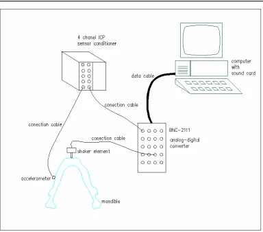
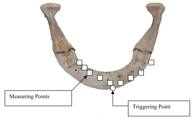
Modal Damping Factor is measured with designed and constructed dedicated device (Fig. 1). For its calculation half power bandwidth method is applied (Dimarogonas, 1992). The sensor used is an accelerometer of 1 gr. Its output signal is transferred through an A/D converter to a PC. The triggering is produced by computer controlled sound electronics and is applied through a metallic stem to the selected anatomic site of the mandible. Modal Damping Factor (MDF) (Dimarogonas, 1992) is extracted by applying FFT. Consecutive signals are acquired (approximately 25) at the same point of the mandible in order to ensure statistically sufficient population. MDF is the average of all measurements. The procedure is repeated for all selected points of the mandible (Fig. 2).
Figure 2. Triggering and measuring points.
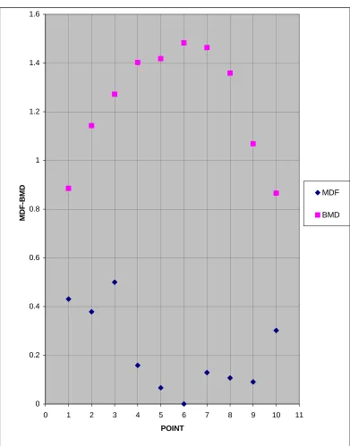
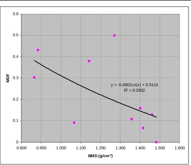
The average of all measured MDF and BMD data in relation to the anatomic site of ent are presented in Fig. 3, while the correlation between MDF and BMD data is ted in Fig. 4.
measurem presen
Measuring Points
0
0 1 2 3 4 5 6
0.2 0.4 0.6 0.8 1 1.2 1.4 1.6
7 8 9 10 11
MDF-BMD MDF
BMD
POINT
y = -0.4901Ln(x) + 0.3114 0.3
0.4 0.5
MD
F
R2 = 0.3302
0.2
0.1
0
0.800 0.900
0.6
1.000 1.100 1.200 1.300 1.400 1.500 1.600
BMD (g/cm2)
Figure 4. Correlation between MDF and BMD for all mandibles.
s of results
bon wer
man
valu is presented in Figure 4.
poin whi valu
3. Comment
In this work, bone density measurements (BMD) with DEXA method, as well as assessment of e mandible structural integrity through measurement of Modal Damping Factor (MDF), e performed.
The average values of all MDF – BMD measurements in relation to the anatomic site of the dibles are presented in Figure 3, while the correlation between all MDF - BMD average es for all mandibles tested
From both Fig. (3, 4), the expected, from our previous research findings (Panteliou et all, 1999 - Panteliou et all, 2004 – Anastassopoulos et all, 2009 – Stavropoulou et all, 2005 – Cristopoulou et all, 2006 – Anastassopoulos et all, 2010), correlation between MDF and BMD is revealed. Specifically, low BMD, which expresses reduced bone density and low bone quality, corresponds to high MDF, which in turn expresses deterioration of bone quality. At this
t, let’s note that MDF is material and system property and index of structural integrity, ch takes values in the range (0-1), with higher values corresponding to low, and lower
4. Conclusions
ur previous resea
In o rch works (Panteliou and Dimarogonas, 1997a,b - Panteliou and
Pant 04 – Anastassopoulos et all, 2009 - Stavropoulou et all, 2005 – Cristopoulou
meth of materials (conventional
on b
meta mparison of measured data with
Ram
expe ssessment tool of the quality of the human
prom be used in
The Patras, Greece, represented by
Dimarogonas, 2000, - Panteliou et all, 2001, - Panteliou et all, 2009 – Panteliou et all, 1999 - eliou et all, 20
et all, 2006 – Anastassopoulos et all, 2010), the application of the thermodynamic damping od as a tool for assessment of structural integrity for a variety
and advanced, i.e. composites) and geometries, has been elaborated. A specific application was ones in order to create a bone quality assessment tool applicable for the monitoring of bolic diseases of bones, especially osteoporosis. The co
MDF and all conventional methods (DEXA, pQCT, biochemical markers, histomorphometry, an Spectroscopy) gave very promising results, which constituted the basis for this initial rimental work, aiming to build an objective a
mandible that will help the decision making during dental implants placement.
The results of this work present a clear correlation in the expected direction. Specifically, MDF and BMD expand in an inverse manner. High BMD values, expressing high bone density, correspond to low MDF values, which in turn express improvement of bone structure and vice versa.
The results from the comparison of the measured data with the two methods constitute a ising basis, which reinforces our belief that we can build an assessment tool to
the process of placement of dental implants.
Acknowledgements
authors acknowledge support of MAGNITIKI S.A.,
Radiologist Medical Doctor Dionissios Karahalios. All measurements presented in this work e performed in MAGNITIKI S.A. (phone +302610365000).
erences wer
Ref
Anastassopoulos G, Panteliou SD, Christopoulou GE, Stavropoulou A, Panagiotopoulos E, Lyritis G, Khaldi L, Varakis J (2009). Vibrational Bone Characteristics vs Bone Density in
Assessing Osteoporosis, Journal of Medical Engineering and Technology (in press).
Anastassopoulos G, Panteliou SD, Panagiotopoulos E, Lyritis G, Raptou P (2010). Damping for
Assessing Osteoporosis. A study in Women. Journal of Medical Engineering and
Technology, (to appear).
Blake, I. Fogelman I (1997). Technical principles of dual energy X-ray absorptionmetry. Semin
Nucl Med, 27:210-228, 1997.
Branemark PI, Zarb GA, Albrektsson T (1985). Tissue Integrated Prostheses, Quintessence
Publishing Co., Inc.
Christopoulou GE, Stavropoulou A, Anastassopoulos G, Panteliou SD, Papadaki E, Karamanos NK, Panagiotopoulos E (2006). Evaluation of modal damping factor as a diagnostic tool for osteoporosis and its relation with serum osteocalcin and collagen I N-telopeptide for
monitoring the efficacy of aledronate in ovariectomized rats, Journal of Pharmaceutical
and Biomedical Analysis, 41, pp. 891-897.
Horner K, Devlin H (1998). The relationship between two indices of mandibular quality and
bone mineral density measured by dual energy X-ray absorptionmetry, Dentomaxillofac
Radiol, 27:17-21.
Molly L (2006). Bone Density and Primary Stability in Implant Therapy. Clin Oral Impl Res,
n objective scale of bone density using the
Res, 12:79-84.
Ostman PO, Hellman M, Sennerby L (2005), Direct implant loading in the edentulous maxilla ry implant stability criteria for
inclusion. Clin Impl Dent Relat Res, 7(supp I):S60-S69).
ow-d Osteoporosis, Journal of Biomechanical
rogonas AD (1997a). Thermodynamic Damping in Porous Materials with
aterials with
Panteliou SD, Chondros TG, Argyrakis VC, Dimarogonas AD (2001). Damping Factor as an
Indicator of Crack Severity, Journal of Sound and Vibration, 235-245, 241(2).
D, Zonios K, Chondrou IT, Fernandes HR, Agathopoulos S, Ferreira JMF (2009).
Damping Factor Associated with Porosity in Alumina, Journal of Mechanics and Materials
17:214-135.
Norton MR, Gamble C (2001). Bone classification: a
computerized tomography scan, Clin Oral Impl
using a bone density-adapted surgical protocol and prima
Panteliou SD, Hasan Abbasi-Jahromi, Dimarogonas AD, Kohrt W, Civitelli R (1999). L Frequency Acoustic Sweep of Bone Integrity an
Engineering, Aug. 99, Vol. 121, pp. 423-431. Panteliou SD, Dima
Spherical Cavities, Journal of Shock and Vibration, Vol. 4, No. 4, pp. 261-268.
Panteliou SD, Dimarogonas AD (1997b). Thermodynamic Damping in Porous M
Ellipsoidal Cavities, Journal of Sound and Vibration, 201(5), 555-565.
Panteliou SD, Dimarogonas AD (2000). Damping Factor Associated with Porosity and Crack in
Solid, Theoretical and Applied Fracture Mechanics, 34, 217-223.
Panteliou S
in Design, 5:167-174.
Panteliou SD, Xirafaki AL, Panagiotopoulos E, Varakis J, Vagenas NV, Kontoyannis CG
(2004). Modal Damping for Monitoring Bone Integrity and Osteoporosis, Journal of
Biomechanical Engineering, Feb 2004, Vol. 126, pp. 1-5.
Stavropoulou A, Christopoulou GE, Anastassopoulos G, Panteliou SD, Lyritis GP, Spiliotis BE, Karamanos NK, Panagiotopoulos E, Lambiris E (2005). Alteration in serum leptin correlates with alterations in serum N-telopeptide of collagen type I and serim osteocalcin
during the progression of osteoporosis in ovariectomized rats, Clinical Cemical Laboratory