R E S E A R C H
Open Access
Asbestos modulates thioredoxin-thioredoxin
interacting protein interaction to regulate
inflammasome activation
Joyce K Thompson, Catherine M Westbom, Maximilian B MacPherson, Brooke T Mossman, Nicholas H Heintz,
Page Spiess and Arti Shukla
*Abstract
Background:Asbestos exposure is related to various diseases including asbestosis and malignant mesothelioma (MM). Among the pathogenic mechanisms proposed by which asbestos can cause diseases involving epithelial and mesothelial cells, the most widely accepted one is the generation of reactive oxygen species and/or depletion of antioxidants like glutathione. It has also been demonstrated that asbestos can induce inflammation, perhaps due to activation of inflammasomes.
Methods:The oxidation state of thioredoxin was analyzed by redox Western blot analysis and ROS generation was assessed spectrophotometrically as a read-out of solubilized formazan produced by the reduction of nitrotetrazolium blue (NTB) by superoxide. Quantitative real time PCR was used to assess changes in gene transcription.
Results:Here we demonstrate that crocidolite asbestos fibers oxidize the pool of the antioxidant, Thioredoxin-1 (Trx1), which results in release of Thioredoxin Interacting Protein (TXNIP) and subsequent activation of inflammasomes in human mesothelial cells. Exposure to crocidolite asbestos resulted in the depletion of reduced Trx1 in human peritoneal mesothelial (LP9/hTERT) cells. Pretreatment with the antioxidant dehydroascorbic acid (a reactive oxygen species (ROS) scavenger) reduced the level of crocidolite asbestos-induced Trx1 oxidation as well as the depletion of reduced Trx1. Increasing Trx1 expression levels using a Trx1 over-expression vector, reduced the extent of Trx1 oxidation and generation of ROS by crocidolite asbestos, and increased cell survival. In addition, knockdown of TXNIP expression by siRNA attenuated crocidolite asbestos-induced activation of the inflammasome.
Conclusion:Our novel findings suggest that extensive Trx1 oxidation and TXNIP dissociation may be one of the mechanisms by which crocidolite asbestos activates the inflammasome and helps in development of MM.
Keywords:Asbestos, Malignant mesothelioma, Thioredoxin, Thioredoxin interacting protein, Inflammasomes
Background
Malignant mesothelioma (MM) is a deadly cancer arising from the mesothelium and its etiology usually involves asbestos exposure [1]. MM is a very invasive and aggres-sive disease that is chemo-resistant to most of the stand-ard chemotherapeutic agents. Patients with MM typically have a poor prognosis with a life expectancy of about 8-12 months after diagnosis [2]. Efforts at understanding how asbestos exposure leads to the development of MM and other malignancies have not definitively determined how
exposure leads to the formation and progression of this unusual neoplasm. Studies have, however, shown that apoptosis followed by compensatory proliferation and chronic inflammation induced by asbestos fibers play a major role in disease progression [3-5]. Chronic inflam-mation induced by asbestos exposure is believed to be involved in the pathogenic process that leads to asbestos related diseases like MM [6,7]. Recent work from our group has demonstrated that asbestos-induced inflam-mation in mesothelial cells and machrophages could, in part, be mediated by activation of the inflammasome, a protein complex involved in the processing of cytokines [8]. The exact mechanism by which asbestos activates * Correspondence:[email protected]
Department of Pathology, University of Vermont, College of Medicine, Burlington, VT 05405, USA
the inflammasome is not completely understood, but reactive oxygen species (ROS) are believed to play a role [5]. It has also been reported that a redox-regulated protein, thioredoxin interacting protein (TXNIP) can bind and activate the Nod-like receptor family pyrin domain containing 3 (NLRP3) inflammasome [9]. ROS induced in response to crocidolite asbestos exposure have been shown to initially deplete intracellular levels of reduced glutathione [10,11], but the effect of crocido-lite asbestos on another major cellular antioxidant, thioredoxin (Trx1) is unknown.
Thioredoxin is a small ubiquitously expressed redox active protein that is important for maintaining the reducing milieu of the cell, in part by reducing protein disulfide bonds that occur in response to oxidative pro-cesses. During reduction of disulfide bonds Trx1 itself becomes oxidized and in turn reduced by thioredoxin reductase (TR) using electrons from reduced nicotinamide adenine dinucleotide phosphate (NADPH) [12]. Trx1 is inhibited by thioredoxin interacting protein (TXNIP) via a redox-dependent interaction [13,14]. TXNIP is only capable of binding to and inhibiting Trx1 in its reduced state [9,14]. In response to oxidative insults, TXNIP has been shown to bind to and activate the NLRP3 inflammasome [9]. Based on these observations and the capacity for crocidolite asbestos fibers to generate ROS intra- and extracellularly, we hypothesized that crocidolite asbestos-induced ROS generation will oxidize Trx1 causing its dissociation from TXNIP. As a result of this dissociation, TXNIP would be free to bind to, and activate, the NLRP3 inflammasome. Here, we show for the first time, that crocidolite asbestos exposure leads to the irreversible oxidation of Trx1 and depletes reduced Trx1 levels in LP9/hTERT cells. We also show that over-expression of Trx1 reduces levels of crocidolite asbestos-induced ROS. Our results indicate that oxidation of Trx1 by crocidolite asbestos results in dissociation of TXNIP and subsequent activation of inflammasomes, as knockdown of TXNIP by siRNA partially reduced cro-cidolite asbestos-induced inflammasome activation as indicated by a reduction in caspase-1 activation.
Materials and methods
Human LP9 mesothelial cells, an hTERT-immortalized cell line that phenotypically and functionally resembles normal human mesothelial cells, were obtained from
Dr. James Rheinwald (Brigham and Women’s Hospital,
Harvard University, Boston, MA). All cells were
incu-bated at 37°C in 5% CO2 and grown to 80–90%
con-fluency as described previously [15]. The physical and chemical characterization of the National Institute on Environmental Health Sciences (NIEHS) reference sample of crocidolite asbestos has been reported previously [16]. The NIEHS chrysotile reference sample was used for
asbestos fiber comparisons. After sterilization under UV light overnight, particulates were suspended in Hank’s balanced salt solution (HBSS) at 1 mg/ml, sonicated for 15 min in a water bath sonicator, and triturated five to ten times through a 22-gauge needle. A volume of this suspension was added to cells in medium to achieve the desired final concentration of 75 × 106 μm2/cm2 dish surface area, a concentration known to cause apoptosis and compensatory proliferation of surrounding rat pleural mesothelial and murine alveolar type II epithelial cells [3,17]. Glass beads (Polysciences Inc, Warrington, PA) were used as a non-pathogenic particle control. Dehy-droascorbic acid (DHA) and 2,4-Dinitro-1-chlorobenzene (DNCB) were purchased from Sigma (St. Louis, MO). Trx1 expression vector (pCMV-SPORT6) and pcDNA (empty vector control) used for over-expression studies were obtained from Dr. Nicholas Heintz.
Western blot analyses
Cells grown in 60 mm culture dishes were washed 3× with ice-cold phosphate buffered-saline (PBS), collected in lysis buffer (20 mM Tris pH 7.6, 1% Triton X-100,
137 mM NaCl, 2 mM EDTA, 1 mM Na3O4V, 10 mM
NaF, 1 mM DTT, 1 mM phenylmethylsulfonyl fluoride,
10μg/ml leupeptin, and 10 μg/ml aprotinin) and
incu-bated on ice for 30 min. Lysates were centrifuged at 14,000 rpm for 15 min at 4°C. Supernatants were col-lected, and protein concentrations were determined using the Bradford assay (Bio-Rad, Richmond, CA).
Cell lysates (40 μg protein per lane) were resolved by
one-dimensional SDS-PAGE and transferred to nitrocel-lulose membranes according to standard procedures. Equal loading of protein was verified byβ-actin (Abcam, Cambridge, MA). Membranes were washed in Tris-buffered saline (TBS), and blocked for 2 h with TBS-Tween (TBST) containing 10% nonfat milk, then incu-bated with rabbit anti-human Trx1 antibody (Abcam, Cambridge, MA) at 1:5000 dilution in TBST containing 5% nonfat milk and 0.01% sodium azide overnight at 4°C. Membranes were washed four times with TBST for 15 min each time prior to incubation with secondary anti-body. Western blots shown are representative blots with their accompanying densitometric analysis.
Determination of the redox state of thioredoxin
The redox state of thioredoxin in response to crocidolite asbestos exposure was determined using the redox Western blot method as previously described [18]. Briefly, cells were lysed in 6 M guanidine HCl buffer (6 M Guanidine HCl; 50 mM Tris/Cl pH 8.3; 3 mM
EDTA, 0.5% Triton X – 100; 10 μg/ml aprotonin and
10μg/ml leupeptin) containing 50 mM iodoacetic acid
in the dark. Excess IAA was removed by spinning lysates on Amicon centrifugal concentrating columns with a 10,000 nominal molecular weight limit (NMWL) (EMD Millipore, Billerica, MA). In order to exchange the IAA containing buffer, the lysates were washed 3 times with a HEPES buffer at pH 7.4 and the concentrated lysates were collected in fresh collection tubes by inverting the columns in the tubes. After protein determination by
the Bradford method (Bio-Rad, Richmond CA), 40μg of
protein was loaded onto a 15% non–reducing native
polyacrylamide gel using a 1× Tris Glycine (pH 8.8) running buffer. The electrophoresis was carried out at 75 V for approximately 3.5 h. The redox gel was then washed in 50 mM Tris (pH 8.3) for 5 min and then equilibrated in 1× transfer buffer by washing in buffer 3 times for 5 min each. Thereafter, the proteins were transferred onto a nitrocellulose membrane by wet transfer at 100 V for 2 h. The nitrocellulose membrane was then blocked with 10% milk in 1× Tris buffered saline with Tween 20 (TBS-T) for 2 h at room temperature (RT) and incubated in anti-Trx1 primary antibody (1:5000 in 5% milk/TBS-T, Abcam, Cambridge, MA) overnight at 4°C. Goat anti–rabbit secondary antibody conjugated to horseradish peroxidase (1:2000 in 1X TBS-T, Jackson ImmunoResearch Laboratories Inc. West Grove, PA) was used and visualization was done by enhanced chemilu-minescence reagents (Amersham Pharmacia Biotech, Piscataway, NJ) on X-ray film. Blots were quantified using Quantity One software (Bio-Rad, Richmond, CA). Dis-tribution of redox states of Trx1 was determined as band intensity of reduced or oxidized Trx/(reduced + semi-oxidized + fully oxidized) as described by Watson et al., (2003) [18].
Real-time quantitative PCR (qRT-PCR)
Total RNA was prepared using an RNeasy plus mini
kit according to the manufacturer’s protocol (Qiagen,
Valencia, CA) as described previously [15]. Total RNA (1μg) was reverse-transcribed with random primers using the Promega AMV Reverse Transcriptase kit (Promega, Madison, WI) according to the recommendations of the manufacturer. To quantify gene expression, the cDNA was amplified by TaqMan Real Time Q-PCR using the 7900HT SequencePrism Detector (Applied Biosystems, Foster City, CA). Duplicate assays were performed with RNA samples isolated from at least four independent experiments. Fold changes in gene expression were calcu-lated using the delta-delta Ct method. The values obtained from cDNA and hypoxanthine phosphoribosyl transferase (HPRT) controls helped determine relative gene expres-sion levels for the gene locus investigated. The Assay on Demand primers and probes used were purchased from Applied Biosystems.
Since exposure of cells to chrysotile asbestos had no effect on the oxidation state of Trx1, all subsequent experiments were performed using crocidolite asbestos. All references to asbestos relate to crocidolite asbestos unless otherwise specified.
Dehydroascorbic acid (DHA) pretreatment
In order to investigate whether the ROS generated by asbestos exposure was responsible for the extensive oxi-dation of thioredoxin, cells where pretreated with the ROS quencher, dehydroascorbic acid (1 mM) for 1 h before exposure to asbestos for 8 h.
Antioxidant pretreatment of cells by N-acetylcysteine (NAC)
To investigate the role of the asbestos-induced ROS on inflammasome activation, cells were pretreated with 2 mM NAC for 20 h as previously described (Shukla et al., 2004). Briefly, cells were grown to 90% confluency in 60 mm dishes and serum starved by replacing complete medium with 0.5% fetal bovine serum (FBS) supplemented medium for 6 h prior to addition of NAC diluted in HBSS at pH 7.4 (cells were maintained in reducing medium for the entire duration of NAC pretreatment). After pre-treatment with NAC, cells were exposed to asbestos for 48 h. Thereafter, inflammasome priming was assessed by qRT-PCR of NLRP3 transcript levels while activation was analyzed by Western blot analysis of caspase-1, p20 fragment.
Treatment with 2,4-Dinitro-1-chlorobenzene DNCB
To obtain a final concentration of 10 μM in culture, 2,4-Dinitro-1-chlorobenzene (DNCB) (an irreversible alkylating inhibitor of TR) was dissolved in DMSO with a final DMSO content of 0.2% which was determined to be non-cytotoxic in previous experiments. For all experiments, cells were pre-treated with DNCB for 1 h prior to exposure to asbestos.
Lactate dehydrogenase (LDH) activity assay
Detection and quantitation of apoptosis
To determine whether modulation of Trx1 protein levels and oxidation state altered cell death in human mesothelial cells, detection of apoptosis was performed using the ApoStain technique as described previously (Shuklaet al., 2003). In brief, cells were grown on glass cover slips and exposed to asbestos with or without
DNCB (10μM) for 8 h. The cover slips were then
proc-essed to determine the numbers of apoptotic cells and total cell numbers per field. Five random fields were evaluated at a magnification of 400× on each cover slip.
Assessment of pyroptosis by asbestos
Since asbestos causes inflammasome activation, as mea-sured by caspase-1 activation [4], we were interested in learning if asbestos-induced cell death may be due in part to pyroptosis (caspase-1 dependent cell death) or not. For this purpose we pretreated LP9 cells with a spe-cific caspase-1 inhibitor (40 μM Caspase-1 inhibitor VI (zYVADfmk), EMD Biosciences, Billerica, CA) for 1 h and subsequently with asbestos for 24 h. The number of viable cells was determined after trypsinization and counting of cells on a hemocytometer at the end of the experiment. Media supernatants were also analyzed for the levels of the p20 subunit of active caspase 1 by Western blot analysis.
Transfection procedures Trx1 over- expression
Cells at 90% confluence were transfected with pcDNA
(empty vector control, 4 μg DNA per 60 mm dish) and
human Trx1 over expression vector (pCMV-SPORT6,
4 μg DNA per 60 mm dish) using Lipofectamine 2000
(10 μl) (Life Technologies, Grand Island, NY), following the manufacturer’s protocol. The efficiency of Trx1 pro-tein over expression was determined by qRT-PCR after 48 and 72 h.
Knockdown of TXNIP
LP9 cells that were 90% confluent were transfected with either ON-TARGET plus smart pool human TXNIP siRNA (siTXNIP) or ON-TARGET plus non-targeting siRNA (siControl) from Dharmacon (Fisher Scientific, Pittsburgh, PA) using Lipofectamine 2000 (Life Tech-nologies, Grand Island, NY) diluted in a final volume of
500 μl Optimem medium (Life Technologies, Grand
Island, NY), as previously described [15]. All siRNA
were reconstituted to 20 μM before transfection and
stored at -20°C until use. The magnitude of TXNIP knockdown was assessed by qRT-PCR.
To confirm observations of the role of TXNIP in inflammasome activation, a human mesothelioma cell line (HMESO) in which the extracellular signal regulated kinase 2 had been stably knocked down (shERK2) was
used. These cells have been previously reported (GSE21750) to have down-regulated expression of TXNIP (several fold). To activate the inflammasome, shERK2 HMESO cells and corresponding control cells stably transfected with non-targeting shRNA (shCon) were treated with
5 μM doxorubicin (Dox) as previously described [19].
After 48 h of treatment the shERK2 and shCon HMESO cells, treated with or without Dox, medium supernatants were harvested by centrifuging medium at 300 × g for 7 min in the cold (4°C) to remove cellular debris. The resulting supernatants, stored in 1 ml aliquots, were concentrated using Amicon centrifugal concentrating columns with a 10,000 nominal molecular weight limit (NMWL) (EMD Millipore, Billerica, MA). 4× sample buffer was added to the concentrated supernatant to a final concentration of 1× before being electrophoresed at 100 V for 2 h on a 15% SDS PAGE gel. Immunoblot-ing was performed as described above with caspase-1 p20 antibody (Cell Signaling, Danvers MA) to detect caspase-1 activation.
Statistical analysis
All data were analyzed by one-way ANOVA compared with the respective control group and a Neuman-Keuls
post test for multiple comparisons or the Student’s
‘t’test. Results are presented as the mean ± SEM. All experiments were repeated at least twice or more.
Comparisons with a p value≤0.05 were considered
statistically significant. Statistical analyses were con-ducted using Graph Pad Prism v6 software.
Results
The effects of crocidolite asbestos exposure on thioredoxin 1 (Trx1) expression in human mesothelial cells
To determine the effect of crocidolite asbestos exposure on the expression levels of Trx1, LP9/h-TERT cells were exposed to 75 × 106μm2/cm2(or 5 μg/cm2) crocidolite for 8 and 24 h and RNA was extracted. Analysis of fold changes in mRNA levels for Trx1 by qRT-PCR revealed a 1.6 fold increase in Trx1 mRNA levels after 24 h of crocidolite asbestos exposure as compared to controls (*p < 0.05) (Figure 1A). An assessment of the total Trx1 protein levels after crocidolite asbestos exposure showed a decrease in protein levels after 24 h (Figure 1B & C). In contrast, glass beads (which were used as a negative control) and crocidolite asbestos at surface area coverage of 15 × 106μm2/cm2(or 1μg/cm2) did not cause a sig-nificant increase in mRNA levels at 24 h as compared to untreated controls.
The oxidation state of thioredoxin 1 (Trx1) after crocidolite exposure
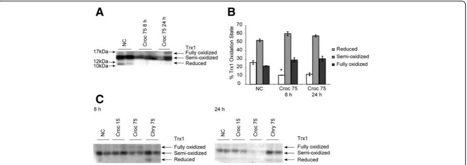
reduced Trx1 as assessed by redox Western analysis as compared to untreated controls (Figure 2A). This decrease in the level of reduced thioredoxin was not observed in cells treated with sodium arsenite, which has been shown previously to oxidize Trx1 in a different cell type [20]. In addition, the total amounts of Trx1 observed in crocidolite asbestos exposed cells appeared to be lower than the levels in the other treatment groups. A third band that repre-sented fully oxidized thioredoxin was present in cells exposed to either crocidolite asbestos or arsenite, but was not observed in lysates from the untreated controls. To
determine whether the oxidation of thioredoxin by cro-cidolite was specific to crocro-cidolite asbestos alone, LP9 cells were also exposed to chrysotile asbestos and glass beads. As assessed by redox Westerns, oxidation of thiore-doxin was specific to the higher concentration (75 × 106 μm2/cm2) of crocidolite asbestos alone (Figure 2C). Levels of reduced and semi-oxidized Trx1 remained the same at 8 and 24 h in response to chrysotile and glass bead exposure.
Inhibition of thioredoxin reductase (TR) by
dinitrochlorobenzene (DNCB) and pretreatment with dehydroascobic acid
To determine whether inhibition of TR by DNCB would increase asbestos-induced oxidation of Trx1, LP9 cells
were pretreated with 10μM DNCB and exposed to 75 ×
106 μm2/cm2 crocidolite asbestos for 8 and 24 h. A
lactate dehydrogenase (LDH) assay performed on the medium from exposed cells showed that LDH levels were reduced in cells pretreated with DNCB when com-pared with cells exposed to asbestos alone (Figure 3A). In addition, redox Western blot analysis of cell lysates indicated that the oxidation of Trx1 by asbestos was ameliorated when cells were pretreated with DNCB be-fore exposure to crocidolite asbestos (Figure 3B). On the contrary, cells exposed to chrysotile asbestos with and without DNCB showed no changes in the redox states of Trx1 when compared to controls (Figure 3C). An Apos-tain assay to detect chromatin condensation and single strand breaks in nuclear DNA as a measure of early apoptotic events was also conducted to determine the effects of pretreatment with DNCB on asbestos-induced apoptosis. Apostain revealed that cells pretreated with DNCB were protected from apoptosis after 8 h of expos-ure to crocidolite asbestos (Figexpos-ure 3D). On the other hand, pretreatment of LP9 cells with DHA before expos-ure to asbestos slightly increased the amount of reduced thioredoxin in cells compared to asbestos exposure alone (Figure 3E). However, there was no significant in-crease when compared to control or asbestos exposure alone.
ROS generation in response to asbestos is modulated by Trx1 in LP9 cells
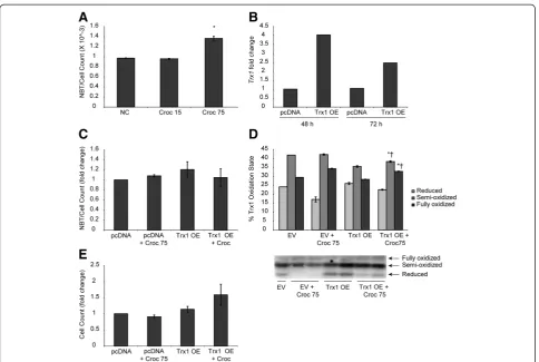
To assess ROS generation after exposure to asbestos, 90% confluent LP9 cells were exposed to 75 × 106μm2/cm2 asbestos for 24 h and incubated with NBT as described in the methods. Spectrophotometric assessment of the solubilized formazan revealed a significant increase in ROS levels when compared to controls (Figure 4A). Quantitative RT-PCR performed on cDNA from Trx1 over-expressing cells and their respective controls indi-cated a 4 fold increase in Trx1 levels after 48 h of trans-fection and this was reduced by approximately 50% 72 h Figure 1Crocidolite asbestos exposure modulates Trx1 levels
post-transfection (Figure 4B). The effect of Trx1 over-expression on asbestos-induced ROS generation and cell survival was assessed by exposing 90% confluent LP9 cells transfected with a Trx1 over-expression vector (pCMV-SPORT6) and empty vector transfected controls (pcDNA) to 75 × 106μm2/cm2crocidolite asbestos for 2 h. The levels of ROS generated in response to asbestos exposure were then measured as described previously. Cells over-expressing Trx1 were found to exhibit a trend of reduced ROS levels compared to the null con-trols exposed to asbestos (Figure 4C). It is to be noted here that the amount of ROS generated by asbestos after 2 h of exposure is significantly lower in magnitude than 24 h after asbestos exposure (Figure 4A). An as-sessment of the redox state of Trx1 in Trx1 over-expressing cells after asbestos exposure indicated that levels of reduced Trx1 were rescued in over-expressing cells as compared to control cells transfected with empty vector (EV) alone (Figure 4D). Additionally, cells over-expressing Trx1 also had increased cell survival after ex-posure to crocidolite asbestos (Figure 4E).
Effect of N-acetyl-cysteine (NAC) on asbestos-induced inflammasome activation
Asbestos exposure results in the generation of ROS in LP9 cells, and ROS has been reported to be one of the activators of the NLRP3 inflammasome [5]. To deter-mine whether asbestos-induced ROS plays a role in inflammasome activation, LP9 cells were pretreated with NAC and analyzed for inflammasome priming and acti-vation by qPCR and Western blot analysis. A decrease in steady-state NLRP3 transcript levels was observed in cells pretreated with NAC prior to asbestos exposure when
compared to cells exposed to asbestos alone (Figure 5A). However, this decrease in NLRP3 transcript levels with NAC pretreatment was not statistically significant. Con-currently, levels of active caspase 1 secreted into the medium after exposure of LP9 cells to asbestos were also significantly reduced after pretreatment with NAC (Figure 5B).
TXNIP down-regulation attenuated asbestos-induced inflammasome activation
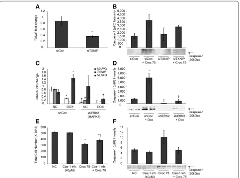
Validation of the knockdown of TXNIP expression by siTXNIP was determined by qRT-PCR and showed that an approximately 50% reduction in TXNIP mRNA levels was achieved after 48 h of transfection (Figure 6A). Western blot analysis of the cell medium after exposure to asbestos for 48 h indicated that active caspase 1 levels (caspase1-p20) were reduced after knockdown of TXNIP when compared to siControl transfected cells (Figure 6B).
In mesothelioma cell lines with a stable knockdown of extracellular signal regulated kinase 2 (shERK 2), the expression of TXNIP was found to be down-regulated 4-fold (Figure 6C). Activation of the NLRP3 inflamma-some by doxorubicin treatment of shERK2 HMESO cells indicated that caspase 1 activation was drastically re-duced as compared to control cells stably transfected with non-targeting shRNA (shCon) (Figure 6D) and there was no priming of NLRP3 as measured by mRNA levels of NLRP3 using qRT-PCR (Figure 6C).
death occurred in response to asbestos-induced inflam-masome activation. To do so, LP9 cells were pretreated
with 40 μM Caspase-1 inhibitor VI (zYVAD-FMK)
before exposure to asbestos. When compared to cells exposed to asbestos alone, there was an 18% increase in cell viability, suggesting this fraction of cells may have undergone pyroptotic cell death upon exposure to asbestos (Figure 6E). Immunoblotting for Cas-1 p20 in concentrated medium supernatants also confirmed that the inhibitor attenuated activation of the caspase-1 as expected (Figure 6F).
Discussion
and extracellulary [24,25]. Mitochondrial generated
super-oxide can react with Fe3+ ions on asbestos fibers to
reduce it to Fe2+[25]. Unfortunately, this enables the Fe2+ to react with any H2O2 present in the cell to produce hydroxyl radicals. This can lead to a cycle of oxidation and reduction of Fe3+to produce more ROS. Additionally, the high aspect ratio of the crocidolite asbestos fibers leads to frustrated phagocytosis as mesothelial cells try to unsuc-cessfully phagocytose the fibers [5]. Frustrated phago-cytosis promotes a sustained production of superoxide through the activity of membrane bound NADPH oxi-dases that are activated during the repeated phagocyt-osis attempts [26]. Asbestos exposure has been shown to cause a depletion of reduced glutathione levels in vitro [10,11], but its effects on Trx, another major antioxidant of the cell, is unknown. The ability of cro-cidolite asbestos fibers to activate the inflammasome in
macrophages and mesothelial cells [4,5] and the in-volvement of the endogenous inhibitor of Trx, TXNIP, in activation of the NLRP3 inflammasome [9] led us to hypothesize that oxidation of Trx by asbestos-induced ROS may cause the dissociation of TXNIP from Trx and lead to activation of the inflammasome. Here we show for the first time that asbestos exposure of human mesothelial cells leads to oxidation of Trx and a com-pensatory increase in Trx1 transcript levels. We also report that TXNIP is involved in asbestos mediated activation of the inflammasome which may be as a result of the oxidation of Trx1 by asbestos-induced ROS generation.
element (ARE) [27]. It is therefore likely that ROS gener-ated in response to asbestos exposure led to an increase in Trx1 expression as a compensatory mechanism by activat-ing ARE-dependent gene expression. It has been reported by our group that cells exposed to asbestos show increased expression of the mitochondrial manganese superoxide dismutase (MnSOD) [11]. Studies also show that increases in expression of cellular antioxidant proteins (MnSOD and Trx1) occur in response to oxidative stress [10,11]. Exogenous thioredoxin as well as endogenously over-expressed Trx has also been shown to increase MnSOD levels in a redox dependent manner [28]. As such, increases in ROS generation in LP9 cells exposed to asbestos coupled with the oxidation of Trx1 and in-creased transcription of Trx1 could be considered an indication of asbestos-induced oxidative stress. To the contrary, the total intracelluar protein levels of Trx1 were reduced by about 67% in response to asbestos exposure after 24 h (Figure 1B, C). This suggests that, ROS generation by asbestos exposure may lead to an
increased oxidation and removal of Trx1 protein as a conjugate, and the cells may increase transcription as a compensatory mechanism. However, Trx1 is secreted into
the culture medium in response to many stimuli–it may
not be degraded [29,30]. Similarly, we have demonstrated previously that asbestos exposure depletes glutathione from cells while increasing the steady-state RNA levels of
γ-glutamylcysteine synthetase (a rate limiting enzyme for glutathione synthesis) at the same time [10].
To determine if asbestos exposure causes changes in the antioxidant capacity of Trx1, the oxidation state of Trx1 was assessed by redox Western blot analysis after exposure of LP9 cells to asbestos. The analysis of the oxidation states of Trx1 after asbestos exposure showed that Trx1 became irreversibly oxidized and levels of reduced Trx1 were drastically decreased compared to control cells. The diminution in the levels of reduced Trx1 may be due to the oxidation of the structural cyst-eine residues (Cys 62, 69 and 73). Oxidation of these structural cysteines can lead to disulfide bond formation between Cys 62 and 69 which has been shown to re-duce the use of Trx1 as a substrate for TR [18]. As a result, Trx1 cannot be reduced to its active form. The formation of disulfide bonds between Cys 73 of two adjacent Trx1 molecules leading to their dimerization has also been shown to occur under extremely oxidiz-ing conditions [31]. This dimerization may lead to con-formational changes in the active site of Trx1 which will make the active site Cys residues inaccessible to TR for reduction. Other studies have shown that strong oxidants can cause the oxidation of Trx1 producing a mixture of Trx1 monomers, dimers and oligomers with no free sulfhydryl groups [32]. These findings support the possibility that asbestos-induced ROS may be in-volved in directly or indirectly altering the oxidation state of Trx1.
Maintenance of the reducing milieu of the cell is also important for cell survival; thus, imbalances in the ratio of cellular antioxidants and oxidants could lead to dele-terious or lethal effects on the cell. To investigate what role perturbations in the redox state of the thioredoxin system plays in asbestos-induced cell death, LP9 cells were pretreated with dinitrochlorobenzne (DNCB), an irreversible alkylating inhibitor of TR [33] and assessed for cell death in response to asbestos exposure. An LDH assay as well as the Apostain assay showed that pretreat-ment with DNCB protected cells from asbestos induced cell death contrary to all reports in the literature in which treatment of cells with DNCB resulted in cell death [34-36]. In contrast, recent studies have shown that inhibition of TR leads to increases in GSH levels and a reduction of Trx1 and Trx2 by glutaredoxin in the absence of a functional TR enzyme [37,38]. It is there-fore likely that the protection of human mesothelial cells Figure 5Asbestos-induced inflammasome priming and
from asbestos-induced cell death after pretreatment with DNCB may be due to increases in GSH and levels of reduced Trx1.
Levels of intracellular GSH are sensitive to ROS levels [39] and are modulated upon exposure of cells to asbes-tos [10,11]. Reactive oxygen species have also been shown to play a role in the activation of the NLRP3 inflammasome after asbestos exposure [6] and may be linked to the effects of GSH levels on Trx1 oxidation state and the availability of TXNIP to bind to and acti-vate the inflammasome. To test the effect of increased GSH or reduced ROS levels on inflammasome activa-tion, LP9 cells were incubated with 2 mM NAC for 20 h
prior to asbestos exposure and this led to a significant decrease in inflammasome activation, as well as a reduc-tion in priming. This suggests that high GSH levels may buffer reduced Trx1 levels [38] and prevent the disso-ciation of TXNIP from Trx1. As such, TXNIP is unable to bind to and activate the inflammasome leading to reduced levels of Caspase-1 p20 peptides in the medium supernatant. The reduction in priming of the NLRP3 may also be due to the decrease in ROS levels as GSH levels increased to buffer cells from the ROS generated in response to asbestos exposure since transcription of
NLRP3 is under the control of NFκB (a redox sensitive
NAC also attenuated the increase in Trx1 mRNA in-duced by asbestos as there was no significant increase when compared to control (data not shown). This implies that the oxidative stress induced by asbestos exposure is ameliorated by NAC.
We have demonstrated previously that crocidolite asbestos causes cell death and compensatory prolifera-tion [3], which may be a required step for crocidolite asbestos-induced cell transformation and MM develop-ment. Our results here, indicate that a fraction of total cell death by asbestos is caused by pyroptosis (caspase-1 dependent cell death, Figure 6), a process known to be regulated in part by TXNIP. However, this observation needs to be confirmed using siRNA mediated knock-down of caspase-1 in future studies. We suspect that pyroptosis is prevented by over-expression of Trx1 (Figure 4) which renders TXNIP unavailable to subse-quently induce inflammasome assembly and thus, caspase-1 activation.
Assessment of the effects of Trx1 over-expression on asbestos-induced ROS generation revealed that LP9 cells over-expressing Trx1 had lower levels of ROS after asbestos exposure when compared to vector transfected cells. Although asbestos induced a significant increase in ROS generation in LP9 cells after 24 h, the trend of reduction in ROS levels with Trx1 over-expression at an earlier time point (2 h) were not statistically significant, but reproducible. The reduction in asbestos-induced ROS generation in LP9s over-expressing Trx1 also cor-responded to a moderate increase in cell survival which also exhibited a trend. Cells undergoing oxidative stress up-regulate the expression of antioxidant proteins like thioredoxin and MnSOD as well as the antioxidant pep-tide glutathione to counter the increase in oxidant levels [10,11,43]. As such, the reduction in asbestos-induced ROS levels upon over-expression of Trx suggests that the increase in Trx1 levels after asbestos exposure may be a compensatory mechanism to restore the antioxidant-oxidant balance that is disrupted by asbestos [21].
Our study also showed that the redox-dependent Trx-TXNIP interaction is involved in asbestos-induced inflammasome activation. When TXNIP, the negative regulator of Trx1 reductase activity, was knocked down in LP9 cells, inflammasome activation was reduced. Cells transfected with siTXNIP had decreased amounts of active Caspase-1 subunit p20 in the medium after exposure to asbestos when compared to control. Add-itionally, activation of the inflammasome by the che-motherapeutic doxorubicin in shERK2 HMESO cells, which have a four-fold lower expression of TXNIP, was attenuated, confirming that TXNIP is required for inflammasome activation. In support of our data, Zhou et al. [9] demonstrated that knockdown of TXNIP by siRNA in beta islet cells reduced activation of the
NLRP3 inflammasome. Thus our findings corroborate the role of TXNIP in inflammasome activation by asbes-tos, and relate inflammasome activation via TXNIP to ROS levels in the cell (Figure 7).
Conclusion
This study has demonstrated that activation of the inflammasome by asbestos is mediated in part by TXNIP as a consequence of alterations in the redox state of Trx1 in the cytosol. Further studies are in progress to offer an understanding of how asbestos-induced activa-tion of the inflammasome and subsequent generaactiva-tion of
biomolecules (IL-1β, IL-18, HMGB1 etc.) may lead to
mesothelial cell transformation events and mesothelioma development.
Abbreviations
TPA:12-O-tetradecanoylphorbol-13-acetate; DNCB: 2,2-Dinitro-1-cholorbenzene; HEPES: 2-[4-(2-hydroxyethyl) piperazin-1-yl]ethanesulfonic acid; ARE: Antioxidant response element; zYVAD-fmk: Carbobenzoxy-tyrosyl-valyl-alanyl-aspartyl-[O-methyl]- fluoromethylketone; Chry: Chrysotile asbestos; Croc: Crocidolite asbestos; DHA: Dehydroascorbic acid; DMSO: Dimethyl sulfoxide; DTT: Dithiotreitol; Dox: Doxorubicin; EV: Empty vector; EDTA: Ethylenediaminetetraacetic acid; ERK2: Extracellular signal regulated kinase 2; GB: Glass beads; HBSS: Hank’s balanced salt solution; HMGB1: High mobility group box 1; LP9/hTERT (LP9): hTERT- immortalized human peritoneal cell line; HPRT: Hypoxanthine phosphoribosyl transferase; IAA: Iodoacetic acid; LDH: Lactate dehydrogenase; MM: Malignant mesothelioma; MnSOD: Manganese superoxide dismutase; NAC: N-acetylcysteine;
NADPH: Nicotinamide adenine dinucleotide phosphate; NLRP3: NLR family pyrin domain containing 3; PBS: Phosphate-buffered saline; qRT-PCR/
qPCR: Quantitative real time PCR; ROS: Reactive oxygen species; shERK2: Short hairpin RNA against ERK2; shCon: Short hairpin RNA non-targeting; SDS PAGE: Sodium dodecyl sulfate polyacrylamide gel electrophoresis; TBST: TBS-Tween; Trx1: Thioredoxin 1; TXNIP: Thioredoxin interacting protein; TR: Thioredoxin reductase; TBS: Tris-buffered saline.
Competing interest
Authors declare no competing interests.
Authors’contributions
JT, performed experiments and wrote the manuscript with the help of AS; CW, performed NAC and inflammasome related studies; MM, performed statistical analysis and made final figures for the publication. MM also provided technical assistance with many experiments; BM, conceived the initial idea and provided intellectual input; NH, provided all the plasmids used in the study and insightful discussion; PS, helped with redox Western blots; AS, conceived the idea, designed experiments, supervised the study, interpreted data, and helped JT in writing the manuscript. All authors have read and approved the manuscript.
Acknowledgement
The authors would like to thank Timothy N. Perkins, Brian Cunniff, Sherrill L. Macura, Mutlay Sayan, Nicholas Fredette and AGTC–DNA core facility, UVM, for their assistance. We thank Dr. Robert Hondal (Biochemistry, UVM) for insightful discussions. We also acknowledge the help of Laura Clayton for preparation of this manuscript. Grant support: NIH grant RO1 ES021110, Mesothelioma Applied Research Foundation (MARF) and the University of Vermont Pathology Department Fellowship.
Received: 16 January 2014 Accepted: 13 May 2014 Published: 20 May 2014
References
1. Mossman BT, Churg A:Mechanisms in the pathogenesis of asbestosis and silicosis.Am J Respir Crit Care Med1998,157:1666–1680.
2. Robinson BW, Lake RA:Advances in malignant mesothelioma.N Engl J Med2005,353:1591–1603.
3. Goldberg JL, Zanella CL, Janssen YM, Timblin CR, Jimenez LA, Vacek P, Taatjes DJ, Mossman BT:Novel cell imaging techniques show induction of apoptosis and proliferation in mesothelial cells by asbestos.Am J Respir Cell Mol Biol1997,17:265–271.
4. Shukla A, Ramos-Nino M, Mossman B:Cell signaling and transcription factor activation by asbestos in lung injury and disease.Int J Biochem Cell Biol2003,35:1198–1209.
5. Dostert C, Petrilli V, Van Bruggen R, Steele C, Mossman BT, Tschopp J: Innate immune activation through Nalp3 inflammasome sensing of asbestos and silica.Science2008,320:674–677.
6. Pinato DJ, Mauri FA, Ramakrishnan R, Wahab L, Lloyd T, Sharma R: Inflammation-based prognostic indices in malignant pleural mesothelioma.
J Thoracic Oncol off Publ Int Assoc Study Lung Cancer2012,7:587–594. 7. Matsuzaki H, Maeda M, Lee S, Nishimura Y, Kumagai-Takei N, Hayashi H,
Yamamoto S, Hatayama T, Kojima Y, Tabata R, Kishimoto T, Hiratsuka J, Otsuki T:Asbestos-induced cellular and molecular alteration of immunocompetent cells and their relationship with chronic
inflammation and carcinogenesis.J Biomed Biotechnol2012,2012:492608. 8. Hillegass JM, Miller JM, MacPherson MB, Westbom CM, Sayan M, Thompson
JK, Macura SL, Perkins TN, Beuschel SL, Alexeeva V, Pass HI, Steele C, Mossman BT, Shukla A:Asbestos and erionite prime and activate the NLRP3 inflammasome that stimulates autocrine cytokine release in human mesothelial cells.Particle Fibre Toxicol2013,10:39.
9. Zhou R, Tardivel A, Thorens B, Choi I, Tschopp J:Thioredoxin-interacting protein links oxidative stress to inflammasome activation.Nat Immunol
2010,11:136–140.
10. Shukla A, Flanders T, Lounsbury KM, Mossman BT:The gamma-glutamylcysteine synthetase and glutathione regulate asbestos-induced expression of activator protein-1 family members and activity.Cancer Res
2004,64:7780–7786.
11. Hillegass JM, Shukla A, MacPherson MB, Lathrop SA, Alexeeva V, Perkins TN, van der Vliet A, Vacek PM, Gunter ME, Mossman BT:Mechanisms of oxidative stress and alterations in gene expression by Libby six-mix in human mesothelial cells.Part Fibre Toxicol2010,7:26.
12. Holmgren A:Redox regulation by thioredoxin and thioredoxin reductase.
Biofactors2000,11:63–64.
13. Junn E, Han SH, Im JY, Yang Y, Cho EW, Um HD, Kim DK, Lee KW, Han PL, Rhee SG, Choi I:Vitamin D3 up-regulated protein 1 mediates oxidative stress via suppressing the thioredoxin function.J Immunol2000, 164:6287–6295.
14. Hwang J, Suh HW, Jeon YH, Hwang E, Nguyen LT, Yeom J, Lee SG, Lee C, Kim KJ, Kang BS, Jeong JO, Oh TK, Choi I, Lee JO, Kim MH:The structural basis for the negative regulation of thioredoxin by thioredoxin-interacting protein.Nat Commun2014,5:2958.
15. Shukla A, Bosenberg MW, MacPherson MB, Butnor KJ, Heintz NH, Pass HI, Carbone M, Testa JR, Mossman BT:Activated cAMP response element binding protein is overexpressed in human mesotheliomas and inhibits apoptosis.Am J Pathol2009,175:2197–2206.
16. BeruBe KA, Quinlan TR, Fung H, Magae J, Vacek P, Taatjes DJ, Mossman BT: Apoptosis is observed in mesothelial cells after exposure to crocidolite asbestos.Am J Respir Cell Mol Biol1996,15:141–147.
17. Buder-Hoffmann S, Palmer C, Vacek P, Taatjes D, Mossman B:Different accumulation of activated extracellular signal-regulated kinases (ERK 1/2) and role in cell-cycle alterations by epidermal growth factor, hydrogen peroxide, or asbestos in pulmonary epithelial cells.Am J Respir Cell Mol Biol2001,24:405–413.
18. Watson WH, Pohl J, Montfort WR, Stuchlik O, Reed MS, Powis G, Jones DP: Redox potential of human thioredoxin 1 and identification of a second dithiol/disulfide motif.J Biol Chem2003,278:33408–33415.
19. Shukla A, Hillegass JM, MacPherson MB, Beuschel SL, Vacek PM, Butnor KJ, Pass HI, Carbone M, Testa JR, Heintz NH, Mossman BT:ERK2 is essential for the growth of human epithelioid malignant mesotheliomas.Int J Cancer
2011,129:1075–1086.
20. Hansen JM, Zhang H, Jones DP:Differential oxidation of thioredoxin-1, thioredoxin-2, and glutathione by metal ions.Free Radic Biol Med2006, 40:138–145.
21. Shukla A, Gulumian M, Hei TK, Kamp D, Rahman Q, Mossman BT:Multiple roles of oxidants in the pathogenesis of asbestos-induced diseases.
Free Radic Biol Med2003,34:1117–1129.
22. Shukla A, Jung M, Stern M, Fukagawa NK, Taatjes DJ, Sawyer D, Van Houten B, Mossman BT:Asbestos induces mitochondrial DNA damage and dysfunction linked to the development of apoptosis.Am J Physiol Lung Cell Mol Physiol2003,285:L1018–L1025.
23. Sullivan DE, Ferris M, Pociask D, Brody AR:The latent form of TGFbeta(1) is induced by TNFalpha through an ERK specific pathway and is activated by asbestos-derived reactive oxygen species in vitro and in vivo.
J Immunotoxicol2008,5:145–149.
24. Srivastava RK, Lohani M, Pant AB, Rahman Q:Cyto-genotoxicity of amphibole asbestos fibers in cultured human lung epithelial cell line: role of surface iron.Toxicol Ind Health2010,26:575–582.
25. Ghio AJ, Stonehuerner J, Richards J, Devlin RB:Iron homeostasis in the lung following asbestos exposure.Antioxid Redox Signal2008,10:371–377. 26. Liu L, Elwing H, Karlsson A, Nimeri G, Dahlgren C:Surface-related
triggering of the neutrophil respiratory burst. Characterization of the response induced by IgG adsorbed to hydrophilic and hydrophobic glass surfaces.Clin Exp Immunol1997,109:204–210.
27. Taniguchi Y, Taniguchi Ueda Y, Mori K, Yodoi J:novel promoter sequence is involved in the oxidative stress-induced expression of the adult T-cell leukemia-derived factor (ADF)/human thioredoxin (Trx) gene.Nucleic Acids Res1996,24:2746–2752.
28. Das KC, Lewis-Molock Y, White CW:Elevation of manganese superoxide dismutase gene expression by thioredoxin.Am J Respir Cell Mol Biol1997, 17:713–726.
29. Nickel W:The mystery of nonclassical protein secretion.A curr View Cargo Proteins Potential Export Routes Eur J Biochem2003,270:2109–2119. 30. Tanudji M, Hevi S, Chuck SL:The nonclassic secretion of thioredoxin is not
sensitive to redox state.Am J Physiol Cell Physiol2003,284:C1272–C1279. 31. Watanabe R, Nakamura H, Masutani H, Yodoi J:Anti-oxidative, anti-cancer
and anti-inflammatory actions by thioredoxin 1 and thioredoxin-binding protein-2.Pharmacol Ther2010,127:261–270.
32. Hashemy SI, Holmgren A:Regulation of the catalytic activity and structure of human thioredoxin 1 via oxidation and S-nitrosylation of cysteine residues.J Biol Chem2008,283:21890–21898.
of both the redox active selenocysteine and its neighboring cysteine residue.J Biol Chem1998,273:10835–10842.
34. Ishikawa A, Kubota Y, Murayama T, Nomura Y:Cell death by 1-chloro-2, 4-dinitrobenzene, an inhibitor of thioredoxin reductase and its dual regulation by nitric oxide in rats.Neurosci Lett1999,277:99–102. 35. Cenas N, Prast S, Nivinskas H, Sarlauskas J, Arner ES:Interactions of
nitroaromatic compounds with the mammalian selenoprotein thioredoxin reductase and the relation to induction of apoptosis in human cancer cells.J Biol Chem2006,281:5593–5603.
36. Seyfried J, Wullner U:Inhibition of thioredoxin reductase induces apoptosis in neuronal cell lines: role of glutathione and the MKK4/JNK pathway.Biochem Biophys Res Commun2007,359:759–764.
37. Britt RD, Velten M, Locy ML, Rogers LK, Tipple TE:The Thioredoxin Reductase-1 Inhibitor Aurothioglucose Attenuates Lung Injury and Improves Survival in a Murine Model of Acute Respiratory Distress Syndrome.Antioxid Redox Signal2013, Epub ahead of print February 6, 2014 doi:10.1089/ars.2013.5332. Online ahead of edits, December 2, 2013. 38. Zhang H, Du Y, Zhang X, Lu J, Holmgren A:Glutaredoxin2 Reduces both
Thioredoxin2 and Thioredoxin1 and Protects Cells from Apoptosis Induced by Auranofin and 4-Hydroxynonenal.Antioxid Redox Signal2013, Epub ahead of print February 4, 2014 doi:10.1089/ars.2013.5499. Online ahead of edits, December 2, 2013.
39. Shin HR, You BR, Park WH:PX-12-induced HeLa cell death is associated with oxidative stress and GSH depletion.Oncology letters2013,6:1804–1810. 40. Qiao Y, Wang P, Qi J, Zhang L, Gao C:TLR-induced NF-kappaB activation
regulates NLRP3 expression in murine macrophages.FEBS Lett2012, 586:1022–1026.
41. Gloire G, Piette J:Redox regulation of nuclear post-translational modifications during NF-kappaB activation.Antioxid Redox Signal2009, 11:2209–2222.
42. Bubici C, Papa S, Dean K, Franzoso G:Mutual cross-talk between reactive oxygen species and nuclear factor-kappa B: molecular basis and biological significance.Oncogene2006,25:6731–6748.
43. Koharyova M, Kolarova M:Oxidative stress and thioredoxin system.
Gen Physiol Biophys2008,27:71–84.
doi:10.1186/1743-8977-11-24
Cite this article as:Thompsonet al.:Asbestos modulates thioredoxin-thioredoxin interacting protein interaction to regulate inflammasome activation.Particle and Fibre Toxicology201411:24.
Submit your next manuscript to BioMed Central and take full advantage of:
• Convenient online submission
• Thorough peer review
• No space constraints or color figure charges
• Immediate publication on acceptance
• Inclusion in PubMed, CAS, Scopus and Google Scholar
• Research which is freely available for redistribution