_____________________________________________________________________________________________________ www.sciencedomain.org
The Cell Based Model of in-vivo Coagulation:
A Work in Progress
Biswaprakash Patri
1, Anubhav Abinash Sahu
1*, Souravi Pal
1and Sukumar Chakravarty
11
Department of Pathology, Hitech-Medical College and Hospital, Bhubaneswar, Odisha, India.
Authors’ contributions
This work was carried out in collaboration between all authors. Author BP identified the study, wrote the protocol and wrote the first draft of the manuscript. Author AAS managed the literature searches and helped in drafting the final manuscript. Author SP managed compiling of the materials and author SC was the guiding force behind the whole process. All authors read and approved the final manuscript.
Article Information
DOI: 10.9734/IBRR/2016/26906
Editor(s):
(1) Ricardo Forastiero, Department of Hematology, Favaloro University, Argentina.
Reviewers:
(1) Fatima Laraba-Djebari, University of Science and Technology Houari Boumediene, Algeria. (2)Jie Fan, Zhejiang University, China. (3)Renshan Sun, Third Military Medical University, Chongqing City, China. Complete Peer review History:http://sciencedomain.org/review-history/15034
Received 9th May 2016 Accepted 9th June 2016 Published 15th June 2016
ABSTRACT
The goal of this article is to review the evolution of theories of coagulation and their proposed models to serve as a tool. When reviewing the research and practice literature that was published in the context of these theories over time, including the critical contribution of cells, we have observed a model based on work of many workers, in which Hemostasis is regulated by various properties of cell surface, which occurs in vivo. The coagulation occurs not as a cascade/waterfall, but occurs in 4 overlapping steps-(i) Initiation, (ii) Amplification, (iii) Propagation, (iv) Termination. Cell Based model has given an insight into the pathophysiological mechanism of certain coagulation disorders which were till now ill understood.
Keywords: Coagulation; coagulation factors and cascade; hemostasis; cell-based theory of coagulation; bleeding disorder.
1. INTRODUCTION
The coagulation cascade was proposed by MacFarlane and Davie & Ratnoff in 1964. This model was used almost for five decades even if it had limitations and can’t explain all the pathophysiology occurring in vivo. Two ‘cascade’ occurs through a series of proteolytic activation of zymogens by plasma proteases, which produces thrombin which then cleaves fibrinogen molecule to fibrin monomers.
Recent advancements had occurred in the coagulation study which occurs in vivo. Newer Models that include the contributions of cells in vitro and systems that involve real-time in vivo imaging of coagulation have significantly modified current understanding of how hemostasis occurs in vivo [1].
2. COAGULATION HISTORY
French Surgeon, Jean-Lousis, Petit in early 1720s noted that after amputation of a limb hemostasis was caused by clot formed in the blood vessels. In 1828 Friedrich Hopff noted that familial bleeding tendency in males was due to hyporcoagulability. In 1860 Rudolf Virchow explained thrombi and its tendency to embolize.
In 1905 Paul Morawitz proposed the classic theory of coagulation. In this theory four coagulation factor are involved. Prothrombin in presence of calcium and thrombokinase is converted into thrombin which in turn converts fibrinogen into fibrin, thus enabling the formation of fibrin clot. Morawitz postulated that all the four coagulation factors were present in circulatory blood but such blood did not normally coagulated due to lack of wettable surface in blood vessels. This theory was used for nearly 40 years.
In 1947 Paul Owren cited that bleeding diathesis in a young female could not be explained by the four factors, the fifth coagulation factors which she lacks in her plasma. After that several coagulation factors were discovered. Importantly, the numeric system that was adopted assigned the number to the factor according to the sequence of discovery and not to the point of interaction in the cascade.
In 1964 MacFarlane proposed cascade model in Journal Nature (I). Then Waterfall Model was shortly reported by Davie and Ratnoff in Journal Science (I). In this model the proezyms (zymogen) are converted to an active enzyme.
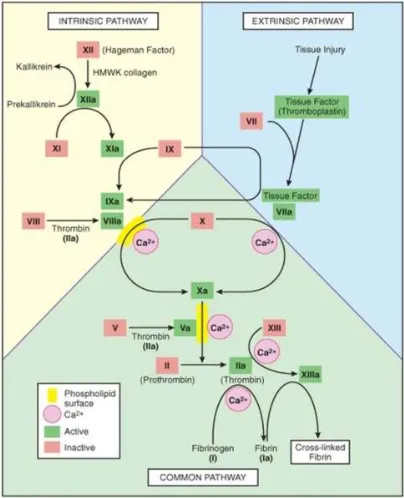
This cascade has three pathway intrinsic pathway, extrinsic pathway and common pathway.
3. OVERVIEW OF HEMOSTASIS
Hemostasis is a dynamic process whereby blood coagulation is initiated and terminated in a rapid and tightly regulated manner. (Nathan, Orkin, Ginsburg & Look, 2003). Hemostasis thus refers to multiple discrete processes that collectively culminate in preservation of vascular integrity[2]. Blood coagulation means the cessation of blood loss from the damaged vessels. It has a very important role on the defence mechanisms. When the blood vessel is injured platelets adhere at the site which then aggregate to form primary hemostatic plug. Blood coagulation occurs when soluble plasma fibrinogen is converted into insoluble fibrin polymer by enzyme thrombin. Subsequently activation of the fibrinolysis system removes the clot, restores blood flow, and initiates tissue repair and regeneration[2].
Hemostatic is regulated mainly by three components which are; vascular wall, platelets and coagulation cascade. Normal Hemostasis has basically two important functions – (1) to maintain blood in a fluid and clot free state (2) to generate a rapid hemostatic plug at the site of injury. Thus balance between the procoagulant, anticoagulant, fibrinolytic and antifibrinololytic process is required to prevent extravascular blood loss, or undesirable intravascular thrombosis[3].
4. MECHANISM OF COAGULATION
CASCADE
The coagulation cascade is a series of amplifying enzymatic reactions that leads to the deposition of an insoluble fibrin clot. Here the zymogens are cleaved by enzymes to generate the next enzyme in cascade. The majority of the steps require calcium which occurs in phospholipid membrane. This cascade is divided into two pathways – (i) Extrinsic pathway - localized outside the blood and consists of Tissue factor and FVIIa, (ii) The intrinsic pathway – localized within the blood having all the components and (iii) Common pathway.
4.1 Extrinsic Pathway
(Thromboplastin) and FVIIa. Nonvascular cells constitutively express the integral membrane protein TF (variably known as FIII or tissue thromboplastin), which is a receptor for the plasma protein FVII (Kumar et al. 2005).
Injury to vessel wall i.e endothelium releases the Tissue factors which activate FVII to FVIIa. The mechanism of the initial conversation of the zymogens FVII to FVIIa is still debated but is most likely due to autocatalytic activation and not a TF effect (M.Hoffman & Monroe, 2005). The FVIIa/TF complex activates FX to FXa in the common pathway. The prothrombin time (PT) assesses the function of the proteins of extrinsic pathway.
4.2 Intrinsic Pathway
The intrinsic pathway has all the factors present within the blood. When in contact with a negatively changed surface such as glass or activated platelate membrane, FXII (Hageman Factor) is activated to FXIIa. A product of platelate known as High molecular weight Kininogen (HMWK) that attaches to platelet membrane helps in anchoring FXII to the charged surface, acting as a cofactor. But this HMWK assisted conversion is limited in speed. When a little amount of FXIIa is produced, this in turn converts prekallikrein to Kallikrein. This newly formed Kallikrein accelerates the conversion of FXII to FXIIa. Thus the above steps are a positive feedback. FXIIa then converts FXI to FXIa. Then FXIa, which is also bound to charged surface by HMWK, cleaves FIX to FIXa.
FIXa, FXa and thrombin cleave FVIII to FVIIIa. Finally FIXa and FVIIIa along with Ca2+ (which comes from activated platelates), and negatively charged phospholipids form a trimolecular complex Tenase. This converts FX to FXa in the common pathway. The activated partial thromboplastin time (aPTT) screens the function of the protein in the intrinsic system.
4.3 Common Pathway
The common pathway starts with the activation of FX from the extrinsic or intrinsic or both pathways. FV is activated to FVa in presence of thrombin, FIIa. FXa, FVa, Ca2+ and phospholipids converts prothrombin (FII) to its active form thrombin (FIIa). The main function of thrombin is to catalyze the proteolysis of soluble plasma protein fibrinogen into soluble fibrin
monomer. Then fibrin monomers polymerizes to fibrin polymers. Thrombin in presence of Ca2+ converts FXIII to FXIIIa which mediates the covalent cross-linking of the fibrin polymers to form a mesh known as stable fibrin. Thrombin can catalyse the formation of cofactor FVa and FVIIIa and new thrombin from prothrombin, which results in efficient amplification of coagulation. Common pathway factor is assessed by PT and aPTT.
Fig. 1. The coagulation cascade-the common link between the intrinsic and extrinsic pathways at the level of factor IX activation.
Factors in red boxes represent inactive molecules; activated factors are indicated in
green box and with a lower-case a
5. USES OF CASCADE MODEL
Ongoing research on cascade model lead to discovery of specific inhibitors of coagulation and cross-interactions between different components of pathway. The cascade model, as modified over the last few decades to include these new interactions between intrinsic and extrinsic pathways, functions reasonably well to explain the way coagulation occurs in plasma or purified protein-based fluid systems where the fluid is static and does not interact with vascular wall or cell surface[4].
6. FALLACY OF CASCADE PATHWAY
The cascade model occurs in plasma based in vitro coagulation, which does not explain hemostasis process in vivo. The model explains that intrinsic and extrinsic pathways which occurs as independent and redundant pathways, but the deficiency of any individual clotting factor contradicts the concept, as bleeding occurs in hemophilic where FVIII or FIX are deficient. Similarly deficiency of enzymes of extrinsic pathways FVII has clinical tendency to bleed even if the intrinsic pathway is intact. This model does not give any explanation for the no cause of bleeding in FXII, High molecular weight Kininogen or prekallikrein deficiencies. FX, FV and FVII deficiency causes serious clinical bleeding syndromes. But FXI deficiency is much less predictable and causes less clinical bleeding tendency than that of FVIII and FIX deficiency.
7. CONTRIBUTION OF PLATELETS TO COAGULATION
The cell based model allows for inclusion of role of specific platelet binding sites for coagulation proteins, such as both activated and inactivated platelets have several binding sites for thrombin, Glycoprotein Ib/IX (GpIb/IX) [5], protease activating receptors (PAR) [6] and etc. GpIb/IX is well characterized as the receptors for VWF, also protein binding sites exist for FIX /IXa on platelet [7-9].There is evidence for a specific, non-lipid activation induced binding site for FVIII on platelet as well [10,11]. There exist a protein receptor on platelet surface for FXa and it has been proposed to be effector protease receptor -1 (EPR--1), FXI can bind to platelet via HMWK [12] but also binds directly to a small number of high affinity sites on platelets through apple -3 – domain [13].
8. SITE OF COAGULATION REACTIONS
Hemorrhage in response to injury is controlled as the, hemostatic activity are required to create an
occlusion at the site of injury. Appropriate hemostasis is important for coagulation control and the regulation has to be localized specifically at the site of injury and that control is accomplished via the contribution of membrane surfaces to coagulation process.
9. INTERACTION OF COAGULATION
FACTORS WITH MEMBRANE
The speed at which many of these enzymatic reactions in coagulation proceed is affected by the presence of an appropriate membrane surface [14]. TF is the only coagulation factor which is permanently attached to the membrane surface .Coagulation factors such as (FVII, FIX, FX, Prothrombin, Protein C, ProteinS) contain glutamic acid residue (Gla Residue), which allows their binding to a membrane surface via interaction between Ca2+ and negatively charged phospholipids [14]. Ca2+ binding to the Gla region of these proteins need these Gla residues to be carboxylated in a post translational modification via vit-K cycle in liver., without complete carboxylation , these Gla –protein lack the ability to properly bind to Ca2+. [14]. The specific regions of Coagualtion factors (FV & FVIII) interact with phospholipids and form a fully functional enzymatic complex on the membrane surface.
10. ROLE OF MEMBRANE SURFACE
All cells are surrounded by lipid bilayers that contain a large number of membrane surface proteins. On resting stage neutral phospholipids such as phosphatidyl choline(PC), sphingomyelin, sphingolipids are localized on the external surface and phosphatidylserine(PS), phosphatidyl ethanolamine(PE) are localized to the inner surface of the membrane. This asymmetry is maintained by various enzymes, like flippase actively transports PS from external to internal leaflet and Flippase transports PC in the opposite direction. These ATP dependent enzymes keep the asymmetry required in the resting stage. When cells are injured or activated PS and PE come to external leaflet of the membrane surface. Enzyme Scramblase actively shuffles the phospholipids between the two surfaces in respose to increased ca2+ concentration in the cytosol [14].
11. ROLE OF PROCOAGULANT
MEMBRANE
properties. PS on external surface hastens up the coagulation reaction (thousands of time faster). When PE is present on external surface the amount of PS needed is less [15]. The Gla– protein of coagulation factors bind to PS clusters on cell surface and PE helps in grouping of PS in to these clusters. Expression of PS (Particualrly with PE) on the external leaflet converts the cell membrane in to a procoagulant surface. Under normal physiologic conditions cell don’t express a pro coagulant membrane. As a result, the ability of cells to regulate the nature of their membrane surface forms a powerful tool of maintaining coagulation reactions [16].
12. ROLE OF MICROPARTICLES
Microparticles are intact vesicles which are derived from cells, and are surrounded by membranes [14]. Their size is 2-20% of the size of RBC. Formed when activate / apoptotic cells gives off bit of their membrane. Cytokines (TNF and IL6), thrombin, sheer stress, Hypoxia are the stimulant for MPs formation. In normal condition MPs are primarily derived from endothelial cells, platelets, monocytes. MPs are derived from granulocytes, erythrocytes in various diseased states. Quantity of circulating MPs increased in various conditions like Diabetes mellitus, sepsis, cardiovascular disease, pathological coagulation in various disorders [17-19]. MPs contain various cell surface proteins such as VWF monomers on endothelial derived MPs, P-selectin on platelet derived MPs, TF on monocyte derived MPs.
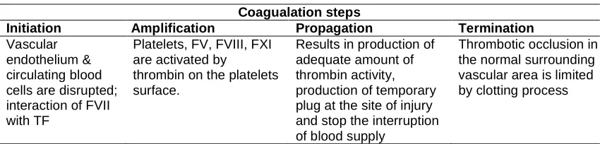
13. THE CELL BASED MODEL OF in-vivo COAGULATION
The current knowledge of the hemostatic process consider the interaction of physical, cellular, biochemical processes in a series of stages or overlapping phases and not as two pathways (intrinsic & extrinsic) as was believed. The stages of initiation, amplification, propagation, termination explains the complex processes that enable the circulation of blood in a liquid form restricted to the vascular bed. Theses 4 stages summarized in Table 1 show the current coagulation theory based on cell surface. There are various mechanisms which control inappropriate coagulation. The inactivation and propagation phases are localized on two different cell surfaces. The plasma protease inhibitors localize the reactions to the cell surface by inhibiting the activated protease that diffuse to the soluble phase. The antithrombotic features of endothelial cells,
prevent initiation of coagulation in intact endothelium.
13.1 Initiation
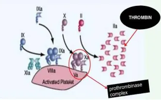
All reports to date suggest that, the relevant initiator of coagulation in vivo is TF bearing cells on their surface, when exposed to blood components at the site of injury. Cells expressing TF are generally localized in extra vasculature that prevents initiation of coagulation under normal flow condition with intact endothelium. Once an injury occurs, the flowing blood is exposed to a TF bearing cells. FVII rapidly binds to the exposed TF. FVII is the sole factor that routinely circulate as an active form (1% of total factor VII, circulate as FVIIa). TF and FVIIa complex, in turn activates additional FVII to FVIIa, this allows for more TF-FVIIa complex action. The TF-FVIIa complex activates small amount of FX and FXI. Then FXa binds with its cofactor FV and activates it to FVa. This complex FXa and FVa is known as PROTHROMBINASE complex, cleaves prothrombin to produce thrombin. Any FXa tries to dissociate from TF bearing cells, undergo deactivation by Antithrombin III (ATIII) or Tissue factor pathway inhibitors (TFPI) but any FIXa tries to dissociate from TF bearing cells to nearby platelets , does so, as it doesn’t get deactivated by TFPI and only slowly deactivated by ATIII. TF is always expressed in the perivascular space .only FVII, has to leave the vasculature through minor gaps in the endothelial barrier, will bind to TF and initiate coagulation.
In normal condition, the gaps in endothelial barrier are very small, whereas some of the downstream coagulation factors are very large (FV-330,000da), this means platelet and large proteins are sequestered from the extravascular space. Coagulation progress beyond generation of small amount of thrombin that happens with initiation only. When the injury allows platelets and large proteins to escape the vascular space and adhere to TF bearing cells in extravascular space [20,21].
13.2 Amplification
coagulant signal by increasing platelet adhesion [22], fully activating platelets and activating FV, FVIII, FIX [23]. Binding of thrombin to Platelet surface receptors PAR [6] causes extreme change in the shape, shuffling of membrane phospholipids to create a pro coagulant membrane, and release of the FV of alpha granules on to their surface in a partially activated form. FV is fully activated by Thrombin or FX [24]. Some of the thrombin bind to non PAR receptors such as GpIb/IX remain active and can activate other coagulation factors on the platelet surface. Ca2+induce clustering of PS and promote binding of coagulation proteins to activated membrane surface of platelet. Thrombin generated during initiation phase also cleaves FXI to produce FXIa and also activates FV to produce FVa. It also cleaves VWF off of FVIII (VWF circulates with FVIII in circulation) releasing VWF to mediate platelet adhesion and aggregation. It also activates FVIII to produce FVIIIa [25]. Once that platelet have been activated and have got activated cofactors FVa and FVIIIa bound on their surface, assembly of pro coagulant complexes results in large scale generation of thrombin begins.
Fig. 2. Initiation phase
Fig 3. Amplification phase
13.3 Propagation
During this phase the TENASE and PROTHROMBINASE complexes are aggregated on the platelet surface and large scale thrombin generation takes place. Platelets express binding sites for FIXa [26], FXa [27], and FXIa [28]. Once few platelets are activated during amplification, the release of granules contents, cause recruitment of additional platelets on the site of injury, propagation phase takes place on the surface of these platelets. Expression of ligands on their surface results in cell to cell interactions which lead to platelet aggregation. FIXa that was generated by TF-FVIIa complex during initiation phase can bind to FVIIIa generated during amplification phase to form an intrinsic tenase complex on the platelet surface. Additional FIXa is generated by FXIa during amplification phase on the platelet surface. Intrinsic tenase complex generated on the activated platelet surface rapidly begins to generate FXa. The FXa then binds to FVa (generated by thrombin during amplification phase). This FXa and FVa complex is known as PROTHROMBINASE complex, cleaves prothrombin to generate thrombin. Thrombin cleaves fibrinopeptide A from fibrinogen. Once enough thrombin is generated at an appropriate speed to result in a critical mass of fibrin, it polymerizes to fibrin strands, resulting in an insoluble fibrin matrix [25].
Fig 4. Propagation Phase
13.4 Termination
Table 1. Summary of current cell-based theory of coagulation
Coagualation steps
Initiation Amplification Propagation Termination
Vascular endothelium & circulating blood cells are disrupted; interaction of FVII with TF
Platelets, FV, FVIII, FXI are activated by
thrombin on the platelets surface.
Results in production of adequate amount of thrombin activity, production of temporary plug at the site of injury and stop the interruption of blood supply
Thrombotic occlusion in the normal surrounding vascular area is limited by clotting process
Thrombin produced in the proximity of HSPGs. Resting endothelial cells produce thrombomodulin (TM). TM binds with thrombin and converts the pro coagulant thrombin to an anticoagulant thrombin, this TM- Thrombin complex activates Protein C (along with its cofactor Protein S), and this irreversibly cleaves FVa & FVIIIa, preventing their further participation in generation of additional thrombin. Protein C is localized to endothelial surface by an endothelial protein receptor 1 (EPCR-1). TFPI prevent additional thrombin generation by inhibiting FXa ,FVIIa. TFPI irreversibly binds FXa and forms a quarternary complex ie(FXa, TFPI, FVIIa, TF) and prevent their further participation in production of additional new thrombin. ADPase activity (CD39) [32], metabolizes ADP released from activated platelets causing in blockade of aggregation response when platelets are in close proximity to healthy endothelium.
14. ADVANTAGES OF THE CELL BASED MODEL OF COAGULATION
This new model of hemostasis enables to explain some clinical aspects of hemostasis that the previous coagulation cascade model lacks. This new model gives a better understanding of the in vivo coagulation process. It is more consistent with clinical observations of several coagulation disorders.
15. RELEVANCE OF THE CELL BASED MODEL OF COAGULATION
15.1 In Laboratory Tests
Classical screening methods include an assessment of blood clotting using activated partial thromboplastin time (aPTT), which assess the intrinsic and common pathway and Prothrombin time (PT), which assess the extrinsic and common pathway [33,34]. The new coagulation pathway has shown both the pathways are not redundant [35]. The extrinsic
pathway takes place on the surface of a TF bearing cell and intensifies the clotting process and the intrinsic pathway operates on the surface of activated platelets to produce an amount of thrombin, in turn results in the formation and stabilization of fibrin clot. PT assesses the level of pro coagulant participated in the initiation phase of coagulation; aPTT assesses the level of pro coagulant generated during the propagation phase. But none of the available tests include the cellular components.
15.2 In Hemophilia
Why do hemophiliacs have bleeding tendencies? The cascade model of coagulation lacks the explanation why the extrinsic pathway can’t produce enough FXa (or partially) to compensate for the lack of FVIII or FIX, i.e. why can’t FXa generated by FVII/TF i.e. extrinsic pathway substitute for the FXa generated by FVIII/FIX i.e. intrinsic pathway?? It is hypothesized that it is because TFPI inhibits the FVII/TF pathway before it can make enough FXa to support generation of hemostatic amount of thrombin. Hoffman & Monroe proposed that the problem in hemophilia is not that enough FXa is not made, but that it is made on the WRONG CELL SURFACE. The cell based model doesn’t suggest that, the FXa generated by FVIIa/TF complex is inadequate in hemophiliacs but it’s poorly expressed on the cell surface. FXa made on a TF bearing cell unable to reach the platelet surface without being inhibited by TFPI/ATIII.
Table 2. A comparison between the coagulation models
Cascade model Cell based model
Principle It’s a cascade of reaction follows an endothelial injury, which in turn causes platelets adhesion and aggregation leading to cessation of bleeding
It’s an in-vivo process of coagulation mediated by tissue factor, platelets and various
coagulation factors in a phase wise manner resulting in cessation of bleeding
Pathways 3 Pathways: Extrinsic, Intrinsic and Common pathways
4 Pathways: Initiation, Amplification, Propagation and Termination
Uses Used for PT and APTT estimation As an potential explanation for the evaluation of certain individual factor deficiencies and bleeding disorder(hemophilia)
FX on platelet surface which lead to failure of platelet surface thrombin generation. Hemophilia patients have a relatively normal coagulation initiation, amplification phase and are capable to form the temporary plug at the site of bleeding [35]. However they are incapable of producing an adequate amount of thrombin on the platelet surface sufficient to stabilize the fibrin clot [36].
17. SUMMARY
The Cell based model of coagulation enables us a great insight into the complete process of coagulation occurring in-vivo by emphasizing the role of cell surface. It also explains the intricacies of mechanisms of bleeding disorder like haemophilia as it was not adequately addressed in the coagulation cascade. Adequate thrombin production directly on the activated platelet surface is the cornerstone in this model of coagulation. This model suggests the different pathways of coagulation cascade occur in parallel, resulting the generation of FXa on two different cell surfaces, rather than in redundant pathways [20]. The advance in both knowledge and understanding this coagulation process will pave the way to novel insight in precise diagnosis and management of various bleeding disorders, as the proposed concept is evolving every day.
CONSENT
It is not applicable.
ETHICAL APPROVAL
It is not applicable.
COMPETING INTERESTS
Authors have declared that no competing interests exist.
REFERENCES
1. Furie B, Furie BC. In vivo thrombus formation. J Thromb Haemost. 2007; 5(Suppl 1):12–17.
2. Wintrobe’s clinical haematology 13th edition, chapter 18th - blood coagulation and fibrinolysis.
3. MacFarlane RG. An enzyme cascade in the blood clotting mechanism and its function as a biochemical amplifier. Nature. 1964;202:498-499.
4. Mann KG, Brummel K, Butenas S. What is all that thrombin for? J Thromb Haemost. 2003;1(7):1504–1514.
5. Dardik R, Varon D, Eskaraev R, Tamarin I, Inbal A. Recombinant fragment of von willebrand factor AR545C inhibits platelet binding to thrombin and platelet adhesion to thrombin-treated endothelial cells. Br J Haematol. 2000;109:512-8.
6. Hung DT, Vu TK, Wheaton VI, Ishii K, Coughlin SR. Cloned platelet thrombin receptor is necessary for thrombin-induced platelet activation. J Clin Invest. 1992; 89:1350-3.
7. Ahmad SS, Rawala-Sheikh R, Walsh PN. Comparative interactions of factor IX and factor IXa with human platelets. J Biol Chem. 1989;264:3244-51.
8. Ahmad SS, Rawala-Sheikh R, Ashby B, Walsh PN. Platelet receptormediated factor X activation by factor IX. High-affinity factor IXa receptors induced by factor VIII are deficient on platelets in Scott syndrome. J Clin Invest. 1998;84:824-8. 9. Hoffman M, Monroe DM, Roberts HR.
10. Nesheim ME, Furmaniak-Kazmiercsak E, Henin C, Cote G. On the existence of platelet receptors for factor V (a) and factor VIII(a). Thromb Haemost. 1993;70:80-6. 11. Ahmad SS, Scandura JM, Walsh PN.
Structural and functional characterization of platelet receptor- mediated factor VIII binding. J Biol Chem. 2000;275:13071-81. 12. Greengard JS, Heeb MJ, Ersdal E, Walsh
PN, Griffin JH. Binding of coagulation factor XI to washed human platelets. Biochemistry. 1986;25:3884-90.
13. Ho DH, Baglia FA, Walsh PN. Factor XI binding to activated platelets is mediated by residues R(250), K(255), F(260), and Q(263) within the apple 3 domain. Biochemistry. 2000;39:316-23.
14. The cell-based model of coagulation. J Vet Emerg Crit Care. (SanAntonio). 2009;19(1):3-10. Review. PubMed PMID: 19691581.
DOI: 10.1111/j.1476-4431.2009.00389.x. 15. Neuenschwander PF, Bianco-Fisher E,
Rezaie AR, et al. Phosphatidylethano-lamine augments factor VIIa-tissue factor activity: Enhancement of sensitivity to phosphatidylserine. Biochemistry. 1995; 34(43):13988–13993.
16. Zwaal RF, Comfurius P, Bevers EM. Surface exposure of phosphatidylserine in pathological cells. Cell Mol Life Sci. 2005; 62(9):971–988.
17. Piccin A, Murphy WG, Smith OP. Circulating microparticles: Pathophysiology and clinical implications. Blood Rev. 2007; 21(3):157–171.
18. Key NS, Geng JG, Bach RR. Tissue factor; from Morawitz to microparticles. Trans Am Clin Climatol Assoc. 2007;118:165–173. 19. Morel O, Morel N, Freyssinet JM, et al.
Platelet microparticles and vascular cells interactions: A checkpoint between the haemostatic and thrombotic responses. Platelets. 2008;19(1):9–23.
20. Hoffman M. Remodeling the blood coagulation cascade. J Thromb Thrombolysis. 2003;16(1–2):17–20. 21. Roberts HR, Hoffman M, Monroe DM. A
cell-based model of thrombin generation. Semin Thromb Hemost. 2006;32(suppl 1): 32–38.
22. Diaz-Ricart M, Estebanell E, Lozano M, Aznar-Salatti J, White JG, Ordinas A, Escolar G. Thrombin facilitates primary
platelet adhesion onto vascular surfaces in the absence of plasma adhesive proteins: Studies under flow conditions. Haematologica. 2000;85:280-8.
23. Monroe DM, Hoffman M, Roberts HR. Transmission of a procoagulant signal from tissue factor-bearing cells to platelets. Blood Coagul Fibrinolysis. 1996;7:459-64. 24. Monkovic DD, Tracy PB. Activation of
human factor V by factor Xa and thrombin. Biochemistry. 1990;29:1118-28.
25. Hoffman M. Remodeling the blood coagulation cascade. J Thromb Thrombolysis. 2003;16(1–2):17–20. 26. Rawala-Sheikh R, Ahmad SS, Monroe
DM, Roberts HR, Walsh PN. Structural requirements for Factor IXa binding to platelets. FASEB J. 1990;4:A2276.
27. Cirino G, Cicala C, Bucci M, Sorrentino L, Ambrosini G, DeDominicis G, Altieri DC. Factor Xa as an interface between coagulation and inflammation. Molecular mimicry of factor Xa association with effector cell protease receptor-1 induces acute inflammation in vivo [see comments]. J Clin Invest. 1997;99:2446-51.
28. Greengard JS, Heeb MJ, Ersdal E, Walsh PN, Griffin JH. Binding of coagulation factor XI to washed human platelets. Biochemistry. 1986;25:3884-90.
29. Sagripanti A, Carpi A. Antithrombotic and prothrombotic activities of the vascular endothelium. Biomed Pharmacother. 2000;54(2):107–111.
30. Levi M, ten Cate H, van der Poll T. Endothelium: Interface between coagulation and inflammation. Crit Care Med. 2002;30(suppl 5):S220–S224. 31. Schouten M, Wiersinga WJ, Levi M, et al.
Inflammation, endothelium, and coagulation in sepsis. J Leukoc Biol. 2008; 83(3):536–545.
32. Marcus AJ, Broekman MJ, Drosopoulos JH, Islam N, Alyonycheva TN, Safier LB, Hajjar KA, Posnett DN, Schoenborn MA, Schooley KA, Gayle RB, Maliszewski CR. The endothelial cell ecto-ADPase responsible for inhibition of platelet function is CD39. J Clin Invest. 1997;99: 1351-60.
33. Hoffman M. Remodeling the blood
coagulation cascade. J Thromb
34. Lewis SM, Bain BJ, Bates I. Hematologia prática de Dacie e Lewis. 9a ed. Porto Alegre: Artmed. 2006;572.
35. A cell-based model of coagulation and its implications. Rev Bras Hematol Hemoter. 2010;32(5):416-421.
36. Hoffman M. A cell-base model of coagulation and the role of factor VIIa. Blood Rev. 2003;17(Suppl 1):S1-5.
_________________________________________________________________________________
© 2016 Patri et al.; This is an Open Access article distributed under the terms of the Creative Commons Attribution License (http://creativecommons.org/licenses/by/4.0), which permits unrestricted use, distribution, and reproduction in any medium, provided the original work is properly cited.
Peer-review history: