Cif\-e^nQrg.pM5igA
jA
4
v o l d j^ eA o p iA ^/uuvioo^
Sy5W_/V\ ,
-Diffusible axon-outgrowth-inhibiting activities in the developing rat
— central nervous system.
A Thesis submitted to the university of London
for the degree of Doctor of Philosophy.
By Nina Uche Orike
BSc, MSc
University College London
ProQuest Number: 10046108
All rights reserved
INFORMATION TO ALL USERS
The quality of this reproduction is dependent upon the quality of the copy submitted.
In the unlikely event that the author did not send a complete manuscript and there are missing pages, these will be noted. Also, if material had to be removed,
a note will indicate the deletion.
uest.
ProQuest 10046108
Published by ProQuest LLC(2016). Copyright of the Dissertation is held by the Author.
All rights reserved.
This work is protected against unauthorized copying under Title 17, United States Code. Microform Edition © ProQuest LLC.
ProQuest LLC
789 East Eisenhower Parkway P.O. Box 1346
ABSTRACT
This work has been directed at demonstrating the existence of diffusible growth-
suppressing signals which may operate during the development of the nervous system.
By co-culturing explants of different brain areas I have produced evidence for the
existence of at least three distinct growth-suppressing signals. Such signals could act
by specifically preventing axons from innervating inappropriate territory in the central
nervous system.
Evidence for the creation of territorial exclusion zones by diffusible factors is novel and
in conjunction with other well described mechanisms could make important
contributions to understanding axon guidance and patterning within the central nervous
system. Identification of diffusible inhibitors of axon growth is not only important to
the basic understanding of how neural networks become organised, but may well be
critical to the problem of regeneration in the adult nervous system. I have therefore
carried out research on the biochemical elucidation of the inhibitory activities. I have
shown that some of these are secreted differentially according to brain area, are likely to
have very short half lives and have a molecular weight range between 15 and 25KDa. If
endogenous inhibitors of axon growth can be identified then there are strong
possibilities for generating soluble antagonists which might eventually be of clinical
TABLE OF CONTENTS
Pages
List of tables 7
List of figures 8
Acknowledgements 10
Chapter one : Introduction
1.1 Historical prespective 11
1.2 Axon guidance mediated by non-diffusible molecules 19
1.21 Contact adhesion mediated by cell adhesion molecules 19
Neural cell adhesion molecuem (NCAM) 19
LI 20
The cadherins 21
The selectins 21
The integrins 22
1.22 Contact adhesion mediated by substrate adhesion molecules 22
Laminin 23
Fibronectin 23
1.23 Contact mediated repulsion 25
The retinotectal pathway 25
Spinal nerve segementation 26
Matrix molecules 27
Myelin proteins (NI-35 and NI-250) 27
Myelin associated glycoprotein 28
1.3 Axon guidance mediated by diffusible molecules 29
1.31 Chemoattraction 29
Developing trigeminal axons 31
Spinal commissural axons 32
Cortical projection axons 32
1.32 Chemorepulsion 34
The lateral olfactory tract 34
Primary sensory afferents 35
Trochlear motor axons 35
Hind brain and spinal developing motor axons 36
1.4 Candidate diffusible guidance molecules 37
1.41 Collapsin 37
1.42 Semaphorins 38
1.43 Netrins 40
1.5 Growth cone motility and guidance cues 41
1.51 Growth cone structure and cytoskeletal arrangement 41
1.52 Signal transduction 43
1.53 How cell adhesion molecules translate their positive growth signals 45
1.54 How inhibitory molecules translate their negative growth signals 46
1.55 How are growth cones able to detect gradients of diffusible molecules 47
1.6 Objectives 4 8
Chapter two : Materials and methods
2.1 Materials 49
2.2 Methods 52
2.21 Embryo dissection 52
2.22 Collagen gel preparation 52
2.3 Bioassay procedure 53
Chapter three : Isochronic co-culture experiments
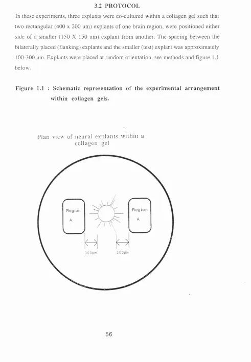
3.1 Introduction 55
3.2 Protocol 56
3.3 Results 59
Chapter four : Conditioned media experiments
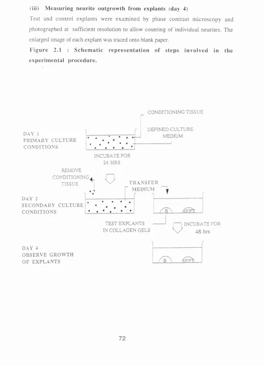
4.1 Introduction 69
4.2 Protocol 71
4.3 Results 74
4.4 Disscusion 77
4.5 Protocol 78
4.6 Results 81
4.7 Discussion 86
Chapter five : Developmental regulation of inhibitory activity
5.1 Introduction 90
5.2 Protocol for heterochronic co-cultures 91
5.3 Results of heterochronic co-cultures 92
5.4 Discussion 95
Chapter six : Biochemical studies
6.0 Introduction 99
Part 1 : Estimation of molecular weight range of inhibitory activity in
superior colliculus-conditioned media 100
6.11 Protocol 102
6.12 Results 104
6.13 Discussion 109
Part 2 : Separation of proteins in conditioned m edia by SDS gel
electrophoresis 111
6.21 Protocol 111
6.22 Results 114
Chapter seven : Final discussion
7.1 Background 117
7.2 Identification of distinct inhibitory activities 118
7.3 Conditioned media - a starting point for preliminary biochemical studies 120
7.4 Molecular weight estimation of inhibitory activity in conditioned medium 121
7.5 What is the chemical nature of the inhibitory activity in conditioned media ? 122
7.6 The inhibitory activity in superior colliculus-conditioned medium is novel 123
7.7 How does inhibitory activity bring about inhibition of neurite extension ? 124
7.8 What is the function of chemorepulsion in the developing nervous system ? 125
LIST OF TABLES
TA BLE 1 : Combinations of E l 5 tissues co-cultured.
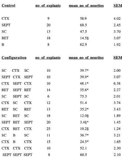
TABLE 2 : Mean numbers of neurites emerging from control and test explants co
cultured with different E l 5 explants.
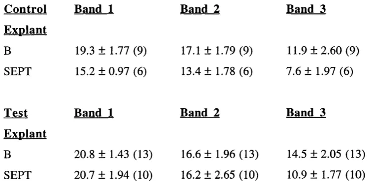
TABLE 3 : Mean numbers of neurites extending within three defined bands from
control explants and test explants cultured in cortex-conditioned media.
TA BLE 4 : Mean numbers of neurites extending within three defined bands from
control explants and test explant cultured in superior colliculus-
conditioned media.
TA BLE 5 : Mean numbers of neurites extending within three defined bands from
control and test explants cultured in continuously conditioned media.
TA BLE 6 : Combinations of E15 and E l 8 tissues co-cultured.
TA BLE 7 : Mean numbers of neurites emerging from control and test explants
co-cultured with different E18 explants.
TA BLE 8 : Mean numbers of neurites extending within three bands from control
and test explants cultured in superior colliculus-conditioned media, with
LIST OF FIGURES
Figure 1.1 : Schematic representation of the experimental arrangement within
collagen gels.
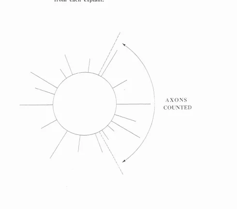
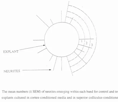
Figure 1.2 : Schematic representation of neurites counted that emerged from each
explant.
Figure 1.3 : Dark field pictures showing a retinal explant co-cultured separately
with two different flanking tissues, septum and superior colliculus.
Figure 1.4 : Phase contrast and dark field pictures showing the effect of the same
flanking explant (superior colliculus) on two different tissues (retina and
bulb).
Figure 1.5 : Phase contrast pictures showing a septal explant co-cultured
separately with two different tissues (cortex and superior colliculus).
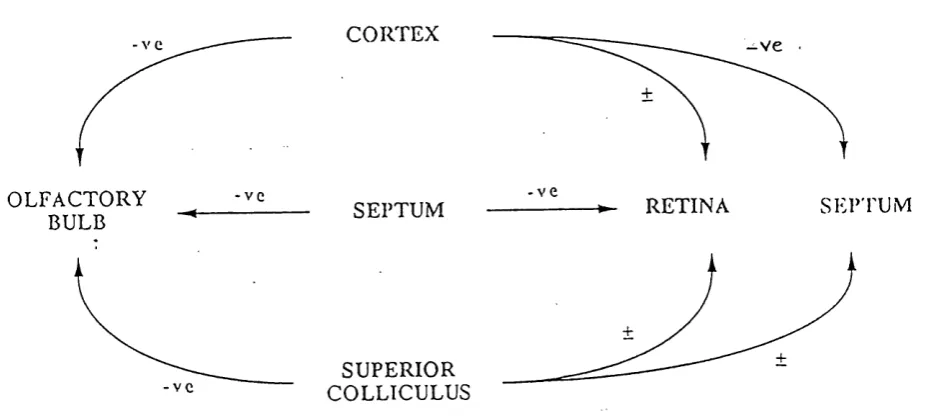
Figure 1.6 : A simple flow diagram showing the relevant tissue interactions
Figure 2.1 : Schematic representation of steps involved in the experimental
procedure.
Figure 2.2 : Schematic representation of an explant and its neurites showing the way
in which neurite lengths were estimated.
Figure 2.3 : Phase contrast pictures of an olfactory bulb explants cultured in superior
colliculus-conditioned medium and in cortex-conditioned medium.
Figure 2.4 : Schematic representation of steps involved in the experimental
procedure.
Figure 2.5 : Dark field pictures of a retinal explant cultured in
septum-conditioned medium and in superior colliculus septum-conditioned medium.
Figure 2.6 : Phase contrast pictures of olfactory bulb explants cultured in septum-
conditioned and in superior colliculus-conditioned media.
Figures 2.7a and b : Flow diagrams summarising the relevant tissue interactions in
the conditioned media and the co-culture experiments.
Figure 3.1 : Dark field pictures of a retinal explant co-cultured with E l5 and with
E18 superior colliculus.
Figure 3.2 a and b : Flow diagrams summarising the relevant tissue interations for
Figure 4.1 : Diagram of the test chamber.
Figure 4.2 : Phase contrast pictures of an olfactory bulb explant cultured in superior
colliculus-conditioned medium in a test chamber separated by two
different molecular weight dialysis membranes.
ACKNOW LEDGEMENTS
I would like to express my sincerest gratitude to my supervisor and friend Adrian Pini
for his guidance, support and constant encouragement throughout the course of this
project. I would also like to thank Julia Nash for her invaluable advice and emotional
support. I am indepted to Anne McLaren for her constant encouragement, interest in my
work and general well being. I thank Andrew Lumsden, Richard Wingate for their
helpful and insightful comments and Bruce Cotsell for the construction of the
“Gizzmo”. Last but not least, I would like to thank my family and friends, particularly
my brother, Donald, my mother, and best friend Bunmi who have endeavoured to
CHAPTER ONE
INTRODUCTION
1.1 HISTORICAL PERSPECTIVE
The nervous system consists of a complex network of neurones which are
interconnected in a highly specific manner. Its development follows a specific sequence
of events which begins with the proliferation of precursor cells to generate
presumptive glial and nerve cells. This is then followed by a period of migration of
these daughter cells to their final locations. During migration, differentiation takes place
in which the nerve cells begin to extend their interconnecting axons and dendrites.
When these processes make contact with one another or their targets, synaptogenesis
may occur. About this time inappropriate and excess synaptic connections are
eliminated. Clearly, the functioning of the nervous system will depend critically on the
specificity of these connections generated during development. Therefore a fundamental
problem in neurobiology is to understand how this specificity is established; i.e. what
are the cues which guide migrating nerve cells to their specific positions within the
nervous system, what is the nature of the signals which govern polarity and direction of
outgrowth, and what are the mechanisms by which these cells actually recognise one
another.
The first suggestion that neurones connect by extending axons, came from observations
made by Ramon y Cajal in 1890. He observed the presence of “ameboid” thickenings,
(growth cones) at the tips of axons, and correctly suggested that since they appeared to
move in an ordered and directed manner, they were involved in axon pathfinding. In
developing his ideas, Cajal had been impressed by the then recent observations that
leukocytes could orient their movement according to gradients of diffusible factors and
proposed that growth cones also responded to gradients of chemicals released by their
target tissues (Cajal 1920).
The dynamic quality of growth cones and evidence supporting Cajal’s initial
observations came with Harrison’s (1907) observations of living axons in tissue
culture for the first time. He proposed that the growth of axons occurred by the
extension of the growth cone. Harrison also suggested that the specificity of axonal
advance to form connections with target organs and then serve as a guide for later
developing axons. He predicted that the energy needed for this initial outgrowth would
come from within the cell and that the direction of the outgrowth was also determined
by factors within the cell. This suggestion, although plausible, was not received
enthusiastically because it did not account for the way in which the pioneer neurones
were able to form the initial appropriate patterns of interconnections. Subsequently, the
full recognition of the part played by pioneer nerves in axon pathfinding only became
evident with their later description in the peripheral (Bate 1976) and central nervous
system (Bate and Grünewald 1981). Nonetheless, Harrison’s contribution to the field
was valuable notably because his recognition that axon outgrowth required a semi-solid
medium and could occur along grooves or small channels of the substratum laid down
one of the earliest fundamental ideas of contact guidance. Since then, evidence
suggesting the importance of the physical structure of the substratum has grown.
Singer and his co-workers (1979) detected continuous extracellular spaces (channels)
between ependymal cells in the spinal cord of embryonic newts and deduced that such
channels provided cues for growing axons. Extracellular channels have also been
suggested to act as ‘guide posts’ for embryonic chick (Krayanek and Goldberg 1981)
and mouse (Silver and Sidman 1980) retinal ganglion cells. However, at present there
is little evidence supporting the existence of such channels in other areas of the nervous
system. There is however, evidence to suggest that growth cones may actively generate
these spaces by releasing specific metallo-proteases which modify their immediate
environment (Pittman 1985).
Up until the 1930’s the idea that specificity of neuronal connections was attributable to
specifically directed axonal outgrowth was still not widely accepted. The concept of
biochemical specificity only fully developed in the second half of the century with the
rise of cell biology and physiological chemistry.
The prevailing view in the 1930’s championed by Paul Weiss was that initial outgrowth
of axons occurred randomly, in an undirected manner. W eiss’ resonance hypothesis
(1936) stated that specificity arose through target responsiveness to only those axons
carrying the appropriate pattern of electrical activity. This hypothesis was derived from
investigations of the development of neuromuscular specificity in relation to the control
of limb movements. In a series of transplant experiments using salamander larvae, a
limb bud from a donor embryo was grafted caudally onto a normal limb bud. Several
weeks later, reinnervation of the grafted limb occurred and it developed movements that
were co-ordinated with that of the host limb, (the homologous response). Weiss
concluded that the appropriate (homologous) patterns of limb movement resulted from
inherent properties which enabled the correct muscles to receive or, ‘resonate’ with
only those motor nerves which carried the normal patterns of activity for movement. He
extended his theory of neuromuscular specificity to account for the general formation of
nerve patterns during development. He suggested that axons initially projected
indiscriminately and those which failed to make appropriate functional connections
were eliminated. Weiss carried out further experiments in tissue culture which led him
to suggest yet another mechanism for neuronal specificity, the contact guidance
hypothesis (Weiss 1941, 1944). He based this theory on his observations of axons in
relation to mechanical features in their environment. He observed that axons grew on
lines of tension in plasma clots and scratches on the surface of culture dishes and
concluded from this that axons did not require chemical cues. He suggested that
guidance simply resulted as a result of the interplay between the motive power of the
cell and contractions provided by adhesive contacts between the cell and the
substratum. The demise of both the resonance and contact hypotheses was largely
attributable to the work of Roger Sperry between the 1940’s and ‘50s. Sperry was
W eiss’ student and his work at the time involved the study of the regenerative capacity
of axons of the retinotectal projection. In these experiments he cut the optic nerve of
newts, rotated the eye through 180®, and waited for regeneration to occur. After optic
nerve regeneration, the animals with rotated eyes behaved as if their visual field was
inverted and shifted to the left, implying that retinal ganglion cell axons had grown back
to their correct targets, even though the connections were behaviourally inappropriate
(Sperry 1944). This, and other similar experiments (Attardi and Sperry 1963) in the
goldfish visual system provided evidence for a high degree o f specificity in the
formation of synaptic connections, and culminated in the chemoaffinity hypothesis
interactions of complimentary chemical affinities present on neurones and their targets.
The establishment of appropriate connections would therefore depend on the correct
and exclusive matching of molecules on pre and post synaptic sites. In this hypothesis,
each neurone would carry some kind of individual identification tag, and therefore
presupposed the existence of multiple distinct ligands and their receptors. Another
assumption of the hypothesis was that once neuronal connections were established,
they would remain rigid and inflexible. This was soon disproved by experiments which
showed that retinal axons readjusted their contact sites in order that they could all be
accommodated by one half of the tectum if the other half had been surgically removed
(Gaze and Sharma 1970, Yoon 1971). Further evidence disproving a rigid matching
proposal also came from experiments in the visual system demonstrating the flexibility
of axonal projections during normal development. During development the initial set of
retinal axons form a succession of temporary ‘incorrect’ connections, which are later
refined (Chung et al. 1974, Gaze et al. 1974). The driving force for these refinements
was proposed to be controlled by electrical activity within the neural pathways
themselves (Fawcett and O’Leary 1985). The molecular complexity inherent in the
chemoaffinity hypothesis, together with the growing evidence for the role of electrical
activity in neuronal patterning (Marsh and Beams 1946, Cohan and Kater 1986),
tempered enthusiasm for it. Consequently some details of Sperry’s hypothesis have
been revised, however the general concept of chemoaffinity is essentially correct, and
his work remains critical in the field of neural development.
Research in the following decade focused on the more specific mechanisms by which
growth cones were guided to their targets.
An early lead to answering these questions came from two major studies during the late
1940’s and early ‘50’s on the behaviour of cells in tissue culture. The first, carried out
by Abercrombie and Heaysman (1953), was a systematic attempt to examine the social
(and antisocial) behaviour of cells. Abercrombie’s observations of monolayering by
fibroblasts and heart myocytes in culture, led him to suggest that the surfaces of cells
were capable of mediating mutual inhibition of cell division on contact. The term
the nervous system. However, in the Abercrombie and Heaysman experiments, the
observed contact inhibition refered to the failure of cells to continue dividing once they
had fully occupied a given space. Nerve cells do not divide; on contact with a non-
permissive substratum, their growth cones collapse or deform and the axon retracts.
Contact-repulsion may therefore be a more accurate description of this behaviour of
nerve cells. Hughes (1953) was the first to observe this phenomenon; he observed that
growth cones retracted when they contacted each other or neurites, the significance of
which with respect to axon guidance only became clear much later.
Following on from Abercrombie’s work. Carter (1965), also investigating cell motility,
took matters a step further and examined the behaviour of cells on various surfaces. He
made substrata of varying adhesiveness by vacuum-evaporating an inert metal on to a
glass slide coated with cellulose acetate, and compared cell motility on these surfaces.
He observed that cell movement ocurred predominantly up the gradients of
adhesiveness and termed this phenomenon haptotaxis i.e. the directed response of cells
to a gradient of adhesiveness. He proposed that all cell movement, including cell
migration involved in morphogenesis could be accounted for by this mechanism.
However, it was not until the advent of time-lapse cinemicrography, when cells were
observed to accumulate on portions of patterned substratum to which they adhered most
strongly, that Carter’s proposal was reinforced (Harris 1973).
The significance of the physical substratum in neuronal morphogenesis was further
inferred by studies carried out by Letourneau in the mid 1970’s. By culturing sensory
neurones on several different substrata, he was able to demonstrate a role for cell-to-
substratum adhesion in the initiation, elongation and branching of axons. In his first
series of experiments, he investigated the adhesive qualities of different surfaces and
the strength with which growth cones attached to them. He observed that growth cones
adhered most strongly to and extended axons at greater rates, on polyornithine coated
surfaces compared with tissue culture plastic (Letourneau 1975a). He suggested that
since there appeared to be a strong correlation between adhesion and enhanced axonal
growth, the degree of adhesive interaction between the growth cone and its
microenvironment must be important for the formation and guidance of neuronal
which are highly positively charged polymers; thus adhesion was probably due to an
electrostatic interaction between the positively charged substrate and the negatively
charged site on the external surfaces of the growth cones. In a second series of
experiments, Letourneau (1975b) further demonstrated the relationship between
adhesion and cell movement by showing that neurites would actively select the path of
greatest adhesiveness when presented with a choice. By the time Letourneau had
carried out these studies, growth cones had already been shown to possess microspikes
which contacted the substratum ahead of the main body of the growth cone and which
advanced by addition of intracellular vesicles at their tips (Bray 1973a). Letourneau
therefore suggested that adhesion could contribute to axonal extenion by promoting the
addition of vesicles to microspikes. This he proposed would lead to expansion of the
microspike and in turn advancement of the growth cone in a specific direction. Thus the
most firmly attached filopodium would be the one most successful at steering the
growth cone. Letourneau’s contribution was to show that growth cones, like motile
cells, could selectively adhere to and advance on specific substrates thus suggesting that
axon guidance resulted from differential adhesion.
Functional evidence implicating cell substrate interactions in the development of the
central nervous system (CNS), came from a description of haptotaxis (of the kind first
described by Carter) in the CNS of the developing wing of the pupal moth (Nardi and
Kafatos 1976a,b). These researchers observed that while sensory neurones from the
tips of the wings grew towards the CNS, they were closely associated with the
extracellular matrix of the upper epithelial layer. When this substratum was altered by
exchanging epithelial grafts, growth cones were able to grow cross both control and
distally positioned grafts, but not those displaced proximally. Supporting evidence for
interactions with substrate came from scanning electron microscopy of the substratum
of different proximo-distal positions which revealed a graded distribution of
extracellular matrix components (Nardi 1983). Berlot and Goodman (1984) have also
described a gradient of adhesion in the development of grasshopper sensory neurones
originating in the antennae and limb buds. By using transmission microscopy and
tissue culture manipulations, they showed that growth cones of pioneer neurones in the
over the surfaces of epithelial cells, suggesting that these axons were guided by an adhesive hierarchy.
Directional adhesive cues need not always be presented in the form of a gradient. A growth cone encountering an environm ent which contains com ponents of varying adhesiveness, could also be guided to its target by recognising specific adhesive molecules distributed at a uniform level along a preformed pathway (see diagram 1). The differential adhesion hypothesis is consistent with Steinberg’s (1978) proposal of morphogenic control of em bryonic tissue. Steinberg’s observation that dispersed populations of embryonic cells could coalesce to form tissue, led him to propose that cells were capable of selecting one another on the basis of the differential adhesive properties on the cell surface. For instance, when presented with the choice of both dorsal and ventral half of the tectum, dissociated retinal ganglion cells from the ventral half of the retina adhere preferentially to the dorsal half of the tectum (Roth and Marchase 1976). Such studies, which followed from H oltfreter’s classical cell sorting studies of the 1930s and 1940s, suggest that differential cell surface affinities play an important part in mediating the organisation of tissues.
1. Diagram illustrating two mechanisms of adhesion
Adhesive gradient Differential adhesion
• • • • •
1 Axon advances up gradient of a single component.
The function of adhesive gradients could be to act more globally, orienting and directing
axons to appropriate intermediate targets within the embryo, whereas the role of
adhesive cues which aline defined tracks (Katz and Lasek 1979, Silver and Rutishauser
1984) may be to direct axons more precisely to their final targets.
Having indicated that adhesive interactions were important for the growth of axons,
researchers began to speculate on the mechanisms by which adhesive interactions could
affect the contractile apparatus of the growth cone in order to regulate growth cone
motility.
Bray suggested that adhesion and tension were linked such that the direction of advance
of a growth cone was determined by the tension existing between it and the rest of the
cell. He based this proposal on calculations of the resultant force on a growth cone from
a vectorial analysis of individual sensory neurones. He deduced that the filopodia of
growth cones evoked a mechanical tension in the growth cones as they pulled against
the substratum (Bray 1979). His later demonstration that mechanical tension directly
applied to a growth cone, by pulling it with a micro-pipette, could initiate elongation of
the growth cone in the direction of the force applied, led him to propose a more
definitive role for mechanical tension in neurite outgrowth (Bray 1984). In his scheme,
a growth cone could determine the levels of adhesion by monitoring the levels of
tension that resulted as it pulled against the substratum. This tension itself could then
directly trigger the contractile elements in the growth cone. If adhesion is weak, forces
exerted on the cytoskeletal elements of the filopodia will be weak and break off,
resulting in retraction of the filopodia. If however adhesion is strong, then tension will
result between the growth cone and the rest of the cell, activating stretch sensitive
systems, which in turn would draw the growth cone forward into the more adhesive
environment.
In summary, over the past century, theories of axon guidance have developed from
those which involved random diffuse outgrowth (Weiss 1936), to those in which
specific chemical signalling is key (Sperry 1963). In parallel, some definitive ideas
about the movement of growth cones emerged (Letourneau 1975, Bray 1979, 1984).
cues involved. Since preferential adhesion was recognised as the major way in which
axons were guided to their targets, subsequent research focused on identifying
molecules that mediated adhesive interactions.
1.2 A X O N G U ID A N C E M E D IA T E D BY N O N -D IF F U S IB L E
MOLECULES
A large number of molecules which are permissive for and promote axon growth have
been identified. They can broadly be divided into two groups; those that mediate
adhesion between cells (cell adhesion molecules), and those that mediate adhesion
between growth cones and the extracellular substratum (substrate adhesion molecules).
I shall now briefly review the pertinent evidence defining their roles in axon guidance.
Although each will be dealt with separately, it is likely that cells interact with different
molecular species simultaneously in vivo.
1.21 Contact adhesion mediated by cell adhesion molecules
There are three major classes of cell adhesion molecules. Those which are calcium-
independent, the immunoglobulin superfamily, those which are calcium-dependent, the
cadherin and selectin families and the integrins.
In general cell adhesion molecules (CAMs) display a wide range of functional
specificity. Edelman proposed that a relatively small number of adhesion molecules
would be sufficient to account for even the most complex of morphogenetic events,
provided they undergo certain changes in expression or chemical properties during
development. He suggested that nature of these changes, collectively termed local cell
surface modification, could involve changes in the number, distribution and chemical
structure of the CAM’s (Edelman 1983).
The Immunoglobulin superfamily
Neural cell adhesion molecule (NCAM)
There are around thirty isoforms of NCAM generated by alternative splicing of a single
gene (Walsh and Doherty 1991), the major three being named after the tissues from
which they were isolated. NCAM, neural cell adhesion molecule (Thiery et al. 1977),
neuron-glia adhesion molecule (Grumet et al 1985 and Edelman 1984).
NCAM is the most prevalent and most extensively studied in vertebrate tissues. It is a
cell surface glycoprotein which mediates homophilic binding with other NCAM
molecules (Edelman 1983). It exists as three major glycoproteins, with molecular
weights of 180, 140, and 120Ka. Each polypeptide form is made up of three domains,
an N-terminal binding domain, a middle poly sialic acid domain and a cytoplasmic
domain. The different forms are identical except for the length of their carboxyl termini
(Edelman 1986). During early development NCAM is expressed by neural epithelial
cells soon after neural induction (Kintner and Melton 1987). However, it is not
expressed during migration of neural crest cells and is re-expressed on sensory
precursor cells (Thiery et al. 1982) suggesting that it is important in cell binding.
Diversity in NCAM function is thought to arise as a result of modification of its
structure as Edelman first suggested. This may be achieved in several ways. For
instance the degree of sialylation of the middle domain modifies the adhesive
properties of the molecule (Hoffman and Edelman 1983, Landmesser et al. 1990). The
cytoplasmic domain of the molecule can also modify the adhesive properties of
NCAMs. Neurones grow relatively poorly on monolayers of 3T3 fibroblast cells
expressing isoforms of NCAM with enlarged cytoplasmic domains generated by
alternative splicing of the cytoplasmic domain (Doherty et al. 1992). In addition to
diversity in NCAM polypeptide structure, differential expression of these molecules
could contibute to their functional diversity particularly in defining boundaries between
groups of cells.
LI
Another molecule similar to NCAM that also belongs to the immunoglobulin super
family is the calcium independent adhesion molecule, LI which is expressed on
postmitotic neurones (Rathjen and Schachner 1984). It appears to be specifically
important in axon-axon binding because LI-blocking antibodies disrupt fasciculation
(Chang et al. 1987 and Fischer et al. 1987). Consistent with this, its expression is
reduced after nerve tract formation, (i.e. after fasciculation has occurred), and increases
after injury in perpherial nerves, when presumably it would be required for re-
The cadherins
The cadherins are a family of transmembrane glycoproteins which mediate
calcium-dependent adhesion (Takeichi 1987). They all share highly conserved transmembrane,
cytoplasmic and extracellular domains. There are at least a dozen subtypes known
(Suzuki et al. 1991) each with a unique tissue distribution. Cells show dynamic
expression of these molecules, some tissues express a single subtype while others
express a few subtypes together (Takeichi 1987). Each subtype displays a different
binding specifity and binding is homophilic (Takeichi et al. 1988). Tissue-specific
expression of cadherin subtypes may explain the segregation of embryonic cell types
first demonstrated by Steinberg (1970) and would be consistent with the observation
that when cell layers are about to separate they express different sets of cadherins
(Takeichi 1988). The differential expression of cadherins is thought to be associated
with morphogenic events occuring in the embryo particularly in relation to cell sorting
and forming and maintaining connections between cells (Hatta et al. 1987, Nagafuchi
et al. 1987, Takeichi et al. 1989, 1990).
The selectins
The selectins are transmembrane proteins which are expressed on blood and endothelial
cells and which are involved in mediating the inflammatory response. Three selectins
L-, P- and B- have been identified (Bevilacqua et al. 1991) which are thought to
mediate cell-cell adhesion by binding to specific carbohydrate groups on neutrophils via
their lectin domains (Hynes and Lander 1992). Endothelial cells release P-selectin
from intracellular stores onto the cell surface following initiation of the inflammatory
response. Since P-selectin recognizes a specific carbohydrate group present on the
surface of neutrophils, it binds to it bringing the neutrophils closer to the endothelial
cells (Geng et al. 1990). Consistent with this role, is the finding that neutrophils
preferentially bind to surfaces coated with purified P-selectin (Geng et al. 1990). L-
selectin is also thought to be important in the selective adhesion of circulating
lymphocytes to endothelial cells of lymphoid organs (Stoolman 1989 ). However, at
The integrins
The integrins are receptors which mediate both cell-cell adhesion and cell-substratum
adhesion and appear to have a much wider distribution than members of the calcium-
dependent adhesion molecules (see later) (Krotoski 1986). They are heterodimers
consisting of two single subunits, alpha and beta, both of which have several
structurally related subunits and there are currently about twenty known functional
pairs of these heterodimers (Hynes 1987). Integrins span the cell membrane, and have
been proposed to act as transmembrane linkers between extracellular ligands and the
cytoskeleton. Integrins bind their ligands by recognising specific amino acid
sequences, particularly those that contain the tripeptide arg-gly-asp (RGD) sequence
which is found on a number of extracellular matrix proteins (Ruoslahti and
Pierschbacher 1987). The intracellular domain of the molecule, more specifically
ih^beta subunit (Buck et al. 1986, Hayashi et al. 1990), interacts with the cytoskeleton
through association with cytoskeletal components such as vinculin and talin (Burridge
et al. 1988). Evidence implicating integrins in promoting cell adhesion have mainly
employed blocking antibodies, for instance the antibody JG22 which recognises the
beta integrin subunit inhibits neural crest cell attachment and migration on laminin and
fibronectin substrata in vitro (Bronner-Fraser 1985). In vivo, neural crest cell
migration in avian embryos is disturbed after injection of the anti-integrin antibodies
into the neural tube at the onset of neural migration (Bronner-Fraser 1986).
1.22 Contact adhesion mediated by substrate adhesion molecules
Neuronal migration and axon elongation occur through the extracellular matrix which
consists of a lattice network of adhesive glycoproteins, collagens, proteoglycans and
glycosaminoglycans. The finding that laminin was the active component in heart-
conditioned media which promoted neurite-promoting ability (Lander et al. 1985),
raised enthusiasm that extracellular matrix molecules could promote axon extension.
Subsequently efforts were made to identify other extracellular matrix molecules capable
Laminin
Laminin, the most abundant glycoprotein in the basement membrane, was first isolated
from the Engelbreth-Holm-Swarm murine tumour (Timpl et al. 1979) and has been
shown to be capable of influencing cell motility. In vitro, laminin is a potent promoter
of neurite outgrowth (Hammerback et al. 1985, Liesi 1985, Gundersen 1987). In vivo
it lines prospective pathways that axons follow to reach their targets (Cohen et al.
1986, Bronner-Fraser 1986, and Riggott and Moody 1987) and plays a prominent role
in neural crest cell migration (Bronner-Fraser 1986, and Bronner-Fraser and Lallier
1988). The interactions of laminin are mediated by integrins (Bozyczko and Horwitz
1986, Tomaselli et al. 1986, Lallier and Bronner-Fraser 1991) which bind to its RGD
sequence (Sasaki et al. 1988). Intially, laminin was thought to promote axon growth by
simply facilitating the adhesion of growth cones to the substrata via integrins.
However, Gundersen (1987) examined the specific involvement of adhesion in
promoting neurite growth on extracellular substrates and found that regardless of how
adhesive an alternative substratum was, laminin was always the preferred substrate for
growth. This indicated that a further factor, not necessarily adhesion, was responsible
for the neurite-promoting ability of laminin. Moreover recently, laminin has been found
to have anti-adhesive effects (Calof and Lander 1991). Thus at present it is still unclear
how laminin mediates its axon outgrowth promoting effects. Since it is less widely
expressed than the CAMs, it may play a more restricted role in axon guidance.
Fibronectin
Fibronectin is a ubiquitous component of the extracellular matrix which has also been
shown to promote the adhesion and migration of developing neurones. It is a large
glycoprotein consisting of a dimer of similar sunbits linked by disulfide bonds. There
are three specific cell-binding regions: a central binding, an alternatively spliced IIICS
region, and a heparin-binlling site (Hynes 1986). Most cells adhere to fibronectin by
binding to the central domain because it contains the crucial RGD amino acid sequence
important for ligand binding (Pierschbacher and Ruoslahti 1984). Fibronectins have
also been shown to be involved in neural crest cell migration (Bronner-Fraser 1986)
however, a specific role of them in neural crest cell migration is questionable because
substrata in the absence of fibronectin (Tucker and Erickson 1984).
Many extracellular matrix molecules are capable of stimulating neuronal outgrowth in
vitro. They are all located in areas of the vertebrate embryo where they could potentially
serve as permissive substrates or provide guidance cues for growing axons. However,
there is little direct evidence to suggest a guidance role for extracellular matrix
molecules in vivo or in vitro. McKenna and Raper (1989) were unable to show that
surface gradients of laminin affected the direction of axon outgrowth, but similar
experiments have not been performed with other extracellular cellular matrix
components.
Recent characterisation of genes that affect growth cone guidance in C.elegans, has
provided some persuasive evidence that extracellular matrix components may function
in guidance. The migration of pioneer axons in between the epidermis and basal lamina
in C.elegans is guided by three characterised genes; UNC 5, UNC 6, and UNC 40
(Hedgecock et al. 1990). UNC 6 encodes the laminin B2 chain, UNC 5 a
transmembrane protein, which has been proposed to be a laminin B2 receptor and
UNC 40 a protein which may also be another laminin B2 receptor. Both ventrally and
dorsally directed circumferential migrations are disrupted in UNC-6 mutants (Culotti et
al. 1994). The identification of such structurally related genes related to laminin which
affect axon guidance specifically, has helped to provide some convincing evidence that
extracellular matrix molecules may play a role in axon guidance in vivo. This
contention is strengthed by identification of the candidate guidance molecule netrin-1,
as a vertebrate homologue of UNC-6 (see later). In addition, homologues of various
ECM components have been identified. For example, merosin is an A-chain
homologue of laminin (Ehrig et al. 1990) and S-laminin a homologue of the laminin B-
chain (Hunter et al. 1989). The existence of these homologues implies that extracellular
matrix molecules have a much more heterogenous distribution than was once thought.
In principle this, together with changes in the levels of their expression make them
good candidates for axon guidance cues. The mechanisms by which extracellular
matrix molecules exert their effects are still unclear but signalling mediated through
1.23 Contact mediated repulsion
During much of the 1980’s, it was generally held that axons were guided to their targets
by differential expression of adhesion molecules (reviewed above). Only in the late
1980’s when it was demonstrated that growth cones could be repulsed on contact with
non-target membranes (Walter et al. 1987, Kapfhammer and Raper 1987a,b) was the
idea that guidance might occur through avoidance seriously considered. Kapfhammer
and Raper examined a wide range of neurones and although these were not
developmentally appropriate, in so far as these neurites did not make contact during
development in vivo, the results were important because they indicated that guidance
could arise as a result of contact repulsion. Crucially they provided a basis for the
development of an assay of growth cone-collapsing activity, which has subsequently
led to the isolation of inhibitory proteins (Davies et al. 1990, Luo et al. 1993).
The retinotectal pathway
In the rat retinotectal projection, anterior and posterior retinal axons project to posterior
and anterior tectal areas respectively (Bonhoeffer and Huf 1982). Using an elegant in
vitro assay system in which retinal axons were made to grow on substrata consisting of
narrow adjacent alternating stripes of anterior and posterior tectal membrane
components, Bonhoeffer and colleagues (Walter et al. 1987) showed that chick
temporal but not nasal retinal axons avoided posterior membranes and grew exclusively
on anterior membranes. However, both types of retinal axon grew equally well on
membranes derived from either tectal region suggesting that inhibition was not
absolute. Bonhoeffer interpreted this behaviour to mean that growth cones actually
detected gradients of inhibitory molecules (at the stripe interfaces) and suggested that
temporal retinal axons were excluded from the posterior tectum on this basis. A
candidate inhibitory molecule was identified as an inositol-linked 33KDa glycoprotein
present in posterior tectal membranes. When incorporated into phospholipid vesicles,
the molecule caused collapse of temporal retinal axons (Stahl et al. 1990). To test
the’gradient reading’ hypothesis, Baier and Bonhoeffer (1992) grew retinal axons on
membrane carpets that contained a gradient of inhibitory tectal membrane components.
Under these conditions, temporal growth cones failed to elongate extensively or
This strongly indicated that it is the gradient of the inhibitory protein which functions to
direct temporal retinal axons away from the posterior tectum.
More recently a 25kDa glycoprotein, repulsive axon guidance signal (RAGS) has been
identified and proposed to be a positional label in the retinotectal system (Drescher et al
1995). However, in the membrane stripe assay made up of alternating RAGS-COS cell
membranes and moek-transfected cell membranes, both nasal and temporal axons
avoided RAGS-containing membrane stripes. Thus while RAGS may have a role in
guidance within the tectum, it does not appear to have the discriminatory properties
required to determine position along the anterior to posterior axis. RAGS shares
significant sequence homology with ligands for the receptor tyrosine kinase of the Eph
subfamily which are the largest of the receptor tyrosine kinase family (Van de Geer et
al. 1994). A recent study by Cheng et al. (1995) has implicated them in mediating
contact-repulsion of axons involved in the formation of the retinotectal system. They
showed that a specific class of these receptors, MeK 4 and its ligand ELF-1 are
expressed in complementary gradients on retinal ganglion and tectal cells respeetively
thus ELF-1 expression is highest in the posterior tectum while correspondingly, MeK 4
expression is highest in the temporal retina. Although the distribution and properties of
this receptor-ligand pair are consistent with a role in the formation of the retinotectal
system, further functional studies will be needed before ELF-1 and other members of
the Eph ligand family can conclusively be described as topographic guidance cues. If
so, this would be the strongest evidence for the original proposal by Sperry of
complimentary lock and key-type chemoaffinities.
Spinal nerve segementatlon
The generation of the segmented pattern of spinal nerves in higher vertebrates in which
sensory and motor nerves are confined to the anterior half of the somites results from
axon repulsion by the posterior half of the somite (Keynes and Stern 1984). Using
lectin-affinity chromatography, two peanut agglutinin-binding inhibitory cell surface
glycoproteins (48 and 55 KDa) have been identified in posterior somite-halves.
Detergent extracts of these proteins cause collapse of sensory growth cones which can
be eliminated by polyclonal antisera raised against these molecules (Davies et al. 1990).
distributed as a gradient but are found exclusively in the posterior somite. In addition,
sensory neurones are not able to grow on surfaces treated with membrane fractions
from posterior somites indicating that inhibition is absolute. The mechanisms by which
these molecules cause growth cones to collapse are at present unknown.
M atrix molecules
Several molecules present in the extracellular matrix inhibit neurite outgrowth. One
such molecule is tenascin. It is a multimeric glycoprotein found in several organs and in
the CNS and PNS (Spring et al. 1989). Its contains several fibronectin and epidermal
growth factor-like (EOF) repeats (Reichardt and Tomaselli 1991). Tenascin is of
particular interest because it appears to have dual function. As a culture substrate, it
mediates adhesion of neurones to astrocytes (Grumet et al. 1985), and promotes neurite
outgrowth (Wehrle and Chiquet 1990) but conversely inhibits migration and axon
outgrowth of other neuronal cells (Pesheva et al. 1989, Faissner and Kruse 1990). The
use of domain specific antibodies strongly suggests that the bifunctionality of tenascin
resides in different binding sites. The monoclonal antibody Jl/tn2 which recognises the
fibronectin repeats, abolishes the neurite promoting activity, but not its inhibitory-
properties (Faissner and Kruse 1990). The growth-promoting effects of tenascin
appear to be associated with the fibronectin repeats and the inhibitory effects appear to
be associated with the EGF-repeats (Friedlander et al. 1988, Spring et al. 1989). There
are several isoforms of tenascin and tenascin-like glycoproteins; Jl-160 and J1 180 are
two such isoforms derived from differential splicing of one gene (Spring et al. 1989).
Judging from their localisation within the developing nervous system, they may be
involved in the formation of boundaries which axons cannot penetrate.
M yelin associated proteins (NI-35 and NI-250)
It has been well established that neurones in the CNS fail to regenerate or regenerate
extremely poorly (Cajal 1928). The reasons for this are still not clearly understood, but
some progress has been made over the past ten years. The pioneering work by Aguago
and colleagues in the early 80’s indicated that the environment was an important
determinant for regeneration (Benfey and Aguago 1982). However, attempts by
Schwab and Thoenen (1985) to produce regeneration by providing injured neurones
that there must be inhibitory activities present in the adult CNS whose effects could not
be overcome by neurotrophic factors. He therefore tested the ability of different glial
cell classes to permit growth of central and peripheral neurones and found that growth
occurred on all glial cell types (astrocytes, immature oligodendrocytes), except mature
oligodendrocytes (Caroni and Schwab 1988a). In addition, conditioned medium made
from oligodendrocytes failed to cause collapse (Brandtlow et al. 1990), suggesting that
inhibition was contact-dependent. Biochemical analysis of oligodendrocyte membrane
fractions and CNS myelin revealed the presence of two inhibitory molecules termed
NI-35 and NI-250 which when incorporated into liposomes inhibited fibroblast-
spreading and neurite outgrowth (Caroni and Schwab 1988a). Treatment of
oligodendrocytes with monoclonal antibodies to these molecules allowed DRG axons
to extend over them, strongly suggesting that these molecules were responsible for the
failure of these neurites to extend over oligodendrocytes (Caroni and Schwab 1988b).
Further compelling evidence supporting the activity of these molecules in preventing
CNS regeneration has come from both in vivo and in vitro studies using neutralizing
antibodies against these molecules. In vitro, the inhibitory effect of myelin is reversed
following the removal of NI-35 and NI-250 with antibodies against them. In vivo, the
regeneration of the disrupted rat corticospinal tract (CST) was increased in both animals
treated with inhibitory neutralizing antibody, IN-1 (Schnell and Schwab 1990), and in
the Olac rat in which oligodendrocytes and myelin are absent (Savio and Schwab
1990). More recently, experiments have shown that regeneration may be slightly
enhanced if IN-1 is delivered to the lesion site in conjuction with the neurotrophic
factor NT3 (Schnell et al. 1994). Although in most experimental cases, regenerating
axons failed to bridge the lesion completely and therefore regeneration was limited, the
results nonetheless provide a potential strategy for facilitating regeneration.
M yelin associated glycoprotein
Recently, another protein associated with CNS and PNS myelin which inhibits neurite
outgrowth has been identified. McKerracher et al (1994) used DEAE anion exchange
chromatography to isolate two peaks of activity from purified bovine CNS myelin,
which inhibited neurite outgrowth for a neuroblastoma cell line. Western blots revealed
myelin-asssociated glycoprotein (MAG), a member of the immunoglobulin-gene super
family. Immunodepletion by anti-MAG antibodies, resulted in loss of neurite inhibitory
activity . More conclusively, when used as a culture substrate, recombinant MAG
inhibited neurite outgrowth from the neuroblastoma cell line (McKerracher et al. 1994).
Similarly, in a separate study, recombinant MAG expressed by Chinese hamster ovary
cells (CHO) inhibited neurite outgrowth from both cerebellar and adult dorsal root
ganglion neurones (Mukhopadhyay et al. 1994). McKerracher and collègues have
suggested that like NI-35 and NI-250, MAG may be a major contributor to the failure
of CNS neurones to regenerate. However unlike NI-35 and NI-250, MAG is also
expressed by PNS neurones, although to a much lesser extent (Quarles et al. 1983).
This raises two interesting questions; first, how much is the failure of CNS axons to
regenerate due to MAG and secondly, since MAG is also present in the PNS, why do
these neurones appear to regenerate better than those of the CNS. It may be critical that
MAG levels are 10-fold higher in CNS than in PNS myelin but in addition,
McKerracher and collègues have suggested that PNS neurones are still able to
regenerate despite the presence of MAG, because of the more rapid removal of
myelinated debris following peripheral damage. Consistent with this proposal is the
recent report that regeneration occurs in unmyelinated nerve cells but not in myelinated
nerve cells in mutant mice in which there is a delay in Wallerian degeneration (Brown et
al. 1994).
1.3 Axon guidance mediated by diffusible molecules
1.31 Chemoattraction
Another way in which growth cone motility can be controlled is by chemotaxis, the
phenomenon by which the direction of cell movement is determined by the
concentration gradient of specific substances. Some well known examples include the
invasion of vascular epithelial cells into tissues which produce angiogenic factors, the
attraction of leukocytes to sites of tissue damage or local infection and the aggregation
of the amoeba, Dictyostelium discoideum, to form a multicellular body (Gerisch et al.
1975, Devroetes 1982). The concept that a gradient of attractive factors released from a
Ramon y Cajal. However, not until fairly recently has there been any evidence to
support this.
The first attempt to demonstrate axonal chemotaxis was prompted by the discovery of
nerve growth factor (NGF), a protein which is secreted by the targets of sympathetic
and sensory neurones and which is required for their survival (Levi-Montalcini and
Angeletti 1963). Evidence indicating that NGF could be a potential chemoattractant
came from both in vitro and in vivo studies. In vivo, sympathetic neurones were
observed to project to an injection site following large doses of NGF into the brain
stem of neonatal rats (Menesini-Chen et al. 1978), and in vitro, regenerating sensory
axons were shown to orient preferentially up a gradient of NGF (Gundersen and
Barett 1979), and also to turn towards a source of NGF (Gundersen and Barett 1980).
Initially, these observations taken together were thought to indicate that NGF could be
a potential chemoattractant in vivo. However, enthusiasm for this soon fell with the
crucial finding that the target cells of these neurones did not produce NGF mRNA until
the first axons reached their targets (Davies et al. 1987). Consequently NGF has been
precluded as an endogenous chemoattractant, nonetheless experiments using NGF
have importantly demonstrated that growth cones can respond chemotactically to
gradients of diffusible molecules. In addition, they have highlighted the key
requirements of potential chemoattractant. These are namely that chemoattractants must
be available at developmentally appropriate times and in sufficient quantity to establish
a suitable gradient in the local cellular environment of developing axons. Axons
exposed to a steep gradient of NGF turned towards the NGF source (Gundersen and
Barrett 1979, 1980). However, only 60% (compared to 50% for random distribution
of orientation) of axons exposed to a shallow gradient of NGF orientated up-gradient
(Letourneau 1978), suggesting that the chemotropic effects of NGF is dependent on the
steepness of the gradient. The importance of the steepness of the gradient has been
further implied by recent studies in which sympathetic neurones of transgenic mice
were made to express NGF during their growth towards their targets. Axon trajectories
in these mice were unaffected but there was a reduction in terminal branching of these
axons. However, by genetically manipulating target cells so that they over-expressed
These researchers have therefore suggested that the initial observed reduction in
terminal branching was due to the abolition of the normal target-derived NGF gradient
as a result of the presence of excess NGF expressed by sensory axons. Thus when the
target cells are made to express increased quantities of NGF, the normal gradient is
restored, and terminal branching is maintained (Hoyle et al. 1993). This study not only
indicates that an NGF gradient could be important for terminal branching and has
indicated a chemotactic role for NGF in the control of terminal arborisations.
A number of neuronal systems have now emerged in which axons have been
demonstrated to grow towards their targets in response to gradients of diffusible
chemoattractants other than NGF (Lumsden and Davies 1983, Tessier-Lavigne et al.
1988, Heffner et al. 1990). The discovery of chemoattraction in now classical studies
by Lumsden and Davies (1983, 1986) took advantage of the fact that neural explants
could be successfully cultured and visualised within three dimensional collagen gel
matrices (Ebendal and Jacobson 1977). The first indication that such a matrix could
provide a stable substratum in which cells could migrate came from initial studies by
Elsdale and Bard (1972).
Developing trigeminal sensory axons
Using a refined collagen gel preparation, Lumsden and Davies (1983, 1986) co
cultured explants of developing trigeminal neurones with their cutaneous target, the
maxillary epithelium, to observe the effects of target-derived diffusible factors on
neurite outgrowth. They showed that trigeminal sensory axons respond chemotactically
to a factor(s), distinct from NGF. ElO trigeminal ganglion explants were cultured
alone, and co-cultured at a distance of about 300-500um, with explants from ElO
maxillary arch. No axon outgrowth was observed from the ganglia cultured alone,
while in over 60% of the cases, axons were directed from ganglia towards the
maxillary epithelium. The chemoattractant has been called Max Factor (MF) and
although it has yet to be characterized, several features are consistent with its role as a
diffusible chemoattractant for developing trigeminal axons. First, MF is expressed at
which most of these sensory axons emerge from the trigeminal ganglion (Lumsden
1988). Secondly, axons turned within the collagen gels towards the target and did not
exhibit radial outgrowth merely on the side of the ganglion facing the target (Lumsden
and Davies 1983). However, since axons emerging from these ganglia were not
retrogradely labelled, it is unknown whether the distribution of their cell bodies is
uniform or is biased towards the target-facing aspect of the ganglia. Thirdly, when two
ganglia were co-cultured in tandem next to the maxillary arch, more axons emerged
from the side of the distal ganglion than from the away-facing aspect of the more
proximal ganglion (Lumsden and Davies 1983). This is powerful evidence for the
operation of a chemotropic gradient. However, the maxillary epithelium clearly
provided trophic support because in its absence early trigeminal ganglia failed to
produce axons. It therefore remains unclear whether MF and this trophic component
are related.
Spinal commissural axons
The floor plate is a neuroepithelial ventral midline stmcture which runs the length of the
developing neural tube. During development, dorsal horn commissural axons travel
laterally and dorso-ventrally. On approaching the floor plate, they turn towards it
cutting through the motor neurone pools. Tessier-Lavigne el al (1988) demonstrated
that E l l dorsal horn explants cultured alone failed to extend commissural axons, but
did so in response to the presence of an ectopic floor plate. Retrograde labelling of
axons showed that they were deflected from their original trajectories and to grow
towards the floor plate, suggesting that the floor plate releases a diffusible
chemoattractant (Placzek et al. 1990). The effect appears to be specific to the floor plate
and could not be mimicked by other explants such as E ll-1 4 dorsal spinal cord or E l l
ventral spinal cord (Tessier-Lavigne et al. 1988). A candidate molecule for the floor
plate-derived chemoattractant, netrin-1, has now been identified (Serafini et al. 1994,
Kennedy et al. 1994 see later).
Cortical projection axons
The basilar pons is innervated by collateral branches of layer 5 pyramidal cell
corticospinal axons. During development corticospinal axons initially grow past the
(O’Leary and Terashima 1988). There is strong evidence to suggest that a diffusible
chemoattractant directs this collateral branching. Postnatal day 0-1 cortex was cultured
either alone, or with target or non-target control tissue, or flanked by both target and
non-target tissues within collagen gels. Compared to controls, a majority of axons
emerged from the proximal face of cortical explants than in controls. The effect is
tropic, since axons which would have missed the pons had they continued in their
normal trajectories, turn preferentially towards it. The cell bodies giving rise to these
axons in the cortical explants correspond to the layer 5 pyramidal cells of the cortex,
suggesting that the chemoattractant is specific for these neurones (Heffner et al. 1990).
Corticothalamic and corticocortical projections
The visual cortex is arranged in distinct cell layers which project axons to different
targets. Cells in the upper layers 2 and 3 project to other cortical areas, whereas cells in
the lower layers 5 and 6 send axons to subcortical targets (Gilbert 1983). Using an
organotypic culture system, Bolz et al (1990) demonstrated that the formation of these
connections is specific and could also be due to axons being chemoattracted towards
their targets. Cortical slices from rat visual cortex were co-cultured on a glass coverslip
with either another visual cortical slice or with lateral thalamus embedded in a plasma
clot. Axons projected from the visual cortical slices and innervated the other co-cultured
explant. Retrograde labelling of these axons revealed that the origin and morphology of
the cells were consistent with their distributions in vivo. Cells that extended axons to
the thalamus were located in the deep cortical layers, whereas cells that extended axons
to other cortical layers were located in the upper layers. Axons were observed to grow
directly towards their appropriate target thus these results indicate that targets of cortical
axons secrete specific chemoattractants which are important in the development of
cortical projections.
Taken together, these studies have provided compelling evidence that certain axonal
targets secrete chemoattractants. Consistent with a role of guiding axons in vivo, they
have been shown to orient specific sets of axons and at developmentally appropriate
times. However, the studies have not provided an indication of the specific contribution