Address for correspondence
Elżbieta Luczaj-Cepowicz
E-mail: [email protected]
Funding sources
none declared
Conflict of interest
none declared
Received on March 27, 2017 Revised on April 13, 2017 Accepted on April 28, 2017
Abstract
Background. Ideal direct pulp capping materials should be biocompatible, have antibacterial and anti-inflammatory properties, promote dental pulp regeneration, provide appropriate sealing, be insoluble in tissue fluids, and have sufficient mechanical properties.
Objectives. The purpose of this study was to observe time-related changes that occur in the pH values of different pulp capping materials.
Material and methods. Nine materials were included in this study: Calcipulpe®, Calcipro®, Biopulp®, Life®, Dycal®, ProRoot MTA Grey®, ProRoot MTA White®, MTA Angelus Grey® and MTA Angelus White®. The pH values were recorded immediately after immersion (baseline) and after 1, 2, 3, 4, 24, 48, 72, 168, 336, and 504 h with a pH-meter, previously calibrated with solutions of known pH levels.
Results. All testing materials had an alkaline pH. Analysis of the pH changes as a function of time showed a gradual rise in the pH of all the materials. Differences in pH values between the first and last experimen-tal period were statistically significant (p < 0.001). The highest pH increase was observed for setting and the lowest for non-setting calcium hydroxide preparations. Based on the obtained results, the preparations were divided into groups relating to their alkaline abilities. The highest pH increase was observed for setting and the lowest for non-setting calcium hydroxide preparations.
Conclusions. In conclusion, we can say that from the point of view of alkalizing properties, all the evalu-ated preparations can be used in vital dental pulp treatment.
Key words: pH, mineral trioxide aggregate (MTA), dental pulp capping materials, calcium hydroxide
Słowa kluczowe: pH, konglomerat trójtlenków metali (MTA), materiały do pokrycia miazgi zęba, wodorotlenek wapnia
DOI
10.17219/dmp/70845
Copyright
© 2017 by Wroclaw Medical University and Polish Dental Society
This is an article distributed under the terms of the Creative Commons Attribution Non-Commercial License (http://creativecommons.org/licenses/by-nc-nd/4.0/)
Evaluation of pH changes produced by dental pulp
capping materials: An in vitro study
Ocena zmian pH wywoływanych przez materiały pokrywające
miazgę zębową – badania in vitro
Elżbieta Łuczaj-Cepowicz
1, A–D, Grażyna Marczuk-Kolada
1, C–F, Małgorzata Pawińska
2, E–F, Marta Obidzińska
1, B, D1 Department of Pedodontics, Medical University of Bialystok, Białystok, Poland 2 Department of Integrated Dentistry, Medical University of Bialystok, Białystok, Poland
A – research concept and design; B – collection and/or assembly of data; C – data analysis and interpretation; D – writing the article; E – critical revision of the article; F – final approval of article
The aim of biological dental pulp treatment is to main-tain its vitality and function. Preserving tooth vitality is very important for physiological tooth growth and main-taining the balance of the oral cavity environment. Direct pulp capping and pulpotomy (partial and total) are used nowadays. These methods are indicated in situations when dentin and pulp are altered by caries, treatment procedures, or traumatic injuries. The primary objec-tive of the application materials in vital pulp therapy is to stimulate the dentinogenic capabilities of pulp cells.1 The
success of such therapy is determined by the location and type of injury, the state of tooth development, the kind of capping material used, and the sealing ability of the res-toration.
A wide spectrum of dental pulp capping materials has been developed. Over the last few decades different preparations have been investigated. Calcium hydroxide (non-setting and setting) and various MTA products are commonly used in clinical practice.
Ideal direct pulp capping materials should be bio-compatible, have antibacterial and anti-inflammatory properties, promote dental pulp regeneration, provide appropriate sealing, be insoluble in tissue fluids and have sufficient mechanical properties.2 The biological
activity of pulp capping agents is determined by the release of hydroxyl and calcium ions. The presence of hydroxyl ions delivered by the material may alter the environmental pH to levels beneficial for cell division and matrix mineralization.3 High pH values induce the
secretion of proteoglycans, growth factors, and met-alloproteinases from the dentin. These molecules may indicate undifferentiated cells to migrate to the lesion site, proliferate, differentiate into odontoblast-like cells to release organic extracellular matrix, and activate min-eralization.4–6 Moreover, high pH contributes to the
an-tibacterial action of pulp capping preparations by having a destructive effect on bacterial cell walls and proteins.1,7
For that reason, the effectiveness of the bioactive action is directly proportional to the ability of hydroxide ions to dissociate.
The purpose of this study was to observe time-related changes that occur in the pH values of different pulp cap-ping materials.
Material and methods
Sample preparation
The following 9 materials were tested in this study: 1. Non-setting calcium hydroxide pastes: Calcipulpe®
(CP) (Septodont, Saint Maur des Fosses, France), Cal-cipro® (CR) (lege artis Pharma GmbH + Co, Detten-hausen, Germany), Biopulp® (B) (Chema-Elektromet, Rzeszow, Poland).
2. Setting calcium hydroxide pastes: Life® (L) (Kerr Ita-lia S.r.l., Salerno, Italy), Dycal® (D) (Dentsply DeTrey GmbH, Konstanz, Germany).
3. Mineral trioxide cements (MTA): ProRoot MTA Grey® (GPMTA), ProRoot MTA White® (WPMTA) (Dentsp-ly Tulsa Dental Specialties, Dentsp(Dentsp-ly International Inc., Johnson City, USA), MTA Angelus Grey® (GAMTA), MTA Angelus White® (WAMTA) (Angelus Industria de Produtos Odontologicos LTDA, Londrina-Parana, Brasil). The materials were mixed immediately before the test, according to the manufacturer’s instructions under asep-tic conditions, except for Calcipulpe which was packaged in a syringe, with no preparation needed pH assay.
Shortly after preparation, 0.1 g of each material was placed into dialysis tubes (Sigma Aldrich Chemie, Stein-heim, Germany) and transferred into separate plastic vials containing 20 mL of deionized water (pH 6.6). A total of 6 samples were used for each material. The vials were her-metically sealed and kept in an incubator at 37°C.
Before each measurement, the vials were shaken for 5 s to ensure uniform hydroxyl ion distribution. The pH values were recorded immediately after immersion (base-line) and after 1, 2, 3, 4, 24, 48, 72, 168, 336, and 504 h with a pH-meter (ISE 710A, Orion Research Inc., Boston, USA), previously calibrated with solutions of known pH levels.8–10 Each sample was measured twice, and the mean
value was recorded. The experiment was performed in static conditions (without changing the deionized water).10
Statistical analysis
All statistical analyses were performed using the STA-TISTICA 8.0 (StatSoft, Inc., Tulsa, USA) software pack-age. One-way analysis of variance, ANOVA, was applied to compare the pH levels of the materials at each time point. If the difference was significant, individual com-parisons were performed using Tukey’s multiple compar-isons test. The level of significance was set at p < 0.05.
Results
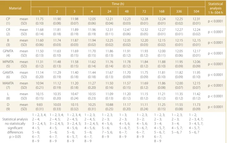
Table 1 presents the means and standard deviations of hydroxyl ion release from the materials at different peri-ods of time. All testing materials used in our study had alkaline pH. Most of the tested materials, besides those marked at the bottom of Table 1, showed statistically sig-nificant differences in terms of pH values (p < 0.05). Anal-ysis of pH changes as a function of time showed a gradual rise in the pH of all materials. Differences in pH values between the first and last experimental period were sta-tistically significant (p < 0.001).
Based on the results obtained, the preparations were di-vided into groups relating to their alkaline abilities (Fig. 1). The greatest similarity in pH was found in the following groups: the first consisted of the most alkaline materials
such as CP, CR and B; the second was composed of 2 MTA groups: GPMTA, WPMTA and GAMTA, WAMTA. The third group contained setting calcium hydroxide prepara-tions (L and D), which were the least alkaline.
The highest pH increase was observed for setting and the lowest for non-setting calcium hydroxide preparations.
Discussion
Release of hydroxyl ions from different dental mate-rials has been investigated by numerous authors.2,10–13
There are no uniform standards for pH assays. Two methods have been described for the evaluation of pH changes: potentiometry and titration curves.2,11–14 The
investigators have also used 2 experimental models: with or without medium replacement after each time period measurement.2,3,10,14–17 We applied the second method
in the present study. Only Liberman et al.18 used both
methods simultaneously. Additionally, the data obtained in the studies was different and not directly comparable because of the various other methodological approaches employed (quantity of sample, time intervals, type of ac-ceptor medium, method of extraction).
The main purpose of vital pulp therapy is to preserve pulp integrity and functionality. Previous reports have suggested that ionic dissociation of hydroxyl ions from contemporary preparations used in dental pulp
protec-Table 1. Mean and standard deviation pH values evaluated over time periods of the experiment
Material
Time (h) Statistical
analysis 1 h–504 h
1 2 3 4 24 48 72 168 336 504
CP (1) mean (SD) 11.75 (0.10) 11.90 (0.08) 11.98 (0.07) 12.05 (0.06) 12.21 (0.04) 12.23 (0.03) 12.28 (0.01) 12.24 (0.01) 12.25 (0.02) 12.31 (0.01) p < 0.0001 CR (2) mean (SD) 11.68 (0.14) 11.81 (0.18) 11.89 (0.19) 11.96 (0.19) 12.31 (0.11) 12.47 (0.06) 12.32 (0.05) 12.27 (0.01) 12.27 (0.01) 12.24 (0.02) p < 0.0001 B (3) mean (SD) 11.60 (0.06) 11.74 (0.03) 11.87 (0.03) 11.94 (0.02) 12.25 (0.02) 12.35 (0.02) 12.20 (0.03) 12.15 (0.02) 12.15 (0.01) 12,.15 (0.01) p < 0.0001 GPMTA (4) mean (SD) 11.50 (0.13) 11.63 (0.10) 11.69 (0.15) 11.70 (0.15) 11.86 (0.13) 11.91 (0.12) 11.93 (0.12) 12.00 (0.11) 12.05 (0.06) 12.17 (0.01) p < 0.0001 WPMTA (5) mean (SD) 11.31 (0.12) 11.48 (0.13) 11.58 (0.15) 11.62 (0.14) 11.76 (0.14) 11.78 (0.12) 11.84 (0.12) 11.88 (0.10) 11.95 (0.09) 12.06 (0.09) p < 0.0001 GAMTA (6) mean (SD) 11.14 (0.20) 11.29 (0.19) 11.40 (0.18) 11.44 (0.18) 11.67 (0.13) 11.70 (0.09) 11.75 (0.09) 11.81 (0.10) 11.82 (0.09) 11.95 (0.10) p < 0.0001 WAMTA (7) mean (SD) 10.94 (0.21) 11.12 (0.19) 11.20 (0.18) 11.27 (0.20) 11.50 (0.16) 11.57 (0.15) 11.69 (0.12) 11.86 (0.08) 12.00 (0.07) 12.15 (0.07) p < 0.0001 L (8) mean (SD) 10,15 (0.15) 10.35 (0.20) 10.47 (0.24) 10.55 (0.23) 11.09 (0.13) 11.20 (0.12) 11.15 (0.12) 11.21 (0.12) 11.35 (0.12) 11.42 (0.12) p < 0.0001 D (9) mean (SD) 9.83 (0.31) 10.03 (0.33) 10.15 (0.32) 10.25 (0.31) 10.88 (0.25) 11.17 (0.20) 11.11 (0.24) 11.25 (0.15) 11.55 (0.08) 11.73 (0.09) p < 0.0001 Statistical analysis no statistically significant differences p > 0.05
1 – 2, 3, 4; 2 – 4; 3 – 2, 4, 5; 4 – 5; 5 – 6; 6 – 7; 8 – 9
1 – 2, 3, 4; 2 – 4, 5; 3 – 2, 4, 5; 4 – 5; 5 – 6; 6 – 7; 8 – 9
1 – 2, 3, 4; 2 – 4, 5; 3 – 2, 4, 5; 4 – 5, 6; 5 – 6; 6 – 5, 7; 8 – 9
1 – 2, 3; 2 – 4, 5; 3 – 2, 4, 5; 4 – 5, 6; 5 – 6; 6 – 7; 8 – 9
1 – 2, 3; 2 – 3; 4 – 5, 6; 5 – 6; 7 – 5, 6; 8 – 9
1 – 3; 2 – 3; 4 – 5, 6; 5 – 6, 7; 6 – 7; 8 – 9
1 – 2, 3; 3 – 2; 4 – 5, 6; 5 – 6, 7; 6 – 7; 8 – 9
1 – 2, 3; 2 – 3; 3 – 4; 4 – 5, 7; 5 – 6, 7; 6 – 7; 8 – 9
1 – 2, 3; 2 – 3; 3 – 4; 4 – 5, 7; 5 – 6, 7
1 – 2; 2 – 3, 4, 7; 3 – 4, 5, 7; 4 – 5, 7; 5 – 6, 7
Fig. 1. pH changes of materials tested during the course of time of the experiment
tion is a major factor in their therapeutic action. Authors emphasize the importance of the vehicle in calcium hy-droxide dissociation. Aqueous solutions enable better dis-sociation and thus an increase in pH.10,13
In this study, we evaluated the long-term pH varia-tions of materials used in vital pulp therapy. The mate-rials tested were able to alkalize deionized water at all the tested time periods and reached the maximum rate of hydroxyl ion release at the final reading. The greatest increase in pH was observed in setting calcium hydrox-ide materials.
Just a few authors have examined different kinds of pulp capping agents under similar experimental conditions as those we applied in this study.10,15,19 However, there are
no papers comparing non-setting and setting calcium hy-droxide materials as well as MTA cements in a single trial. We showed that the highest pH level throughout the whole observation period was achieved for non-setting calcium hydroxide preparations. Zmener et al.19
demon-strated similar results in relation to that group of materi-als, which had yielded a permanent increase in pH for up to 30 days. In contrast, Tamburic et al.found maximum release of hydroxyl ions at 8 h of the experiment.20
More-over, studies in other experimental conditions confirmed an increase in the pH of these materials.12,21
In the present study, all 4 MTA materials showed al-kalizing abilities. Unfortunately, there are no reports comparing these identical MTA products simultaneously. Our results revealed no variations in hydroxyl ion release between white and gray MTA from the same commer-cial brand throughout the duration of the whole experi-ment. In contrast, Chng et al.demonstrated a significantly higher increase in the pH of white MTA in comparison with gray MTA after 70 min.16 In addition, our findings
revealed significant differences between GPMTA and GAMTA as well as between WPMTA and WAMTA. This can probably be attributed to variations in the composi-tion of different commercial MTA brands.
Setting calcium hydroxide cements exhibited continu-ous hydroxyl ion release resulting in a rise in pH until the end of our experiment. Liberman et al.18 and Gençay et
al.22 obtained similar results in shorter 2-hour and 7-day
studies. In contrast, Tamburič et al.indicated maximum pH of Dycal at 24 h with subsequent declines at 48 and 72 h.20 Liberman et al.examined the short-release rate
of OH– ions into aqueous solutions of 7 calcium
hydrox-ide-containing lining materials, with and without replac-ing the medium.18 In the first group, a decline in pH
val-ues was recorded, and in the second one an increase in pH values was observed, which caused accumulation of OH ions in the unchanged medium. Maintaining an alkaline pH of setting calcium hydroxide preparations, despite re-placement of the medium in long-term evaluations, was also noted by other authors.2,3,12,23
It should be remembered that extrapolation of in vitro investigations to a clinical situation has some limitations.
It is essential to emphasize the fact that there is a per-manent exchange of tissue fluids at the material interface that will reduce pH.3 The buffering effect of dentin also
has clinical significance.24 However, we can assume that
the use of materials with a prolonged alkaline effect may be more advantageous in terms of anti-inflammatory and mineralization activity.
Conclusions
In conclusion, we can say that, from the point of view of alkalizing properties, all of the preparations evaluated can be used in vital dental pulp treatment, but in view of the limitations of this study, further investigation into the conditions that mimic clinical situations is required.
References
1. Camargo SEA, Camargo CHR, Hiller KA, Rode SM, Schweikl H, Schmaltz G. Cytotoxicity and genotoxicity of pulp capping mate-rials in two cell lines. Int Endod J. 2009;42:227–237.
2. Duarte MAH, Martins CS, De Oliveira Cardoso Demarchi AC, De Godoy LF, Kuga MC, Yamahshita JC. Calcium and hydroxide release from different pulp-capping materials. Oral Surg Oral Med Oral
Pathol Oral Radiol Endod. 2007;104:e66–e69.
3. Abbasi J, Barkhordar RA. The pH variation in calcium hydroxide lin-ers. Quintessence Int. 1987;18:225–226.
4. Ferracane JL, Cooper PR, Smith AJ. Can interaction of materials with the dentin-pulp complex contribute to dentin regeneration?
Odontol. 2010;98:2–14.
5. Smith AJ, Murray PE, Sloan AJ, Matthews JB, Zhao S. Trans-dentinal stimulation of tertiary dentinogenesis. Adv Dent Res. 2001;15:51–54. 6. Smith AJ, Sheven BA, Takashi Y, Ferracane JL, Shelton RM, Coo-per PR. Dentine as a bioactive extracellular matrix. Arch Oral Biol.
2012;57:109–121.
7. Hauman CHJ, Love RM. Biocompatibility of dental materials used in contemporary endodontic therapy: A review. Part 1. Intracanal drugs and substances. Int Endod J. 2003;36:75–85.
8. Mohammadi Z, Dummer PM. Properties and applications of calci-um hydroxide in endodontics and dental tracalci-umatology. Int Endod J.
2011;44:697–730.
9. Okabe T, Sakamoto M, Takeuchi H, Matsushima K. Effects of pH on mineralization ability of human dental pulp cells. J Endod.
2006;32:198–201.
10. De Andrade Ferreira FB, De Almeida Rodriguez Silva E Souza P, Sampaio Do Vale M, De Moraes Ig, Granjeiro JM. Evaluation of pH levels and calcium ion release in various calcium hydroxide end-odontic dressings. Oral Surg Oral Med Oral Pathol Oral Radiol Endod.
2004;97:388–392.
11. Natale LC, Rodrigues MC, Xavier TA, Simoes A, De Souza DN, Braga RR. Ion release and mechanical properties of calcium silicate and calcium hydroxide materials used for pulp capping. Int Endod J.
2015;48:89–94.
12. Murray PE, Lumley PJ, Smith AJ, Ross HF. The influence of sample dimensions of hydroxyl ion release from calcium hydroxide prod-ucts. Endod Dent Traumatol. 2000;16:251–257.
13. Pacios MG, De La Casa ML, De Los Angelos Bulacio M, Lopez ME. Influence of different vehicles on the pH of calcium hydroxide pastes. J Oral Sci. 2000;46:107–111.
14. Camilleri J, Pitt Ford TR. Evaluation of the effect of tracer pH on the sealing ability if glass ionomer cement and mineral trioxide aggre-gate. J Mater Sci Mater Med. 2008;19:2941–2948.
15. Pawinska M, Skrzydlewska E. Release of hydroxyl ions from calci-um hydroxide preparations used in endodontic treatment. An Acad
Med Bial. 2003;48:145–149.
16. Chng HK, Islam I, Yap AUJ, Tong YW, Koh ET. Properties of a new root-end filling material. J Endod. 2005;31:665–668.
17. Duarte MAH, Midena RZ, Zeferino MA, et al. Evaluation of pH and calcium ion release of calcium hydroxide pastes containing differ-ent substances. J Endod. 2009;35:1274–1277.
18. Liberman R, Ben-Amar A, Lupo N. The rate of OH– ion release
from lining materials containing calcium hydroxide. J Oral Rehab.
1995;22:809–815.
19. Zmener O, Pameijr CH, Banegas G. An in vitro study of the pH of three calcium hydroxide dressing materials. Dent Traumatol.
2007;23:21–25.
20. Tamburič SD, Vuleta GM, Ogjanowič JM. In vitro release of calcium and hydroxyl ions from two types of calcium hydroxide prepara-tion. Int Endod J. 1993;26:125–130.
21. Ordinola-Zapata R, Monteiro Bramante C, García-Godoy F, et al. The effect of radiopacifiers agents on pH, calcium release, radiopacity, and antimicrobial properties of different calcium hydroxide dress-ings. Microsc Res Tech. 2015;78:620–625.
22. Gençay K, Seymen F, Selvi S, Kiziltan B. In vitro evaluation of pH changes induced by calcium hydroxide liners. Quintessence Int.
2004;35:560–562.
23. Gandolfi MG, Siboni F, Primus CM, Prati C. Ion release, porosity, solubility, and bioactivity of MTA Plus tricalcium silicate. J Endod.
2014;40:1632–1637.
24. Haapasalo HK, Siren EK, Waltimo TMT, Orstavik D, Haapasalo MTT. Inactivation of local root canal medicaments by dentine: An in vitro