ARTIGO ORIGINAL
The Experience of a Protocol for the Management of
Pediatric Minor Head Injury: A Three Years Longitudinal
Study
A Experiência duma Norma de Atuação no Traumatismo
Crânio-Encefálico Ligeiro em Idade Pediátrica: Estudo
Longitudinal de Três Anos
1. Serviço de Pediatria. Hospital Garcia de Orta. Almada. Portugal.
2. Departamento de Pediatria. Hospital de Santa Maria. Centro Hospitalar de Lisboa Norte. Lisboa. Portugal. 3. Serviço de Pediatria. Hospital dos Lusíadas. Lisboa. Portugal.
4. Serviço de Pediatria. Hospital de Cascais Dr. José de Almeida. Cascais. Portugal. 5. Serviço de Neurocirurgia. Hospital Garcia de Orta. Almada. Portugal.
Autor correspondente: Joana Matias. [email protected]
Recebido: 05 de fevereiro de 2017 - Aceite: 24 de julho de 2017 | Copyright © Ordem dos Médicos 2017
Joana MATIAS1, Sofia ALMEIDA2, Sofia FERRITO1, Ana Margarida QUEIROZ1, Ana DIAS ALVES3, Ana TAVARES4,
Andreia AMORIM5, Paulo CALHAU1, Isabel SARAIVA DE MELO1
Acta Med Port 2017 Oct;30(10):704-712 ▪ https://doi.org/10.20344/amp.8795
RESUMO
Introdução: Os traumatismos crânio-encefálicos são frequentes em Pediatria, habitualmente ligeiros e sem lesões intracranianas. A
tomografia computorizada crânio-encefálica é o exame de eleição, implicando radiação ionizante, não existindo consenso nas indica-ções nos traumatismos crânio-encefálicos ligeiros. Uma atitude expectante é uma opção adequada na maioria dos casos. Os autores pretenderam comparar os doentes internados e submetidos a tomografia computorizada crânio-encefálica com os doentes internados sem tomografia computorizada crânio-encefálica, inferindo a segurança da norma de atuação em vigor na nossa instituição.
Material e Métodos: Estudo analítico longitudinal retrospetivo, durante três anos, de doentes com menos de 15 anos admitidos na
Urgência Pediátrica e internados por traumatismos crânio-encefálicos ligeiros. Amostra organizada num grupo A (internados com to-mografia computorizada crânio-encefálica) e num grupo B (internados sem toto-mografia computorizada crânio-encefálica).
Resultados: Amostra de estudo constituída por 206 doentes: 81 (39%) grupo A e 125 (61%) grupo B. Sintomas, nomeadamente
vómi-tos, foram mais frequentes no grupo B (91% e 61% vs 75% e 35%, p < 0,05); hematoma epicraniano volumoso e afundamento ósseo no grupo A (11% e 12% vs 0%, p < 0,05). Realizou-se tomografia computorizada crânio-encefálica em 39% dos doentes estudados (crianças com sinais de alarme à observação ou evolução não favorável durante o internamento); 43% tinham alterações (29 doentes apresentavam fratura, 18 doentes apresentavam lesões intracranianas). Três doentes foram submetidos a neurocirurgia. Não registá-mos óbitos, reinternamentos ou sequelas neurológicas.
Discussão: As lesões intracranianas clinicamente significativas foram pouco frequentes. A atitude preconizada de vigilância hospitalar
das crianças e adolescentes com traumatismos crânio-encefálicos ligeiros sintomáticos, sem alterações significativas ao exame obje-tivo, não pareceu ter implicado riscos acrescidos.
Conclusão: A utilização criteriosa da tomografia computorizada crânio-encefálica permitiu reduzir o número de exames e a exposição
a radiação ionizante.
Palavras-chave: Criança; Lesões Encefálicas/diagnóstico por imagem; Tomografia Computorizada; Traumatismos Craniocerebrais
ABSTRACT
Introduction: Head injury is common in children, with mostly being minor and not resulting in intracranial injury. Computerized
tomography head scan is the preferred exam, but implies exposure to radiation; the indications for computerized tomography head scan in minor injuries are not consensual. An expectant approach is a good option in most cases. The aim was to compare the patients hospitalized and subjected to computerized tomography head scan with patients hospitalized but not subjected to computerized tomography head scan in order to assess the safety of our institution’s practice protocol.
Material and Methods: Analytical longitudinal retrospective study, during three years, including patients younger than 15 years of age
with minor head injury, admitted for in hospital surveillance through a paediatric emergency room. We defined two study groups: group A (hospitalized with computerized tomography head scan) and group B (hospitalized without computerized tomography head scan).
Results: Study sample consisting of 206 patients: 81 (39%) group A and 125 (61%) group B. Symptoms, including vomiting, were
more frequent in group B (91% and 61% vs 75% and 35%, p < 0.05); large scalp hematoma and palpable fracture in group A (11% and 12% vs 0%, p < 0.05). We performed computerized tomography head scan in 39% of the study patients (children with red flags in the physical examination or unfavourable course during hospitalization); 43% had traumatic brain injury (29 patients had fracture, 18 patients had intracranial injury). Three patients underwent neurosurgery. We did not register deaths, readmissions or neurologic sequelae.
Discussion: Significant intracranial injury was infrequent. The hospitalization and surveillance of children and adolescents with
symptomatic minor head injury, without red flags in the physical examination, did not seem to result in additional risks.
Conclusion: The careful selection of patients for computerized tomography head scan enabled a decrease in the number of these
exams and the exposure to ionizing radiation.
ARTIGO ORIGINAL INTRODUCTION
Traumatic brain injuries (TBI) are frequent and poten-tially severe events in childhood and a public health issue1-3
with a real incidence that is difficult to be determined.4-6
Recent data published by the National Institute for Health and Care Excellence showed that children aged under 15 years are involved in 33-50% of the 1,4 million admissions to emergency and of the 200,000 hospital admissions for TBI occurring annually in the UK.7,8 The incidence of TBI in
Portugal is currently unknown while a 137 per 100,000 rate has been found between 1996 and 1997, all age groups involved.5,6
Between 75 and 90% of the patients with TBI having at-tended paediatrics emergency with mild TBI (i.e., Glasgow coma scale (GCS) scores ≥ 13).2,4,7,9,10 Low morbidity and
mortality rates have been reported and deadly and compli-cated events usually occur in patients presenting with mod-erate or severe TBI, related to clinically-important traumatic brain injury (ci-TBI).4,7,11
Computed tomography (CT) scanning of the head/ brain is the main imaging test used for the identification of ci-TBI.11-17 The indications for a CT head scan in patients
with mild TBI is the current challenge.3,8,16,18 A very low
in-cidence of ci-TBI has been found in patients with mild TBI, mainly considering the presence of any damage with the need for an urgent surgical approach.3,9,18-21 In addition, the
deleterious and cumulative effects of radiation exposure in childhood must be taken into consideration, involving an increased risk of cancer (leukaemia and brain tumours), global developmental delay, endocrine disorders and moy-amoya disease. Therefore, careful management of neuro-imaging is crucial in this population.16,22-25
Different clinical algorithms for the use of CT head scan-ning in children with mild TBI have been recommended by different population-based studies such as PECARN ( Pedi-atric Emergency Care Applied Research Network, carried out in North-America), CHALICE (Children’s Head Injury Algorithm for the Prediction of Important Clinical Events, in
the United Kingdom) and CATCH (Canadian Assessment of Tomography for Childhood Head Injury, in Canada) over the past few years.8,11,16,19,24-28 Recommendations have not been
uniform, though.13,18,20,21,28 CT head scan is recommended (i)
by the PECARN study in children aged < 2 years with mild TBI and presenting with GCS scores ≤14, palpable skull fracture, scalp haematoma in occipital, parietal or tempo-ral regions, ≥5 second loss of consciousness, severe injury mechanism or suspected ill-treatment, as well as in chil-dren aged ≥ 2 with mild TBI, GCS scores <14, with signs of basilar skull fracture, severe symptoms at admission (loss of consciousness, vomiting or headache) associated with worsening symptoms or signs during emergency depart-ment observation and by physician or parental preference,11
(ii) by the CHALICE study in patients with mild TBI and >5 minute loss of consciousness, > 5 min amnesia, drowsi-ness, vomiting ≥3 times, suspicion of non-accidental injury, seizures, GCS score <14 (or <15 in children aged under 1 year), penetrating or depressed skull fracture suspected or
tense fontanelle, signs of basilar skull fracture, focal neuro-logical deficits, > 5cm scalp haematoma or contusion in chil-dren less than one year of age or high-impact mechanism of injury (high-speed car accident as a pedestrian, bicyclist or passenger, fall of more than 3 m or head struck by a high-speed object)25 and (iii) by the CATCH study in patients
with mild TBI, GCS score <15 at 2 hours after injury, sus-pected open or depressed skull fracture, history of worsen-ing headache, irritability on examination, any sign of basilar skull fracture, large haematoma of the scalp or high-impact mechanism injury (motor vehicle crash, fall from elevation ≥ 0.91 m or fall from a bicycle with no helmet).19
Recent studies showed that the presence of drowsi-ness, vomiting or headache is not associated with higher probability of ci-TBI, mainly when these were transitory and self-limited.3,13,17,30,33,34
Portuguese scientific literature is still scarce in this area. A guideline aimed at the approach to mild TBI is in use since 2004 in our hospital, with the recommendation for hospital admission and monitoring of any patient presenting with certain red-flag signs at the initial medical examination, as an alternative to the immediate use of a CT head scan (Table 1).
This study aimed at the analysis of a group of children and adolescents admitted to a polyvalent paediatric emer-gency department with mild TBI. The comparison of clini-cal characteristics and progression of in-patients submitted
vs. those non-submitted to a CT head scan was the study’s main endpoint, showing the safety of complying with the current guideline. The evaluation of the frequency of skull fracture and/or ci-TBI and the evaluation of the use of head X-ray imaging in hospitalised patients with mild TBI were the secondary endpoints.
MATERIAL AND METHODS
This was a three-year (1 Jan 2011 to 31 Dec 2013) ana-lytical retrospective longitudinal study of a group of patients aged under 15 years attending the paediatric emergency department of a level-II hospital with polyvalent emergency (including a neurosurgery department) and admitted with mild TBI for monitoring at the Emergency Short-Stay Unit (ESSU).
Mild TBI has been defined as a blunt head trauma with preserved or mildly depressed level of consciousness (GCS score 13-15) at the initial medical examination. In line with the PECARN study, a ci-TBI has been defined as the pres-ence of life-threatening brain damage, with an indication for surgery, associated with the use of invasive ventilation for over a 24-hour period or associated with a long hospital stay (at least two nights) for clinical or imaging monitoring.11
ARTIGO ORIGINAL
involving the following recommendations:
• Admission to the ESSU for clinical monitoring (monitoring of heart rate, blood pressure and GCS score) and analgesia in patients with mild TBI and GCS score 13-14, < 5-min loss of consciousness, amnesia, persistent vomiting or headache. • CT head scan in patients with mild TBI and > 5-min
loss of consciousness, neurological focal deficits, post-TBI seizures, indirect signs of skull fracture, head X-ray showing signs of fracture or clinical worsening during monitoring. The presence of any large haematoma of the scalp (mainly in children under the age of 2), bilateral periorbital or retro-auricular haematoma, presence of cerebrospinal fluid leakage from the nose or ear, otorrhagia or haemotympanum and the presence of depressed skull are considered as indirect signs of head frac-ture.
• Head X-ray in children under the age of 2 with mild TBI and presenting with a large epidural haema-toma.
• Discharge home without any imaging test and with an indication for monitoring of red-flag signs in pa-tients with TBI related to low-impact mechanism, GCS score of 15 at admission, haemodynamic stability, normal neurological examination and no indirect signs of skull fracture.
The following study inclusion criteria were considered: patients with a TBI admitted to the ESSU in our hospital during the study period. The presence of GCS score ≤ 12 at the initial hospital examination, patient referral from another hospital for neurosurgery examination, personal history of
chronic neurological disorder, blood dyscrasia and the use of anticoagulant or antiplatelet drugs were defined as exclu-sion criteria from the study.
Patient identification was based on the ESSU clini-cal database (computer record including personal record number, identification and diagnosis regarding admitted pa-tients). Data have been collected from hospital computer clinical records (involving the analysis of emergency epi-sodes of TBI and any subsequent emergency episode and/ or outpatient visit in order to rule out any re-admission and the presence of neurological complications in patients hav-ing at least once subsequently attended our hospital). All information regarding patients with TBI involving no admis-sion to the ESSU was obtained from the hospital’s patient management database.
Our group of patients was divided into two groups: in-patients having (group A) and not having been submitted to a CT head scan (group B).
Demographic (gender and age) and clinical variables were analysed: injury mechanism (fall from one’s own height, fall of < or > 3 m in height, pedestrian struck by a vehicle, traffic accident as bicyclist, as a passenger or other injury mechanism); location of scalp haematoma (frontal, parietal, temporal, occipital or ≥ 2 locations); post-TBI symp-toms (vomiting, sleepiness, < or > 5-min loss of conscious-ness, amnesia, dizziconscious-ness, vision deficit or disorientation); abnormalities in initial examination (< or > 5 cm epidural haematoma, depressed skull, bruises or neurological focal deficits).
Imaging data were also recorded, including head X-ray imaging and any abnormality found (presence or absence of signs of fracture), CT head scanning and abnormality
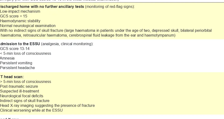
Table 1 – Clinical guideline in use at the paediatric emergency department in our hospital for the approach to patients with mild traumatic brain injury (i.e. with GCS scores 13-15 in the initial medical examination)
Discharged home with no further ancillary tests (monitoring of red-flag signs):
- Low-impact mechanism - GCS score = 15 - Haemodynamic stability - Normal neurological examination
- With no indirect signs of skull fracture (large haematoma in patients under the age of two, depressed skull, bilateral periorbital haematoma, retroauricular haematoma, cerebrospinal fluid leakage from the ear and haemotympanum)
Admission to the ESSU (analgesia, clinical monitoring):
- GCS score 13-14
- < 5-min loss of consciousness - Amnesia
- Persistent vomiting - Persistent headache
CT head scan:
- > 5-min loss of consciousness - Post-traumatic seizure - Suspected ill-treatment - Neurological focal deficits - Indirect signs of skull fracture
- Head X-ray imaging suggesting the presence of fracture - Clinical worsening while at the ESSU
Head X-ray:
- Age under 2 years and large epidural haematoma
ARTIGO ORIGINAL
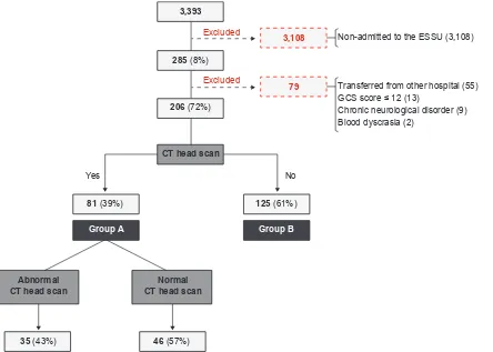
(8%) were admitted to the ESSU (Fig. 1). A total of 79 pa-tients (28%) were excluded from the initial group: 55 were referred to neurosurgery at another hospital, 13 presented with GCS score <13 (i.e. with no mild TBI), nine with a his-tory of chronic neurologic disorder and two patients with a history of blood dyscrasia.
A percentage of 98% of the patients who attended the paediatric emergency department over the study period due to head injury presented with mild TBI (3,314). A 6% admis-sion rate has been found in patients with mild TBI (n = 206). Apart from the patients admitted to the ESSU, 40 patients underwent a CT head scan at the paediatric emergency department and were not admitted (1.2% of the total num-ber of patients with mild TBI), according with the protocol in use at the department. In total, 121 of the patients with mild TBI (3.6%) examined at the paediatric emergency over the study period underwent a CT head scan.
A final group of 206 patients was included in the study and patients were subsequently ranked into two groups (Fig. 1): in-patients submitted (group A, n = 81) and non-submitted to CT head scan (group B, n = 125). The demo-graphic, clinical and imaging data of our group of patients are shown in Table 2.
Considering the total group of patients, 61% (126 / 206) were male and a median age of 6.5 years has been found (range 11 days – 14 years and 11 months), with a mean of found (head fracture, epidural haematoma, subdural
hae-matoma or cerebral contusion), as well as an indication of the unit where the patient was admitted to (ESSU, Paediat-rics ward and/or PaediatPaediat-rics ICU), patient’s outcome (per-sistent or worsening symptoms, deterioration of the level of consciousness or new neurological signs), need for neuro-surgery approach, re-admission and presence of post-TBI neurological consequences (in patients with subsequent ex-amination at the emergency department or at outpatients). A univariate statistical analysis has been carried out by using Chi-square test for the comparison of categorical variables from two independent groups and Mann-Whitney test for the comparison of numerical variables from two independent groups. A significance threshold of 0.05 has been considered and the IBM® SPSS® Statistics 20.0 for
Windows® software has been used.
RESULTS
In total, 128,554 children and adolescents aged under 15 years were admitted to the Paediatric Emergency De-partment over the three-year study period. Approximately 3% of the total number of admissions (3,393) presented with TBI (unspecified head injury diagnosis: this was the generic term used by the encoder paediatricians who were responsible for the codification of patients admitted to the paediatric emergency department). A total of 285 patients
Figure 1 – Group of patients and study groups. In total, 3,393 patients were admitted to the paediatric emergency department over the study period diagnosed with unspecified traumatic brain injury.
ESSU: Emergency Short-Stay Unit
3,393
3,108
CT head scan
Normal CT head scan Abnormal
CT head scan
Group A Group B
Excluded
Yes No
Non-admitted to the ESSU (3,108)
Transferred from other hospital (55) GCS score ≤ 12 (13)
Chronic neurological disorder (9) Blood dyscrasia (2)
Excluded
79
285 (8%)
206 (72%)
81 (39%)
35 (43%) 46 (57%)
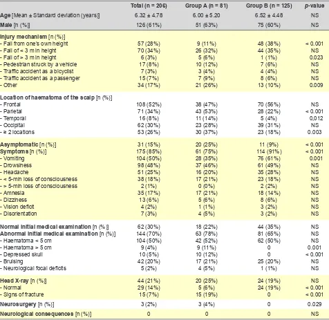
ARTIGO ORIGINAL 6.32 years (standard deviation: 4.78 years). Thirty-one per cent of the patients (64 / 206) were aged under 2 years.
No significant differences regarding patient’s gender or age group were found between both groups.
Falls of <3m in height (34%) and from one’s own height (28%) were the most frequently found injury mechanisms. Traffic road accidents (as pedestrian, bicyclist or passen-ger) corresponded to 18%. An 18-month old patient at-tended emergency due to having suffered from closed-fist injuries to the head, face and thorax (victim of ill-treatment). Falls from one’s own height were significantly more frequent in patients who did not undergo a CT head scanning [38% (48 / 125) in group B vs. 11% (9 / 81) in group A (p < 0.001)] while falls of >3m in height were significantly more frequent in patients who did not undergo a CT head scan (group A)
[6% (5 / 81) vs. 1% (1 / 81) in group B (p = 0.023)]; ‘other mechanisms’ were also significantly more frequent in the group of patients who underwent a CT head scan [26% (21 / 81) in group A vs. 10% (13 / 125) in group B (p = 0.009)]. Frontal scalp haematoma (52% of the patients) was most frequently found in patients with TBI, while parietal was significantly more frequent in group A patients (submit-ted to a CT head scan): 53% (43 / 81) vs. 22% (28 / 125) in group B (p < 0.001), such as temporal: 14% (11 / 81) in group A vs. 4% (5 / 125) in group B (p = 0.012) and ≥ 2 loca-tions: 37% (30 / 81) in group A and 18% (23 / 125) in group B (p = 0,003).
The presence of post-TBI symptoms was found in 85% of the patients: 50% presented with vomiting (104 / 206), 48% with sleepiness (98 / 206) and 25% with headache (51
Table 2 –Characteristics of our group of patients (patients with mild TBI admitted for monitoring to the ESSU) and both study groups: in-patients who underwent (group A) and who did not undergo a CT head scan (group B)
Total (n = 206) Group A (n = 81) Group B (n = 125) p-value
Age [Mean ± Standard deviation (years)] 6.32 ± 4.78 6.00 ± 5.20 6.52 ± 4.48 NS
Male [n (%)] 126 (61%) 51 (63%) 75 (60%) NS
Injury mechanism [n (%)]
- Fall from one’s own height - Fall of < 3 m in height - Fall of > 3 m in height - Pedestrian struck by a vehicle - Traffic accident as a bicyclist - Traffic accident as a passenger - Other 57 (28%) 70 (34%) 6 (3%) 17 (8%) 7 (3%) 15 (7%) 34 (17%) 9 (11%) 26 (32%) 5 (6%) 10 (12%) 3 (4%) 7 (9%) 21 (26%) 48 (38%) 44 (35%) 1 (1%) 7 (6%) 4 (4%) 8 (6%) 13 (10%) < 0.001 NS 0.023 NS NS NS 0.009
Location of haematoma of the scalp [n (%)]
- Frontal - Parietal - Temporal - Occipital - ≥ 2 locations
108 (52%) 71 (34%) 16 (8%) 62 (30%) 53 (26%) 38 (47%) 43 (53%) 11 (14%) 23 (28%) 30 (37%) 70 (56%) 28 (22%) 5 (4%) 39 (31%) 23 (18%) NS < 0.001 0,012 NS 0.003
Asymptomatic [n (%)]
Symptoms [n (%)]
- Vomiting - Drowsiness - Headache
- < 5-min loss of consciousness - > 5-min loss of consciousness - Amnesia
- Dizziness - Vision deficit - Disorientation 31 (15%) 175 (85%) 104 (50%) 98 (48%) 51 (25%) 38 (18%) 2 (1%) 35 (17%) 13 (6%) 4 (2%) 7 (3%) 20 (25%) 61 (75%) 28 (35%) 37 (46%) 16 (20%) 17 (21%) 0 (0%) 17 (21%) 5 (6%) 1 (1%) 4 (5%) 11 (9%) 114 (91%) 76 (61%) 61 (49%) 35 (28%) 23 (18%) 2 (2%) 18 (14%) 8 (6%) 3 (2%) 3 (2%) < 0.001 < 0.001 0.001 NS NS NS NS NS NS NS NS
Normal initial medical examination [n (%)]
Abnormal initial medical examination [n (%)]
- Haematoma < 5 cm - Haematoma > 5 cm - Depressed skull - Bruising
- Neurological focal deficits
62 (30%) 144 (70%) 104 (50%) 9 (4%) 10 (5%) 42 (20%) 5 (2%) 18 (22%) 63 (78%) 42 (52%) 9 (11%) 10 (12%) 17 (21%) 4 (5%) 44 (35%) 81 (65%) 62 (50%) 0 0 25 (20%) 1 (1%) NS NS NS 0.001 < 0.001 NS NS
Head X-ray [n (%)]
- Normal - Signs of fracture
44 (21%) 29 (14%) 15 (7%) 20 (25%) 5 (6%) 15 (19%) 24 (19%) 24 (19%) 0 NS < 0.001 < 0.001
Neurosurgery [n (%)] 3 (2%) 3 (4%) 0 0.029
Neurological consequences [n (%)] 0 0 0 NS
ARTIGO ORIGINAL
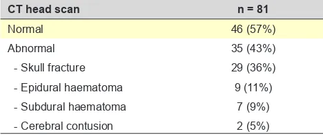
Table 3 – CT head scan obtained in group A patients (in-patients with mild TBI who underwent a CT head scan) and major abnor-malities found
CT head scan n = 81
Normal 46 (57%)
Abnormal 35 (43%)
- Skull fracture 29 (36%)
- Epidural haematoma 9 (11%)
- Subdural haematoma 7 (9%)
- Cerebral contusion 2 (5%)
/ 206). The presence of symptoms was significantly more frequent in in-patients who were not submitted to a CT head scan: 91% in group B (114 / 125) vs. 75% (61 / 81) in group A (p < 0.001), namely the presence of vomiting: 61% in group B (76 / 125) vs. 35% (28 / 81) in group A (p = 0.001). The presence of abnormalities in the initial medical ex-amination has been found in 70% of the patients: an epi-dural haematoma was mostly found, in 55% (113 / 206) of the patients. The presence of a >5 cm diameter haematoma was significantly more frequent in the group of patients who were submitted to a CT head scan: 11% in group A (9 / 81)
vs. 0% (0 / 125) in group B (p = 0.001); the presence of de-pressed skull fracture was also more frequent in group A pa-tients: 12% (10 / 81) vs. 0% (0 / 125) in group B (p < 0.001). Only five patients were found with neurological focal deficits in the initial examination, all of whom underwent a CT head scan, except an eight-year old child with dysmetria in the finger-to-nose test which improved over the first hour within the ESSU. Two patients with > 5-min loss of consciousness were not submitted to a CT head scan, as both showed a favourable clinical progression over the monitoring period. The patients who underwent a CT head scan (group A) were on average submitted to the test 4.95 hours upon the head injury event and neuroimaging abnormalities were found in 35 patients (43%; 35 / 81) (Table 3): skull fracture in 36% (29 / 81) and ci-TIB in 25% (20 / 81). The presence of ci-TIB was mainly found in patients with epidural haema-toma (9), subdural haemahaema-toma (7) and cerebral contusion (4). As regards abnormal CT head scans, 77% (27 / 35) were found in a group of children under the age of 2 (p <
0.001).
A total of 44 patients (21%; 44 / 206) underwent a head X-ray.
A total of 22 patients in this group were under the age of 2 and 17 presented with an epidural haematoma at the medical examination (one patient presented with a 5 cm haematoma) showing that the guideline criteria were met by 53% of the patients who underwent a head X-ray (23 / 44). The presence of signs suggesting a skull fracture was found in 34% (15 / 44) of the children who underwent an X-ray, which was confirmed in all these patients with the CT head scan. Six patients presented with an epidural haematoma and two patients with a subdural haematoma related to the fracture found in the CT head scan. In addition, two patients
with basilar skull fracture (both were ci-TBI-related) were not identified by the X-ray imaging and were only found in the CT head scan: an eight-year-old child who underwent a CT head scan due to worsening of vomiting during the period of clinical monitoring and a six-year-old child with an epidural haematoma at the examination who underwent a CT head scan due to an exuberant imaging; none of these patients presented with any clinical sign suggesting the presence of a basilar skull fracture.
A total of 190 patients (92%; 190 /206) from our group of patients were specifically admitted to the ESSU and re-mained for less than a 24-hour monitoring. A total of 16 pa-tients in group A with an abnormal CT head scan needed for a longer monitoring period: one patient admitted to the PICU (six-year-old child with fracture and epidural haema-toma with no indication for surgery) and 15 to the paediatric ward.
Three patients (1.5% of our group of patients and 0.1% of the total of attending patients with mild TBI over the study period) underwent cranioplasty: a five-month-old baby with a parietal TBI (fall of < 3m in height), < 5-min loss of con-sciousness, < 5cm epidural haematoma with depressed skull fracture and CT head scan showing an isolated pa-rietal fracture; 10-month-old baby with a frontal and pari-etal TBI (fall of < 3m in height), incoercible vomiting, normal examination and CT head scan showing a parietal fracture and epidural haematoma; asymptomatic 10-month-old-ba-by with a parietal TBI (direct injury against a hard surface) and signs of depressed skull fracture in the examination and CT head scan showing an isolated parietal fracture. These three patients showed a favourable outcome with no postoperative complications.
No deceased patients were found over the study period. Emergency episodes and post-TBI hospital attendances regarding over 95% of the patients were analysed and no patients with TBI were re-admitted to hospital or presented with any neurological consequences.
DISCUSSION
TBI was the most frequent reason for attending the pae-diatric emergency department over the study period and 98% of the patients presented with mild TBI (3,314 / 3,393). This was a higher percentage than those described in litera-ture (75 to 90% with mild TBI).2,4,7,9,10
Patients admitted to the ESSU with mild TBI were in-cluded into our group of patients, corresponding to 6% of all patients with TBI having attended the hospital over that pe-riod of time (206 / 3,393). In line with other studies, mostly male patients were involved and more than one quarter of the patients were under the age of two.1,4,13,14,18,29 Only 39%
(81 / 206) of these underwent a CT head scan.11,13,21
ARTIGO ORIGINAL by the clinical guideline in use at our department, those lo-cations were more frequently related to the presence of a
large haematoma and/or signs of depressed skull fracture in the medical examination and therefore with a higher risk for the presence of fracture, explaining for the use of a CT head scan. This may be explained by the mechanism of in-jury that is usually associated with these locations in a TBI: unsupported backwards falls and road traffic accidents, for instance.
The presence of symptoms was more frequent in pa-tients not submitted to a CT head scan (mainly with vomit-ing and headache). This difference is explained by the fact that the admission and monitoring of patients with symp-toms (as long as no red-flag signs existed at the medical examination) has been favoured rather than submitting the patients to any early neuroimaging assessment. These pa-tients showed a clinical improvement during monitoring and upon analgesia and therefore had no indication for a CT head scan. In fact, even though the presence of vomiting has been frequently found, different studies have suggested that this symptom is not a strong indicator of the presence of ci-TBI in children.3,13,17,30 The presence of headache is also
frequently found and often severe, persistent and difficult to manage.6,10,31,32,33 However, different studies have proven
that this symptom, in the absence of any other red-flag sign, is also not related to ci-TBI.33,34
In addition, the presence of any abnormality in medical examination mainly regarding large epidural haematoma (> 5cm) and signs of depressed skull fracture was associated with the use of a CT head scan, reflecting the compliance with the guideline in use at the paediatric emergency de-partment. Patients who were not submitted to a CT head scan presented with minimal abnormalities in the medical examination (nearly always bruising and small scalp hae-matomas). Different studies have shown that large haema-tomas and depressed skull fracture are risk factors for the presence of fracture and/or ci-TBI.3,13,19,35 In fact, these signs
are considered as red-flag signs by the three mostly used clinical algorithms for the approach to mild TBI in childhood (PECARN, CHALICE and CATCH).11,19,25
The fact that two patients presenting with >5-min loss of consciousness and one with neurological focal deficits at admission were not submitted to a CT head scan should be mentioned, as the guideline has not been complied with in these cases. Nevertheless, a favourable progression has been found, with no further neurological consequences. In addition, only half of the children having been submitted to a head X-ray had an indication for its use, according with the guideline. This appears to reflect old practices non-evi-dence based and not included in the current guideline. In total, 35 of the 81 patients who underwent a CT head scan (43%) and 17% of the patients in our study group (in-patients with mild TBI) (17%; 35 / 206) presented ei-ther with ci-TBI or skull fracture. The comparison between these results and those found in literature is not feasible due to the heterogeneous definition of the different study samples. Most studies were aimed at the determination
of the frequency of fracture and/or ci-TBI in patients with mild TBI who did not undergo a CT head scan (around 5% rate).18,30 Large international series, including PECARN and
CATCH studies also found lower frequencies of abnormal CT head scan when compared to our study. The presence of an abnormal CT head scan (intracerebral haemorrhage or contusion, cerebral oedema, post-traumatic cerebral infraction, diffuse axonal injury, sigmoid sinus thrombosis, midline shift of intracranial contents or signs of brain hernia-tion, diastasis of the skull, pneumocephalus and depressed skull fracture) has been found in 4-10% of the patients with mild TBI in PECARN study. The presence of abnormalities in CT head scan (epidural haematoma, cerebral contusion, pneumocephalus, subdural haematoma, diffuse cerebral oedema, cerebellar haematoma and intraventricular haem-orrhage) has been found in 4% of the patients with mild TBI in CATCH study.11,19 The higher frequency of abnormalities
found in our study reflects the careful criteria used in the selection of patients with an indication for neuroradiology imaging. Altogether, these results become more relevant in the absence of any re-admission or neurological con-sequence, at least in patients in whom the analysis of the emergency episode or subsequent medical examination in our hospital was available (over 95% of the total group of patients).
The presence of a skull fracture was the most frequently found abnormality in neuroimaging, in 36% (29 / 81) of the patients who underwent a CT head scan and in 14% (29 / 206) of the patients in the whole study group. A 22% rate of ci-TBI (18 / 81) has been found in patients who underwent a CT head scan and 9% in our group of patients (18 / 206). In line with other studies, most patients with abnormal CT head scan were aged under two years.9,14 The clinical
as-sessment is more difficult in this age group, there is a higher risk of skull fracture and TBI (even with mild TBI) and ci-TBI is often asymptomatic. In addition, there is a higher risk of ill-treatment in this age group.1,3,16
A head X-ray has been performed in a significant num-ber of patients, leading to a suspected fracture in 15 pa-tients, eight from which presented with ci-TBI in the CT head scan. Two patients presented with a basilar skull frac-ture and ci-TBI in the CT head scan, with normal X-ray. In fact, the interpretation of the head X-ray imaging is never an easy task and associated with scarce benefits for the initial assessment of mild TBI, due to the fact that any im-age suggesting the presence of a fracture does not allow for the identification of any ci-TBI and a normal X-ray does not rule it out.3,9,14 The usefulness of the head X-ray seems
scarce for the assessment of children and adolescents with TBI considering these data. Therefore, the use of a CT head scan is recommended in children under the age of two and with a large scalp haematoma. The head X-ray may be useful in institutions unable to perform a CT head scan.3,6,14
ARTIGO ORIGINAL
This percentage is lower than those described in literature (1 to 5%),3,11,13,30 showing a low risk for severe head injuries
in mild TBI affecting children.
There is still no consensually recommended duration for clinical monitoring in the hospital. This was individually tak-en into consideration, according with patitak-ent’s clinical pro-gression. Some studies have proven that a six-hour period would be enough, considering that clinically-important TBI would produce some manifestation throughout that period of time.15,16
Finally, the limitations of the study are worth mentioning. This was a retrospective study not always based on fully-completed computerized clinical records. The study sample was small and patients with mild TBI and who were not ad-mitted to hospital (the largest group of patients with such a diagnosis) were not analysed. Finally, the methodology that was used in the study, involving the search for any subse-quent emergency episode or medical examination in order to rule out any neurological consequence, despite most of the patients had subsequently at least once attended our department, does not allow for the identification of mild or late consequences in patients that did not re-attend our hospital.
Further prospective studies involving a larger group of patients and including subsequent neurological and neu-ropsychological assessment are crucial in order to rule out the presence of consequences, allowing for checking the safety of this conservative attitude and for establishing the optimal duration of the monitoring period at the hospital.
CONCLUSION
The presence of clinically-important brain injury related to mild TBI is not common. The careful use of a CT head scan in patients with mild TBI either with relevant abnormali-ties in the medical examination or unsolved symptoms while
staying at the hospital allowed for a higher percentage of patients diagnosed with clinically-important brain injury in the CT head scan, reducing any unnecessary exposure to radiation. No additional risk seems to have been related to the wait-and-see approach involving the clinical monitoring of children and adolescents with mild TBI presenting with symptoms and with no significant abnormalities in the medi-cal examination. The use of the head X-ray imaging was not useful in these patients.
ACKNOWLEDGMENTS
The authors wish to acknowledge António Gomes for his participation in the critical revision of the manuscript and to Paula Azeredo for her enthusiasm regarding the prepara-tion of the clinical guideline for mild TBI management in use at our hospital up to the present study.
HUMAN AND ANIMAL PROTECTION
The authors declare that the followed procedures were according to regulations established by the Ethics and Clini-cal Research Committee and according to the Helsinki Dec-laration of the World Medical Association.
DATA CONFIDENTIALITY
The authors declare that they have followed the proto-cols of their work centre on the publication of patient data. CONFLICTS OF INTEREST
The authors declare that there were no conflicts of inter-est in writing this manuscript.
FINANCIAL SUPPORT
The authors declare that there was no financial support in writing this manuscript.
REFERENCES
1. Atabaki SM. Pediatric head injury. Pediatr Rev. 2007;28:215-24. 2. Cassidy JD, Carroll LJ, Peloso PM, Borg J, Holst H, Holm L, et al.
Incidence, risk factors and prevention of mild traumatic brain injury: Results of the WHO collaborating centre task force on mild traumatic brain injury. J Rehabil Med Suppl. 2004;43:28-60.
3. Schutzman SA, Greenes DS. Pediatric minor head trauma. Ann Emerg Med. 2001;37:65-74.
4. Trefan L, Houston R, Pearson G, Edwards R, Hyde P, Maconochie I, et al. Epidemiology of children with head injury: a national overview. Arch Dis Child. 2015;0:1.6.
5. Santos ME, Sousa L, Castro-Caldas A. Epidemiologia dos traumatismos crânio-encefálicos em Portugal. Acta Med Port. 2003;16:71-6. 6. Oliveira E, Lavrador JP, Santos MM, Lobo Antunes J. Traumatismo
crânio-encefálico: abordagem integrada. Acta Med Port. 2012;25:179-92.
7. Head injury: triage, assessment, investigation and early management of head injury in children, young people and adults. NICE clinical guideline, January 2014. [accessed 2016 Mar 25]. Available from: https://www. nice.org.uk/guidance/cg176.
8. Pickering A, Harnan S, Fitzgerald P, Pandor A, Goodacre S. Clinical decision rules for children with minor head injury: a systematic review. Arch Dis Child. 2011;96:414-21.
9. Muñoz-Santanach D, Sainz de la Maza VT, Forster GE, Cubells LC. Ninos con traumatismo craneal leve en urgencias: inverted question markes necesaria la radiografia de craneo en pacientes menores de 2
anos? Neurocirurgia. 2014;25:149-53.
10. Kacperski J, Arthur T. Management of post-traumatic headaches in children and adolescentes. Headache. 2016;56:36-48.
11. Kuppermann N, Holmes JF, Dayan PS, Hoyle JD Jr, Atabaki SM, Holubkov R, et al. Identification of children at very low risk of clinically-important brain injuries after head trauma: A prospective cohort study. Lancet. 2009;374:1160-70.
12. Hennelly KE, Mannix R, Nigrovic LE, Lee LK, Thompson KM, Monuteaux MC, et al. Pediatric traumatic brain injury and radiation risks: a clinical decision analysis. J Pediatr. 2013;162:392-97.
13. Fundarò C, Caldarelli M, Monaco S, Cota F, Giorgio V, Filoni S, et al. Brain CT scan for pediatric minor acidental head injury. An Italian experience and review of literature. Childs Nerv Syst. 2012;28:1063-8. 14. Esteves I, Crispim J, Farela Neves J, Cunha F. Abordagem do
traumatismo craniano ligeiro na idade pediátrica: Neuro-imagem ou atitude conservadora? Acta Pediatr Port. 2009;40:197-202.
15. Schonfeld D, Fitz BM, Nigrovic LE. Effect of the duration of emergency department observation on computed tomography use in children with minor blunt head trauma. Ann Emerg Med. 2013;62:597-603.
16. Wing R, James C. Pediatric head injury and concussion. Emerg Med Clin N Am. 2013;31:653-75.
17. Bainbridge J, Khirwadkar H, Hourihan MD. Vomiting – is this a good indication for CT head scans in patients with minor head injury? Br J Radiol. 2012;85:183-6.
ARTIGO ORIGINAL
G, et al. Variation in utilization of computed tomography scanning for the investigation of minor head trauma in children: A Canadian experience. Acad Emerg Med. 2000;7:739-44.
19. Osmond MH, Klassen TP, Wells GA, Correll R, Jarvis A, Joubert G, et al. CATCH: A clinical decision rule for the use of computed tomography in children with minor head injury. CMAJ. 2010;182:341-8.
20. Nigrovic LE, Lee LK, Hoyle J, Stanley RM, Gorelick MH, Miskin M, et al. Prevalence of clinically important traumatic brain injuries in children with minor blunt head trauma and isolated severe injury mechanisms. Arch Pediatr Adolesc Med. 2012;166:356-61.
21. Mannix R, Meehan WP, Monuteaux MC, Bachur RG. Computed tomography for minor head injury: Variation and trends in major United States pediatric emergency departments. J Pediatr. 2012;160:136-9. 22. Rice HE, Frush DP, Farmer D, Waldhausen JH. Review of radiation
risks from computed tomography: essentials for the pediatric surgeon. J Pediatr Surg. 2007;42:603-7.
23. Mathews JD, Forsythe AV, Brady Z, Butler MW, Goergen SK, Byrnes GB, et al. Cancer risk in 680,000 people exposed to computed tomography scans in childhood or adolescence: Data linkage study of 11 million Australians. BMJ. 2013;21:1-18.
24. Tavor O, Boddu S, Kulkarni AV. Presenting characteristics of children who required neurosurgical intervention for head injury. Childs Nerv Syst. 2016;3.
25. Dunning J, Daly JP, Lomas JP, Lecky F, Batchelor J, Mackway-Jones K, et al. Derivation of the children’s head injury algorithm for the prediction of important clinical events decision rule for head injury in children. Arch Dis Child. 2006;91:885-91.
26. Lyttle MD, Crowe L, Oakley E, Dunning J, Babl FE. Comparing CATCH, CHALICE and PECARN clinical decision rules for paediatric head injuries. Emerg Med J. 2012;29:785-94.
27. Easter JS, Bakes K, Dhaliwal J, Miller M, Caruso E, Haukoos JS. Comparison of PECARN, CATCH, and CHALICE rules for children with minor head injury: A prospective cohort study. Ann Emerg Med. 2014;64:145-52.
28. Maguire JL, Boutis K, Uleryk EM, Laupacis A, Parkin PC. Should a head-injured child receive a head CT scan? A systematic review of clinical prediction rules. Pediatrics. 2009;124:145-54.
29. Samuel N, Jacob R, Elion Y, Mashiach T, Shavit I. Falls in young children with minor head injury: a prospective analysis of injury mechanisms. Brain Inj. 2015;29:946-50.
30. Alharthy N, Al Queflie S, Ayousef K, Yunus F. Clinical manifestations that predict abnormal brain computed tomography (CT) in children with minor head injury. J Emerg Trauma Shock. 2015;8:88-93.
31. Nigrovic LE, Schunk JE, Foerster A, Cooper A, Miskin M, Atabaki SM, et al. The effect of observation on cranial computed tomography utilization for children after blunt head trauma. Pediatrics. 2011;127:1067-73. 32. Eisenberg MA, Meehan WP, Mannix R. Duration and course of
post-concussive symptoms. Pediatrics. 2014;133:999-1006.
33. Dayan PS, Holmes JF, Hoyle J Jr, Atabaki S, Tunik MG, Lichenstein R, et al. Headache in traumatic brain injuries from blunt head trauma. Pediatrics. 2015;135:504-12.
34. Chelse AB, Epstein LG. Blunt head trauma and headache. Pediatr Neurol Briefs. 2015;29:30.
35. Burns EC, Grool AM, Klassen TP, Correll R, Jarvis A, Joubert G, et al. Scalp hematoma characteristics associated with intracranial injury in pediatric minor head injury. Acad Emerg Med. 2016;7.