195
DIVERSITY OF ROOT NODULE BACTERIA FROM LEGUMINOUS CROPS
Pooja Agrawal1 and Shruti Shukla2,*
1 Department of Microbiology and Biotechnology, Dr. H. S. Gour Vishwavidyalaya, Sagar 470002, MP, India
2Department of Food Science and Technology, School of Biotechnology, Yeungnam University, Gyeongsan, Gyeongbuk
712-749, Republic of Korea
*Corresponding author: [email protected]
Received: May 4, 2015; Revised: June 25, 2015; Accepted: June 26, 2015; Published online: December 23, 2015
Abstract:In the present study, a total of 353 nodule-associated bacteria were isolated from 220 legume plant samples
belonging to Cicer arietinum (85), Glycine max (74), Vigna radiata (21) and Cajanus cajan (40). A total of 224 bacteria were identified as fast-growing Rhizobium spp. on the basis of differential staining (Gram staining and carbol fuchsin staining) and biochemical tests. All the isolates were tested for indole acetic acid production (IAA), phosphate solubilization and siderophore production on plate assay. To examine the effect of volatile organic metabolites (VOM) and water soluble soil components (WSSC) on nodule bacteria, culture conditions were optimized by observing the effects of various parameters such as pH, salt content and temperatures on the growth of bacteria. Selected rhizobia were subjected to random amplified polymorphic DNA (RAPD) and amplified ribosomal DNA restriction analysis (ARDRA) analysis to identify their species. On the basis of RAPD and ARDRA, 10 isolates were identified as Rhizobium meliloti. In this study, Rhizobium GO4, G16, G20, G77, S43, S81, M07, M37, A15 and A55 were observed as the best candidates among the tested bacteria and can be further used as potent bioinoculants.
Key words: legumes; Rhizobium; RAPD; ARDRA; rhizobacteria
INTRODUCTION
Rhizobium is a well-known symbiotic nitrogen fixer and hence the species belonging to this genus are widely used as biofertilizers and bioinoculants. Rhizobium species are also reported as a source of various biologically active compounds such as poly-β-hydroxybutyrate, indole acetic acid and siderophore (Ilic et al., 2007). Antifungal and antibacterial com-pounds including other plant growth promoting substances have also been developed from Rhizo-bium species for sustainable agricultural uses (Ilic et al., 2007). In recent years, researchers are focusing on co-inoculation of PGPR and Rhizobium and it is becoming the most popular approach for improving the growth of economically important legumes. Plant growth-promoting rhizobacteria (PGPR) may increase the efficiency of Rhizobium inoculation in legumes through the production of antibiotics, siderophore
prob-lem, statistical experimental designs have been sug-gested for an optimization strategy by other research-ers (Kim et al., 2005; Lee and Gilmore, 2005).
In the present study, we isolated, identified and characterized various isolates of Rhizobium spp. In order to ensure the survival of soil microorganisms, a number of soluble components including organic me-tabolites are produced, which may affect soil microbial populations by inhibiting or stimulating the growth of other microorganisms. Therefore, an attempt has also been made to evaluate the effect of water soluble organic compounds of soil on the survival and growth of selected isolates of nodule bacteria.
MATERIALS AND METhODS Selection and collection of crop plants
Four legume crop plants were selected for the isolation of rhizobia from their root nodules: Cicer arietinum (Chick pea), Glycine max (Soybean), Vigna radiata (Moong) and Cajanus cajan (Arhar), collected from six different sites in the Sagar district (Madhya Pradesh), India. A total of 85, 74, 21 and 40 samples of Cicer arietinum, Glycine max, Vigna radiata, Cajanus cajan, respectively, were collected for the isolation of rhizobia.
Surface sterilization
The surface of root nodules was sterilized with 1% HgCl2 for about 5 min and the root nodules were washed with sterile distilled water and placed in 70% ethanol for 3 min followed by washing with sterile distilled water. Preparations of root nodule suspen-sions followed according to the modified methods referred to previously (Subba Rao, 1995; Agrawal and Jain, 2008; Agrawal and Jain, 2009).
Isolation of bacteria
For the isolation of root-nodulating bacteria, the root nodule suspension was serially diluted to a concentra-tion of 10-4 in sterile distilled water and streaked on yeast extract mannitol agar (YEMA) plates (Vincent,
1970). In order to reduce the risk of isolating con-taminants, Congo red was added to the YEMA plates. Plates were incubated at 30°C for 3 days. Well grown colonies were taken and purified by streaking on fresh slants containing YEMA (Agrawal and Jain, 2009).
Characterization of bacterial isolates
The morphological, biochemical and physiologi-cal characteristics of the authenticated isolates were previously studied by our research group (Agrawal, 2012). Gram-staining reaction was carried out using a loop-full of pure culture grown on YEM broth and stained as per the standard Gram procedure. The puri-fied isolates were characterized based on the shape of bacterial cells, i.e., rod shapes in single, paired chains and dense clusters that stained Gram-negative were considered rhizobia. Further, carbol fuchsin staining was carried out and the cells were characterized by their shape and color, and dark purple rounded bodies were considered as rhizobia.
Biochemical examination Ketolactose agar test
Lactose utilization by test bacteria was tested using YEMA medium. For this test, mannitol was replaced by lactose (10g/L) in the composition of the YEM agar (Subbarao, 1993). Similarly, the ketolactose test was also performed on the YEMA plates and overlaid with Benedict’s reagent to distinguish Agrobacterium and Rhizobium. The absence of a yellow zone after using Benedict’s reagent was considered for the presence of rhizobium. Other physiological and biochemical tests such as the catalase production test, oxidase test, cit-rate utilization, nitcit-rate reduction, motility test, growth on different salt concentrations and various pH condi-tions were also performed according to the previously described method (Agrawal et al., 2011).
Phosphate solubilization
Re-sults were previously discussed and reported by our research group (Agrawal et al., 2011).
Determination of siderophore production
Siderophore production by test rhizobia was deter-mined by chrome azurol sulfonate (CAS) assay as described by Schwyn and Neiland (1987). The test strains were then streaked over CAS agar medium and incubated for 3 days at 30ºC. After incubation, the formation of a yellow-orange halo zone around the colony was observed as an indication of siderophore production.
Determination of indole acetic acid (IAA) production
To determine the indole acetic acid (IAA) produc-tion by test bacteria, a mineral salt broth containing 0.5% glucose and 500 µg/mL tryptophan was prepared (Agrawal et al., 2011). The growth and culture of bac-teria were maintained on Luria Bertani (LB) broth, and incubated at 28°C for 24 h at 120 rpm. To obtain the cell-free supernatant, the culture from the expo-nential phase was centrifuged at 10000 g for 15 min at 4°C. For confirmation of IAA production by bacterial isolates, 2 drops of o-phosphoric acid were added to the supernatant of each bacterial isolate to generate a pink color as confirmation of IAA production.
Effect of volatile organic metabolites (VOM) of soil on the growth of rhizobia
To study the effect of volatile organic metabolites (VOM) of the soil on the growth of rhizobacteria, the test organisms were exposed to volatile emanations of different soils. The experiments were conducted using a set of 250-mL Erlenmeyer flasks with a side connecting tube. A set of two flasks was used for each test organism. In one flask, a soil sample (200 g) was taken and moistened with small amount of sterile water to activate microbial activity. These flasks were made air-tight and allowed to stand for 24 h at room temperature before connecting to culture flasks. The flasks were run as soil-filled volatile chambers. On the
other hand, the flasks of the second set were used for the cultivation of test rhizobacteria in culture broth. An aliquot (50 mL) of the broth of YEMA culture medium was taken in a culture flask and both open-ings cotton-plugged. Flasks were then autoclaved and inoculated with 0.2 mL of cell suspension of the test rhizobacteria. Inoculated flasks were then connected to the soil-filled volatile chambers and incubated for 48 h. A set of flasks was also run as control for each test organism. After 48 h at 35°C of incubation, the growth of bacteria in each test flask and control was noted in terms of increase or decrease in turbidity by the nephelometric method utilizing 1-100 NTU (nephelometric turbidity units) and 400 NTU stand-ard suspensions.
Effect of water soluble soil components (WSSC) on the growth of rhizobia
The effect of water soluble soil components (WSSC) was studied using soil extract in the growth medium. Briefly, 100 g of test soil sample was suspended in 300 mL of double-distilled water in a 500-mL flask and the flask was kept for 24 h. After 24 h, when the soil par-ticles settled to the bottom of the flask, approximately 50 mL of water was removed and centrifuged at 10000 rpm for 10 min. The supernatant was collected and filter-sterilized using a syringe filter. An amount of 2.5 mL filter sterilized soil solution was dispensed in each test flask containing 50 mL of YEMA broth. The flasks were then inoculated with 1 mL cell suspension of test rhizobacteria and incubated at 30°C for 48 h. A set of flasks was also run as a control with only YEMA broth for each test organism. The growth of each test organism in the control and test flasks was then deter-mined by turbidimetric methods as described earlier.
Optimization of conditions for the growth of rhizobium
Random amplified polymorphic DNA (RAPD)
RAPD finger printing was done followed by Sajjad et al. (2008) to assess diversity among root nodule rhizobiaisolated from four different crop plants.
Genomic DNA and template DNA preparation
The total genomic DNA and template DNA of Rhizo-bium strains were isolated following the method of Ivanova et al.(2000), with slight modifications. The rhizobia cultures were grown in yeast extract mannitol broth on a rotatory shaker at 125 rpm and 37ºC for 48 h. About 5 mL of actively grown rhizobial cultures were pelleted by centrifugation at 13000 rpm for 10 min at 4oC to harvest the cells. The cells were sus-pended in 500 μl of TEN buffer (50 mM Tris-HCl, 20 mM disodium EDTA, 50 mM NaCl, pH 8.0) and 0.5 mL of 1-butanol was mixed well. The contents were centrifuged at 6000 rpm for 5 min at 4°C and the supernatant was discarded. The cell pellets were then resuspended in 2 mL of TE buffer and again centrifuged at 6000 rpm for 5 min at 4°C to remove traces of butanol. The cell pellets covered with 1 mL of TE buffer and 100 µL lysozyme solution (10 mg/mL freshly prepared) and incubated at room temperature for 5 min. One hundred µL of 10% SDS and 25 µL of 100 µg/mL proteinase K were added, mixed well and incubated at 37°C for 1 h. After incubation, the cell lysates were combined with 200 µL of 5 M NaCl, 150 µL of CTAB (10% stock) and incubated at 65°C for 10 min. The cell lysate was deproteinized with 1 mL of phenol:chloroform mixture (24:1 v/v) and centrifuged at 6000 rpm for 10 min at 4°C. The aqueous layer was transferred carefully to new 2.0-mL microcentrifuge tubes. To this, 1/10 volume of sodium acetate and 600 µL of ice-cold isopropanol were added and incubated at -20˚C overnight. The precipitated DNA was sedi-mented at 12000 rpm for 15 min at 4°C. After discard-ing the supernatant, the pellet was allowed to dry for 30 min at room temperature and resuspended in 100 µL of TE buffer (pH 8.0). One µL of DNase free RNase (10 mg /mL stock) was mixed, incubated at 37°C and stored at -20°C for further use.
PCR amplification
PCR amplification of isolated template DNA was car-ried out with two oligonucleotides primers, GLA11-(5’CAATCGCCGT3’) and GLC19-(5’GTTGCCAGCC 3’), following the method as described by Sajjad et al. (2008). The amplification reactions were performed in an automated thermal cycler (Corbett, Germany), in 25 μl of reaction mixture containing 2.5 µL of 10 X buffer (pH 8.4), 3 µL of MgCl2 (25 mM), 1 μl of dNTPs mix (2.5 mM each), 2 μl primer (15 ng/µL), 2.5 μL of genomic DNA (15 ng/µL), 0.2 µL of (1U) of Taq DNA polymerase and 13.8 µL of deionized water. A negative control was maintained containing all com-ponents except template DNA. The reaction mixture was overlaid with two drops of mineral oil, incubated for 5 min at 95ºC for initial denaturation, and then amplified for 35 cycles consisting of 1 min at 94˚C, 1 min at 36˚C and 2 min at 72ºC followed by a final extension of 10 min at 72˚C.
Electrophoresis and pattern analysis
Ten mL of amplified product were loaded in 0.8% aga-rose gel with a 100-bp DNA ladder as a marker. After electrophoresis, the gel was stained with 0.5 µL/mL of ethidium bromide and photographed under UV light in a Transilluminator (Bangalore Genie, India). A culture of Rhizobium leguminosarum (MTCC-99) was used as a standard strain. The data are presented in binary code i.e., 0 for absence of band and 1 for presence of band, and analyzed using NTSYSpc 2.02i package.
Amplified rDNA restriction analysis (ARDRA)
Amplified ribosomal DNA restriction analyses was performed as follows:
PCR amplification of 16s rDNA genes
mix (2.5 mM each) and 0.5 μL of Taq DNA polymer-ase (3 U/ μL). The final volume was brought to 50 μL by adding sterilized distilled water. The temperature program for amplification was as follows: after initial denaturation at 94˚C for 3 min, the reaction mixture was run through 35 cycles of denaturation at 94°C for 1 min, annealing at 55˚C for 1 min, extension at 72°C for 1 min and with a final extension at 72°C for 10 min. Primers were derived from the conserved region present at the edges of the 16S rDNA(Weisburg et al., 1991). The primers were f D1 (5’-AGAGTTTGATC-CTGGCTCAG-3’) and r D1 (5’-AAGGAGGTGATC-CAGCC-3’). An aliquot of 5 μL of each PCR product was electrophoresed on 1% agarose gel in 1 X TAE buffer for 1 h at 90 V/cm and stained with ethidium bromide to confirm the amplification of DNA.
Restriction endonuclease digestion
Restriction digestion was carried out using three re-striction endonucleases − Hinf III, Hae II and Rsa I, at 37ºC for 3 h. Briefly, a 20-μL volume of reaction mix-ture was prepared with 10 μL 16 S rRNA gene product (amplified/ PCR product), 2 μL 10 X incubation buffer, 1 μL enzyme (10 U/ μL) restriction endonuclease, and 7 μL distilled water, and used for further steps.
Electrophoresis and pattern analysis
Restriction fragment patterns were analyzed by gel electrophoresis at 95 V/cm for 3 h in 2.5% agarose gel in 1 X TAE buffer. The gels were stained with 0.5 µL/mL of ethidium bromide. Gels were photographed using a digital camera system (Nikon, cool pI x 995, Japan) and the patterns were compared for the size of restriction fragments with a 100-bp DNA molecular marker (Bangalore Genei, India).
Cluster analysis
Multivariate analysis was conducted to generate a similarity matrix using NTSYSpc software version 2.02, based on unweighted pair-group method using arithmetic means (UPGMA) to estimate genetic dis-tance and relatedness of rhizobial strains.
RESULTS
Isolation, characterization and biochemical examination of rhizobia
In the present study, a total of 353 nodule-associated bacteria were isolated from 220 plant samples including 85 plant samples of Cicer arietinum, 74 samples of Gly-cine max, 21 samples of Vigna radiata and 40 samples of Cajanus cajan. These samples were collected from different localities of the Sagar district, MP, India fol-lowing the standard protocols. A total of 353 nodule bacteria were isolated, which included 125 isolates from Cicer arietinum, 106 from Glycine max, 58 from Vigna radiata and 64 from Cajanus cajan. Identification of rhizobia was done based on their Gram-staining re-action, presence of poly-β-hydroxy butyrate granules, motility, lactose agar test, growth on N2 free medium, oxidase and catalase test, and citrate utilization. The fol-lowing characters were considered typical for rhizobia and accordingly isolated bacteria were identified.
Phosphate solubilization
In all, 224 rhizobium isolates including 71 from Cicer arietinum, 70 from Glycine max, 43 from Vigna radia-ta and 40 from Cajanus cajan were used as test organ-isms. Phosphate solubilization activity was noted in 59 rhizobacteria of Cicer arietinum, 55 of Glycine max, 30 of Vigna radiata and 28 of Cajanus cajan. Phosphate solubilizing activity was found to be widely distributed in the strains of nodule rhizobia of all the test plants. Although some test strains including all the rhizobia isolated from C. arietinum and V. radiata and some isolates of G. max and C. cajan showed phosphate solubilization activity with the production of acid, 11 test rhizobia from C. cajan and 7 of G. max showed phosphate solubilization with the production of an alkaline reaction during the acid production test using bromothymol blue as indicator in YEMA.
Siderophore and IAA production
crop plants either directly or indirectly. The produc-tion of siderophore, phosphate solubilizing efficiency and plant growth hormones by inoculants and their growth stimulation by volatile organic metabolites (VOM) and water soluble soil components (WSSC) can be of special significance for crop productivity. The abovementioned properties of the rhizobia, se-lected on the basis of screening results of phosphate solubilizing potential, were studied in the present work. In all 10 rhizobium strains, including 4 isolates from C. arietinum (strains GO4, G16, G20 and G77), 2 from G. max (S43 and S81), 2 from V. radiata (M07 and M37) and 2 of C. cajan (A15 and A55) were stud-ied for abovementioned properties. Test Rhizobium sp. A15 showed maximum phosphate solubilization activity. All the other test strains showed 10-44 mg/ mL Ca3(PO4)2 solubilization. All these strains were found to produce siderophore when tested using CAS assay medium. The test strain Rhizobium sp. MO7 showed an excellent production of indole acetic acid (6.5916 µg/mL) in its culture grown on mineral salt broth containing 0.5% glucose and 500 µg/mL tryp-tophan (Table 1).
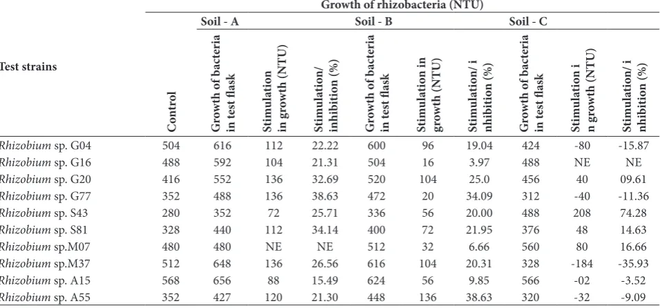
Effect of VOM and WSSC of different crop field soil on the growth of isolated rhizobacteria
To study the effect of VOM and water soluble com-ponents of soils, three different soil samples collected from different localities of the Sagar district were used. The VOM of two test soils (i.e., soil A and soil B) stim-ulated the growth of almost all test Rhizobium species. Table 1. Siderophore and IAA production by 10 root nodule iso-lates.
No. Strain number zone around the Yellow orange colony (mm)
Indole acetic acid (µg/mL)
1 Rhizobium sp. G04 1.82 3.594
2 Rhizobium sp. G16 2.6 5.4340
3 Rhizobium sp. G20 2.22 1.0200
4 Rhizobium sp. G29 1.0 0.196
5 Rhizobium sp. G30 - 0.120
6 Rhizobium sp. G45 - 1.011
7 Rhizobium sp. G75 1.12
-8 Rhizobium sp. G77 2 2.869
9 Rhizobium sp. G88 -
-10 Rhizobium sp. G98 -
-Table 2. Effect of Volatile organic metabolites (VOM) of different crop field soil on the growth of selected rhizobacteria isolated from root nodules of 4 crops.
Test strains
Growth of rhizobacteria (NTU)
C
on
tr
ol
Soil - A Soil - B Soil - C
G ro wth o f b ac te ri a in t es t fl as k St im ul at io n in g ro wth (NTU) St im ul at io n/ in hi bi tio n (%) G ro wth o f b ac te ri a in t es t fl as k St im ul at io n in gr ow th (NTU) St im ul at io n/ i nhi bi tio n (%) G ro wth o f b ac te ri a in t es t fl as k St im ul at io n i n g ro wth (NTU) St im ul at io n/ i nhi bi tio n (%)
Rhizobium sp. G04 504 616 112 22.22 600 96 19.04 424 -80 -15.87
Rhizobium sp. G16 488 592 104 21.31 504 16 3.97 488 NE NE
Rhizobium sp. G20 416 552 136 32.69 520 104 25.0 456 40 09.61
Rhizobium sp. G77 352 488 136 38.63 472 20 34.09 312 -40 -11.36
Rhizobium sp. S43 280 352 72 25.71 336 56 20.00 488 208 74.28
Rhizobium sp. S81 328 440 112 34.14 400 72 21.95 376 48 14.63
Rhizobium sp.M07 480 480 NE NE 512 32 6.66 560 80 16.66
Rhizobium sp.M37 512 648 136 26.56 616 104 20.31 328 -184 -35.93
Rhizobium sp. A15 568 656 88 15.49 624 56 9.85 566 -02 -3.52
Rhizobium sp. A55 352 427 120 21.30 448 136 38.63 320 -32 -9.09
+ Stimulation in growth, - inhibition in growth, NE − no effect on growth.
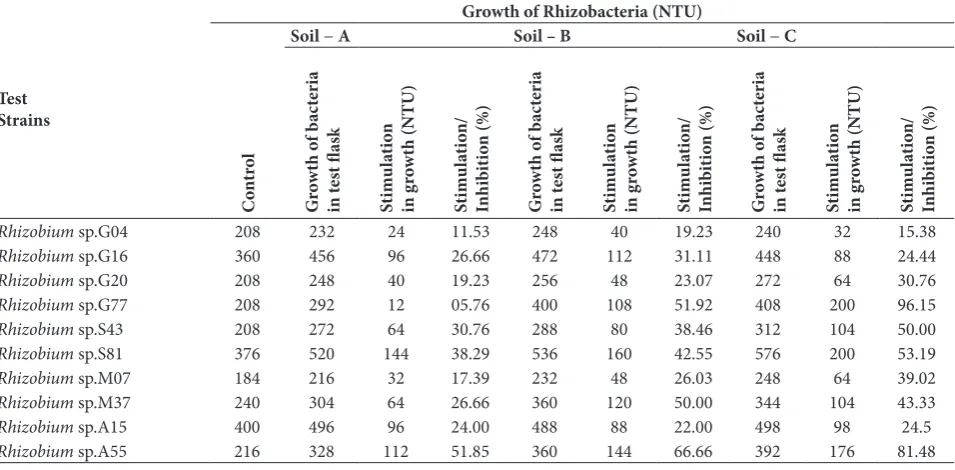
On the other hand, the VOM of soil C caused both stimulation and/or inhibition in the growth of test rhizobia. It is interesting that the WSSC of all the soil samples caused growth stimulation in all the tested rhizobacteria (Table 2 and 3).
Random amplified polymorphic DNA (RAPD)
Analysis of the 10 isolates revealed that the rhizo-bacteria produced several RAPD band patterns by using two oligonucleotides primers, GLA-11-5’CAATCGCCGT3’ and GLC-19-5’GTTGCCAGCC 3’. PCR amplification with these primers indicated that each primer template yielded distinct, easily detect-able bands of varidetect-able intensities (data not shown). Ten isolates generated 4 RAPD profiles when using the GLA-11 primer. The dendrogram obtained using NTSYSpc software showed total relatedness at 1.75 (Fig. 1a). Similarly, all the isolates generated 5 RAPD profiles when using the GLC-19 primer. In this case, the isolates showed total relatedness at 2.51 (Fig. 1b).
Amplified ribosomal DNA restriction analysis (ARDRA)
Test isolates of rhizobacteriawere subjected to PCR-RFLP with 16S rDNA genes using three restriction enzymes, Hinf I, Hae III and Rsa I. This resulted in separate restriction patterns for each enzyme (data not shown). The ARDRA produced fragments in the range of 100-900 bp. The number of bands detected was higher when the rDNA regions of test rhizobia were digested with Hae III (53 bands) compared to Hinf I (45 bands)and Rsa I (46 bands). A total of 144 bands were found in ARDRA. The relationship among test rhizobium isolates based on ARDRA is depicted in the dendogram (Fig 2a, 2b and 2c). The UPGMA cluster analysis shows 100% similarity at a coefficient of 3.15, 2.05 and 2.93 when Hinf I, Hae III and Rsa I restriction enzymes were used, respectively. Rhizobium species showed band patterns similar to the standard culture of rhizobacteriawhen digested with Hinf I,ranging from 100-700 bp (data not shown). 16S rDNA digested with Hea III showed bands of 100-250 base pairs (data not Table 3. Effect of Water soluble soil components (WSSC) of different crop field soil on the growth of selected rhizobacteria isolated from root nodules of 4 crops.
Test Strains
Growth of Rhizobacteria (NTU)
C
on
tr
ol
Soil − A Soil – B Soil − C
G ro wth o f b ac te ri a in t es t fl as k St im ul at io n in g ro wth (NTU) St im ul at io n/ In hi bi tio n (%) G ro wth o f b ac te ri a in t es t fl as k St im ul at io n in g ro wth (NTU) St im ul at io n/ In hi bi tio n (%) G ro wth o f b ac te ri a in t es t fl as k St im ul at io n in g ro wth (NTU) St im ul at io n/ In hi bi tio n (%)
Rhizobium sp.G04 208 232 24 11.53 248 40 19.23 240 32 15.38
Rhizobium sp.G16 360 456 96 26.66 472 112 31.11 448 88 24.44
Rhizobium sp.G20 208 248 40 19.23 256 48 23.07 272 64 30.76
Rhizobium sp.G77 208 292 12 05.76 400 108 51.92 408 200 96.15
Rhizobium sp.S43 208 272 64 30.76 288 80 38.46 312 104 50.00
Rhizobium sp.S81 376 520 144 38.29 536 160 42.55 576 200 53.19
Rhizobium sp.M07 184 216 32 17.39 232 48 26.03 248 64 39.02
Rhizobium sp.M37 240 304 64 26.66 360 120 50.00 344 104 43.33
Rhizobium sp.A15 400 496 96 24.00 488 88 22.00 498 98 24.5
Rhizobium sp.A55 216 328 112 51.85 360 144 66.66 392 176 81.48
+ Stimulation in growth, - inhibition in growth, NE − no effect on growth.
shown). When digested with Rsa I, bands ranging from 100-900 base pairs were obtained (data not shown).
The UPGMA-based dendrogram obtained by AR-DRA with Hinf I indicated three strains: M37, G04 and G20 in one cluster and 4 strains, A15, M07, G77 and A55 in another cluster, while other species showed varied band patterns. Using the restriction enzyme Hae III 6, test rhizobia fell in one cluster (i.e., isolates S81, G20, G77, A55, M07 and A55). Cluster analysis of the rhizobium isolates on the basis of ARDRA with Rsa I indicated 100% similarity at a coefficient of 1.46 with two distinct clusters, i.e., cluster I (G77, A55 and M07) and cluster II (G16 and G20), which showed 100% similarity with cluster III (S43, S81 and A15) at a coef-ficient of nearly 1.95 (Fig. 1 and 2).
Optimized conditions for the growth of rhizobia
Rhizobium sp. G16 showed potential to be developed as a bioinoculant and was thus selected for studies related to optimization of the best culture conditions in regard
Fig. 2. UPGMA-based dendogram showing cluster analysis of the Rhizobium isolates on the basis of random amplified ribosomal DNA restriction analysis (ARDRA) with [A], HinfI; [B], Hae III; and [C], Rsa I.
Fig. 1. UPGMA-based dendogram showing cluster analysis of the
to incubation period, pH of the medium and tempera-ture for growth. The best incubation period was found to be 3 days for this organism. The best temperature was found to be 37ºC and the organism was found to achieve its maximum growth, i.e. equivalent to NTU 497.6, in 3 days. The obtained data indicated growth of organisms at acidic pH (<7.0 pH) and at alkaline pH (>7.0 pH) while the optimum pH was found to be 7.0 in the present study (data not shown). The organ-ism achieved an increased growth under pH 7.0, tem-perature 37ºC and a 3-day incubation period. Under these culture conditions, the test strain Rhizobium sp. G16, an isolate of the root nodules of G. max, achieved growth equivalent to NTU 501.6. The results obtained during our investigation were of great fundamental and applied value. A number of rhizobial strains were collected which showed value-added properties such as the production of IAA and siderophore as well as the ability to solubilize phosphates.
DISCUSSION
In the present study, we studied the phosphate-solu-bilizing efficiency of root-nodulating bacteria isolated from four different legume crop plants. This prop-erty in Rhizobium inoculants may provide additional advantages to crop plants. Strains from the genera Pseudomonas, Bacillus and Rhizobium are consid-ered the most powerful phosphate solubilizers and efforts should be made to find N2-fixing bioinoculants with phosphate-solubilizing properties (Tambekar et al., 2009). In the present study, on the root nodule rhizobia of four legume crops properties such as acid production and phosphate solubilization were consid-ered basic desirable characteristics. Depending on the extent of phosphate-solubilizing capacity, 10 strains of Rhizobium were selected during screening studies and further studied to quantify this ability. Wani et al. (2007) reported that among phosphate-solubilizing (PS) bacteria, Bacillus PSB1 and Bacillus PSB10 pro-duced a clear halo zone 4 and 5 mm in size, respec-tively, on solid Pikovskaya medium.
Iron is another vital element required by vir-tually all living organisms including bacteria with
the exception of a few taxa (Posey and Gherardini, 2000). Microbial siderophores generally stimulate plant growth directly by increasing the availability of iron in the soil surrounding the roots. Marschner and Romheld (1994) reported that plants may also utilize siderophores synthesized by microorganisms present in the rhizosphere as they can be a source of soluble iron for the host plant. Strain-specific production of siderophore in Rhizobium species was first reported by Schwyn and Neilands (1987). Gaonkar et al. (2012) studied siderophore producing Bacillus sp. and Pseu-domonas aeruginosa produced a yellowish fluorescent siderophore identified as pyoverdine. According to Guerinot et al. (1990), variation in the production of siderophores by different strains of rhizobia may be a strain-specific characteristic. Although siderophore production is not essential for both nodule formation and nitrogen fixation, it may significantly increase the efficiency of nitrogen fixation. Siderophore-mediated competition for iron appears to be a widespread phe-nomenon in microbial systems. Arora et al. (2001) reported siderophore production by 2 out of 12 rhizo-bial isolates, i.e., RMP3 and RMP5,using CAS agar medium. Guerinot et al. (1990) first reported citric acid release in response to iron stress by a rhizosphere bacterium. The siderophore produced by the test strain 61A152 was found to be citric acid (Guerinot et al., 1990). Moreover, strain-specific production of siderophore has been observed by a number of other researchers (Ames-Gottfred et al., 1989).
avail-able to the plant; however, it depends greatly on the interactions between the plant and the microorgan-isms (Patten and Glick, 1992) and also on the inter-actions among the microorganisms. Datta and Basu, (2000) reported the production of 28.0 µg IIA/mL in a 72-h-old culture of Rhizobium strain 13 isolated from Cajanus cajan. They also reported maximum production of IAA in a glucose-containing medium by a rhizobium isolate. In addition, Qureshi et al. (2012) observed the improved yield of Vigna mungo by the co-inoculation of phosphate-solubilizing bacteria and Rhizobium in the presence of L-tryptophan, whereas Iqbal et al. (2012) found an improved yield of lentil by integrated use of Rhizobium and PGPR.
The past decades have witnessed an indiscrimi-nate use of chemical fertilizers to improve crop pro-ductivity; however, it has been realized that chemicals not only affect soil flora and fauna but also impose the threat of pollution to soil and water bodies, eventually leading to ecological imbalance. Therefore, microbial biofertilizers offer a suitable alternative to agro-chem-icals and chemical fertilizers. Rhizobium is one of the main symbiotic bioinoculants used. Many rhizobial species have been tested for enhanced crop productiv-ity and promising strains have been applied in field studies. However, the failure of available biofertiliz-ers to substantially increase productivity is posing a severe problem with regard to their practicability in agriculture. This study will lead to industrial and re-gional economic growth through the development of region-specific native strains of Rhizobium for their potential use as biofertilizers, which may enhance the productivity of different leguminous crops.
Acknowledgments: We are grateful to the Madhya Pradesh Council of Science and Technology, Bhopal for financial assist-ance, and to Prof. Anjana Sharma, R.D. University, Jabalpur, MP, India for the molecular studies carried out in her laboratory.
Authors’ contribution: PA and SS conceived and designed the experiments and analyzed the data. PA wrote the paper.
Conflict of interest disclosure: Authors declare that there is no conflict of interest.
REFERENCES
Agrawal, P. and P.C.Jain (2008). Isolation of native bioinoculant from the different legumes of district Sagar (M.P.) India. J. Curr. Sci.12, 799-804.
Agrawal, P., Arti, A. and P.C.Jain (2011). Microbiological study of root nodule bacteria from Wild legumes. Asian J. Biosci.
6, 74-78.
Agrawal, P. and P.C.Jain (2009). Study of native bioinoculants from the mung bean of district Sagar (M.P.) India. Int. J. Plant Sci. 4, 521-523.
Abdel, F., Yasser, R. and A.Olama Zakia (2002). L-asparaginase production by Pseudomonas aeruginosa in solid-state cul-ture: evaluation and optimization of culture conditions using factorial designs. Process Biochem. 38, 115-122.
Agrawal, P. and P.C. Jain (2012) Optimization of growth param-eters of Rhizobium meliloti strain G16. Search and Research Journal. 1, 64-67.
Agrawal, P. (2012). Molecular identification of root nodule bacte-ria from chickpea. Res. J. Biotechnol. 7, 73-79.
Ames-Gottfred, N.P., Christie, B.R. and D.C.Jordan (1989). Use of the chrome azurol S agar plate technique to differenti-ate strains and field isoldifferenti-ates of Rhizobium leguminosarum
biovar trifolii. Appl. Environ. Microbiol. 55, 707-710.
Arora, N.K., Kang, S.C. and D.K.Maheshwari (2001). Isolation of siderophore producing strains of Rhizobium meliloti and their biocontrol potential against Macrophomina phaseo-lina that causes charcoal root of groundnut. Curr. Sci. 81, 673-677.
Costacurta, A., Keijers, V. and J.Vanderleyden (1994). Molecular cloning and sequence analysis of an Azospirillum brasilense
indole-3-pyruvate decarboxylase gene. Mol. Gen. Gene.
243, 463-472.
Datta, C. and P.S. Basu (2000). Indole acetic acid production by a
Rhizobium species from root nodules of a leguminous shrub
Cajanus cajan. Microbiol. Res. 155, 123-127.
Garcia-Fraile, P., Carro, L., Robledo, M., Ramírez-Bahena, M.H., Flores-Felix, J.D., Fernandez, M.T., Mateos, P.F., Rivas, R., Igual, J.M., Martínez-Molina, E., Peix, A. and E.Velazquez
(2012). Rhizobium promotes non-legumes growth and quality in several production steps: towards a biofertiliza-tion of edible raw vegetables healthy for humans. PLoS One.
7, e0038122.
Guerinot, M.L., Meidi, E.J. and O.Plessner (1990). Citrate as a siderophore in Bradyrhizobium japonicum. J. Bacteriol. 172, 3298-3303.
Ivanova, E.G., Doronina, N.V. and Y.A.Trotsenko (2000). Aerobic methylobacteria are capable of synthesizing auxins. Micro-biol. 70, 392-397.
Ilic, S.B., Konstantinovic, S.S., Todorovic, Z.B., Lazic, M.L., Veljkovic, V.B., Jokovic, N. and B.C. Radovanovic (2007). Characterization and antimicrobial activity of the bioac-tive metabolites in streptomycete isolates. Mikrobiologiia.
Iqbal, M.A., Khalid, M., Shahzad, S.M., Ahmad, M., Soleman, N.
and N.Akhtar (2012). Integrated use of Rhizobium legumi-nosarum, plant growth promoting bacteria and enriched compost for improving growth, nodulation and yield of lentil (Lensculinaris medic). Chil.J. Agric. Res. 72, 104-110
Kim, H.M., Kim, J.G., Cho, J.D. and J.W.Hong (2003). Optimi-zation and characteriOptimi-zation of UV-curable adhesives for optical communications by response surface methodology.
Polym Test. 22, 899-906.
Lee, K.M. and D.F.Gulmore (2005). Formulation and process modeling of biopolymer (polyhydroxyalkanoates: PHAs) production from industrial wastes by novel crossed experi-mental design. Process Biochem.40, 229-246.
Lambrecht, M., Okon, Y., Broek, A.V. and J.Vanderleyden (2000). Indole-3-acetic acid: a reciprocal signaling molecule in bacteria-plant interactions. Trends Microbiol. 8, 298-300.
Marshnner, T. and V.Romheld (1994). Strategies of plant for acquisition of iron. Plant Soil. 165, 261-274.
Patten, C.L. and B.R. Glick (2002). Regulation of indoleacetic acid production in Pseudomonas putida GR12-2 by tryptophan and stationary-phase sigma factor RpoS. Can. J. Microbiol.
48, 635-642.
Pikovskaya, R.I. (1948). Mobilization of phosphorus in soil in connection with vital activity of some microbial species.
Mikrobiologiya. 17, 362-370.
Posey, J.E. and F.C.Gherardini (2000) Lack of a role of iron in the time disease pathogen. Science.288, 1651-1653.
Ravi Kumar, P. and M. Raghu Ram (2012). Production of indole acetic acid by Rhizobium isolates from Vigna trilobata (L) Verdc. African J. of Microbiol. Res. 6(27), 5536-5541
Sajjad, M., Malik, T.A., Arshad, M., Zahir, Z.A., Yusuf F. and S. Rahman (2008). PCR studies on genetic diversity of rhizo-bial strains. Int. J. Agric. Biol. 10, 1814-9596.
Subba Rao, N.S. (1995). Soil microorganism and plant growth, 3d rev Ed. New Delhi: Oxford & IBH Publishing Co.
Schwyn, B. and J.B.Neilands (1987). Universal chemical assay for the detection and determination of siderophores. Anal. Biochem.160, 47-56.
Tambekar, D.H., Gulhane, S.R., Somkuwar, D.D., Ingle, K.B., Kachalwar, S.W.P., Upadhye, M.A. and U.A.Bidwai (2009). Potential Rhizobium and phosphate solubilizers as a bio-fertilizers from saline Belt of Akola and Buldhara districts (India). Res. J. Agric. Biol. Sci.5, 578-582.
Wani, P.A., Khan, M.S. and A.Zaidi (2007). Co-inoculation of Nitrogen fixing and phosphate solubilizing bacteria to pro-mote growth yield and nutrient uptake in chickpea. Acta Agro. Hungaric.55, 315-323.
![Fig. 1. UPGMA-based dendogram showing cluster analysis of the Rhizobium isolates on the basis of Random Amplified Polymor-phic DNA using [A] GLA 11; and [B] GLC 19.](https://thumb-us.123doks.com/thumbv2/123dok_us/7821361.2087656/8.595.105.491.423.683/dendogram-showing-analysis-rhizobium-isolates-random-amplified-polymor.webp)
