Impaired left atrial reservoir function in
metabolic syndrome predicts symptoms in
HFpEF patients
Gëzim Berisha
1, Gani Bajraktari
1,2,3, Pranvera Ibrahimi
1,3, Ibadete Bytyçi
1,
Nehat Rexhepaj
1, Shpend Elezi
2, Michael Y. Henein
31. Clinic of Cardiology, University Clinical Centre of Kosovo, Prishtina, Republic of Kosovo 2. Medical Faculty, University of Prishtina “Hasan Prishtina”, Prishtina, Republic of Kosovo
3. Department of Public Health and Clinical Medicine, Umeå University and Heart Centre, Umeå, Sweden
Introduction
Metabolic syndrome (MetS) presents a significant group of risk factors for cardiovascular morbidity and mortality1-4 and constitutes a major public health problem with increasing incidence and prevalence, in recent decades5-6. MetS has been shown to be associated with structural and functional heart disturbances7-8, including left ventricular (LV) diastolic dysfunction9-10, coronary artery disease11-12 and arterial and ventricular stiffness13-14. Left atrial (LA) function15-16 has been reported to be abnormal due mainly to increased LV filling pressure, which is a reflection of LV cavity stiffness17-20, and is manifested in the form of increased LA volume21, and reduced systolic strain, reservoir and overall pump function15. These deteriorated heart function seems to be directly related to severity of MetS15. LV22-23 and LA24-25 functions are also known to be abnormal, in heart failure (HF) with preserved LV ejection fraction (HFpEF). However, the potential additional effect of MetS on such abnormalities in HFpEF remains unknown. The aim of this study, therefore, was to investigate the additional effect of MetS on LA reservoir function in HFpEF patients and its potential relationship with symptoms.
Methods
Study population
We studied 194 consecutive patients (mean age 62 ± 9 years, 54% female) with clinical diagnosis of HF, who were in New York Heart Association (NYHA) functional class I-III. Patients were referred to the Service of Cardiology, Internal Medicine Clinic, University Clinical Centre of Kosovo, between February 2013 and November 2013. At the time of the study all patients were on conventional medical treatment, optimized at least 2 weeks prior to enrollment. Based on patient’s symptoms and renal function, 88% were receiving ACE inhibitors or ARB, 66% beta-blockers, 45% diuretics and 23% Calcium channel antagonists. Of the studied cohort, 89% had hypertension, 29% diabetes and 25% smoked.
Patients with clinical evidence for severe cardiac
decompensation, with chronic renal failure with a stage >2 (glomerular filtration rate ≥ 89 mL/min), chronic obstructive pulmonary disease, recent acute coronary syndrome, stroke or anemia, were excluded. According to NCEP-ATP III criteria
Abstract
Background and Aim
The hospital readmission rate has been thought to reflect the quality of patient care. Understanding the risk factors for these can guide strategies to reduce them.
Methods
This study included 194 consecutive patients (age 62 ± 9 years) with stable HFpEF. LV dimensions, ejection fraction (EF), mitral annulus peak systolic excursion (MAPSE), myocardial velocities (s’, e’ and a’), LA dimensions and volumes were measured. Total LA emptying fraction (LA EF) was measured by Simpson rule volumes. Based on the NCEP-ATP III criteria, patients were divided into two groups; MetS (n=89) and non-MetS (n=105) and were compared with 34 age and gender matched controls.
Results
Age and gender were not different between patients and control neither between MetS and non-Met. LV dimensions, EF and longitudinal function indices were also not different. The MetS patients had higher LV mass index (p=0.038), lower septal and lateral e’ (p=0.003 and p=0.001, respectively) velocities, larger LA minimal volume (p=0.007) and lower LA EF (p<0.001) compared with the non-MetS patients. Age, LA EF and MetS independently predicted the NYHA class.
Conclusions
Despite no difference in LV systolic function, patients with HFpEF and MetS have worse LA emptying fraction, compared with HFpEF and non-MetS patients. In addition, LA reservoir function impairment and MetS independently predict patients limiting symptoms, thus add to a better understanding of HFpEF.
Key words: Metabolic syndrome, left atrial function, heart failure.
Citation: Berisha G., Bajraktari G., Ibrahimi P., et al. Impaired left atrial reservoir function in metabolic syndrome predicts symptoms in HFpEF patients International Cardiovascular Forum Journal. 2015;4: 37-42 DOI: 10.17987/icfj. v4i0.171
for metabolic syndrome, the presence of 3/5 of the following criteria classified the patients into MetS and non-MetS26: 1) abdominal obesity i.e. waist circumference ≥ 94cm for male and ≥ 80cm for female; 2) triglycerides level ≥1.7 mmol; 3) HDL cholesterol <1.0 mmol/L for male and <1.3 mmol/L for female; 4) raised blood pressure ≥130/85 mmHg or antihypertensive medications; and 5) insulin resistance/glucose intolerance (fasting plasma glucose ≥ 6.1 mmol/L). A total of 34 age- and gender matched healthy individuals (age 58±10 years), who served as controls, were also studied using the same Doppler echocardiographic protocol, as well as clinical and biochemical assessment. None of the controls had any cardiovascular risk or systemic disease. Patients and controls gave a written informed consent to participate in the study, which was approved by the local Ethics Committee.
Data collection
Detailed history and clinical assessment were obtained in all patients, in whom routine biochemical tests were also performed including lipid profile, blood glucose level, and kidney function tests. Estimated body mass index (BMI) was calculated from weight and height measurements. Waist, hip measurements were also made and waist/hip ratio calculated.
Echocardiographic examination
A single operator performed all echocardiographic examinations using a Philips Intelligent E-33 system (Philips Healthcare, The Netherlands) with a multi-frequency transducer, and harmonic imaging as appropriate. Images were obtained with the patient in the left lateral decubitus position and during quiet expiration. LV end-systolic and end-diastolic dimension measurements were made from the left parasternal long axis view with the M-mode cursor positioned by the tips of the mitral valve leaflets. LV volumes and emptying fraction were calculated from the apical 2 and 4 chamber views using the modified Simpson method. MAPSE was studied by placing the M-mode cursor at the lateral and septal angles of the mitral annulus which displayed the long axis motion in systole and diastole. Total amplitude of long axis motion (MAPSE) was measured
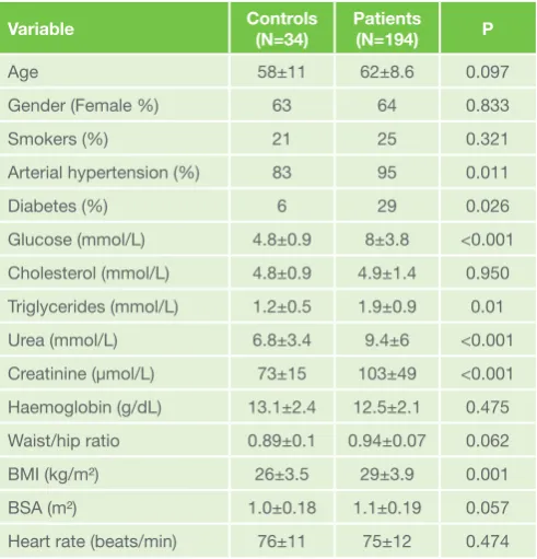
as previously described27 from peak inward to peak outward points. LV long axis myocardial velocities were also studied using Doppler myocardial imaging technique. From the apical 4-chamber view, longitudinal velocities were recorded with the Table 1: Comparison of clinical and biochemical data between
controls and patients
Variable Controls (N=34) Patients(N=194) P
Age 58±11 62±8.6 0.097
Gender (Female %) 63 64 0.833
Smokers (%) 21 25 0.321
Arterial hypertension (%) 83 95 0.011
Diabetes (%) 6 29 0.026
Glucose (mmol/L) 4.8±0.9 8±3.8 <0.001 Cholesterol (mmol/L) 4.8±0.9 4.9±1.4 0.950 Triglycerides (mmol/L) 1.2±0.5 1.9±0.9 0.01
Urea (mmol/L) 6.8±3.4 9.4±6 <0.001
Creatinine (μmol/L) 73±15 103±49 <0.001 Haemoglobin (g/dL) 13.1±2.4 12.5±2.1 0.475 Waist/hip ratio 0.89±0.1 0.94±0.07 0.062
BMI (kg/m²) 26±3.5 29±3.9 0.001
BSA (m²) 1.0±0.18 1.1±0.19 0.057
Heart rate (beats/min) 76±11 75±12 0.474
Table 2: Comparison of Echocardiographic data between controls and patients
Variable Controls (N=34) Patients(N=194) P
LV structure
IVSd (cm) 1.0±0.1 1.2±0.3 <0.001 LVPW (cm) 0.9±0.13 1.0±0.14 <0.001
LV EDD (cm) 4.9±0.5 5.0±0.7 0.113
LV ESD (cm) 3.2±0.6 3.3±0.6 0.114
LV EDV (ml) 112±26 120±34 0.146
LV ESV (ml) 41±13 47±20 0.070
LVMI (g/m2.7) 50±16 56±19 0.001
Systolic LV function
LV EF (%) 63±8 61±9 0.243
LV SF (%) 35±6 33±6 0.274
Aorta (cm) 3.1±0.4 3.4±0.4 0.003 S’septal (cm/s) 5.3±0.9 5.2±1.6 0.961 S’lateral (cm/s) 6.1±1.5 6.0±1.7 0.693 MAPSE lateral (cm) 1.5±0.2 1.3±0.3 0.002 MAPSE septal (cm) 1.4±0.2 1.1±0.3 <0.001 Diastolic LV function
A (cm/s) 59±21 73±21 <0.001
Lateral a’ (cm/s) 9.0±2.6 8.6±2.9 0.536
E/A ratio 1.1±0.3 0.9±0.5 0.022
Septal a’ (cm/s) 8.2±2.5 8.0±2.5 0.765
Septal e’ (cm/s) 7.5±2.4 5.8±2.2 0.022
E (cm/s) 61±16 60±22 0.643
Lateral e’ (cm/s) 10±4 7.4±3 0.022
FT (ms) 425±108 403±110 0.316
E wave DT (ms) 179±39 188±55 0.320
Global LV function
TIVT (s/min) 7.7±2.1 9.2±4.3 0.005
Tei index 0.37±0.1 0.47±0.3 0.001
E/e’ ratio 7.5±2.5 9.8±4.9 <0.001
Left atrium
LA diameter (mm) 39±5 42±6 <0.001
LA max volume (ml) 47±18 56±19 0.010 LA min volume (ml) 17±8 21±12 0.019
Total LA EF (%) 64±11 60±12 0.031
pulsed wave Doppler sample volume placed at the basal part of LV lateral and septal segments. Systolic (s’) and early and late (e’ and a’) diastolic myocardial velocities were measured with the gain optimally adjusted to obtain the thinnest velocity envelope thickness. Mean value of the lateral and septal LV velocities was calculated.
Diastolic LV function was assessed from filling velocities using spectral pulsed wave Doppler with the sample volume positioned at the tips of the mitral valve leaflets, during a brief apnea. Peak LV early (E wave), and late (A wave) diastolic velocities were measured and E/A ratio was calculated. The E/e’ was also calculated as the ratio between transmitral E wave and mean lateral and septal e’ wave velocities. The isovolumic relaxation time was measured as the time interval between aortic valve closure and mitral valve opening, from the pulsed wave Doppler recording. LV filling pattern was considered ‘restrictive’ when E/A ratio was >2.0, E wave deceleration time < 140 ms and the transverse left atrium diameter more than 40 mm28. Mitral regurgitation severity was assessed by color and continuous wave Doppler and was graded as mild, moderate, or severe according to the relative jet area to that of the left atrium as well as the flow velocity profile, in line with the recommendations of the American Society of Echocardiography29. Likewise, tricuspid regurgitation was assessed by color Doppler and continuous-wave Doppler. Retrograde trans-tricuspid pressure drop>35 mmHg was taken as an evidence for pulmonary hypertension30. All M-mode and Doppler recordings were made at a fast speed of 100 mm/s with a superimposed ECG (lead II).
LV dyssynchrony measurements
Indirect assessment of LV dyssynchronous function was obtained by measuring total isovolumic time (t-IVT) and Tei index. Total LV filling time was measured from the onset of the E wave to the end of the A wave and ejection time from the onset
to the end of the aortic Doppler flow velocity. Total isovolumic time (t-IVT) was calculated as 60 - (total ejection time + total filling time) and was expressed in s/min31. Tei index was calculated as the ratio between t-IVT and ejection time32. Table 3: Comparison of clinical and biochemical data between
patient’s groups
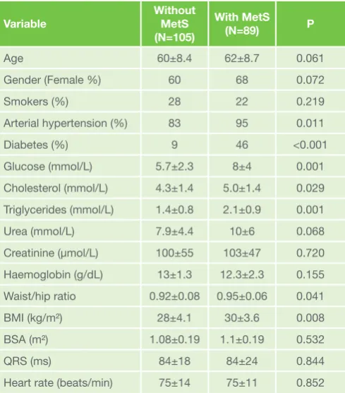
Variable Without MetS (N=105)
With MetS (N=89) P
Age 60±8.4 62±8.7 0.061
Gender (Female %) 60 68 0.072
Smokers (%) 28 22 0.219
Arterial hypertension (%) 83 95 0.011
Diabetes (%) 9 46 <0.001
Glucose (mmol/L) 5.7±2.3 8±4 0.001 Cholesterol (mmol/L) 4.3±1.4 5.0±1.4 0.029 Triglycerides (mmol/L) 1.4±0.8 2.1±0.9 0.001
Urea (mmol/L) 7.9±4.4 10±6 0.068
Creatinine (μmol/L) 100±55 103±47 0.720
Haemoglobin (g/dL) 13±1.3 12.3±2.3 0.155 Waist/hip ratio 0.92±0.08 0.95±0.06 0.041
BMI (kg/m²) 28±4.1 30±3.6 0.008
BSA (m²) 1.08±0.19 1.1±0.19 0.532
QRS (ms) 84±18 84±24 0.844
Heart rate (beats/min) 75±14 75±11 0.852 BMI: body-mass index; BSA: body-surface area.
Table 4: Comparison of Echocardiographic data between patient’s groups
Variable LV structure
Without MetS (N=105)
With MetS
(N=89) P
IVS (cm) 1.1±0.2 1.2±0.2 0.011
LVPW (cm) 1.0±0.1 1.1±0.1 0.005
LV EDD (cm) 5.1±0.7 4.9±0.6 0.138
LV ESD (cm) 3.4±0.6 3.2±0.6 0.107
LV EDV (ml) 125±37 113±28 0.033
LV ESV (ml) 49±23 44±16 0.132
LVMI (g/m2.7) 50±16 56±19 0.038
Systolic LV function
LV EF (%) 61.3±8.6 61±9.2 0.945
LV SF (%) 33.3±6 33.2±7 0.937
Aorta (cm) 3.4±0.4 3.4±0.3 0.231 S’septal (cm/s) 5.3±1.6 5.1±1.5 0.399 S’lateral (cm/s) 6.1±1.7 5.7±1.7 0.107 MAPSE lateral (cm) 1.3±0.3 1.2±0.3 0.260 MAPSE septal (cm) 1.16±0.3 1.1±0.3 0.222 Diastolic LV function
A (cm/s) 72±21 75±21 0.331
Lateral a’ (cm/s) 8.7±2.7 8.3±3 0.412
E/A ratio 0.9±0.4 0.8±0.6 0.611
Septal a’ (cm/s) 8.2±2.4 7.8±2.4 0.194
Septal e’ (cm/s) 6.2±2.2 5.3±2 0.003
E (cm/s) 61±23 57±21 0.299
Lateral e’ (cm/s) 8±3 6.7±2.3 0.001
FT (ms) 407±115 397±104 0.507
E wave DT (ms) 186±56 189±54 0.767
Global LV function
TIVT (s/min) 9.1±4.4 9.2±4.1 0.929
Tei index 0.4±0.2 0.4±0.3 0.920
E/e’ ratio 9.4±4.8 10.3±4.9 0.166
Left atrium
LA diameter (mm) 39±5 41±6.3 0.093
Measurements of left atrial dimensions and function LA diameter was measured from aortic root recordings with the M-mode cursor positioned at the level of the aortic valve leaflets. LA volumes were measured using the area-length method from the apical four and two chamber views, according to the recommendations of the European Association of Echocardiography33. LA maximum volume (LA end-systolic volume) was measured at the end of LV systole, just before the opening of the mitral valve, LA minimum volume (LA end-diastolic volume) was measured at end diastole, at the time of closure of the mitral valve, and total LA EF was calculated using the formula Total LA EF = Vmax – Vmin / Vmax33,34.
Statistical analysis
Data are presented as mean ± SD or proportions (% of patients). Continuous data was compared with two-tailed unpaired Student t test and discrete data with Chi-square test. Patients were divided according to the presence of the MetS into HFpEF with MetS and non-MetS, and were compared using unpaired Student t test. For the multivariate prediction of NYHA class, the ordinal regression was used.
Results
Clinical and biochemical data in HFpEF patients vs.
Controls
Patients with HFpEF had more prevalent diabetes (p=0.026), and had higher glycaemic levels (p<0.001), triglycerides (p=0.01), urea and creatinine (<0.001, for both) and BMI (p=0.001) compared with controls (Table 1). All other clinical and biochemical data were not different between the two groups. LV and LA structure and function in HFpEF patients vs. Controls Patients with HFpEF had thicker interventricular septum and posterior LV wall (p<0.011) for both), higher LV mass index (p=0.001), larger aortic root (p=0.003), reduced septal and lateral MAPSE (p=0.002 and p<0.001, respectively), higher E wave (p<0.001), lower E/A ratio (p=0.022), lower lateral and septal e’ (p=0.022, for both), higher E/e’ (p<0.001), longer t-IVT (p=0.005) and higher Tei index (p=0.001) compared to controls. LV dimensions and EF were not different between groups (Table 4). LA diameter, its maximal and minimal volume were significantly larger (p<0.001, p=0.01 and p=0.019, respectively), and LA EF lower in HFpEF patients compared to controls (Table 2).
Clinical and biochemical data in HFpEF patients, MetS
vs non-MetS
Patients with MetS had more prevalent diabetes (p<0.001), higher glycaemic levels (p=0.001), arterial hypertension
(p=0.011), cholesterol and triglycerides (p=0.029 and p=0.001, respectively), BMI (p=0.008) and waist/hip ratio (p=0.041) compared with non- MetS (Table 3). The other clinical and biochemical data were not different between groups.
LV and LA structure and function in HFpEF patients,
MetS vs No-MetS
Patients with MetS had thicker interventricular septum and posterior LV wall (p=0.011 and p=0.005, respectively), larger end-diastolic volume (p=0.033), higher LV mass index (p=0.038) and lower lateral and septal e’ (p=0.001 and p=0.003, respectively) compared with non-MetS, but LV dimensions and EF were not different (Table 4). In the MetS group, LA minimal volume was also larger (p=0.007) and LA EF lower (p<0.001, Table 2, Figure 1), than those with non-MetS (Table 4).
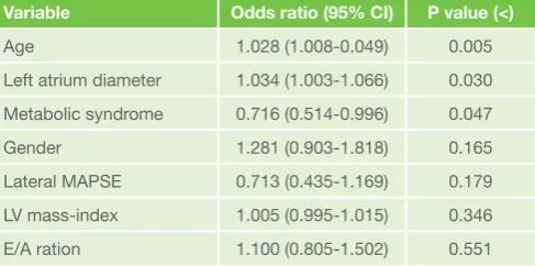
Predictors of NYHA class
In ordinal regression multivariate analysis, age (p=0.005), LA diameter (p=0.03) and the presence of MetS (p=0.047) were independent predictors of NYHA class in HFpEF patients (Table 5).
Discussion
Findings
The main finding of our study was that despite no difference in LV systolic function between patients with HFpEF and controls, the former had clear evidence for increased LV wall thickness and overall cavity mass, compromised long axis amplitude and velocities as well as worse diastolic function in the form of higher E/A, E/e’ and dyssynchrony as shown by high Tei index and longer t-IVT. LA volumes were also larger and its emptying fraction lower than controls. In addition, patients with MetS had worse LA structure and reservoir function compared to non-MetS. Finally, age, LA diameter and MetS were the only independent predictors of NYHA class in this HFpEF population.
Data interpretation
Our findings confirm previous data in showing maintained LV systolic function in HFpEF patients compared to controls despite having myocardial hypertrophy. LV subendocardial function in the form of long axis amplitude of motion and Table 5: Multivariate predictors of NYHA class in HFpEF patients
Variable Odds ratio (95% CI) P value (<)
Age 1.028 (1.008-0.049) 0.005
Left atrium diameter 1.034 (1.003-1.066) 0.030 Metabolic syndrome 0.716 (0.514-0.996) 0.047
Gender 1.281 (0.903-1.818) 0.165
Lateral MAPSE 0.713 (0.435-1.169) 0.179 LV mass-index 1.005 (0.995-1.015) 0.346
E/A ration 1.100 (0.805-1.502) 0.551 HFpEF: heart failure with preserved ejection fraction; LV: left ventricle; MAPSE: mitral annular plane systolic excursion;
velocities were significantly reduced in patients, particularly those with MetS. These findings are also similar to what has previously been found in HFpEF35, 36, thus suggesting a significant pathology that affects the subendocardial layer of the myocardium rather than the transmural function. Of note, the same subendocardial disturbances were worse in the MetS patients compared to non-MetS, again suggesting an even better explanation for those abnormalities. In the absence of significant epicardial coronary artery disease, a number of mechanisms could be playing a part in this pathology, specifically those related to MetS. Metabolic syndrome induces multiple complex metabolic disturbances including altered insulin signalling, glyco- and lipotoxicity, increased cytokine activity and intramyocyte and/or interstitial deposition of triacylglycerol and AGEs, which may all directly or indirectly affect myocardial function37. These factors also trigger endothelial dysfunction38 which causes dysregulation of vascular permeability, inflammatory responses, and vascular remodeling, mediated by increased vascular tone and arterial stiffness, blood pressure and pulse pressure39. The increased afterload, caused by the latter, results in increased myocardial work and consecutively oxygen consumption, resulting in increased energy demand, impaired myocardial perfusion and reduced cardiac efficiency40. This energy demand/ supply mismatch induces myocardial hypertrophy, autonomic dysfunction and LV diastolic dysfunction, which was profound in our HFpEF patients, particularly those with MetS.
The second interesting finding of this study was the enlarged LA with reduced LA emptying fraction. This finding suggest an alteration in pulmonary capillary wedge pressure independent of LV systolic function, as was previously shown41. Anatomical studies show that LV long axis (subendocardial) function is closely related to LA function42. This dependency relationship comes about through the shared mitral annulus as the site of insertion for the longitudinal ventricular (subendocardial) fibers as well as the longitudinal LA myocardial fibers. Indeed, Sir Arthur Keith, over a century ago, had shown this anatomical relationship and described the function of one cavity to depend on that of the other, with one contracting while the other is relaxing and vice versa43. With the ventricular longitudinal function impaired in MetS, as explained above, that of the LA is inevitably impaired too, which eventually results in established LA enlargement, being the collecting chamber, with its reservoir function reduced. This finding is also supported by Laplace law44, and together explains the high prevalence of atrial fibrillation in HFpEF patients as previously shown45.
The third finding in this study is the evidence for LV remodeling which is shown in the form of prolonged t-IVT and raised Tei index, the two are conventionally taken as markers of dyssynchrony and remodeling, which are affected by age46. Despite their presence, it seems that the extent of LV remodeling in this cohort was not strong enough to be able to predict severity of patients’ symptoms as shown by the NYHA class, when compared with LA dysfunction and the presence of MetS. Indeed the enlargement of the LA diameters and volumes as well as the fall in its emptying fraction suggests and evidence for LA remodeling which itself predicted NYHA class of our patients. This finding is supported by several other studies which showed that LA enlargement and/or its dysfunction are independent predictors of clinical outcome in HF patients34,
47, 48, as well as exercise capacity49-51. While the latter
disturbances can be explained on the basis of MetS affecting LV subendocardium, a direct effect of MetS on LA myocardium, through its microcirculation remains to be explored.
Study limitations
The relatively small number of patients in the two subgroups MetS and non-MetS is an important limitation of this study. Also, information on the duration of MetS individual risk factors was not available, this would have likely added to a precise determination of their predictive value. Assessment of LA intrinsic myocardial function by speckle tracking could have helped in differentiating primary LA pathology from a secondary one that is influenced by LV log axis dysfunction.
Clinical implications
In patients with HFpEF, the additional MetS adds to further deterioration of cardiac function, particularly the long axis function as well as left atrial function. The enlarged left atrial volume and reduced emptying fraction predict patient’s limiting symptoms according to NYHA class. Thus, assessing MetS and LA reservoir function routinely in breathless patients should shed light on its exact mechanism.
Conclusions
Metabolic syndrome worsens LV subendocardial function as well left atrial reservoir function. While the two could be interrelated, direct effect of MetS on LA reservoir function remains to be explored in view of their significant prediction of patients’ symptoms. Also, monitoring these two variables in HFpEF should add more insight into the pathophysiology of the condition.
Statement of ethical publishing
The authors agree to abide by the requirements of the “Statement of publishing ethics of the International Cardiovascular Forum Journal”52.
Conflict of interest:
The authors declare that they have no conflict of interest.
Address for correspondence:
Dr. Gani Bajraktari, MD, FESC
Clinic of Cardiology, University Clinical Centre of Kosovo, “Rrethi i Spitalit”, p.n., Prishtina, Kosovo
Tel: + 377 45 800 808
E-mail:[email protected]
References:
1. Boulogne A, Vantyghem MC. Epidemiological data and screening criteria of the metabolic syndrome. Presse Med 2004; 33: 662-5. doi: 10.1016/S0755-4982(04)98711-8.
2. Isomaa B, Almgren P, Tuomi T, Forsén B, Lahti K, Nissén M, et al. Cardiovascular morbidity and mortality associated with the metabolic syndrome. Diabetes Care 2001; 24: 683-9. doi: 10.2337/diacare.24.4.683. 3. Ivanovica BA, Tadic MV, Simic DV. Predictors of global left ventricular
function in metabolic syndrome. Arq Bras Cardiol 2001; 96: 377-84. doi: 10.1590/S0066-782X2011005000039.
4. Lakka HM, Laaksonen DE, Lakka TA, Niskanen LK, Kumpusalo E, Tuomilehto J, et al. The metabolic syndrome and total and cardiovascular disease mortality in middle-aged men. JAMA 2002; 288: 2709-16. doi: 10.1001/jama.288.21.2709.
5. McCullough AJ. Epidemiology of the metabolic syndrome in the USA. J Dig Dis 2011; 12: 333-40. doi: 10.1111/j.1751-2980.2010.00469.x.
6. Meiqs JB. Epidemiology of the metabolic syndrome. AM J Manag Care 2002; 8: 283-92.
7. Ferrara LA, Giuda L, Ferrera F, De Luca G, Staiano L, Calentano A, Mancini M. Cardiac structure and function and arterial circulation in hypertensive patients with and without metabolic syndrome. J Hum Hypertens 2007; 21: 729-35. doi: 10.1038/sj.jhh.1002222.
RJ, et al. Metabolic syndrome is associated with abnormal left ventricular diastolic function independent of left ventricular mass. Eur Heart J 2007; 28: 553-559. doi: 10.1093/eurheartj/ehl526.
9. Penjaskoviγ D, Sakac D, Dejanoviγ J, Zec R, Zec Petkoviγ N, Stojsiγ Milosavljeviγ A. Left ventricular diastolic dysfunction in patients with metabolic syndrome. Med Pregl 2012; 65: 18-22.
10. Masugata H, Senda S, Goda F, Yoshihara Y, Yoshikawa K, Fujita N, et al. Left ventricular diastolic dysfunction as assessed by echocardiography in metabolic syndrome. Hypertens Res 2006; 29: 897-903. doi:10.1291/ hypres.29.897.
11. Fiuza, M. Metabolic syndrome and coronary artery disease. Rev Port Cardiol 2012; 3: 779-82.
12. Malik S, Wong ND, Franklin SS, Kamath TV, L’italien GJ, Pio JR, et al. Impact of the Metabolic Syndrome on Mortality From Coronary Heart Disease, Cardiovascular Disease, and all Causes in United States Adults. Circulation 2004; 110: 1245-50. doi: 10.1161/01.CIR.0000140677.20606.0E. 13. Li SH, Yang B, Gong HP, Tan HW, Zhong M, Zhang Y, et al. Impaired atrial
synchronicity in patients with metabolic syndrome associated with insulin resistance and independent of hypertension. Hypertens Res 2009; 32: 791-61. doi: 10.1038/hr.2009.105.
14. Crendel E, Walther G, Dutheil F, Courteix D, Lesourd B, Chapier R, et al. Left ventricular myocardial dyssynchrony is already present in nondiabetic patients with metabolic syndrome. Can J Cardiol 2014; 30: 320-4. doi: 10.1016/j.cjca.2013.10.019.
15. Kurt M, Tanboγa IH, Büyükkaya E, Karakaγ MF, Akçay AB, Sen N, et al. Relation of presence and severity of metabolic syndrome with left atrial mechanics in patients without overt diabetes: a deformation imaging study. Anadolu Kardiyol Derg 2014; 14: 128-33. doi: 10.5152/akd.2014.4686. 16. Yilmaz M, Ozlem AO, Akgumus A, Peker T, Karaagac K, Vatansever F, et al.
Left atrial mechanical functions in patients with the metabolic syndrome. Acta Cardiol 2013; 68: 133-7.
17. Kurt M, Wang J, Torre-Amione G, Nagusha SG. Left atrial function in diastolic heart failure. Circ Cardiovasc Imaging 2009; 2: 10-5. doi: 10.1161/ CIRCIMAGING.108.813071.
18. Teo SG, Yang H, Chai P, Yeo TC. Impact of left ventricular diastolic dysfunction on left atrial volume and function: a volumetric analysis. Eur J echocardiogr 2010; 11: 38-43. doi: 10.1093/ejechocard/jep153.
19. Tsang TS, Barnes ME, Gersh BJ, Bailet KR, Sewerd JB. Left atrial volume as a morphophysiologic expression of left ventricular diastolic dysfunction and relation to cardiovascular risk burden. Am J Cardiol 2002; 90: 1284-9. doi: 10.1016/S0002-9149(02)02864-3.
20. Chinali M, de Simone G, Roman MJ, Bella JN, Liu JE, Lee ET, et al. Left atrial systolic force and cardiovascular outcome. The Strong Heart Study. Am J Hypertens 2005; 18: 1570-6. doi: 10.1016/j.amjhyper.2005.05.036. 21. Koprowski P, Kostkiewicz M, Leγniak-Sobelga A. Echocardiographic
assessment of left atrial volume in asymptomatic ambulatory patients with metabolic syndrome and/or arterial hypertension - is it parameter worth into considerate? Przegl Lek 2012; 69: 1199- 204.
22. Maeder MT, Rickli H. Heart failure with preserved left ventricular ejection fraction. Praxis (Bern 2014) 2013; 102: 1299-307. doi: 10.1024/1661-8157/ a001439.
23. Borlaug BA, Paulus WJ. Heart failure with preserved ejection fraction: Pathophysiology, diagnosis, and treatman. Eur Heart J 2011; 32: 670-9. doi: 10.1093/eurheartj/ehq426.
24. Guan Z, Zhang D, Huang R, Zhang F, Wang Q, Guo S. Association of left atrial myocardial function with left ventricular diastolic dysfunction in subjects with preserved systolic function: a strain rate imaging study. Clin Cardiol 2010; 33: 643-9. doi: 10.1002/clc.20784.
25. Bilen E, Kurt M, Tanboγa IH, Kocak U, Ayhan H, Durmaz T, et al. Assessment of left atrial phasic functions in heart failure patients with preserved or low ejection fraction. Turk Kardiyol Dern Ars 2012; 40: 122-8. doi: 10.5543/ tkda.2012.01802.
26. Grundy SM, Breweber HB Jr, Cleeman JI, Smith SC Jr, Lenfant C, American Heart Association; National Heart, Lung and Blood Institute. Definition of metabolic syndrome: Report of the National Heart, Lung and Blood Institute/ American Heart Association conference on scientific issues related to definition. Circulation 2004; 109: 433-8. doi: 10.1161/01. CIR.0000111245.75752.C6
27. Höglund C, Alam M, Thorstrand C. Atrioventricular valve plane displacement in healthypersons. An echocardiographic study. Acta Med Scand 1988; 224: 557-62.
28. Appleton CP, Hatle LK, Popp RL. Relation of transmitral flow velocity patterns to leftventricular diastolic function: new insights from a combined hemodynamic and Doppler echocardiographic study. J Am Coll Cardiol 1988; 12: 426-40. doi: 10.1016/0735-1097(88)90416-0
29. Zoghbi WA, Enriquez-Sarano M, Foster E, Grayburn PA, Kraft CD, Levine RA, et al; American Society of Echocardiography. Recommendations for evaluation of the severity of native valvular regurgitation with two-dimensional and Doppler echocardiography. J Am Soc Echocardiogr 2003; 16: 777-802. doi: 10.1016/S0894-7317(03)00335-3.
30. Gardin JM, Adams DB, Douglas PS, Feigenbaum H, Forst DH, Fraser AG, et al; American Society of Echocardiography. Recommendations for a standardized report for adult transthoracic echocardiography: a report from
the American Society of Echocardiography’s Nomenclature and Standards Committee and Task Force for a Standardized Echocardiography Report. J Am Soc Echocardiogr 2002; 15: 275-90. doi: 10.1067/mje.2002.121536. 31. Duncan AM, Francis DP, Henein MY, Gibson DG. Importance of left
ventricular activation in determining myocardial performance (Tei) index: comparison with total isovolumic time. Int J Cardiol 2004; 95: 211-7. doi: 10.1016/j.ijcard.2003.07.007.
32. Tei C, Ling LH, Hodge DO, Bailey KR, Oh JK, Rodeheffer RJ, et al. New index of combined systolic and diastolic myocardial performance: a simple and reproducible measure of cardiac function - a study in normals and dilated cardiomyopathy. J Cardiol 1995; 26: 357–366.
33. Galderisi M, Henein MY, D’hooge J, Sicari R, Badano LP, Zamorano JL, et al; European Association of Echocardiography. Recommendations of the European Association of Echocardiography: how to use echo-Doppler in clinical trials: different modalities for different purposes. Eur J Echocardiogr 2011; 12: 339-53. doi: 10.1093/ejechocard/jer051.
34. Jarnert C, Melcher A, Caidahl K, Persson H, Ryden L, Eriksson MJ. Left atrial velocity vector imaging for the detection and quantification of left ventricular diastolic function in type 2 diabetes. European Journal of Heart Failure 2008; 10: 1080-7. doi: 10.1016/j.ejheart.2008.08.012.
35. Yip G, Wang M, Zhang Y, Fung JW, Ho PY, Sanderson JE. Left ventricular long axis function in diastolic heart failure is reduced in both diastole and systole: time for a redefinition? Heart 2002; 87: 121-5. doi: 10.1136/ heart.87.2.121.
36. Bajraktari G, Berbatovci-Ukimeraj M, Hajdari A, Ibraimi L, Daullxhiu I, Elezi Y, et al. Predictors of increased left ventricular filling pressure in dialysis patients with preserved left ventricular ejection fraction. Croat Med J 2009; 50: 543-9. doi: 10.3325/cmj.2009.50.543.
37. Fang NN, Sui DX, Yu JG, Gong HP, Zhong M, Zhang Y, et al. Strain/strain rate imaging of impaired left atrial function in patients with metabolic syndrome. Hypertens Res 2015 Jul 16. doi: 10.1038/hr.2015.76. 38. Oktay AA, Rich JD, Shah SJ. The emerging epidemic of heart failure with
preserved ejection fraction. Curr Heart Fail Rep 2013; 10: 401-10. doi: 10.1007/s11897-013-0155-7.
39. von Bibra H, St John Sutton M. Diastolic dysfunction in diabetes and the metabolic syndrome: promising potential for diagnosis and prognosis. Diabetologia 2010; 53: 1033-45. doi: 10.1007/s00125-010-1682-3. 40. Widlansky ME, Gokce N, Keaney JF Jr, Vita JA. The clinical implications
of endothelial dysfunction. J Am Coll Cardiol 2003; 42:1149–1160. doi: 10.1016/S0735-1097(03)00994-X
41. Henein, M., Tossavainen, E., Söderberg, S., Grönlund, C., Gonzalez, M., & Lindqvist, P. Left atrial strain rate estimates PCWP. International cardiovascular forum journal 2013; 1: 25-30. http://dx.doi.org/10.17987/icfj. v1i1.11
42. Santos AB, Kraigher-Krainer E, Gupta DK, Claggett B, Zile MR, Pieske B, et al. Impaired left atrial function in heart failure with preserved ejection fraction. Eur J Heart Fail 2014; 16: 1096-103. doi: 10.1002/ejhf.147.
43. Keith A. Harveian Lecture ON THE FUNCTIONAL ANATOMY OF THE HEART. Br Med J 1918 Mar 30;1(2987):361-3.
44. Li JK. Comparative cardiac mechanics: Laplace’s Law. J Theor Biol. 1986; 118: 339-43.
45. Zakeri R, Chamberlain AM, Roger VL, Redfield MM. Temporal relationship and prognostic significance of atrial fibrillation in heart failure patients with preserved ejection fraction: a community-based study. Circulation. 2013; 128: 1085-93. doi: 10.1161/CIRCULATIONAHA.113.001475.
46. Bajraktari, G., Lindqvist, P., & Henein, M. Y. Left ventricular global dyssynchrony is exaggerated with age. International Cardiovascular Forum Journal. 2013; 1: 47-51. http://dx.doi.org/10.17987/icfj.v1i1.16
47. Rossi A, Gheorghiade M, Triposkiadis F, Solomon SD, Pieske B, Butler J. Left atrium in heart failure with preserved ejection fraction: structure, function, and significance. Circ Heart Fail. 2014; 7: 1042-9. doi: 10.1161/ CIRCHEARTFAILURE.114.001276.
48. Zamora E, Lupón J, López-Ayerbe J, Urrutia A, González B, Ferrer E, et al. Left atrium diameter: a simple echocardiographic parameter with high prognostic value in heart failure. Med Clin (Barc) 2007; 129: 441-5. 49. Bajraktari G, Fontanive P, Qirko S, Elezi S, Simioniuc A, Huqi A, et al.
Independent and incremental value of severely enlarged left atrium in risk stratification of very elderly patients with chronic systolic heart failure. Congest Heart Fail 2012; 18: 222-8. doi: 10.1111/j.1751-7133.2011.00280.x. 50. Yamaguchi K, Yoshitomi H, Ito S, Ito S, Adachi T, Sato H, et al. Left atrial
remodeling and recurrence of congestive heart failure in patients initially diagnosed with heart failure. Echocardiography 2014; 31: 936-40. doi: 10.1111/echo.12497.
51. Ceresa M, Capomolla S, Pinna GD, Febo O, Caporotondi A, Guazzotti GP, et al. Left atrial function: bridge to central and hormonal determinants of exercise capacity in patients with chronic heart failure. Monaldi Arch Chest Dis 2002; 58: 87-94.