Factors associated with left ventricular
hypertrophy in adults with surgically repaired
coarctation of the aorta
Daniel Rinnström MD
1, gunnar Engström MD, phD
2, Martin ugander MD, phD
3and Bengt
Johansson MD, phD
11 Cardiology, Heart Centre and Department of Public Health and Clinical Medicine, Medicine, Umeå University, Umeå, Sweden 2 Cardiothoracic surgery, Heart Centre and Department of Perioperative sciences, Umeå University, Umeå, Sweden
3 Department of Clinial Physiology, Karolinska Institutet and Karolinska University Hospital, Stockholm, Sweden
Introduction
Coarctation of the aorta (Coa) is a congenital heart lesion associated with an increased risk of hypertension, left ventricular (lV) hypertrophy, and other adverse cardiovascular events, even after the Coa has been surgically repaired1-3. lV hypertrophy is important from a clinical perspective, since it is considered an adaptive response to chronically increased afterload4, and has been shown to be highly related to cardiovascular morbidity5. Furthermore, studies have shown that treatments reducing lV mass decreases the risk of future complications6.
It has previously been shown that patients with repaired Coa are at risk of developing increased lV mass due to impairment of vascular mechanics, and abnormal blood pressure
regulation7, 8, especially in those with an angulated aortic arch, which in turn is associated with late hypertension9. However, little is known about how other factors interact with blood pressure and lV mass in this particular population. Therefore, the aim of the current study was to identify factors associated with high lV mass, assessed by cardiovascular magnetic resonance, among patients with Coa.
Material and methods
Patients
In this retrospective and cross-sectional study, all consecutive patients with Coa evaluated by cardiovascular magnetic resonance imaging (MRI) at our institution during 2008-2012 were identified. In general, these patients were followed in the cardiology clinic and underwent at least one MRI examination as adults. Clinical data and echocardiography reports were reviewed.
only patients with previous surgical treatment of Coa were included (n = 59). patients were excluded; if the MRI examination was undertaken below 18 years of age (n = 6), if the MRI data was insufficient (n = 1) or if data regarding body-surface area (n = 1) were missing, thus leaving 51 patients for analysis. The study complied with the Declaration of Helsinki, and was approved by the Regional Ethical Review Board in umeå, sweden (Dnr 09-194M).
Abstract
Introduction: Most patients with repaired coarctation of the aorta (Coa) live normal lives and have good physical
performance. However, even after a successful surgical intervention, long-term cardiovascular risks including left ventricular hypertrophy remain. The aim of the study was to identify factors associated with increased left ventricular mass (lVM) in patients with surgically repaired Coa.
Methods: Consecutive cardiovascular magnetic resonance investigations in 51 patients with surgically repaired Coa (age 37+/-15 years, age at intervention 9.7 ± 6.8 years, 45% female) were reviewed. lVM was measured and indexed to body surface area. The association between increased lVM index and clinical, anatomic and functional variables was investigated with logistic regression analysis.
Results: In this population, 14/51 (27%) patients had a lVM index above normal limits. Factors associated with an increased lVM index in univariate analysis were higher systolic blood pressure (odds ratio (oR) = 1.04, 95 % confidence interval (CI) 1.00-1.08, p = 0.03), descending aortic diameter (oR = 1.48, CI 1.14-1.90, p = 0.003) and more than mild aortic valve disease or previous aortic valve intervention (oR = 15.1, CI 2.50-48.4, p=0.002), but not diastolic blood pressure, diameter of ascending aorta, diameter or ratio of Coa, velocity in descending aorta, smoking or bicuspid aortic valve (p > 0.05 for all). In multivariate analysis, only systolic blood pressure (p = 0.05) and aortic valve disease (p = 0.006) remained significant, yielding R2 = 0.47, p = 0.002 for the model.
Conclusion: Increased lVM is a common late finding after surgically repaired Coa. This study showed that lVM was associated with modifiable factors; systolic blood pressure and aortic valve disease. as most patients are young, and increased lVM will eventually affect ventricular function, close attention to blood pressure optimization may be of particular importance in the surgically repaired Coa population.
Cardiovascular magnetic resonance
The investigations were performed using a philips 1.5 T Intera or achieva scanner (philips, Best, The Netherlands). Most examinations included a transaxial stack of T2 weighted “black-blood” images covering the heart and the great vessels. Cine steady-state free-precession (ssFp) images included the two-chamber view, the four-chamber view, a short-axis stack covering the lV from the apex to the base, and three contiguous image planes perpendicular to the aortic root covering the aortic valve and sinus of Valsalva. In 37 patients, a a contrast enhanced MRI angiogram of the thoracic aorta was performed.
Measurement of ventricular mass
analysis of lV volumes, ejection fraction and mass was performed using segment v1.9 R1942 (http://segment.heiberg. se)10. The delineation of lV endocardial and epicardial borders was performed manually, and included papillary musculature as a part of the lV myocardial mass (Figure 1). Measurements of lV mass were performed at end systole and end diastole and expressed as the mean of the two measurements. a difference of less than 5% between end-systolic and end-diastolic mass was accepted, otherwise technical reasons were sought for and corrected. The resulting mass measurements were indexed to body surface area.
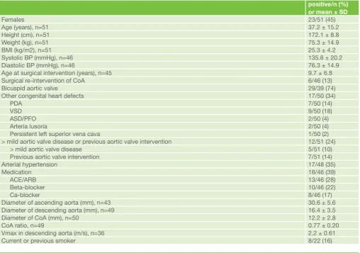
Table 1: overview of demographic and clinical data. BMI = body mass index, Bp = arterial blood pressure measured in the right arm, Coa = coarctation of the aorta. pDa = presistent ductus arteriosus, VsD = ventricular septal defect. asD/pFo = atrial septal defect/persistent foramen ovale, aCE/aRB = angiotensin converting enzyme inhibitors /angiotensin receptor blockers, Vmax = peak velocity. Coa ratio is defined as the ratio between the minimum dimension at the level of coarctation and that of the distal descending aorta.
positive/n (%) or mean ± SD
Females 23/51 (45)
age (years), n=51 37.2 ± 15.2
Height (cm), n=51 172.1 ± 8.8
Weight (kg), n=51 75.3 ± 14.9
BMI (kg/m2), n=51 25.3 ± 4.2
systolic Bp (mmHg), n=46 135.8 ± 20.2
Diastolic Bp (mmHg), n=46 76.3 ± 14.9
age at surgical intervention (years), n=45 9.7 ± 6.8
surgical re-intervention of Coa 6/46 (13)
Bicuspid aortic valve 29/39 (74)
other congenital heart defects 17/50 (34)
pDa 7/50 (14)
VsD 9/50 (18)
asD/pFo 2/50 (4)
arteria lusoria 2/50 (4)
persistent left superior vena cava 1/50 (2)
> mild aortic valve disease or previous aortic valve intervention 12/51 (24)
> mild aortic valve disease 5/51 (10)
previous aortic valve intervention 7/51 (14)
arterial hypertension 17/48 (35)
Medication 18/46 (39)
aCE/aRB 13/46 (28)
Beta-blocker 10/46 (22)
Ca-blocker 8/46 (17)
Diameter of ascending aorta (mm), n=43 30.6 ± 5.6
Diameter of descending aorta (mm), n=49 16.4 ± 3.5
Diameter of Coa (mm), n=50 12.2 ± 2.8
Coa ratio, n=49 0.77 ± 0.20
Vmax in descending aorta (m/s), n=36 2.2 ± 0.61
Current or previous smoker 8/22 (16)
Measurements of aortic dimensions
The diameter of the ascending aorta was measured in transaxial T2-weighted “black-blood” images at level of the pulmonary artery bifurcation, whereas the diameter of the descending aorta, and the minimum diameter of the aorta at the site of coarctation, was measured in maximum intensity projection reconstructions of the aorta from the contrast enhanced MRI angiography images (n = 37) or in orthogonal cine images at the site of coarctation (n = 12).
Aortic valve function
Information concerning aortic valve function (degree of stenosis or regurgitation) was primarily gathered from the patients’ MRI report, and, when necessary, from the echocardiography report. aortic valve function was determined using standard definitions and was graded as mild, moderate or severe. In the statistical analyses, patients were classified into two groups; those with moderate or severe aortic valve disease or previous aortic valve intervention, and those with mild lesions or normal aortic valve function.
Statistics
all calculations were performed using spss 19 (IBM, armonk, NY, usa). Each patient’s lV mass was indexed to body surface area and compared to the average value of a healthy population, defined by sex and age11. For the statistical analyses, those with a lV mass index exceeding the expected mean by more than two standard deviations were grouped together, and compared with the remaining patients in a series of univariate logistic binary regressions. Casewise deletion was used for missing numeric data. For categorical data (e.g., smoking pattern), missing data were substituted with an inert category that was checked for significance, to be indicated if so.
Factors with p < 0.15 in the univariate regressions were included in a multivariable binary logistic regression model. Variables exhibiting co-linearity were rejected, In post-hoc analysis, an additional multivariate model was constructed with systolic blood pressure categorized into the following groups:
≤ 120 mmHg, [120; 140) mmHg, [140; 160) mmHg and > 160 mmHg. The group ≤ 120 mmHg served as reference. The null-hypothesis was rejected for p < 0.05.
Table 2: uni- and multivariate logistic regressions, with left ventricular mass index divided into two groups: ≤ upper reference limit, and > upper reference limit, as the dependent variable. Coa ratio is defined as the ratio between the minimum dimension at the level of coarctation and the distal descending aorta. y denotes yes indicating analysis according to presence of the measure. CI high and CI low represent the upper and lower 95% confidence limits, respectively. oR denotes odds ratio. The multivariate model included 46 patients, and had a Nagelkerke R2 of 0.47, and p = 0.002.
Univariate Multivariate
Variable Unit N Wald OR CI low CI high p-value Wald OR CI low CI high p-value
Hypertension y 48 2.56 2.92 0.78 10.9 0.11
systolic Bp, right arm mmHg 46 4.83 1.042 1.00 1.08 0.03 3.96 1.054 1.00 1.11 0.05 Diastolic Bp, right arm mmHg 46 0.005 1.00 0.96 1.04 0.94
Diameter of ascending aorta
mm 43 3.37 1.13 0.99 1.29 0.07
Diameter of descending aorta
mm 49 8.89 1.48 1.14 1.90 0.003
Diameter of Coa mm 50 0.79 1.11 0.88 1.40 0.39
Coa ratio 49 2.93 0.045 0.001 1.57 0.09
Vmax in aorta descendens
m/s 36 2.57 2.96 0.79 11.1 0.11
> mild aortic valve disease or previous aortic valve intervention
y 51 11.3 15.1 2.50 48.4 0.002 7.68 16.3 2.26 117 0.006
Current or previous smoker
y 29 3.52 5.33 0.93 30.64 0.06 0.09 1.49 0.11 19.4 0.76
Bicuspid aortic valve y 39 0.13 0.74 0.15 3.67 0.72
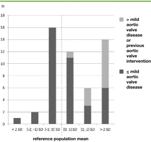
Figure 2: The distribution of left ventricular mass index in relation to reference mean values. sD denotes standard deviation. Note that all patients with > mild aortic valve disease or previous aortic valve intervention, have left ventricular mass index above the mean of normal values.
reference population mean
> mild aortic valve
disease or previous aortic valve intervention
Results
Patients
Table 1 shows the patient demographics and clinical
characteristics. In summary, 45% of the patients were female, the mean age was 37 years, and approximately one third was treated for arterial hypertension. More than mild aortic valve disease was present in 5 patients, and 7 patients had undergone aortic valve intervention. approximately one third had associated simple congenital heart defects.
Left ventricular mass
The lV mass index was grouped for distribution in relation to normal values as shown in Figure 2. Nineteen patients (37 %) had a lV mass index below the expected mean of healthy control subjects while 32 patients (63 %) had a lV mass index above this mean. as a result, the distribution of the mass index was somewhat skewed. Fourteen patients (27 %) had lV mass index more than two standard deviations above the normal mean, 8 (57 %) of whom had a more than mild aortic valve disease or had undergone previous aortic valve intervention.
Factors associated with increased LV mass
as detailed in Table 2, factors significant at univariate analysis were systolic blood pressure measured, diameter of descending aorta, and > mild aortic valve disease or previous aortic valve intervention. In multivariable analysis, factors independently associated with an increased ventricular mass were; systolic blood pressure, and more than mild aortic valve disease or previous aortic valve intervention. The multivariable model included 46 patients, and yielded R2 = 0.47, p = 0.002.
Blood pressure and LV mass
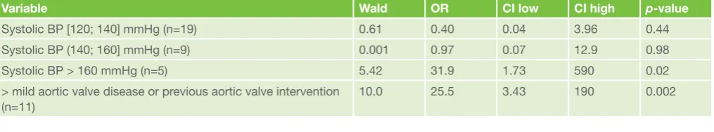
In post hoc analysis, systolic blood pressure was categorized into four groups and analysed together with aortic valve status. The group with the highest systolic blood pressure (> 160 mmHg) and more than mild aortic valve disease or previous aortic valve intervention remained significantly associated with an increased lV mass index, and the model yielded R2 = 0.53 and p = 0.002.
Discussion
In this population with surgically repaired coarctation of the aorta undergoing regular medical follow-up, we found that approximately one quarter of these patients had lV mass index above normal limits. High blood pressure and aortic valvular disease were independently associated with increased lV mass index. Both these factors are potentially modifiable by intervention. It is important to note that even after surgical intervention on the aortic valve, many patients still remain with increased lV mass.
Before surgical intervention was possible, patients with significant coarctation of the aorta often died from lV failure secondary to increased afterload12. Despite the fact that surgical intervention of aortic coarctation has been practiced for almost 70 years13, persistent lV hypertrophy is still a concern for approximately one quarter of all patients with coarctation of the aorta. The long-term clinical significance of such hypertrophy in young and middle-aged patients is unknown, but treatments targeting and preventing development of increased lV mass are likely justifiable, in order to avoid the well-known cardiovascular morbidity5. similarly, a reduction in lV mass is an identified favourable prognostic marker during treatment of essential hypertension14.
all patients with more than mild aortic valve disease had a lV mass index above the normal mean, with the majority exceeding the upper normal limit. Thus, many patients may potentially benefit from valve intervention. However, the majority of patients with significant valve disease had undergone surgical correction at some point in their lives. This observation suggests that the risk of lV hypertrophy is not completely eliminated by surgical valve intervention. Furthermore, it may be speculated that aortic valve disease in coarctation of the aorta is associated with general pathology in the ascending aorta and aortic arch, which might contribute to lV hypertrophy9. Interestingly, neither peak velocity over the coarctation, nor the absolute luminal diameter at the site of coarctation, both expected to affect systolic blood pressure, were found to be associated with increased lV mass index. In this context, it must be emphasized that there was no haemodynamically significant recoarctation in the population. There was an association between the diameter of descending aorta and lV hypertrophy, but this association did not persist in multivariate analysis. Furthermore, it appears that this was the result of a methodological indexing since the difference disappeared in the multivariable models that compensated for; age, height, and weight (data not shown).
It has long been known that there is an association between blood pressure and lV mass15. It is also known that
hypertension in patients with initially asymptomatic mild to moderate aortic stenosis is associated with a more abnormal lV structure, as well as with increased cardiovascular morbidity and mortality16. Hereby we show that systolic blood pressure, as measured at a regular clinical visit, is highly associated with lV hypertrophy, defined as a mass index exceeding the normal limits. of interest, a prior diagnosis of arterial hypertension was not, in itself, a determinant of hypertrophy, suggesting that successful blood pressure treatment might alleviate the risk. In post-hoc analysis, we found that the association with lV hypertrophy was only valid for the highest quartile of systolic blood pressures exceeding 160 mmHg. From this observation one might conclude that the actual blood pressure at a clinical visit is strongly associated with lV hypertrophy, but this Table 3: post hoc multivariate logistic regression, with left ventricular mass index divided into two groups: ≤ upper reference limit, and > upper reference limit, as dependent variable. The multivarite model includes 46 patients, and has a Nagelkerke R2 of 0.53, and p = 0.002.
Variable Wald OR CI low CI high p-value
systolic Bp [120; 140] mmHg (n=19) 0.61 0.40 0.04 3.96 0.44
systolic Bp (140; 160] mmHg (n=9) 0.001 0.97 0.07 12.9 0.98
systolic Bp > 160 mmHg (n=5) 5.42 31.9 1.73 590 0.02
> mild aortic valve disease or previous aortic valve intervention (n=11)
applies only to very high blood pressures (> 160 mmHg). Blood pressures in the range of 120-to-160 mmHg did not appear to indicate any association with ventricular hypertrophy, although that analysis was likely underpowered. Moreover, this finding does not mean that systolic blood pressures of medium range should be clinically accepted. our observation is valid only for the tested association between systolic blood pressure and lV hypertrophy, and in a highly selected population of adult patients with surgically repaired coarctation of the aorta. In conclusion, a more than mild aortic valve disease or previous aortic valve intervention is associated with an increased lV mass index. There is also an association between ventricular mass and systolic blood pressure, particularly values exceeding 160 mmHg. Controlled hypertension appeared to protect against the development of lV hypertrophy in these patients.
Correspondence to: Bengt Johansson
Cardiology, Heart Centre, umeå university Hospital, sE-90185, umeå, sweden
References
1. guntheroth Wg. Coarctation of the aorta: long-term follow-up and prediction of outcome after surgical correction. Circulation. 1990;81(4):1441. Epub 1990/04/01.
2. o’sullivan JJ, Derrick g, Darnell R. prevalence of hypertension in children after early repair of coarctation of the aorta: a cohort study using casual and 24 hour blood pressure measurement. Heart. 2002;88(2):163-6. Epub 2002/07/16.
3. Celermajer Ds, greaves K. survivors of coarctation repair: fixed but not cured. Heart. 2002;88(2):113-4. Epub 2002/07/16.
4. Ruilope lM, schmieder RE. left ventricular hypertrophy and clinical outcomes in hypertensive patients. american journal of hypertension. 2008;21(5):500-8. Epub 2008/04/26.
5. Verdecchia p, Carini g, Circo a, Dovellini E, giovannini E, lombardo M, et al. left ventricular mass and cardiovascular morbidity in essential hypertension: the MaVI study. Journal of the american College of Cardiology. 2001;38(7):1829-35. Epub 2001/12/12.
6. Cuspidi C. lowering left ventricular mass in hypertension: a way to improve cardiovascular prognosis? american journal of hypertension. 2010;23(8):818. Epub 2010/07/21.
7. leandro J, smallhorn JF, Benson l, Musewe N, Balfe JW, Dyck JD, et al. ambulatory blood pressure monitoring and left ventricular mass and function after successful surgical repair of coarctation of the aorta. Journal of the american College of Cardiology. 1992;20(1):197-204. Epub 1992/07/01.
8. Crepaz R, Cemin R, Romeo C, Bonsante E, gentili l, Trevisan D, et al. Factors affecting left ventricular remodelling and mechanics in the long-term follow-up after successful repair of aortic coarctation. Cardiology in the young. 2005;15(2):160-7. Epub 2005/04/23.
9. ou p, Celermajer Ds, Raisky o, Jolivet o, Buyens F, Herment a, et al. angular (gothic) aortic arch leads to enhanced systolic wave reflection, central aortic stiffness, and increased left ventricular mass late after aortic coarctation repair: evaluation with magnetic resonance flow mapping. The Journal of thoracic and cardiovascular surgery. 2008;135(1):62-8. Epub 2008/01/09.
10. Heiberg E, sjögren J, ugander M, Carlsson M, Engblom H, arheden H. Design and validation of segment--freely available software for cardiovascular image analysis. BMC medical imaging. 2010;10:1. Epub 2010/01/13.
11. Maceira aM, prasad sK, Khan M, pennell DJ. Normalized left ventricular systolic and diastolic function by steady state free precession cardiovascular magnetic resonance. Journal of cardiovascular magnetic resonance : official journal of the society for Cardiovascular Magnetic Resonance. 2006;8(3):417-26. Epub 2006/06/08.
12. Jurcut R, Daraban aM, lorber a, Deleanu D, amzulescu Ms, Zara C, et al. Coarctation of the aorta in adults: what is the best treatment? Case report and literature review. Journal of medicine and life. 2011;4(2):189-95. Epub 2011/07/22.
13. Crafoord C. The surgical treatment of coarctation of the aorta. surgery. 1947;21(1):146. Epub 1947/01/01.
14. Verdecchia p, schillaci g, Borgioni C, Ciucci a, gattobigio R, Zampi I, et al. prognostic significance of serial changes in left ventricular mass in essential hypertension. Circulation. 1998;97(1):48-54. Epub 1998/01/27.
of electrocardiographic left ventricular hypertrophy in human hypertension: an updated review. Journal of hypertension. 2012;30(11):2066-73. Epub 2012/08/24.
16. Rieck aE, Cramariuc D, Boman K, gohlke-Barwolf C, staal EM, lonnebakken MT, et al. Hypertension in aortic stenosis: implications for left ventricular structure and cardiovascular events. Hypertension. 2012;60(1):90-7. Epub 2012/06/01.