Adrenomedullin Signaling in the Growth and Function of Adult Lymphatic Vessels
Samantha Lynn Hoopes
“A dissertation submitted to the faculty of the University of North Carolina at Chapel Hill in partial fulfillment of the requirements for the degree of Doctor of Philosophy in
the Department of Cell and Molecular Physiology.”
Chapel Hill 2013
Approved by:
ii ©2013
iii Abstract
SAMANTHA LYNN HOOPES: Adrenomedullin Signaling in the Growth and Function of Adult Lymphatic Vessels
(Under the direction of Dr. Kathleen Caron)
The highly conserved peptide, adrenomedullin (Adm=gene; AM=peptide), is known to play a role in the development and permeability of the lymphatic vascular system. AM signals through a G-Protein coupled receptor, calcitonin receptor-like receptor (Calcrl), associated with a receptor activity modifying protein (Ramp2/3).
To determine the role of AM signaling during adulthood, several studies were undertaken using mice with temporal deletion of Calcrl and mice haplo-insufficient for Adm. Temporal loss of Calcrl in adult mice resulted in multi-organ
lymphangiectasia associated with increased lymphatic permeability and disrupted lymphatic function. These mice also exhibited reduced body weight due to impaired lipid absorption and protein-losing enteropathy. Therefore, AM signaling is required for preservation of normal lymphatic function and permeability. An ear wound assay was performed on mice haplo-insufficient for Adm to stimulate lymphangiogenesis as well as angiogenesis. Genetic reduction of Adm impaired lymphangiogenesis in response to the wound, but angiogenesis was unaffected. AM is highly expressed in all endothelial cells, but from these studies as well as previous developmental
studies we have found that there is an enhanced response to AM signaling in lymphatic endothelial cells that is imparted by increased levels of receptor
iv
blood endothelial cells that could contribute to the different responses to AM
signaling, we explored the expression and function of the serum binding protein for AM, complement factor H. Previous studies showed that AM activity is enhanced by complement factor H and previous microarray data revealed that the complement factor H gene is upregulated in lymphatic endothelial cells. Here we show that
complement factor H protein expression is increased in lymphatic endothelial cells and it enhances the AM-induced migration response to a scratch assay in lymphatic endothelial cells. Overall, results from these studies established the importance of AM signaling in lymphatic vessels during adulthood and identified a factor
v
vi
Acknowledgements
I would like to thank members of the Caron lab for stimulating and insightful discussions. With respect to information presented in Chapter I, I respectfully acknowledge the international community of investigators studying adrenomedullin signaling and regret that space limitations precluded me from referencing all relevant studies in this chapter. Regarding the scientific data presented in Chapter III, I would like to thank: Dr. Terete Borrás and members of her laboratory (UNC-CH Dept. of Ophthalmology) for their helpful discussions and assistance with intraocular pressure measurements, Dr. Nobuyo Maeda and Dr. Raymond Fox (UNC-CH
Pathology Dept.) for assistance and advice with acid steatocrit and lipase
experiments, Dr. Hyung-Suk Kim (UNC-CH) with the Animal Clinical Chemistry and Gene Expression Core and Kirk McNaughton (UNC-CH) with the Histology
vii
Table of Contents
List of Tables ... x
List of Figures ... xi
List of Abbreviations ... xiii
Chapter I. Introduction: Adrenomedullin Function in Vascular Endothelial Cells: Insights from Genetic Mouse Models ... 1
Overview ... 1
The Multifunctional Adrenomedullin Peptide ... 1
Adrenomedullin GPCR-Mediated Signaling in Endothelial Cells ... 2
Development ... 6
Physiology and Pathophysiology ... 11
Summary and Future Directions ... 19
Author Contributions ... 20
Tables ... 21
Figures ... 23
II. The Lymphatic Vascular System ... 25
Function of the Lymphatic System ... 25
Lymphatic Specification and Sprouting ... 26
viii
Lymphatic Plexus Remodeling and Maturation ... 28
Summary ... 28
Figures ... 30
III. Characteristics of Multi-Organ Lymphangiectasia Resulting from Temporal Deletion of Calcitonin Receptor-like Receptor in Adult Mice ... 31
Overview ... 31
Introduction ... 32
Methods ... 35
Results ... 41
Discussion ... 49
Author Contributions ... 54
Figures ... 55
IV. Haplo-insufficiency of Adrenomedullin Impairs Wound-Induced Lymphangiogenesis in Adult Mice in Response to a Minimally Invasive Wound ... 64
Overview ... 64
Introduction ... 65
Methods ... 66
Results ... 68
Discussion ... 70
Author Contributions ... 72
ix
V. Complement Factor H is Enriched in Lymphatic Endothelial Cells and Increases Adrenomedullin-
Induced Migration ... 77
Overview ... 77
Introduction ... 78
Methods ... 81
Results ... 84
Discussion ... 86
Author Contributions ... 88
Figures ... 89
VI. Conclusions and Future Directions ... 93
Summary ... 93
Current State of the Lymphatic Field ... 93
Future Directions ... 97
x
List of Tables Table
1. Gene Targeted Mouse Models for Studying
Adrenomedullin Signaling ... 21 2. Vascular Assays for Studying Adrenomedullin
xi
List of Figures Figure
Chapter I. Introduction: Adrenomedullin Function in Vascular Endothelial Cells: Insights from Genetic Mouse Models
1.1 Fold change in plasma adrenomedullin levels in a
variety of human conditions ... 23 1.2 Adrenomedullin signaling in development and
vascular biology ... 24 Chapter II. The Lymphatic Vascular System
2.1 Stepwise Process of Lymphangiogenesis ... 30 Chapter III. Characteristics of Multi-Organ Lymphangiectasia
Resulting from Temporal Deletion of Calcitonin Receptor-like Receptor in Adult Mice
3.1 Acute-onset eye phenotype, eye inflammation, edema and enlarged lymphatic vessels in the
Calcrlfl/fl/CAGGCre-ERTM mice ... 55
3.2 Dilated lacteals and submucosal lymphatics in the Calcrlfl/fl/CAGGCre-ERTM mice and chyle-filled
lymphatics after short-term Western diet ... 57 3.3 Calcrlfl/fl/CAGGCre-ERTM mice exhibit reduced
body weight due to impaired lipid absorption ... 58 3.4 Dilated dermal lymphatic capillaries with
exacerbated and prolonged edema ... 59 3.5 Increased lymphatic vascular permeability without
change to blood vascular permeability ... 60 3.6 Inhibition of AM signaling disrupts lymphatic
endothelial cell-cell junctions ... 61 3.S1. The acute onset eye phenotype with temporal
deletion of Calcrl is not associated with
glaucoma-like characteristics ... 62 3.S2. Calcrl gene expression in lung and heart tissue
xii
Chapter IV: Haplo-insufficiency of Adrenomedullin Impairs Wound-Induced Lymphangiogenesis in Adult Mice in Response to a Minimally Invasive Wound
4.1 Ear wound closure is unaffected with a
50% genetic reduction in AM ... 73 4.2 Ear wound-induced angiogenesis is not altered in
Adm+/- adult mice ... 74 4.3 Lymphangiogenesis is significantly reduced in
response to an ear wound in Adm+/- mice ... 75
4.4 Lymphatic vessel diameter in the healing margin of an ear wound is significantly smaller in Adm+/- mice
relative to control adult mice after 28 days ... 76 Chapter V: Complement Factor H is Enriched in Lymphatic
Endothelial Cells and Increases Adrenomedullin-Induced Migration. 5.1 CFH excretion is significantly enhanced in human LECs
as compared to HUVECs... 89 5.2 CFH enhances AM-induced migration of LECs in response
to a scratch wound ... 90 5.3 CFH treatment in the presence of AM does not affect LEC
proliferation or cell viability after 18 hours... 91 5.4 Rap1 is expressed in mouse thoracic duct and AM
treatment induces active Rap1 protein expression
xiii
List of Abbreviations GPCR (G-Protein coupled receptor)
CGRP (calcitonin gene-related peptide) AM (adrenomedulin peptide)
Adm (adrenomedullin gene)
CLR (calcitonin receptor-like receptor protein) Calcrl (calcitonin receptor-like receptor gene) RAMP (receptor activity modifying protein protein) Ramp (receptor activity modifying protein gene) CFH (complement factor h protein)
Cfh (complement factor h gene)
PAMP (proadrenomedullin N-terminal 20 amino acid peptide) LEC (lymphatic endothelial cell)
BEC (blood endothelial cell)
vSMC (vascular smooth muscle cell)
HUVEC (human umblical vein endothelial cell) HIF-1 (hypoxia inducible factor-1)
NO (nitric oxide)
Chapter I. Introduction: Adrenomedullin Function in Vascular Endothelial Cells: Insights from Genetic Mouse Models1
Overview
Adrenomedullin is a highly conserved peptide implicated in a variety of physiological processes ranging from pregnancy and embryonic development to tumor progression. This chapter highlights past and current studies that have
contributed to our current appreciation of the important roles adrenomedullin plays in both normal and disease conditions. There is a particular emphasis on the functions of adrenomedullin in vascular endothelial cells and how experimental approaches in genetic mouse models have helped to drive the field forward.
The Multifunctional Adrenomedullin Peptide
Adrenomedullin (gene=Adm; protein=AM) is a highly conserved
multifunctional peptide that is implicated in a wide variety of physiological processes including angiogenesis and cardiovascular homeostasis [2]. For over a decade, the association of ~2-fold elevations in plasma levels of AM peptide with a wide variety of cardiovascular disease conditions has prompted intense inquiry into
1
2
understanding the functions and roles of AM in human disease (Figure 1-1).
Moreover, the recent development of highly precise methods for the quantitation of midregional proadrenomedullin (MR-proADM) as a reliable surrogate of mature AM plasma levels [3], has paved the way for the introduction of AM as a clinically useful biomarker for the staging of adverse cardiovascular events, including myocardial infarction, sepsis and community acquired pneumonia [4-7]. While it is clear that AM can elicit powerful effects on vascular smooth muscle cells and thus acutely
modulate vascular tone, numerous studies in the past 5 years have elucidated essential functions of AM on vascular endothelial cells. In the following sections information is summarized pertaining to the multi-faceted role of AM in endothelial cells during development, how perturbations in AM signaling may lead to vascular pathologies, and recent discoveries regarding AM that have contributed in
substantial ways to the broader field of vascular biology. Much of these discoveries have been unraveled through the use of sophisticated genetic animal models
(Tables 1 and 2), and so special emphasis has been placed on describing the merits and shortcomings of these approaches and also highlighting current questions that are of predominant interest to the field today.
Adrenomedullin GPCR-Mediated Signaling in Endothelial Cells
G-protein coupled receptors (GPCRs) are widely expressed proteins that span the cell membrane 7 times and respond to a variety of stimuli including
3
protein=CLR). The discovery of a novel class of GPCR associated proteins called receptor activity-modifying proteins (gene=Ramp; protein=RAMP) [8] provided insight into how GPCRs signal. The RAMPs are single-pass transmembrane accessory proteins that regulate the translocation of GPCRs to the plasma membrane as well as provide ligand specificity to these receptors. The tissue specific and temporal expression pattern of RAMPs determines the responsiveness of GPCRs to particular ligands. For example, AM binds to the CLR receptor when CLR is associated with either RAMP2 or RAMP3. However, co-expression of CLR with RAMP1 changes the ligand specificity to another potent vasodilator called calcitonin gene-related peptide (CGRP), a related family member of the AM peptide. The ability of CLR to bind multiple ligands provides a unique mechanism by which the receptor can initiate a variety of signaling pathways. Since the AM receptor CLR and the 3 mammalian RAMPs are highly expressed in the vasculature, this cell signaling paradigm is being intensely investigated to determine how it can be exploited for the potential treatment of conditions such as pulmonary hypertension [9], cardiovascular disorders [10], and the inhibition of cancer metastasis [11].
The binding of AM to its receptor CLR results in a myriad of downstream effects including modulation of endothelial cell survival, proliferation, and vessel permeability. For example, AM-induced proliferation and migration of lymphatic endothelial cells is mediated in part by cAMP and downstream MEK/ERK pathways [12]. Similar results were shown using cultured HUVECs. AM-mediated induction of HUVEC proliferation and migration through activation of PKA, PI3K, and focal
4
studies [13,14]. AM induced the proliferation and migration of cultured human umbilical vein endothelial cells (HUVECs) [13] and numerous studies have shown a direct role for AM in endothelial growth and survival [15-17].
Using in vitro experiments, AM was found to regulate the permeability and migration of HUVECs [18]. Previous studies indicated that adult Ramp2+/- mice had increased vascular permeability and overexpression of Ramp2 in BECs resulted in reduced permeability [19]. AM also reduces the permeability of HUVECs and pulmonary artery endothelial cells treated with permeabilizing agents including hydrogen peroxide and thrombin [20]. AM has been shown to regulate the transport of molecules across the blood brain barrier in cerebral endothelial cells by
modulating permeability [21]. In cerebral endothelial cells, AM regulated various functions of the blood brain barrier including increasing transendothelial electrical resistance, reducing fluid-phase endocytosis, and reducing permeability for sodium fluorescein which indicate that the cerebral endothelial cell junctions are tightened by AM [21]. Also in an in vivo model, AM treatment reduced lung vascular permeability resulting from ventilator use in a mouse model where prolonged mechanical
ventilation was administered [22]. Overall, these data provide evidence for the role of AM as a junctional tightening factor to help regulate endothelial cell permeability.
5
receptor components, Calcrl and Ramp2 [23-25]. This increase in Calcrl expression is mediated in part by induction of the transcriptional regulator of lymphatic
specification, Prox1 [23]. It is therefore not surprising that loss of any component of the AM signaling axis (Adm, Calcrl, or Ramp2) results in embryonic lethality
associated with profound lymphatic vascular defects [23]. Furthermore, several in vitro and in vivo experiments reveal that AM controls lymphatic permeability and flow
through reorganization of junctional proteins ZO-1 and an adherens protein VE-Cadherin, independent of changes in junctional protein gene expression [26]. Administration of AM to a monolayer of LECs resulted in tightening of the lymphatic endothelial barrier by reorganization of a tight junction protein at the plasma
membrane to form continuous cell-cell contacts. Through the use of in vivo tail
microlymphography, local administration of AM in a SvEv129/6 mouse tail resulted in decreased velocity of lymph uptake from the interstitial space and movement
through the lymphatic dermal capillaries in the tail [26]. Thus, it becomes critically important to consider the pleiotropic effects of AM not just on blood endothelial cells, but also on neighboring lymphatic vessels—a dynamic that may ultimately help resolve the complex functions of AM peptide in cardiovascular disease, tumor progression and inflammation.
6
differentiation system to study mechanisms of arterial-venous specification. They demonstrated that coordinated signaling of AM/cAMP, VEGF, and Notch induces arterial endothelial cell differentiation from vascular progenitors [29]. Furthermore, GPCR-induced transactivation of receptor tyrosine kinases is another mechanism that allows interaction between signaling molecules. Evidence exists that AM and VEGF pathways are likely to interact in endothelial cells. Although an earlier study claimed that AM-induced capillary tube formation in HUVECs was independent of VEGF activation [15], a more recent study by Guidolin et al. demonstrated that VEGFR2 inactivation inhibited AM-mediated angiogenesis in HUVECs [30]. This latter finding suggests that the pro-angiogenic effects of AM require transactivation of the receptor tyrosine kinase VEGFR2. Although controversy still exists regarding the degree of cooperation between pathways, it is certainly intriguing to consider that regulation of endothelial cell biology may very likely involve coordination of multiple signaling molecules. We now must begin to unravel these complexities and
elucidate whether these interactions occur differentially in blood and lymphatic endothelial cells and identify the intermediate molecular players involved in pathway cross-talk in the vasculature.
Development
Endothelial Adrenomedullin Signaling is Essential for Embryonic Development
7
genetic ablation of Adm [31-33], Calcrl [34], Ramp2 [19,23,35] or the enzyme responsible for functional AM amidation, peptidylglycine alpha-amidating
monooxygenase (PAM) [36] all result in midgestational lethality associated with
severe interstitial edema and cardiovascular defects. The conserved phenotypes between these knockout (KO) mice is compelling genetic evidence that the
CLR/RAMP2 complex is the main receptor of AM during development, and also is the first in vivo confirmation that RAMP2 functionally interacts with CLR [23].
Although the overt phenotypes of these KO mice are conserved, the
physiological cause of edema and lethality is still debated. One possible hypothesis is that loss of AM signaling causes developmental cardiac abnormalities that lead to heart failure, thus resulting in edema and death that is similar to previously
characterized KO mice with developmental heart failure [37-39]. Supporting this line of thought, our lab showed that Adm-/-, Calcrl-/-, and Ramp2-/- mice have smaller hearts due to decreased myocyte proliferation and increased apoptosis.
8
fact that Tie2-Cre mediated excision also occurs in developing endocardial cells. Therefore, to definitively determine if cardiac abnormalities contribute to this phenotype the reverse experiment using Cre lines specific to cardiac myocytes would be beneficial.
Although vascular defects are responsible for the edema in these KO mice, it remained unclear whether defects in the blood or lymphatic endothelium were the principle cause of the phenotypes. Given the role of AM in regulating vascular permeability, it seems reasonable that loss of AM signaling could lead to increased vascular permeability and a resulting build up of interstitial fluid. In support of this idea, the KO mice have thinner aorta and carotid artery walls due to a decrease in vSMC proliferation [19,31,34], although the endothelium lining the aorta appeared to be normal [34]. There are reported abnormalities in endothelial basement
membranes and a down-regulation of junctional proteins in Adm-/- and Ramp2 -/-embryos that may lead to increased vascular permeability and hemorrhage [19,32], but these phenotypes were observed in a small proportion of animals and not conserved in all studies. In addition, the severity of the edema and their survival beyond e10.5 does not resonate with other knockout mouse models with established vascular permeability defects [40-42]. In contrast, the onset (Calcrl=E13.5,
9
function during lymphatic vascular development [23], which is further described in Chapter II. It is most likely that a combination of both blood and lymphatic defects leads to the edema and lethality in the KO mice given the integrated physiology between the two vasculatures. However, more specialized genetic assays are required to resolve the relative contributions of each vasculature within these KO mice [46].
An alternative approach to assess the role of AM signaling in development would be to use transgenic mouse models that overexpress Adm, Calcrl, or Ramp2. Interestingly, no developmental phenotypes have been reported in gain-of-function mouse models of AM signaling, either by vascular Adm overexpression [47] or vSMC-specific Ramp2 overexpression [48], though these models displayed adult cardiovascular phenotypes. Given the essential nature of AM signaling within the endothelium, it would be interesting to over-express Calcrl or Ramp2 specifically in the endothelium, which to our knowledge, has not yet been reported.
Adrenomedullin vs. Proadrenomedullin
One potential caveat with the majority of Adm-/- studies is that the gene targeting strategies delete the entire Adm coding sequence [31,32], which results in the genetic KO of two functionally active peptides, AM and proadrenomedullin N-terminal 20 amino acid peptide (PAMP) [49]. PAMP is small peptide that is produced during post-transcriptional splicing of preproadrenomedullin and has
10
out whether the observed phenotypes in the KO animals were due to loss of AM, PAMP, or both [31,32]. This controversy was partially resolved using a third independent Adm-/- mouse, which left PAMP intact, and illustrated that loss of AM alone was enough to recapitulate embryonic lethality [33]. However, these mice lacking only AM had a milder phenotype (less edema and no cardiovascular abnormalities) when compared to KO mice lacking both peptides. This
inconsistency in phenotypes could be attributed to differences in mouse strain and/or gene targeting approach [33]. However, a more intriguing hypothesis, which
remains to be vigorously experimentally addressed, is that AM and PAMP may have non-redundant functions during cardiovascular development [53].
Developmental Role of RAMP2 vs. RAMP3
While Ramp2-/- mice recapitulated the Adm-/- and Calcrl-/- phenotypes, it appears that RAMP3, another RAMP that associates with CLR and binds AM, is not essential for embryonic survival since Ramp3-/- mice develop normally to adulthood. There also appears to be no functional redundancy between RAMP2 and RAMP3 in development, since there is no transcriptional compensatory mechanism of either RAMP in response to loss of the other [19,35]. Although RAMP3 has been
11
New Developmental Insights of Adrenomedullin Pathway
A recent study by Nicoli et al. expanded our knowledge regarding the role of CLR during embryonic vascular development using a zebrafish model. By knocking down crlr they showed drastic vascular defects due to decreased expression of vegf. While vegf appears to be the critical mediator in the vascular development since overexpression of vegf is able to rescue the crlr knockdown phenotype, it still appears that crlr is essential for appropriate levels of vegf. This study provides in vivo evidence that crlr is downstream of sonic hedgehog, but upstream of vegf and
notch signaling in arterial differentiation and development [56]. Modulation of vegf levels by AM signaling were previously reported in mice [57] but a complete
characterization of AM and VEGF interactions is not well understood. It is novel that sonic hedgehog appears to regulate crlr expression and further dissection of this pathway in animal models would improve our understanding of how CLR is
regulated during development. The zebrafish model system has recently been used to study lymphatic development [58-60] and it would be interesting if phenotypes seen in Adm-/-, Calcrl-/-, and Ramp2-/- mice could be recapitulated in zebrafish.
Physiology and Pathophysiology Adrenomedullin Signaling in Pregnancy
12
mice expressing 50% less adrenomedullin revealed that there is disrupted fertility, placentation, uterine receptivity, and fetal growth resulting from reduced AM expression [69]. AM signaling components are also expressed in the trophoblast cells [70-75], which take on an endothelial-like function during the process of
decidual maternal spiral artery remodeling during pregnancy. The trophoblast giant cells deriving from the trophectoderm invade and replace the vascular wall by inducing a loss of endothelial cells and smooth muscle cell coverage to allow for higher blood flow to the fetus through the spiral arteries. Failure of this remodeling process to occur is a hallmark feature of pre-eclampsia. Further research needs to be performed to determine the extent to which AM signaling affects trophoblast cells in the process of maternal spiral artery remodeling during pregnancy.
Adrenomedullin Signaling and Cardiovascular Biology
AM has been reported to be upregulated in various cardiovascular conditions [2,76,77] and is a potent angiogenic factor as well as a cardioprotective factor [2]. Plasma AM increases 2-fold in conditions such as essential hypertension, renal failure and congestive heart failure [78,79] (Figure 1-1). Previous studies with gene-targeted KO mice for Adm and Calcrl indicated that AM signaling is important for cardiovascular development [23,31,34]. Genetic reduction of Adm results in
enhanced cardiovascular damage including increased cardiac hypertrophy in male Adm+/- mice [80] and marked perivascular fibrosis, coronary artery intimal
13
hypertension and cardiac hypertrophy, myocardial infarction, heart failure and atherosclerosis [81,82], but the exact mechanisms of AM-mediated cardioprotection have not been fully elucidated. A comprehensive review of the cardioprotective function of AM during hypertension and heart failure has recently been provided by several groups [10,83].
14
Also, in a rat model of sepsis induced by cecal ligation and puncture, administration of AM and AM-binding protein (AMBP-1 also known as complement factor H) were shown to prevent against endothelial cell dysfunction and decreased endothelium-dependent vascular relaxation in thoracic aorta [96]. These studies implicate AM as having a protective role in cardiovascular disease and endothelial dysfunction, but further research needs to be performed to investigate how AM directly impacts on the cardiac endothelial cells to regulate their function.
The Role of Adrenomedullin Signaling in Response to Injury, Vascular Dysfunction and Wound Healing
Endothelial proliferation and angiogenesis are known to be impacted by AM signaling. In a hind-limb ischemia model, AM promotes endothelial cell proliferation and capillary formation and conversely, Adm+/- mice showed reduced blood flow and capillary development [57]. Other whole animal studies using matrigel plugs to assess vascular growth demonstrated the role of AM in vascular regeneration because AM increased blood flow and capillary densities through PKA- and PI3K-dependent pathways [13,14]. AM also induced tube-formation of HUVECs cultured on matrigel [15]. Another study pertaining to RAMP2 expression also revealed similar findings. An aortic ring assay and matrigel plug assay with adult Ramp2+/- mice revealed that with decreased RAMP2 expression there was reduced
neovascularization in response to growth factor stimulation [19]. Collectively, these studies indicate the importance of AM in endothelial cell proliferation and
15
AM signaling is known to impact the blood and lymphatic vasculature in other physiological processes and pathological conditions. In a pathological mouse model of subcortical vascular dementia (chronic cerebral hypoperfusion), AM was shown to promote arteriogenesis and angiogenesis as well as inhibit oxidative stress and preserve white matter in the brain [97]. AM signaling can also induce anti-apoptotic and anti-inflammatory effects in response to injury. In the sinusoidal endothelial cells of the liver, AM helps to protects these cells from cold injury during the process of cold preservation for a liver transplant by decreasing endothelial cell apoptosis and inflammation [89]. Conversely, in Adm+/- and Ramp2+/- mice there is increased apoptosis of the sinusoidal endothelial cells in the liver after cold injury [89] further indicating that AM signaling helps to regulate apoptosis. Wound healing is an essential physiological process that requires angiogenesis and lymphangiogenesis for proper healing. Since AM is a known angiogenic factor and lymphangiogenic factor [23], it is not surprising that AM signaling is necessary in the wound healing process. In an ischemia/reperfusion mouse model of a pressure ulcer, AM
administration reduced the wound area and accelerated angiogenesis as well as lymphangiogenesis [98]. Also in a wounded HUVEC monolayer, AM promoted vascular regeneration via activation of endothelial Akt in a PKA- PI3K- dependent manner [13]. Lymphedema is a hallmark condition of lymphatic dysfunction resulting in the swelling of one or more limbs due to accumulation of interstitial fluid. In
16
endothelial cell function in both physiological and pathological states to regulate apoptosis, inflammation, and lymphangiogenesis as well as angiogenesis.
An important issue to still address is to determine the exact role of AM
signaling during adulthood by using temporal and spatial KO mice for components of the AM signaling system to evaluate physiology and function of the vascular beds in these mice. Previous studies with genetic KO mice for the AM signaling system reveal an enhanced impact of AM on lymphatic vascular development relative to blood vascular development [23]. It has also been shown that the gene expression of AM receptor components, Calcrl and Ramp2, are enhanced in LECs compared to BECs [24,25]. Due to these known differences of AM signaling between BECs and LECs, it would be interesting to determine whether there is also an enhanced effect of AM on the lymphatic vasculature in adult physiology and pathology. The
underlying mechanisms through which AM impacts the lymphatic vasculature, blood vasculature as well as the more specialized cardiac tissue during adulthood also needs to be identified.
Adrenomedullin Expression in Tumor Progression
The AM peptide was initially isolated from a human adrenal tumor
17
been implicated in a variety of pro-tumor functions including acting as an autocrine growth factor [101-103], apoptosis survival factor [16], promoter of tumor cell motility and invasion [103-105], and molecular intermediate to enhance communication between tumor cells and immune cell infiltrates [106]. Furthermore, it has been suggested that the presence of AM in tumors may signify a more aggressive tumor phenotype due to correlation between Adm gene expression and histological tumor grade [103,107].
The mechanism(s) by which Adm gene expression is transcriptionally regulated in tumors remains unclear. It is likely that AM can be both an autocrine and paracrine factor [108] by providing tumor cells a growth advantage in addition to acting on surrounding endothelial cells to promote proliferation and changes in vessel permeability to perhaps facilitate metastasis. Moreover, it has been suggested that hypoxia may play a role in AM production [9,109]. Tumors often develop hypoxic zones in areas where blood flow is inadequate to supply the necessary oxygen required for the growing tumor cells. As a result of this unfavorable state, hypoxia inducible factor-1 (HIF-1) is activated which in turn upregulates a number of genes to compensate for the reduced oxygen
microenvironment. Interestingly, a HIF-1 dependent mechanism was found to increase the expression of Adm in hypoxic human tumor cell lines [110]. Furthermore, Adm and Calcrl were found to be upregulated in microvascular
18
hypoxic conditions and may provide a mechanism for elevated AM levels in a tumor setting.
Although the precise role of AM in tumor development and progression is still unresolved, significant progress has been made to better understand how AM affects not only a tumor cell, but also the endothelial cells in the surrounding microenvironment. Analysis of immunohistochemical staining of human ovarian cancer found that in addition to tumor cells, AM was also localized to the endothelial cells of the surrounding stroma [107]. Furthermore, an in vitro co-culture system found that HUVECs became activated upon exposure to tumor cells and
consequently increased transcriptional activity of Adm, among other factors [112]. Since AM directly impacts endothelial cell proliferation and permeability, AM induced modulation of vessels may affect the spread of cancer cells to distant sites via blood or lymphatic vasculature. Research groups have been performing the in vivo studies necessary to confirm that AM promotes tumor progression through its known
angiogenic properties. Several reports have shown that inhibition of AM action by neutralizing antibodies or AM antagonist AM22-52 have reduced the growth of tumor
xenografts in vivo by suppressing vascular development [57,113,114]. While much of the focus in understanding the process of tumor
19
Since the embryonic lethal phenotype of Adm-/- mice makes studying this signaling pathway more complicated, novel genetic mouse models (Table 1) using conditional alleles [19,23,115] and vascular endothelium specific Cre animals are a starting point for such tumor studies. Furthermore, these mouse models will be needed to refine our understanding of the metastatic process. Given the knowledge that AM can act on both the blood and lymphatic endothelium, a key question that remains to be answered is by what mechanisms do tumor cells disseminate into the blood and/or lymphatic vessels.
Summary and Future Directions
The use of genetic animal models in the field of AM research has produced significant contributions toward understanding the biology of this pleiotropic
molecule, with a renewed appreciation for is critical regulation of endothelial cells function during development and vascular diseases. To date, AM has been implicated in lymphatic vascular development, in proper functioning of blood and lymphatic endothelial cells and in a variety of conditions such as pregnancy,
cardiovascular disease, and tumor progression (Figure 1-2). Despite the strides that have been made, there is much more to learn regarding the mechanisms mediating AM function and regulation. With the generation of additional sophisticated
molecular biology tools such as genetic mouse models, we are poised to refine our current knowledge as well as discover other novel roles for this peptide and
20 Author Contributions
All co-first authors including NOK, SLH, and DOK contributed equally to the manuscript entitled “Adrenomedullin Function in Vascular Endothelial Cells: Insights from Genetic Mouse Models.” The design and layout of the chapter was drafted by NOK, SLH, and DOK. The idea for the mouse model figure was designed by NOK, SLH, and DOK and prepared by DOK. NOK contributed sections in the chapter related to adrenomedullin GPCR-mediated signaling in endothelial cells and tumor pathology. DOK contributed the sections on development. SLH contributed the sections on adrenomedullin in pregnancy, cardiovascular biology, injury/wound healing of endothelial cells, and the tables related to mouse models and vascular assays pertaining to adrenomedullin signaling. PML contributed the graph of AM plasma levels. KMC helped design and edit the review article.
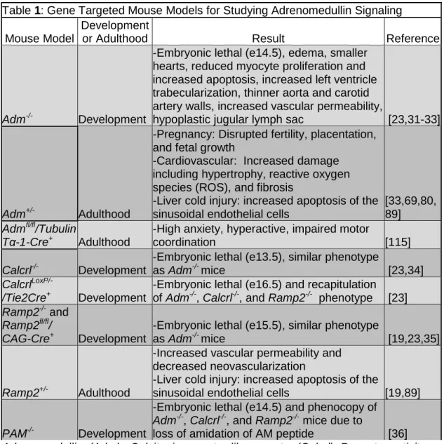
21 Tables:
Table 1: Gene Targeted Mouse Models for Studying Adrenomedullin Signaling
Mouse Model
Development
or Adulthood Result Reference
Adm-/- Development
-Embryonic lethal (e14.5), edema, smaller hearts, reduced myocyte proliferation and increased apoptosis, increased left ventricle trabecularization, thinner aorta and carotid artery walls, increased vascular permeability,
hypoplastic jugular lymph sac [23,31-33]
Adm+/- Adulthood
-Pregnancy: Disrupted fertility, placentation, and fetal growth -Cardiovascular: Increased damage
including hypertrophy, reactive oxygen
species (ROS), and fibrosis -Liver cold injury: increased apoptosis of the
sinusoidal endothelial cells
[33,69,80, 89] Admfl/fl/Tubulin
Tα-1-Cre+ Adulthood
-High anxiety, hyperactive, impaired motor
coordination [115]
Calcrl-/- Development
-Embryonic lethal (e13.5), similar phenotype
as Adm-/- mice [23,34]
Calcrl
LoxP/-/Tie2Cre+ Development
-Embryonic lethal (e16.5) and recapitulation of Adm-/-, Calcrl-/-, and Ramp2-/- phenotype [23] Ramp2-/- and
Ramp2fl/fl/
CAG-Cre+ Development
-Embryonic lethal (e15.5), similar phenotype
as Adm-/- mice [19,23,35]
Ramp2+/- Adulthood
-Increased vascular permeability and
decreased neovascularization -Liver cold injury: increased apoptosis of the
sinusoidal endothelial cells [19,89]
PAM-/- Development
-Embryonic lethal (e14.5) and phenocopy of Adm-/-, Calcrl-/-, and Ramp2-/- mice due to
22
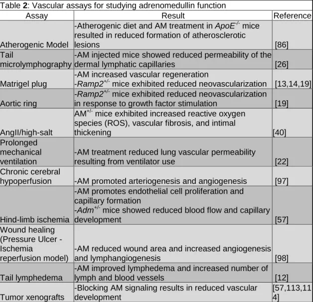
Table 2: Vascular assays for studying adrenomedullin function
Assay Result Reference
Atherogenic Model
-Atherogenic diet and AM treatment in ApoE-/- mice resulted in reduced formation of atherosclerotic
lesions [86]
Tail
microlymphography
-AM injected mice showed reduced permeability of the dermal lymphatic capillaries [26]
Matrigel plug
-AM increased vascular regeneration -Ramp2+/- mice exhibited reduced neovascularization [13,14,19]
Aortic ring
-Ramp2+/- mice exhibited reduced neovascularization in response to growth factor stimulation [19]
AngII/high-salt
AM+/- mice exhibited increased reactive oxygen species (ROS), vascular fibrosis, and intimal
thickening [40]
Prolonged mechanical ventilation
-AM treatment reduced lung vascular permeability
resulting from ventilator use [22] Chronic cerebral
hypoperfusion -AM promoted arteriogenesis and angiogenesis [97]
Hind-limb ischemia
-AM promotes endothelial cell proliferation and
capillary formation -Adm+/- mice showed reduced blood flow and capillary
development [57]
Wound healing (Pressure Ulcer
-Ischemia
reperfusion model)
-AM reduced wound area and increased angiogenesis
and lymphangiogenesis [98]
Tail lymphedema
-AM improved lymphedema and increased number of
lymph and blood vessels [12]
Tumor xenografts
-Blocking AM signaling results in reduced vascular development
23 Figures
Figure 1-1. Fold Change in Plasma Adrenomedullin Levels in a Variety of Human Conditions.
24
Figure 1-2. Adrenomedullin Signaling in Development and Vascular Biology. (A.) Loss of AM signaling causes embryonic lethality due to severe edema
Chapter II: The Lymphatic Vascular System
Function of the Lymphatic System
The lymphatic vascular system is a network of blind-ended microvessels that is critical for regulating and maintaining fluid homeostasis. Tissue fluid, proteins, lipids, and cells are unidirectionally transported through the lymphatic vessels from the interstitial space back to the circulatory system via the thoracic duct. White blood cells and antigen-presenting cells are transported through the lymphatic
vessels to the lymphoid organs. The initial lymphatic capillaries, lacking a basement membrane, consist of a single layer of overlapping endothelial cells that stretch apart to uptake interstitial fluid and proteins. The initial lymphatics lead to the larger
26 Lymphatic Specification and Sprouting
Only in the past few years has there been an increase in the understanding of the development of the lymphatic vascular system with the advent of genetically engineered mouse models. From characterizing the phenotypes of numerous mouse models, several genes have been found to be involved at sequential stages in the development of the lymphatic system as described in several recent
comprehensive review articles [197-199] (Figure 2-1). The initial lymphatic
endothelial cells (LECs) are derived from a subset of venous endothelial cells. This subset of cells begins to express Sox18 at E9.0 [44] that then activates the
expression of Prox1 at E9.5, the transcription factor that is the master genetic regulator of LEC fate, in a polarized manner [200].
In order for LECs to bud away from the venous endothelial cells, VEGFR3 expression becomes localized to the polarized area of LECs [201]. The vascular smooth muscle cells (vSMCs) and mesenchymal cells near the LECs begin to express VEGFC, the ligand for VEGFR3 [202]. Then, LECs begin to respond to VEGFC via VEGFR3 to sprout away from the cardinal vein initiating lymph sac
formation.
Lymph Sac Formation and Proliferation
27
between E11-E14.5. Numerous genes have been found to play a role in the formation of the lymph sacs and primary lymphatic plexus including Adm, Calcrl, Ramp2, Spred-1/2, Slp-76, Syk, and Plcγ2.
Platelets are known to be important in the development of the lymphatic vascular system because of their ability to activate the hematopoietic SKY-SLP-76 signaling pathway. The C-type lectin receptor on platelets binds to podoplanin, expressed on LECs, to activate the SKY-SLP-76 signaling [204]. A study by
Bertozzi, et al showed that disruption of this process in lymphatics results in aberrant vascular connections and blood-lymphatic mixing [204]. Therefore, platelets and SYK-SLP-76 signaling are necessary for proper embryonic blood-lymphatic vascular separation.
The Caron Lab identified Adm, Calcrl, and Ramp2 as a novel G-protein coupled receptor (GPCR) pathway that is a critical regulator of lymphatic vascular development [23]. Adm is temporally and spatially expressed on the endothelium of the jugular vein in a polarized fashion towards the budding primary lymph sac in vivo, which is identical to the lymphatic-specific transcriptional regulator, Prox1
[43,205,206]. Moreover, Calcrl and Ramp2 are preferentially up-regulated in LECs, partially under the control of the transcription factor, Prox1 [23]. Gene knockout mice for Adm [31], Calcrl [34], and Ramp2 [23,35] all exhibit mid-gestation embryonic lethality characterized by hydrops fetalis due to defects in the
28
decreased LEC proliferation [23]. These results indicate that AM is necessary for proper morphological development and proliferation of the embryonic lymphatics.
Lymphatic Plexus Remodeling and Maturation
After formation of the primary lymphatic plexus, maturation of the lymphatic vascular network occurs with the involvement of various genes, some of which include Neuropilin-2, Foxc2, Lyve-1, Ephrin B2, Angiopoietin-2, Fiaf, and Integrin-α9. The maturation process continues several days postnatally to generate the different types of LECs from small capillaries to larger collecting vessels with valves and vSMC coverage. Interestingly, a recent study has implicated the interaction of vSMCs with LECs to be important in the development of the larger collecting lymphatic vessels [207]. This study showed that recruitment of vSMCs to LECs enhanced signaling of an extracellular matrix glycoprotein, Reelin, that is expressed from LECs and regulates lymphatic development and function [207]. After
development of the lymphatic system is completed, the vessels are typically quiescent unless growth is triggered in response to a stimulus, such as a wound/injury.
Summary
29
deletion of Calcrl during adulthood resulted in overall lymphangiectasia, dilation of lymphatic vessels, with disruption in the function and permeability of the vessels. A second study using an ear wound assay in adult mice haplo-insufficient for Adm determined that there is reduced lymphangiogenesis, while angiogenesis is unaffected in response to the wound. The third and last study in this dissertation focused on understanding why there are differential responses to AM signaling in the lymphatic and blood vascular systems. While both systems express AM receptors and respond to the ligand, there is an enhanced effect of AM signaling on the
lymphatic system. This study indicated that complement factor H (CFH), the binding protein for AM, is upregulated in LECs as compared to blood endothelial cells
30 Figures
Figure 2-1. Stepwise process of lymphangiogenesis. The development of the lymphatic vasculature begins with specification of lymphatic endothelial cells from venous precursors. The chemoattractant and growth promoting properties of numerous growth factors, including VEGFC, causes lymphatic endothelial cells to sprout and separate from veins to form primitive lymph sacs. Proliferation of lymph sacs leads to the formation of a primary lymphatic plexus which is later remodeled into the mature lymphatic vascular system. Below each step is a list of genes for which a functional role has been demonstrated using genetically engineered mouse models. Reprinted with permission from [199].
Chapter III: Characteristics of multi-organ lymphangiectasia resulting from temporal deletion of calcitonin receptor-like receptor in adult mice2
Overview
Adrenomedullin (AM) and its receptor complexes, calcitonin receptor-like receptor (Calcrl) and receptor activity modifying protein 2/3, are highly expressed in
lymphatic endothelial cells and are required for embryonic lymphatic development. To determine the role of Calcrl in adulthood, we used an inducible Cre-loxP system to temporally and ubiquitously delete Calcrl in adult mice. Following tamoxifen injection, Calcrlfl/fl/CAGGCre-ERTM mice rapidly developed corneal edema and inflammation that was preceded by and persistently associated with dilated corneoscleral lymphatics. Lacteals and submucosal lymphatic capillaries of the intestine were also dilated, while mesenteric collecting lymphatics failed to properly transport chyle after an acute Western Diet, culminating in chronic failure of
Calcrlfl/fl/CAGGCre-ERTM mice to gain weight. Dermal lymphatic capillaries were also dilated and chronic edema challenge confirmed significant and prolonged dermal lymphatic insufficiency. In vivo and in vitro imaging of lymphatics with either genetic or pharmacologic inhibition of AM signaling revealed markedly disorganized lymphatic junctional proteins ZO-1 and VE-cadherin. The maintenance of AM
2
32
signaling during adulthood is required for preserving normal lymphatic permeability and function. Collectively, these studies reveal a spectrum of lymphatic defects in adult Calcrlfl/fl/CAGGCre-ERTM mice that closely recapitulate the clinical symptoms of patients with corneal, intestinal and peripheral lymphangiectasia.
Introduction
33
Failure of lymphatic vessels to function properly in adults can result in numerous types of clinical conditions, including primary and secondary
lymphedema, which can have a broad range of clinical presentations and associated correlates[211,212]. Some congenital forms of primary lymphedema are associated with lymphangiectasia, which is typically characterized as dilation and enlargement of lymphatic vessels. Interestingly, there are a few organ systems, including the intestine[213], the conjunctiva of the eye[214] and the dermis[215], that are particularly prone to developing lymphangiectasia. While the pathophysiological mechanisms leading to lymphangiectasia are not well understood, it is likely that dilated lymphatic vessels are the result of lymphatic obstruction and improper drainage or lymph stasis. The consequences of persistent lymphangiectasia include, on a cellular level, increased permeability of dilated lymphatic vascular beds, and on a systemic level, protein-losing enteropathy, limb lymphedema and ocular irritation with dryness. Although lymphangiectasia can be associated with a variety of primary, congenital lymphedema syndromes, there is currently no known genetic pathway that directly and predominantly contributes to lymphangiectasia.
34
referred to as the adrenomedullin 1 (AM1) receptor, while the CLR and RAMP3 complex is referred to as the AM2 receptor; both of which bind AM peptide, but differ in their relative binding affinities [216]. Gene knockout mice for Adm[31], Calcrl[34], and Ramp2[23,35] all exhibit mid-gestational embryonic lethality characterized by hydrops fetalis, or marked edema, that is associated with arrested lymphatic vascular development. Conditional deletion of Calcrl in endothelial cells confirmed that AM signaling, and its downstream activation of the MAPK/ERK signaling cascade, is required for normal lymphatic endothelial cell proliferation during development.
AM signaling through Calcrl/Ramp2 also has robust effects on endothelial cell permeability. For example, AM can abrogate the permeabilizing effects of hydrogen peroxide and thrombin on human umbilical vein endothelial cells[20] and it can retard the transport of molecules across the blood brain barrier by tightening the permeability of cerebral endothelial cells[21,217]. Similarly, we have shown that AM can impact the permeability and function of lymphatic endothelial cells (LECs). Treatment of cultured LECs with AM significantly and functionally reduced their permeability by causing a subcellular reorganization of the junctional proteins ZO-1 and VE-Cadherin[26]. Furthermore, in vivo tail microlymphography reinforced these findings since mice injected with AM showed reduced lymph velocity through dermal lymphatic capillaries, indicative of functionally reduced permeability[26].
35
[24,25,218]. Consistent with this notion, continuous administration of AM promoted lymphangiogenesis and ameliorated secondary tail lymphedema in a surgical injury mouse model [12]. Whether the maintained expression of Calcrl in adult animals is also required for appropriate lymphatic function remains unclear. To address this question, we used a ubiquitously expressed, tamoxifen-inducible Cre transgenic mouse line (CAGGCre-ERTM) to delete a floxed Calcrl gene in 3-4 month old animals and thus explore the role of Calcrl during adulthood. Our results continue to support a preferential role for Calcrl in the lymphatic vasculature and reveal that Calcrl expression in adult animals is critical for maintaining the proper function of lymphatic vessels in a wide variety of organs.
Methods Animals
Mice used in these studies were generated from crossing Calcrlfl/fl [23] mice (N7-10 on C57BL/6 background) to CAGGCre-ERTM mice (The Jackson Laboratory, Bar Harbor, ME 004682, B6.Cg-Tg(CAG-Cre/Esr1)5Amc/J). Male and female adult mice aged 3-4 months were administered tamoxifen (Sigma) consecutively for 5 days (5mg/40g body weight; IP). Mice were genotyped for the floxed and Cre alleles as well as the excised allele after tamoxifen injection. Primer sets (5’-3’) P1:
36
For Western Diet studies, mice were fed Teklad Adjusted Calories Diet (TD.88137; 42% from fat; Harlan Laboratories) for 1½ weeks and then housed in metabolic cages for 24 hours during which food intake, urine, and fecal samples were measured. Weights of mice were also recorded before tamoxifen injection, after tamoxifen injection, and after Western Diet.
All experimental procedures involving mice were approved by the Institutional Animal Care and Use Committee of The University of North Carolina Chapel Hill and all efforts were made to minimize suffering.
Cell Culture
Human adult dermal lymphatic endothelial cells (HMVEC-dLyAd-Der Lym Endo Cells, Lonza) of 8 passages or less were maintained using EGM-2MV media with bullet kit (Lonza). Cells were seeded in 6 well plates at 100,000 cells/well and grown on acid washed coverslips until monolayers formed. Treatment conditions included no treatment (control), 10nM AM (American Peptide Co., Inc.), 1µM AM22-52 (AM antagonist; American Peptide Co., Inc.) or AM+AM22-AM22-52. Cells were
37
with 2% normal donkey serum (NDS) for 10 minutes, followed by incubation with secondary antibody for 1 hour at room temperature, rinsed 3x5 minutes with PBS and then mounted on slides using Mowiol.
Immunohistochemistry and Immunofluorescence
Tissues were dissected, fixed with 4% PFA overnight and embedded in paraffin or protected in 30% sucrose and embedded in OCT (Tissue-Tek) for sectioning. Sections were permeabilized using 0.1% Triton X-100 (in 0.01M PBS; pH 7.2; 15 minutes), blocked with 5% NDS (in 0.1% Triton X-100; 30 minutes), incubated overnight in primary antibodies, PBS rinsed (3x5minutes), blocked with 5% NDS (30 minutes), incubated with secondary antibodies (2 hours), rinsed with PBS and coverslipped with Mowiol. Primary antibodies included: LYVE-1 (1:200; polyclonal rabbit α mouse; Fitzgerald, Acton, MA), podoplanin (1:200, Syrian hamster α mouse, Developmental Studies Hybridoma Bank, Univ. Iowa), ZO-1 (1:200, monoclonal rat α mouse; clone R40.76, Millipore, Billerica, MA) and VE-Cadherin (1:200, goat polyclonal; sc-6458, Santa Cruz Biotechnology, Santa Cruz, CA). Secondary antibodies included Alexa Fluor 594, Alexa Fluor 488 and Cy3 (1:200, Jackson Immunoresearch) and nuclear marker DAPI (1:1000, bisbenzimide 33258; Sigma, St. Louis, MO). TUNEL staining was performed using the ApopTag Fluorescein In Situ Apoptosis Detection Kit (S7110, Chemicon International)
38 Tonometry
Tonometry was performed in anesthetized adult mice using a TonoLab tonometer (Colonial Medical Supply) as described previously [219,220]. After avertin injection, a drop of tetracaine hydrochloride 0.5% (Alcon) was placed on the eye as a local anesthetic. Eyes were lubricated throughout testing with TEARS Naturale FORTE (Alcon). At least six readings were recorded per eye and averaged.
Tail Microlymphography and Vessel Diameter
Three to four months post tamoxifen injection, adult mice were used for tail microlymphography as described previously[26] with several modifications. FITC-conjugated dextran (200kDa; 1µl; Molecular Probes, Invitrogen Detection
Technologies) was injected intradermally into the mouse tail using a 5µl Hamilton syringe fitted with a 30 gauge needle. Images were taken every minute for 15 minutes and image analysis was performed using Adobe Photoshop 7.0 and Image J.
Lymphatic and Blood Permeability Assays
An ear lymphatic permeability assay was performed as previously
described[221] with minor modification. Ears of anesthetized mice were injected intradermally with 2µl of 0.5% Evan’s Blue dye (in saline) with a 10ul Hamilton
39
modifications to the protocol [222]. Anesthetized mice were retro-orbitally injected with 200µl 0.5% Evan’s Blue dye (in saline). After 30 minutes, the mice were perfused with saline and the liver, lung, adductor muscle, spleen, intestine, heart, and brain were harvested. Tissues were weighed and desiccated overnight at 55ºC followed by formamide extraction (55ºC, overnight) and 100µl was used for
absorbance reading at 600nm.
Acid Steatocrit/Lipase/Triglyceride Measurements
Fecal samples collected after Western Diet were examined by testing for fecal steatocrit and fecal lipase as previously described[223] with recent
modifications[224]. Fecal specimens were powdered and mixed with 1N perchloric acid and 0.5% oil red O and placed in a capillary tube and centrifuged. Steatocrit was calculated as 100 x {length of fatty layer/(length of solid layer+length of fatty layer)} . Fecal lipase and serum triglycerides were analyzed at the Animal Clinical Chemistry and Gene Expression Labs (UNC-CH).
Dot Blot Assay
40
(1:2000 dilution) was dotted onto a nitrocellulose membrane. The membrane was blocked (TBS+3% nonfat dry milk) for 2 hours at room temperature, rinsed with TBST (1x5minutes), then incubated overnight with primary antibody (mouse anti alpha-1 antitrypsin-1:500; Novus and mouse anti-actin-1:10000; Sigma) in TBST+ 3% nonfat dry milk at 4oC. The membrane was rinsed with TBST (3x10 minutes), incubated with secondary antibody in TBST (HRP goat anti-mouse; 1:2000; Upstate) for 45 minutes at room temperature followed by TBST (3x5 minutes) rinses and a final TBS (1x5 minutes) rinse . The membrane was then developed with film (GeneMate) using WesternBright ECL reagents (Advansta). Analysis of integrated density was performed using Image J.
Edema Formation Assay
Anesthetized mice were injected with 10µl of 4µg/µl Complete Freund’s Adjuvant (CFA) in one hind paw. The other hind paw served as an internal control. Paw thickness was measured with calipers before injection of CFA and every other day after injection up to 21 days.
RNA and qRT-PCR
41
using Assay-on-Demand for Calcrl (Mm00516986_m1; Applied Biosystems). The comparative quantitation (∆∆CT) method was used to determine the relative level of
Calcrl expression in the tissues compared to mouse embryo total RNA calibrator
(Ambion). All assays were repeated at least three times and run in duplicate.
Statistical analysis
All experiments were repeated at least 3 times and data are expressed as means with SEM values. Student t tests (tails=2, type=3) and two-way ANOVA were performed and P≤0.05 was considered significant.
Results
Temporal deletion of Calcrl results in acute onset eye phenotype with enlarged corneoscleral lymphatic vessels
Tamoxifen injection resulted in a significant reduction of Calcrl gene
expression in Calcrlfl/fl/CAGGCre-ERTM animals compared to Calcrlfl/fl animals and to Calcrlfl/fl/CAGGCre-ERTM non-injected animals (control animals) as indicated by qRT-PCR of lung and heart tissue (Figure 3-S2). Within 7 to 10 days of tamoxifen
42
To this end, TUNEL staining indicated no difference in retinal ganglion cell death between Calcrlfl/fl/CAGGCre-ERTM and Calcrlfl/fl control mice (Figure 3-S1A and 2-S1B). We also found no significant histological differences in the optic nerve of Calcrlfl/fl/CAGGCre-ERTM mice compared to Calcrlfl/fl control mice (data not shown). Finally, we found no significant difference in the intraocular pressure of
Calcrlfl/fl/CAGGCre-ERTM and Calcrlfl/fl control mice when compared either before injection or after injection of tamoxifen (Figure 3- S1C) and all intraocular pressure measurements were within the normal range for C56BL/6 mice[219]. Taken together, these data rule out the possibility that the acute-onset eye phenotype in Calcrlfl/fl/CAGGCre-ERTM mice is associated with classical features of glaucoma. However, hematoxylin and eosin staining of eyes revealed marked changes in histology of the Calcrlfl/fl/CAGGCre-ERTM corneas relative to those of Calcrlfl/fl control mice. The corneas of Calcrlfl/fl/CAGGCre-ERTM mice were thickened and edematous and often showed a disrupted and damaged epithelial lining (Figure 3-1 C,D, arrow). We also observed pronounced inflammation in the anterior chamber and cornea of Calcrlfl/fl/CAGGCre-ERTM mice (Figure 3-1 E,F, arrowhead).
Based on the well-established role of Calcrl in lymphatic vascular
43
corneoscleral junction of Calcrlfl/fl/CAGGCre-ERTM mice were significantly dilated and twice the size of corneoscleral lymphatics in Calcrlfl/fl control mice (Figure 3-1 I,J,K), similar to the phenotype observed in humans with conjunctival lymphangiectasia. More importantly, the eyes of Calcrlfl/fl/CAGGCre-ERTM mice that did not present with the overt corneal pathology (approximately 1/3rd of the mice), either because they failed to develop the phenotype or they were euthanized prior to the onset of the phenotype, still showed significantly dilated lymphatics at the corneoscleral junction. Taken together, these data demonstrate that an abnormal lymphatic vessel phenotype precedes the onset of acute corneal pathology in Calcrlfl/fl /CAGGCre-ERTM mice.
Calcrlfl/fl/CAGGCre-ERTM mice exhibit enlarged submucosal lymphatic vessels and lacteals in the intestine with dysfunctional mesenteric collecting lymphatic vessels.
Since dilated lymphatic vessels were observed at the corneoscleral junction in the Calcrlfl/fl/CAGGCre-ERTM mice, we wanted to assess the morphology and
function of lymphatic vessels in other lymphatic vascular beds, for example, within the intestine. The overall histology of the intestines of Calcrlfl/fl/CAGGCre-ERTM mice was normal when compared to that of Calcrlfl/fl control mice under normal conditions (Figure 3-2A,B). Lymphatic vessels within the intestine were identified with LYVE-1 and podoplanin staining, showing co-localization within the lacteals and the
44
submucosal lymphatics and a greater proportion of villi sections revealing enlarged, LYVE-1-positive lacteals. Once again, these dilated lymphatics vessels are
reminiscent of the dilated lymphatics observed in human patients with intestinal lymphangiectasia.
Intestinal lymphatics are required for normal lipid absorption, and patients with intestinal lymphangiectasia often present with weight loss as a result of lipid malabsorption [227]. Therefore, the function of these vessels was evaluated by placing Calcrlfl/fl/CAGGCre-ERTM and Calcrlfl/fl control mice on a short term Western Diet following an overnight fast. After 1½ hours of Western Diet, the
Calcrlfl/fl/CAGGCre-ERTM mice exhibited chyle-filled mesenteric lymphatic vessels which were not visible in the Calcrlfl/fl control mice (Figure 3-2H,I). Chyle-filled submucosal lymphatic vessels were also visibly distinguishable in
45
Reduced body weight and impaired lipid absorption with protein-losing enteropathy
in Calcrlfl/fl/CAGGCre-ERTM mice
We next wanted to assess the impact of a longer term high fat diet on intestinal lipid absorption in the Calcrlfl/fl/CAGGCre-ERTM mice. There were no significant differences in body weights between 3-4 month old, male or female Calcrlfl/fl/CAGGCre-ERTM mice and Calcrlfl/fl control mice before the injection of
tamoxifen (Figure 3-3A,B). However, 3-4 months after the injection of tamoxifen, we found that the Calcrlfl/fl/CAGGCre-ERTM mice weighed significantly less than their control counterparts (Figure 3-3A,B), indicating that the tamoxifen-induced loss of Calcrl contributes to a failure of Calcrlfl/fl/CAGGCre-ERTM to gain weight and thrive. The failure of Calcrlfl/fl/CAGGCre-ERTM mice to gain weight and thrive was
significantly exacerbated (Figure 3-3A,B) and visibly apparent (Figure 3-3C) when the mice were fed a Western Diet for 1½ weeks.
Moreover, fecal acid steatocrit levels, representative of lipid excretion levels, were significantly elevated in Calcrlfl/fl/CAGGCre-ERTM animals maintained on a Western Diet for 1½ weeks compared to similarly fed Calcrlfl/fl control animals (Figure 3-3D), demonstrating reduced lipid absorption. Consistently, fecal
46
mice and supporting the conclusion that their failure to gain weight is due to
abnormal lipid absorption in the intestine. Finally, fecal samples of Western Diet-fed Calcrlfl/fl/CAGGCre-ERTM mice contained a significantly elevated level of alpha-1 antiptrysin—a clinical diagnostic marker for protein-losing enteropathy—compared to similarly fed Calcrlfl/fl control mice (Figure 3-3G,H).
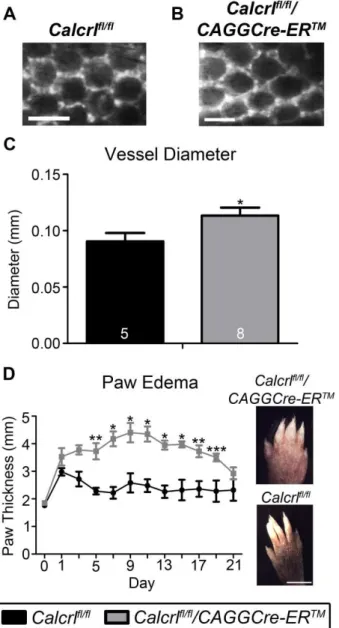
Temporal deletion of Calcrl results in increased dermal lymphatic capillaries with exacerbated and prolonged edema.
The dermal lymphatic capillaries of Calcrlfl/fl/CAGGCre-ERTM mice also exhibited significant dilation and dysfunction. Specifically, intradermal injection of a large molecular weight (200kDa) FITC-dextran into the subdermal area of the tail tip revealed significantly enlarged dermal capillaries in Calcrlfl/fl/CAGGCre-ERTM mice compared to Calcrlfl/fl control mice (Figure 3-4A,B,C). Despite this dermal
lymphangiectasia, we noticed that at the basal or quiescent state,
47
(Figure 3-4D). These data demonstrate that the expression of Calcrl is required for maintaining highly effective lymphatic function under conditions of edema and inflammation.
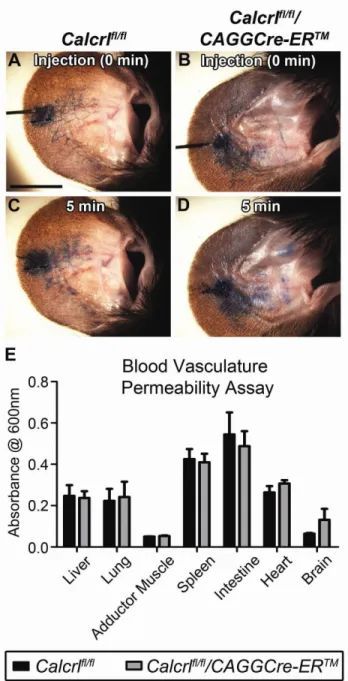
Calcrlfl/fl/CAGGCre-ERTM mice exhibit increased lymphatic capillary permeability with no apparent disruption of blood vascular permeability.
To better evaluate the permeability of lymphatic and blood vasculatures in Calcrlfl/fl/CAGGCre-ERTM and Calcrlfl/fl mice, we used the small molecular weight Evan’s blue dye which can freely penetrate in and out of dermal capillaries. Injection of 0.5% Evan’s blue dye intradermally in the ear showed rapid uptake of the dye by dermal lymphatics in both Calcrlfl/fl/CAGGCre-ERTM and Calcrlfl/fl mice (Figure2- 5A,B). However after 5 minutes, Calcrlfl/fl/CAGGCre-ERTM mice exhibited increased leakage of the dye from the lymphatic vessels, as evidenced by the diffuse
spreading of the dye and poorly demarcated lymphatics throughout the ear region compared to the Calcrlfl/fl control mice (Figure 3-5C,D). To determine whether this lymphatic permeability defect was impacted or perhaps confounded by a
48
Calcrl in adult animals results in increased lymphatic capillary permeability with no
overt or functional changes in blood vascular permeability.
Inhibition of AM signaling results in disorganization of lymphatic endothelial cell junctions.
To elucidate the molecular mechanisms contributing to the lymphatic
dysfunction in Calcrlfl/fl/CAGGCre-ERTM mice, we evaluated VE-Cadherin expression and localization in mesenteric lymphatic vessels of Calcrlfl/fl/CAGGCre-ERTM and Calcrlfl/fl control mice that had been fed a high fat diet for 1½ hours. VE-Cadherin expression was visibly disrupted in lymphatic vessels of Calcrlfl/fl/CAGGCre-ERTM mice (Figure 3-6B,D) compared to control mice (Figure 3-6A,C). More specifically, while the relative expression levels of VE-cadherin appeared similar between genotypes, the VE-cadherin in mesenteric lymphatic vessels of Calcrlfl/fl /CAGGCre-ERTM appeared as punctate lobules throughout the cells and was not localized to well-defined cell boundaries, as seen in the Calcrlfl/fl control mice.
49
resulted in highly disorganized and jagged junctional protein configurations (Figure 3-6G,H). Taken together, these results demonstrate that in vitro and in vivo inhibition of Calcrl signaling, either by antagonist treatment or by genetic deletion, results in a profound loss of junctional protein organization, likely resulting in increased
permeability of lymphatic endothelial cell barriers.
Discussion
These studies demonstrate that temporal loss of murine Calcrl in adulthood causes lymphatic insufficiency in a wide range of organs, representing functional similarities to the sequelae observed in patients with a variety of lymphangiectasia conditions. Consistently, the lymphatic vessels in the eye, intestine and skin of Calcrlfl/fl/CAGGCre-ERTM mice were dilated, had irregular junctional protein
organization and were dysfunctional when challenged with either fat absorption or edema and inflammation. Taken together, these data identify an important new role for AM signaling as a potent regulator of lymphatic vascular drainage and
permeability in adult animals.