Guidance for the treatment of deep vein thrombosis
and pulmonary embolism
Michael B. Streiff1• Giancarlo Agnelli2• Jean M. Connors3•Mark Crowther4• Sabine Eichinger5•Renato Lopes6•Robert D. McBane7•Stephan Moll8• Jack Ansell9
Published online: 16 January 2016
ÓThe Author(s) 2016. This article is published with open access at Springerlink.com
Abstract This guidance document focuses on the diag-nosis and treatment of venous thromboembolism (VTE). Efficient, cost effective diagnosis of VTE is facilitated by combining medical history and physical examination with pre-test probability models, D dimer testing and selective use of confirmatory imaging. Clinical prediction rules, biomarkers and imaging can be used to tailor therapy to disease severity. Anticoagulation options for acute VTE include unfractionated heparin, low molecular weight heparin, fondaparinux and the direct oral anticoagulants (DOACs). DOACs are as effective as conventional therapy
with LMWH and vitamin K antagonists. Thrombolytic therapy is reserved for massive pulmonary embolism (PE) or extensive deep vein thrombosis (DVT). Inferior vena cava filters are reserved for patients with acute VTE and contraindications to anticoagulation. Retrievable filters are strongly preferred. The possibility of thoracic outlet syn-drome and May-Thurner synsyn-drome should be considered in patients with subclavian/axillary and left common iliac vein DVT, respectively in absence of identifiable triggers. The optimal duration of therapy is dictated by the presence of modifiable thrombotic risk factors. Long term antico-agulation should be considered in patients with unprovoked VTE as well as persistent prothrombotic risk factors such as cancer. Short-term therapy is sufficient for most patients with VTE associated with transient situational triggers such as major surgery. Biomarkers such as D dimer and risk assessment models such the Vienna risk prediction model offer the potential to customize VTE therapy for the indi-vidual patient. Insufficient data exist to support the inte-gration of bleeding risk models into duration of therapy planning.
Keywords Anticoagulant therapyVenous
thromboembolismDeep vein thrombosis Pulmonary embolism NOACs DOACs
Introduction
Venous thromboembolism (VTE) which consists princi-pally of deep vein thrombosis (DVT) and pulmonary embolism (PE) is a common cause of morbidity and mor-tality. Consequently, health care providers in all clinical settings will be faced with managing patients with this illness. Numerous evidence-based guidelines are available
& Michael B. Streiff [email protected]
1 Division of Hematology, Department of Medicine and Pathology, The Johns Hopkins University School of Medicine, Baltimore, MD, USA
2 Stroke Unit, Department of Internal Medicine, University of Perugia, Perugia, Italy
3 Hematology Division, Brigham and Women’s Hospital, Dana Farber Cancer Institute, Harvard Medical School, Boston, MA, USA
4 Departments of Medicine and Pathology and Molecular Medicine, McMaster University, Hamilton, Canada
5 Department of Medicine, Medical University of Vienna, Vienna, Austria
6 Division of Cardiology, Department of Medicine, Duke University Medical Center, Durham, NC, USA
7 Cardiovascular Division, Department of Medicine, Mayo Clinic, Rochester, MN, USA
8 Department of Medicine, University of North Carolina School of Medicine, Chapel Hill, NC, USA
9 Department of Medicine, Hofstra North Shore/LIJ School of Medicine, Hempstead, NY, USA
to assist providers in clinical decision-making. However, there are many clinical scenarios where a paucity of data exist. The purpose of this guidance document is to provide advice to providers on all aspects of the treatment of VTE based upon the best available information including situa-tions where evidence is limited.
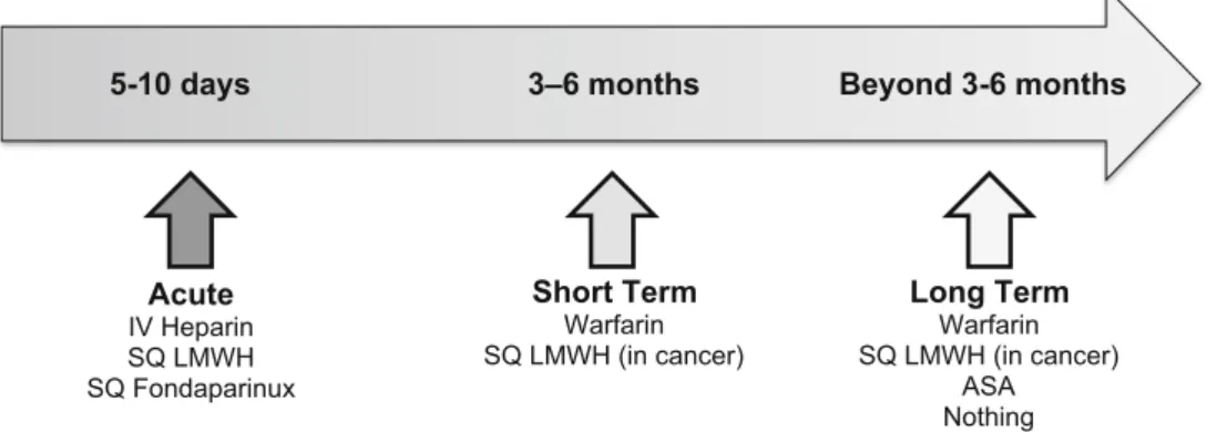
Many authorities divide the therapy of VTE into various phases of treatment following the initial diagnosis based upon the risk of recurrence. For the purposes of this guidance document, we consider the initial treatment of VTE, the ‘‘acute’’ phase, to encompass the first 5–10 days which corresponds to the time period when patients his-torically have been treated with parenteral therapy. The next 3–6 months, we consider the ‘‘short term’’ treatment phase of therapy. After 3–6 months, we apply the term ‘‘long term’’ treatment of VTE when the benefit/risk of continued treatment becomes a critical aspect of the
decision making process. Figure 1 illustrates this contin-uum of care.
Methods
To provide guidance on the management of VTE, the authors developed a list of important management ques-tions to be considered in this document (Table1). Ques-tions were developed by consensus of all the authors. To answer these questions, a literature search of MEDLINE and EMBASE from January 2004 to August 2014 was conducted. The following search terms were used and combined: anticoagulant treatment, anticoagulant therapy, antithrombotic treatment, heparin, low molecular weight heparin, enoxaparin, nadroparin, dalteparin, certoparin, bemiparin, tinzaparin, parnaparin, reviparin, vitamin K Acute
IV Heparin SQ LMWH SQ Fondaparinux
Short Term
Warfarin SQ LMWH (in cancer)
5-10 days 3–6 months Beyond 3-6 months
Long Term
Warfarin SQ LMWH (in cancer)
ASA Nothing
Fig. 1 The different phases of treatment and traditional therapies in venous thromboembolism
Table 1 Guidance questions to
be considered How is the diagnosis of deep vein thrombosis and pulmonary embolism established?
Which patients require hospitalization versus initial outpatient therapy for the management of VTE? What are the therapeutic options for the acute treatment of venous thromboembolism?
Which patients are candidates for a DOAC?
What is the role of vena cava filters if the patient is not a candidate for anticoagulation? How is upper extremity VTE treated?
When is ambulation/exercise safe after DVT/PE?
Is the use of graduated compression stockings safe after acute DVT/PE? What is the recommended duration of therapy for VTE?
What is the recommended duration of therapy for a patient with distal DVT?
What is the recommended duration of therapy for a patient with a surgically provoked VTE? What is the recommended duration of therapy for a pregnancy or estrogen-associated VTE?
What is the recommended duration of therapy for a medical illness-associated VTE? What is the recommended duration of therapy for a travel-associated VTE? What is the recommended duration of therapy for a malignancy-associated VTE? What is the recommended duration of therapy for a patient with unprovoked DVT/PE? What are the therapeutic options for long term treatment of DVT/PE?
antagonists, warfarin, acenocoumarol, phenprocoumon, thrombolysis, thrombolytic treatment, fibrinolytic agent, fibrinolysis, urokinase, tenecteplase, alteplase, rtPA, tPA; aspirin, ticlopidine, clopidogrel; venous thromboembolism, venous thrombosis, deep venous thrombosis, deep vein thrombosis, superficial venous thrombosis, superficial venous thrombophlebitis; diagnosis. The search strategy was restricted to papers published in English. Detailed information on the results of the literature search is avail-able upon request.
For papers published before 2004, we only considered the most important studies that were likely to influence our responses to the questions. These studies were selected and suggested by the authors of this guidance document.
Guidance
(1) How is the diagnosis of deep vein thrombosis and pulmonary embolism established?
Deep vein thrombosis should be suspected in any patient who presents with unexplained extremity swelling, pain, warmth or erythema. Pain associated with DVT is often described as being a cramp or ache in the calf or thigh. Pulmonary embolism is often heralded by development of dyspnea and pleuritic chest or back pain. Pulmonary embolism can also cause progressive fatigue, dyspnea on exertion, syncope or pre-syncope or sudden death. Since these symptoms can be caused by many diseases, the likelihood of VTE can be estimated by assessing a patient’s thrombosis risk factors (Table2) [1, 2]. The presence of these disease processes should be elicited in the history when assessing a patient for VTE.
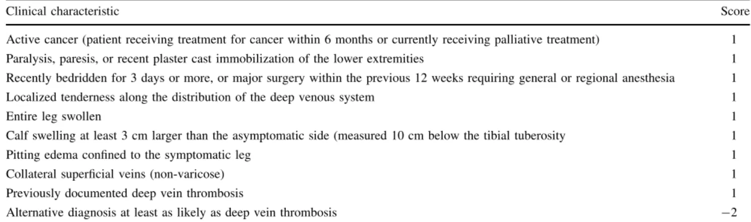
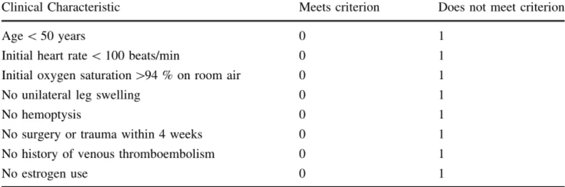
Pre-test probability models have been developed to facilitate a consistent and structured approach to the diagnosis of VTE. The best studied and validated models are the Wells’ criteria for DVT and PE diagnosis and the Geneva Score for PE diagnosis (Tables 3, 4, 5) [3–5]. In conjunction with D dimer testing, these models have been demonstrated to safely exclude a DVT or PE without use of objective diagnostic imaging in outpatients presenting with suspected VTE. A wide variety of D dimer assays are available on the market for use in VTE diagnosis. Highly sensitive assays include enzyme-linked immunofluores-cence assays (sensitivity 96 %; 95 % CI 89–98 %), microplate enzyme-linked immunosorbent assays (ELI-SAs) (sensitivity 94 %; 95 % CI 86–97 %), and quantita-tive latex or immunoturbidimetric assays (sensitivity 93 %; 95 % CI 89–95 %). Whole blood red cell agglutination assays (sensitivity 83 %; 95 % CI 67–93 %) and semi-quantitative latex bead agglutination assays (sensitivity 85 %; 95 % CI 68–93 %) are considered moderately sen-sitive D dimer assays. Since the sensitivity of D dimer assays varies considerably, it is important to follow man-ufacturer recommendations closely when using D dimer assays in the diagnosis of VTE, [6]. The Pulmonary Embolism Rule-out Criteria (PERC) is a clinical decision support tool developed by Kline and coworkers to identify outpatients presenting with chest pain who are thought to be at low risk for PE in whom further diagnostic testing can be avoided (Table6) [7, 8]. A recent metaanalysis of 12 studies encompassing over 14,000 patients confirmed the accuracy of the PERC [9]. Consequently, the PERC was included in the American College of Physician’s Practice Guideline on the diagnosis of pulmonary embolism [10]. A Table 2 Risk factors for first episode of venous thromboembolism
Genetic Risk Factors
Antithrombin deficiency Protein C deficiency Protein S deficiency
Factor V Leiden
Prothrombin gene mutation Non-O ABO blood group Dysfibrinogenemia Elevated Factor VIII Elevated Factor IX Elevated Factor XI
Hyperhomocysteinemia (including homocystinuria)
Acquired Risk Factors
Increasing age Cancer
Antiphospholipid syndrome Infections (HIV, Sepsis, etc.)
Inflammatory disorders (e.g. SLE, IBD, vasculitis, etc.) Nephrotic syndrome
Obesity
Smoking Environmental
Surgery (major inpatient, ambulatory) Trauma
Immobilization Central venous catheter Pregnancy/post-partum
Hormonal therapy (e.g. oral, transcutaneous, vaginal ring contraceptive, Depot progestin injections, hormone replacement, etc.)
Chemotherapy
schematic depiction of the use of the Wells criteria and the Geneva Score in conjunction with the PERC and D dimer testing in the diagnosis of DVT and PE is displayed in Figs.2and3[11]. A recent patient level meta-analysis of
studies using the Wells rule in exclusion of DVT found that in conjunction with a negative D dimer test, the Wells Score was safe and efficient in men and women, both inpatients and outpatients. A notable exception was Table 3 Wells clinical DVT model
Clinical characteristic Score
Active cancer (patient receiving treatment for cancer within 6 months or currently receiving palliative treatment) 1 Paralysis, paresis, or recent plaster cast immobilization of the lower extremities 1 Recently bedridden for 3 days or more, or major surgery within the previous 12 weeks requiring general or regional anesthesia 1
Localized tenderness along the distribution of the deep venous system 1
Entire leg swollen 1
Calf swelling at least 3 cm larger than the asymptomatic side (measured 10 cm below the tibial tuberosity 1
Pitting edema confined to the symptomatic leg 1
Collateral superficial veins (non-varicose) 1
Previously documented deep vein thrombosis 1
Alternative diagnosis at least as likely as deep vein thrombosis -2
A score ofB0 indicates that a low pretest probability of deep vein thrombosis. A score of 1 or 2 points indicates a moderate risk of DVT and a score of 3 or higher indicates a high risk of deep vein thrombosis [152]
Table 4 Wells clinical pulmonary embolism model
Clinical characteristic Score
Active cancer (patient receiving treatment for cancer within 6 months or currently receiving palliative treatment) 1 Surgery or bedridden for 3 days or more during the past 4 weeks 1.5
History of deep venous thrombosis or pulmonary embolism 1.5
Hemoptysis 1
Heart rate[100 beats/min 1.5
Pulmonary embolism judged to be the most likely diagnosis 3
Clinical signs and symptoms compatible with deep venous thrombosis 3
A score of\2 indicates a low probability of pulmonary embolism. A score of 2–6 indicates an intermediate probability of PE. A score of more than 6 indicates a high probability of pulmonary embolism. Kearon C, Ginsberg JS, Douketis J, et al. (2006) An evaluation of D-dimer in the diagnosis of pulmonary embolism: a randomized trial. Ann Intern Med Jun 6; 144(11):812-21
Table 5 Revised Geneva Score Pulmonary Embolism Model (Simplified version)
Clinical characteristic Score
Previous PE or DVT 1
Heart rate
75-94 beats/min 1
C95 beats/min 2
Surgery or fracture within last month 1
Hemoptysis 1
Active cancer 1
Unilateral lower limb pain 1
Pain on lower limb deep venous palpation and unilateral edema 1
Age[65 years 1
patients with cancer [12]. Age-adjusted D dimer thresholds have been prospectively demonstrated to increase the efficiency of exclusion of PE without increasing the rate of missed diagnoses. If the diagnosis of DVT or PE is con-firmed, treatment is initiated as outlined below [13]. In patients at moderate or high pre-test probability of DVT or PE, if diagnostic testing must be delayed, some experts have recommended that therapy should be initiated until the diagnosis can be confirmed [14].
In patients with renal insufficiency in whom intravenous contrast is contraindicated, PE should be evaluated with ventilation perfusion imaging. If non-diagnostic, a negative proximal leg duplex study rules out the diagnosis of PE in patients with a low pre-test probability. In patients at moderate or high pretest probability, additional imaging
should be considered to confirm the diagnosis (e.g. whole leg duplex or echocardiography) [14]. In the meantime, treatment should continue until the diagnosis is excluded.
For the diagnostic approach to cancer-associated VTE and pregnant patients with suspected VTE see the papers by Khorana et al. and Bates et al., respectively, in this issue.
Guidance Statement We suggest the use of validated pre-test probability models in conjunction with D dimer testing and selective use of objective diagnostic imaging to increase the cost-efficiency and accuracy of VTE diagnosis.
(2) Which patients require hospitalization versus initial outpatient therapy for the management of VTE?
The availability of LMWH, fondaparinux and direct oral anticoagulants has increased the options for acute Table 6 Pulmonary embolism
rule-out criteria Clinical Characteristic Meets criterion Does not meet criterion
Age\50 years 0 1
Initial heart rate\100 beats/min 0 1
Initial oxygen saturation[94 % on room air 0 1
No unilateral leg swelling 0 1
No hemoptysis 0 1
No surgery or trauma within 4 weeks 0 1 No history of venous thromboembolism 0 1
No estrogen use 0 1
Pretest probability with a score of 0 is less than 1 %. Derived from Kline JA, Courtney DM, Kabrhel C, et al. (2008) Prospective multicenter evaluation of the pulmonary embolism rule-out criteria. J Thromb Haemost 6(5):772–780
Fig. 2 A diagnostic approach to DVT. HS High sensitivity, MS moderate sensitivity,USUltrasound,WLwhole leg. High sensitivity D dimer assays include enzyme-linked immunofluorescence assays, microplate enzyme-linked immunosorbent assays (ELISAs) and quantitative latex or immunoturbidimetric assays. Moderate
outpatient treatment of DVT and PE. Contraindications to outpatient management of DVT and PE are listed in Table7. Outpatient management of DVT has been com-pared to inpatient management in six randomized con-trolled trials that included 1708 participants. These studies found that patients treated at home with LMWH were less likely to suffer recurrent VTE (fixed effect relative risk (RR) 0.61; 95 % confidence interval (CI) 0.42–0.90) and major bleeding (RR 0.67; 95 % CI 0.33–1.36) and had
lower mortality (RR 0.72; 95 % CI 0.45–1.15). However, it is important to note that these studies had high exclusion rates and many patients who received outpatient treatment were initially managed as inpatients [15].
A number of different approaches have been taken to identify PE patients at low risk for adverse outcomes who might be safely managed as outpatients including use of clinical risk assessment models (PESI, Hestia, Geneva), laboratory biomarkers of right ventricular strain (e.g. Fig. 3 Diagnostic approach to PE. PERC Pulmonary Embolism
Rule-out Criteria,HSHigh sensitivity,MSModerate sensitivity,CTA CT Angiography. High sensitivity D dimer assays include enzyme-linked immunofluorescence assays, microplate enzyme-linked immunosorbent assays (ELISAs) and quantitative latex or immuno-turbidimetric assays. Moderate sensitivity assays include whole blood
red cell agglutination assays and semiquantitative latex bead agglu-tination assays. * Using the lab designated threshold for DVT/PE diagnosis NOT the lab normal range for the D dimer assay. If the threshold for DVT/PE diagnosis is not reported by the lab, contact the lab for more information
Table 7 Contraindications to outpatient treatment of venous thromboembolism
Active or high risk of bleeding
Recent surgery (within 7 days) Cardiopulmonary instability
Severe symptomatic venous obstruction High risk pulmonary embolism*
Thrombocytopenia (platelets\50,000/lL)
Other medical or surgical condition requiring inpatient management Medical non-compliance
Geographical or telephone inaccessibility
Poor hepatic function (International Normalized Ratio (INR)C1.5) Unstable renal function (e.g. rising serum creatinine)
Poor home health care support environment
troponin, NT pro-BNP) and imaging studies (CT or echocardiogram assessment of right ventricular overload) [16]. The four chamber cardiac view on chest CT can be used to identify right ventricular pressure overload. In a retrospective study of 431 patients with PE, RV enlarge-ment on CT was an independent predictor of 30 day mortality (hazard ratio: 5.17;95 % CI 1.63–16.35) [17]. However, a meta-analysis of 10 studies of normotensive PE patients determined that although CT RVD was associated with an overall increased risk of death (OR 1.8 95 % CI 1.3–2.6), with death resulting from PE(OR 7.4; 95 % CI 1.4–39.5), and with PE-related complications (OR 2.4; 95 % CI 1.2–4.7), CT only demonstrated modest utility in assessing risk for adverse outcomes and thus should not be used in isolation for determining management [18].
Echocardiographic evidence of RV dysfunction has been identified as an independent predictor of adverse outcomes. However, a meta-analysis noted that echocar-diography had an unsatisfactory negative likelihood ratio for early all-cause mortality (0.62; 95 % CI 0.41–0.92) and PE-related mortality (0.36; 95 % CI 0.20–0.80). This result may be due to the lack of standardized echocardiographic criteria for RV dysfunction and the difficulty inherent in attempting to differentiate between acute and chronic RV overload [19]. Therefore, it is currently premature to rely upon echocardiography to identify low risk patients with PE.
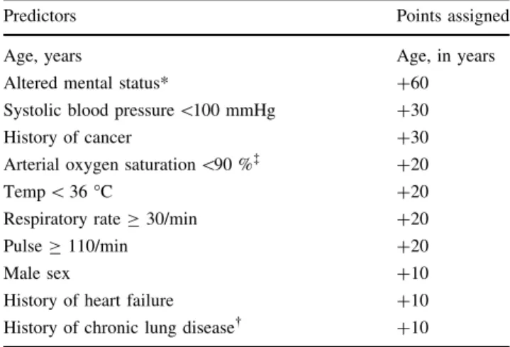
Several clinical prediction models have been developed to determine the outcome of patients with acute PE including the Pulmonary Embolism Severity Index (PESI) score, the Geneva score and the Hestia criteria (Tables8,9,
10). Of these, the PESI score and a simplified version, sPESI, have been the most extensively validated. In a multicenter prospective open randomized clinical trial of inpatient versus outpatient management of low risk PE patients as determined by the PESI score, Aujesky et al. found that there was no difference between outpatients and inpatients in recurrent VTE (1 of 171, 0.6 % vs. 0 of 168; 95 % upper CI limit 2.7 %), major bleeding (3 of 171, 1.8 % vs. 0 of 168, 0 %, 95 % upper CI limit 4.5 %) and 90 day mortality (1 of 171, 0.6 % vs. 1 of 168, 0.6 %; 95 % upper CI limit 2.1 %). These data indicate that out-patient management of low risk PE out-patients (as identified by the PESI score) is feasible and associated with excellent outcomes [20]. The HESTIA criteria have also been demonstrated to be useful in identifying patients for out-patient management [21].
Cardiac biomarkers that are released from myocytes during right ventricular strain have also proven useful for identification of PE at risk for adverse outcomes. In a multicenter prospective study of cardiac biomarkers for risk stratification of PE, Vuilleumier and colleagues found that a NT-pro-BNP level\300 pg/mL had a negative
predictive value of 100 % (95 % CI 91–100) for adverse outcomes at 3 months. Troponins have also been identified as useful biomarkers for risk stratification in PE [22]. High sensitivity assays for troponin I and T have also been useful in identification of low risk patients with PE. In a prospective validation study of 526 normotensive patients with PE, Lankeit et al. noted that only 4 of 214 (1.9 %) patients with a high sensitive troponin T\14 pg/mL had adverse outcomes at 30 days. When combined with a simplified Pulmonary Embolism Severity Index (sPESI) score of zero, none of 127 patients with this combination had adverse outcomes [23]. A combination of clinical and laboratory biomarkers may represent the ideal strategy for identification of normotensive patients at low risk for adverse outcomes. Jimenez et al. conducted a multicenter cohort study of normotensive PE patients to identify a multi-marker prognostic score for risk stratification. The Table 8 Pulmonary embolism severity index (PESI) score
Predictors Points assigned
Age, years Age, in years
Altered mental status* ?60
Systolic blood pressure\100 mmHg ?30
History of cancer ?30
Arterial oxygen saturation\90 %à ?20
Temp\36°C ?20
Respiratory rateC30/min ?20
PulseC110/min ?20
Male sex ?10
History of heart failure ?10 History of chronic lung disease ?10
A total point score for a given patient is obtained by summing the patient’s age in years and the points for each applicable predictor. Points assignments correspond with the following risk classes: Class 1 (very low risk):B65; Class II (low risk): 65–85; Class III (interme-diate risk): 86–105; Class IV (high risk): 106–125; Class V (very high risk):[125
Chronic obstructive pulmonary disease
à With and without supplemental oxygen administration
* Altered mental status was defined as confusion, disorientation, somnolence, lethargy, stupor, or coma
Table 9 Simplified PESI score
Predictors Points assigned
Age[80 years 1
History of cancer 1
History of heart failure 1
Pulse[110 beats/min 1
Systolic blood pressure\100 mmHg 1 Arterial oxygen saturation\90 % 1
combination of a sPESI and a BNP level\100 pg/mL was associated with a negative predictive value of 99 and 100 % in the derivation and validation cohorts [24].
A recent systematic review of outpatient treatment of PE including 11 studies and 1258 patients noted that the rates of recurrent VTE (1.47 %; 95 % CI 0.47–3.0 %), fatal PE (0.47 %; 95 % CI 0.16–1.0 %), major bleeding (0.81 %; 95 % CI 0.37–1.42 %) and mortality (1.58 %; 95 % CI 0.71–2.80 %) were low, similar to the rates identified in inpatient treatment studies. Furthermore, the authors found that both ‘‘clinical gestalt’’ and standardized risk assess-ment models appeared to be equally useful in identifying low risk patients appropriate for outpatient management. However, they recommended that future studies comparing formal risk stratification models and ‘‘clinical gestalt’’ should be conducted since there was more heterogeneity in the studies on clinical gestalt [25].
Management of patients with PE should be guided by an assessment of their risk for adverse outcomes (Table11). Normotensive patients in PESI Class I or II or simplified PESI Class 0 do not need further risk stratification with imaging (e.g. echocardiography) and can be considered for outpatient management. Normotensive patients in PESI ClassCII or simplified PESIC1 should undergo
additional imaging and laboratory risk assessment and warrant initial inpatient management until the results of these studies are complete. Patients in this group who have no sign of right ventricular dysfunction on echocardiography or abnormal cardiac biomarkers are considered at low inter-mediate risk for adverse outcomes. This group of patients can be considered for early discharge from the hospital. Patients with abnormal echocardiography or cardiac biomarkers are consider intermediate-low risk patients and are often man-aged in the hospital. Patients with abnormal echocardiogra-phy and cardiac biomarkers are considered at intermediate high risk of adverse outcomes and are generally managed as inpatients. Intermediate high risk PE patients are considered for thrombolytic therapy on a case-by-case basis. PE patients with hypotension are at high risk for adverse outcomes. They routinely undergo echocardiography and are strongly con-sidered for thrombolytic therapy [14]. Further discussion of PE management can be found in the accompanying paper by Vedantham et al.
Guidance Statement We suggest that most patients with DVT and many patients with PE can be managed as out-patients. PE patients should be risk stratified to determine appropriate management. A variety of laboratory tests and
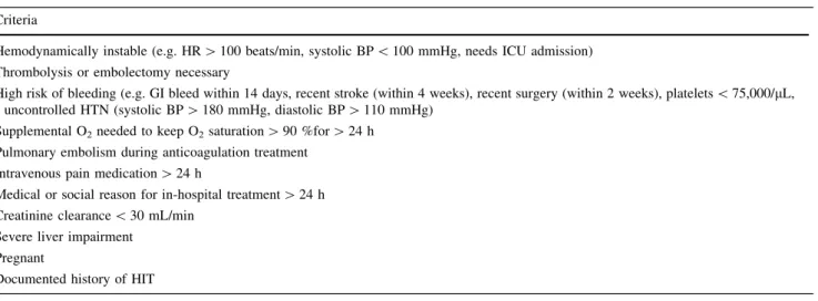
Table 10 Hestia criteria
Criteria
Hemodynamically instable (e.g. HR[100 beats/min, systolic BP\100 mmHg, needs ICU admission) Thrombolysis or embolectomy necessary
High risk of bleeding (e.g. GI bleed within 14 days, recent stroke (within 4 weeks), recent surgery (within 2 weeks), platelets\75,000/lL, uncontrolled HTN (systolic BP[180 mmHg, diastolic BP[110 mmHg)
Supplemental O2needed to keep O2saturation[90 %for[24 h Pulmonary embolism during anticoagulation treatment
Intravenous pain medication[24 h
Medical or social reason for in-hospital treatment[24 h Creatinine clearance\30 mL/min
Severe liver impairment Pregnant
Documented history of HIT
The presence of any criterion precludes outpatient treatment
Table 11 Mortality risk categories for patients with acute pulmonary embolism
30 day mortality risk Risk factors
Hypotension PESI Class III through V RV dysfunction Abnormal cardiac biomarkers
High Present Optional assessment Present Optional test
Intermediate-high Absent Present Present Present
Intermediate-low Absent Present Either one or neither present
Low Absent Absent Absent but test not necessary Absent but test not necessary
imaging modalities as well as clinical risk prediction models are available to identify PE patients who are suitable for outpatient management. Further research is needed to identify the optimal approach to risk stratifica-tion of PE patients.
(3) What are the therapeutic options for the acute treatment of venous thromboembolism?
Anticoagulation (AC) is the primary approach to ther-apy during all three phases of VTE treatment (acute, short term and long term). For those with life- or limb-threat-ening thrombosis or in patients with significant thrombus burden, systemic (for PE) or catheter-directed thrombolysis in conjunction with mechanical thrombectomy can be considered in the acute phase of treatment. Application of these therapies in the short term treatment phase of therapy is associated with a less favorable benefit:risk ratio as the thrombus becomes better organized and correspondingly less amenable to lysis/fragmentation.
In patients with contraindications to anticoagulation, placement of a vena cava filter can be considered in patients at risk for PE. In patients with distal ‘‘calf’’ DVT, serial duplex studies can be considered to determine if clot extension occurs that would place the patient at risk for PE warranting filter placement if AC is still contraindicated.
The acute treatment phase corresponds to the initial 5–10 days of therapy when parenteral therapy is
traditionally used during the transition to vitamin K antagonists which were the primary therapy during the short term and long term phases of therapy for VTE (Fig.1). The goals during the acute phase are to rapidly extinguish thrombin and fibrin clot generation. Achieving this goal reduces the symptoms associated with acute VTE and prevents thrombus extension and embolization. Prevention of further thrombus formation also allows the body’s fibrinolytic system to begin the process of thrombus dissolution.
For patients with acute VTE who are candidates for anticoagulation, multiple therapeutic options are now available to the clinician (Fig.4). If the patient is hospi-talized, unfractionated heparin (UFH) or low molecular weight heparin (LMWH) are generally utilized given their shorter elimination half-lives that facilitate peri-procedural management (Table12). In patients felt to be at high bleeding risk, unfractionated heparin may be preferable due to its shorter half-life and complete reversibility. UFH may also be preferable in special patient populations such as morbidly obese (BMI C40 kg/M2) and underweight patients (weight\50 kg) as well as patients with severe renal impairment or unstable renal function (creatinine clearance\30 mL/min). The disadvantages of intravenous unfractionated heparin are significant inter-individual dose requirements that make close laboratory therapeutic mon-itoring a necessity. Since the sensitivity of different aPTT
Start therapy Day 1 – no heparin lead-in
Start therapy Day 1 – no heparin lead-in
Heparin lead-in required 5-10 days Heparin lead-in required 5-10 days Rivaroxaban
Apixaban
Dabigatran
Edoxaban Acute
IV Heparin SQ LMWH SQ Fondaparinux
DOAC
Short Term
Warfarin SQ LMWH (in cancer)
DOAC
5-10 days 3–6 months Beyond 3-6 months
Long Term
Warfarin SQ LMWH (in cancer)
DOAC ASA Nothing
?
reagents to UFH varies substantially, it is important for each laboratory to establish its own therapeutic range based upon UFH levels as measured by protamine titration or chromogenic anti-Xa levels [26]. Observational studies have demonstrated that optimal management of UFH is difficult to achieve in routine clinical practice [27]. In
addition, UFH poses an 8-10-fold higher risk for heparin-induced thrombocytopenia (HIT) than LMWH [28,29].
Given the disadvantages associated with UFH, LMWH is often preferred outside of special hospitalized patient populations. Fondaparinux can also be employed as a parenteral agent for hospitalized patients in whom Table 12 Treatment options for VTE
Acute VTE treatment options Elimination half-life
Unfractionated heparin: 80 U/kg intravenous bolus followed by 18 U/ km/h infusion adjusted to activated partial thromboplastin time (aPTT) ratio
1 h
Low molecular weight heparin
Dalteparin 100 U/kg subcutaneously every 12 h or 200 U/kg subcutaneously every 24 h
Renal dosing: no official recommendation-use with caution, consider LMWH anti-Xa levels monitoring and dose adjustment
3–5 h (Half-life 5.7 h after IV administration of 5000 units in hemodialysis patients compared with 2.1–2.3 h in normal renal function)
Enoxaparin 1 mg/kilogram subcutaneously every 12 h or 1.5 mg/ kilogram subcutaneously every 24 h
FDA approved renal dosing-1 mg/kg sc q24hours (CrCl\30 mL/min)
4.5–7 h (17 % lower clearance with mild renal impairment-CrCl 50–80 mL/min; 31 % lower clearance with moderate renal impairment-CrCl 30–50 mL/min 44 % lower with severe renal impairment-CrCl\30 mL/min)
Tinzaparin 175 U/kg subcutaneously every 24 h
Renal dose: same (no evidence of bioaccumulation in the IRIS study)
3-4 h (24 % reduced clearance in severe renal impairment-CrCl\30 mL/min)
Pentasaccharide
Fondaparinux 5–10 mg subcutaneously every 24 h (5 mg for weight
\50 kg, 7.5 mg for weight 50–100 kg and 10 mg for weight[100 kg)
Renal dosing: Avoid in patients with CrCl\30 mL/min; caution in patients with CrCl 30–50 mL/min
17–21 h (25 % lower clearance with mild renal insufficiency-CrCl 50–80 mL/min; 40 % lower with moderate renal impairment-CrCl 30–50 mL/min; 55 % lower with severe renal impairment-CrCl\30 mL/min)
Direct oral anticoagulants
Apixaban (oral direct factor Xa inhibitor) 10 mg orally BID X 7 days then 5 mg po BID
In patients with at least 2 of the following characteristics:
ageC80 years, body weightB60 kg, or serum creatinineC1.5 mg/ dL, the recommended dose is 2.5 mg orally BID.
Would avoid in patients with CrCl\25 mL/min or sCr[2.5 mg/dL or hepatic dysfunction (AST/ALT[29ULN or bilirubin[1.5X ULN)
12 h
Dabigatran (oral direct thrombin inhibitor)
150 mg orally BID after 5–10 days of initial parenteral anticoagulation (Avoid in patients with CrCl\30 mL/min and liver impairment with
transaminase[2x ULN))
13 h (CrClC80 mL/min) 15 h (CrCl 50–79 mL/min) 18 h (CrCl 30–49 mL/min) 27 h (CrCl 15–29 mL/min) Edoxaban (oral direct factor Xa inhibitor)
60 mg orally once daily
30 mg once daily if CrCl 15–50 mL/min or body weightB60 kg or Avoid in patients with CrCl\15 mL/min or Child-Pugh class B/C
hepatic impairment
10–14 h (Total systemic exposure increased by 32 % (CrCl 50–79 mL/ min),
74 % (30–49 mL/min), 72 % (CrCl\30 mL/min), and 93 % (peritoneal dialysis), respectively)
Rivaroxaban (oral direct factor Xa inhibitor)
15 mg orally BID X 3 weeks followed by 20 mg once daily Avoid in patients with CrCl\30 mL/min and Child-Pugh class B/C
5–9 h (age 20–45 years) 11–13 h (ageC65 years)
transition to a vitamin K antagonist (VKA) is anticipated. A distinct advantage for fondaparinux is an extremely low incidence of HIT. However, fondaparinux has several limitations as an anticoagulant for inpatients including its long half-life (17–21 h with normal renal function) and lack of an antidote [26]. Detailed information about the pharmacology and clinical use of UFH, LMWH and fon-daparinux can be found in the accompanying papers by Nutescu et al. and Smythe et al.
If a VKA is anticipated to be the agent for the short term phase of treatment, initiation of VKA therapy should be delayed until all planned invasive procedures are com-pleted and the patient has resumed regular oral intake. If these conditions are satisfied, VKA therapy can begin as soon as therapeutic levels of UFH/LMWH are achieved. Parenteral therapy with UFH or LMWH should continue for at least 5 days of overlap and until an INR of 2 or more is achieved for 24 h. Both these goals should be achieved before discontinuation of parenteral therapy [30]. Detailed information about warfarin dosing and its management can be found in the accompanying paper by Witt et al.
Direct oral anticoagulants (DOACs) are also an option for the treatment of VTE in hospitalized patients. While DOACs are advantageous because they do not require monitoring, they are not easily reversible, have longer elimination half-lives (7–15 h) than UFH or LMWH and could accumulate in patients with suboptimal renal (esti-mated creatinine clearance\30 mL/min) or hepatic func-tion (Child-Pugh class B or C). In addifunc-tion, experience with perioperative management is limited. Therefore, DOACs are optimized for outpatient rather than inpatient use [31]. If either dabigatran or edoxaban are chosen, therapy must include 5 days of parenteral anticoagulation prior to beginning these agents. In contrast, rivaroxaban and apix-aban can both be used for acute treatment of VTE without initial parenteral therapy.
Thrombolytic therapy is an important management option in patients with acute extensive proximal lower extremity DVT or patients with proximal DVT that fails to respond to initial anticoagulation. Catheter-directed phar-macomechanical thrombolysis/thrombectomy is typically employed in patients with acute (within 2 weeks) proximal (ilio-femoral) deep vein thrombosis at significant risk for long term post-thrombotic complications or poor outcomes with conventional anticoagulation who are at low risk for bleeding complications. May-Thurner syndrome (MTS) (iliac vein compression syndrome) is a congenital anatomic alteration in which the left iliac vein is compressed between the right iliac artery and the lumbosacral spine. Compression results in intravascular strictures that slow venous flow which may precipitate thrombus formation [32]. Consequently, catheter-directed pharmacomechanical thrombolysis and thrombectomy in conjunction with
angioplasty and venous stenting has been advocated to reduce the risk of recurrent thrombosis although well designed studies supporting this contention are lacking [33]. Further investigation in this area is warranted. Until these data are available, patients with May-Thurner syn-drome associated iliac vein deep venous thrombosis should be managed on a case-by-case basis. Irrespective of inter-ventional management, therapeutic anticoagulation is required.
In patients with PE, systemic thrombolytic therapy is generally reserved for patients with massive pulmonary embolism (i.e. high risk pulmonary embolism with sys-temic hypotension and right ventricular dysfunction). Thrombolytic therapy is applied in a case-by-case basis in patients with sub-massive PE (i.e. intermediate risk pul-monary embolism in normotensive patients with right ventricular dysfunction) who are at low risk for bleeding complications. Catheter-based and surgical thromboem-bolectomy are other options available to providers for patients with hemodynamically significant PE [14]. A complete discussion of thrombolytic therapy for PE and DVT can be found in the accompanying paper by Vedan-tham et al.
Guidance Statement With the variety of treatment options available, we recommend that the acute therapy of VTE should be customized to suit the unique clinical cir-cumstances of the individual patient. We suggest that unfractionated heparin may be preferable for inpatients with planned invasive procedures, recent major bleeding episodes or severely impaired renal function as well as underweight and morbidly obese patients although several members of panel felt there were insufficient data to sup-port this suggestion. LMWHs are convenient options for inpatient and outpatient therapy. DOACs are optimized for outpatient therapy of VTE.
We suggest that systemic and catheter-directed phar-macomechanical thrombolytic therapy are effective options for treatment of massive PE and acute extensive proximal DVT that can rapidly reduce thrombus burden. Given the greater risks of bleeding associated with these approaches, we recommend that a careful assessment of the risks and benefits of therapy should be performed in each patient prior to the initiation of thrombolytic therapy.
(4) Which patients are candidates for a DOAC?
thrombocytopenia, high bleeding risk and potent drug–drug interactions were excluded from participation in the phase 3 VTE studies. In addition, certain patient populations were not well represented in these studies such as patients with active cancer. Therefore, it is important to consider the inclusion and exclusion criteria and the enrolled popula-tions in the published studies when considering a DOAC for treatment of VTE. In addition, 2 of the DOACs (dabigatran and edoxaban) were studied using acute treat-ment with a parenteral agent (dabigatran median duration 9 days; edoxaban median duration 7 days). Therefore, these agents should be used only after an initial period of parenteral therapy for acute VTE (Fig.4).
Dabigatranis an oral direct thrombin inhibitor that has
been compared to warfarin in the short term treatment and warfarin and placebo in long term treatment of VTE in 3 double blind randomized controlled trials, the RECOVER, REMEDY and RESONATE studies. In the RE-COVER study, 2564 patients with acute symptomatic objectively documented proximal lower extremity DVT or PE were randomized to either dabigatran 150 mg twice daily or adjusted-dose warfarin (INR range 2–3) after acute treat-ment with unfractionated or low molecular weight heparin (median parenteral treatment duration=9 days). Seven patients in the dabigatran group and 18 in the warfarin group did not receive study medication leaving a total of 1274 dabigatran patients and 1265 warfarin patients in the population for efficacy analysis. In the warfarin group, the time in therapeutic range over the duration of the study was 60 % (53 % month 1, 66 % in the last month). Thirty of 1274 patients on dabigatran (2.4 %) and 27 of 1265 war-farin recipients (2.1 %) suffered recurrent VTE (0.4 % absolute risk difference; 95 % CI for non-inferiority-0.8 to 1.5). The hazard ratio (HR) with dabigatran was 1.10 (95 % CI 0.65–1.84). Major bleeding occurred in 20
patients assigned to dabigatran (1.6 %) and in 24 patients taking warfarin (1.9 %) for a hazard ratio with dabigatran of 0.82 (95 % CI 0.42–1.48) (Table13). There was no difference in mortality, acute coronary events or abnormal liver function tests [34].
These results were confirmed in RECOVER II, a ran-domized double-blind double dummy study that compared dabigatran 150 mg twice daily with warfarin (INR 2–3) after median of 9 days of parenteral therapy. Recurrent symptomatic objectively confirmed VTE occurred in 30 of 1279 dabigatran patients (2.3 %) and 28 of 1289 warfarin patients (2.2 %) (HR 1.08; 95 % CI 0.64–1.80). Major bleeding occurred in 15 dabigatran patients (1.2 %) and 22 warfarin patients (1.7 %) (HR 0.69; 95 % CI 0.36–1.32) (Table13). Pooled analysis with the RECOVER and RECOVER II studies produced a hazard ratio for recurrent VTE of 1.09 (95 % CI 0.76–1.57), major bleeding of 0.73 (95 % CI 0.48–1.11) and for any bleeding of 0.70 (95 % CI 0.61–0.79) [35]. These studies demonstrate that dabigatran is at least as effective as warfarin for short term treatment of VTE. Compared with warfarin, dabigatran was associ-ated with an increased risk of myocardial infarction or acute coronary syndrome in a meta-analysis of the ran-domized clinical trials (RCT) leading to its approval (dabigatran, 237 of 20,000 [1.19 %] vs. control, 83 of 10,514 [0.79 %]; OR 1.33; 95 % CI 1.03–1.71; P =0.03) [36]. However, no difference was seen in a recent large new user cohort of 134,414 propensity-matched elderly US Medicare patients (Hazard ratio 0.92 (95 % CI 0.78–1.08) perhaps due to clinical differences in the two study popu-lations [37]. Until this issue is further clarified, prescribers should use caution when prescribing dabigatran in elderly patients at risk for acute coronary syndrome.
GI bleeding also appears to be more common in higher risk patients treated with dabigatran compared with
Table 13 Results of randomized controlled trials of DOACs versus conventional therapy for VTE
Study Treatment Patients Recurrent VTE1 Major bleeding
RE-COVER, 2009
Dabigatran 150 mg BID vs. VKA 1273/ 1266
30 (2.4 %) vs. 27 (2.1 %) (HR 1.10; 95 % CI-0.8 to 1.5)
20 (1.6 %) vs. 24 (1.9 %) (HR 0.82; 95 % CI 0.45–1.48) RE-COVER
II, 2014
Dabigatran 150 mg BID vs. VKA 1279/ 1289
30 (2.3 %) vs. 28 (2.2 %) (HR 1.08; 95 % CI 0.64–1.80)
15 (1.2 %) vs. 22 (1.7 %) (HR 0.69; 95 % CI 0.36–1.32) EINSTEIN
DVT, 2010
Rivaroxaban 15 mg BID X 3 weeks, then 20 mg daily vs. Enoxaparin/VKA
1731/ 1718
36 (2.1 %) vs. 51 (3.0 %) (HR 0.68; 95 % CI 0.44–1.04)
14 (0.8 %) vs. 20 (1.2 %) (HR 0.65; 95 % CI 0.33–1.30) EINSTEIN
PE, 2012
Rivaroxaban 15 mg BID X 3 weeks, then 20 mg daily vs. Enoxaparin/VKA
2419/ 2413
50 (2.1 %) vs. 44 (1.8 %) (HR 1.12; 95 % CI 0.75–1.68)
26 (1.1 %) vs. 52 (2.2 %) (HR 0.49; 95 % CI 0.31–0.79) AMPLIFY,
2013
Apixaban 10 mg BID X 7 days then 5 mg BID vs. Enoxaparin/Warfarin
2609/ 2635
59 (2.3 % vs. 71 (2.7 %) (RR 0.84; 95 % CI 0.60–1.18)
15 (0.6 %) vs. 49 (1.8 %) (RR 0.31; 95 % CI 0.17–0.55)
HOKUSAI-VTE, 2013
Edoxaban 60 mg daily (or 30 mg daily) vs. warfarin
4118/ 4122
130 (3.2 %) vs. 146 (3.5 %) (HR 0.89; 95 % CI 0.70–1.13)
56 (1.4 %) vs. 66 (1.6 %) (HR 0.84; 0.59–1.21)
warfarin. The risk of GI bleeding with dabigatran was significantly higher than warfarin in the RE-LY RCT in atrial fibrillation (RR 1.30; 95 % CI 1.07–1.56) but similar in VTE (RECOVER RR1.79; 95 % CI 0.60–5.32; RECOVER II 0.60; 95 % CI 0.22–1.66; REMEDY RR 0.62; 95 % CI 0.22–1.90) [38]. This difference likely reflects differences in study populations as the AF patients tended to be older and/or on concomitant antiplatelet agents more commonly than VTE patients. This interpre-tation is borne out by a new-user Medicare cohort of AF patients in which dabigatran was associated with an increase in major GI bleeding (RR 1.28 (95 % CI 1.14–1.44) [37]. It is important to note that there is a dose-related difference in the risk of GI bleeding between dabigatran 150 mg twice daily (Relative risk 1.50; 95 % CI 1.19–1.89) and 110 mg twice daily (RR 1.10; 95 % CI 0.86–1.41) [39]. Only the 150 mg dose is available in the United States. Given the GI bleed data from the RE-LY study, it may be worthwhile considering another DOAC than dabigatran for older patients with VTE.
Guidance Statement When used after a 5–10 day initial course of parenteral anticoagulation, dabigatran is as effective as warfarin in the acute and short term treatment of VTE. We suggest dabigatran as an alternative to vitamin K antagonists for the short term therapy of VTE. In some studies, dabigatran has been associated with an increased risk of acute coronary syndrome and gastrointestinal bleeding compared with vitamin K antagonists.
Rivaroxaban, an oral direct factor Xa inhibitor, has been
compared with conventional therapy for acute, short term and long term treatment of VTE in the EINSTEIN DVT and PE trials as well as the EINSTEIN Extension trial [40,
41]. In contrast to the dabigatran VTE studies, the EIN-STEIN DVT and PE trials were open-label event driven randomized controlled trials. Patients were randomized within 48 h of diagnosis to either conventional therapy (enoxaparin transitioned to adjusted dose warfarin or acenocoumarol INR 2–3) or rivaroxaban 15 mg twice daily for 3 weeks followed by 20 mg once daily. The median duration of enoxaparin in the EINSTEIN DVT study was 8 days and 80.8 % of patients had an INR of 2 or more at the end of treatment. The overall time in therapeutic range was 57.7 % (54.1 % in month 1 and 66.4 % in month 10). Recurrent VTE occurred in 36 rivaroxaban patients (2.1 %) and 51 enoxaparin/VKA patients (3.0 %) (HR 0.68; 95 % CI 0.44–1.04). Major bleeding occurred in 14 rivaroxaban patients (0.8 %) and 20 enoxaparin/VKA patients (1.2 %) (HR 0.65; 95 % CI 0.33–1.30). The principal safety out-come (major or clinically relevant non-major bleeding) was also similar between groups (rivaroxaban, 139 [8.1 %] vs. enoxaparin/VKA 138 [8.1 %]; HR 0.97 [95 % CI 0.76–1.22]) [40]. The EINSTEIN PE trial had a similar
design to the EINSTEIN DVT trial. The median duration of enoxaparin therapy in the enoxaparin/VKA arm was 8 days and 83 % of patients achieved an INR of 2.0 or more by the end of enoxaparin treatment. The time in therapeutic range for VKA patients over the course of the study was 62.7 % (57.8 % during the first month and 72.7 % during month 11). Symptomatic recurrent VTE occurred in 50 patients taking rivaroxaban (2.1 %) and 44 patients who received enoxaparin/VKA (1.8 %) (HR 1.12; 95 % CI 0.72–1.68). Major or clinically relevant non-major bleeding occurred in 249 rivaroxaban patients (10.3 %) and 274 (11.4 %) enoxaparin/VKA patients (HR 0.90; 95 % CI 0.76–1.07). Major bleeding occurred in 26 rivaroxaban patients (1.1 %) and 52 enoxaparin/VKA patients (2.2 %) (HR 0.49; 95 % CI 0.31–0.79) (Table13). These studies demonstrate that rivaroxaban is a safe and effective alternative for acute and short term therapy of VTE. Major bleeding was similar or lower with rivaroxaban compared with conventional ther-apy. No increase in gastrointestinal bleeding or acute coronary events was seen. However, patients age 75 and older appear to be at increased risk of GI bleeding with rivaroxaban compared with warfarin, therefore caution is warranted in these patients [38,42,43].
Guidance Statement Rivaroxaban is as effective as LMWH/VKA in the treatment of DVT and PE. We suggest rivaroxaban as an alternative to LMWH/VKA for the acute and short term treatment of VTE in appropriate patients. No increase in acute coronary syndrome has been seen with rivaroxaban, however GI bleeding may be more common in patients age 75 and older.
Apixaban, an oral direct factor Xa inhibitor, was
than conventional therapy patients (9.7 %) (RR 0.44; 95 % CI 0.36–0.55). All-cause mortality was similar between groups (1.5 % vs. 1.9 %; RR 0.79; 95 % CI 0.53–1.19) [44] (Table13). These results indicate that apixaban, like rivaroxaban is an attractive one drug treatment for acute and short term therapy of VTE compared to conventional therapy. No increase in acute coronary events was seen compared to warfarin [42].
Guidance Statement Apixaban is as effective as LMWH/ VKA in the treatment of DVT and PE and associated with less major bleeding and major or clinically relevant non-major bleeding. We suggest apixaban as an alternative to LMWH/VKA in the acute and short term treatment of VTE in appropriately selected patients. No increase in acute coronary syndrome or gastrointestinal bleeding has been seen with apixaban.
Edoxaban is a direct oral inhibitor of factor Xa that is
capable of inhibiting free and bound factor Xa. Edoxaban was compared with warfarin in the treatment of VTE in the HOKUSAI-VTE study, a large randomized double-blind non-inferiority study conducted in 8292 patients enrolled in 439 centers in 37 countries [46]. After a median of 7 days of parenteral therapy (unfractionated or low molecular weight heparin) following enrollment, patients were ran-domized to edoxaban 60 mg once daily (30 mg once daily if creatinine clearance 30–50 mL/min, body weight of 60 kg or less or concomitant therapy with a potent P-gly-coprotein inhibitor) or placebo and warfarin or matching placebo. A total of 4921 patients had a DVT and 3319 had PE. Extensive thrombus burden (common femoral vein or iliac vein DVT or PE with involvement of multiple lobes with 25 % or more of the entire pulmonary vasculature) was present in 743 (45 %) edoxaban patients and 778 (46.6 %) warfarin patients. Right ventricular dysfunction was noted in 172 edoxaban PE patients (34.5 %) and 179 warfarin PE patients (35.5 %). Recurrent symptomatic VTE occurred in 130 (3.2 %) edoxaban patients and 146 (3.5 %) warfarin patients (HR 0.89; 95 % CI 0.70–1.13). Among patients who qualified for the edoxaban 30 mg daily dose, recurrent VTE occurred in 22 of 733 (3.0 %) edoxaban patients and 30 of 719 (4.2 %) warfarin patients (HR 0.73; 95 % CI 0.42–1.26). The rate of recurrent symptomatic VTE in patients with PE and right ventricular strain was 3.3 % in edoxaban patients and 6.2 % in war-farin patients (HR 0.52; 95 % CI 0.28–0.98). The primary safety outcome (major or clinically-relevant non-major bleeding) occurred in 349 (8.5 %) edoxaban patients and 423 (10.3 %) warfarin patients (HR 0.81; 95 % CI 0.71–0.94). Major bleeding occurred in 56 (1.4 %) edox-aban patients and 66 (1.6 %) warfarin patients (HR 0.84; 95 % CI 0.59–1.21). Among patients who fulfilled criteria for the 30 mg edoxaban dose, 58 of 733 (7.9 %) edoxaban
patients and 92 of 719 (12.8 %) warfarin patients (HR 0.62; 95 % CI 0.44–0.86) developed clinically relevant non-major bleeding [46] (Table13). The Hokusai VTE study confirms that once daily edoxaban is as effective as war-farin in the prevention of recurrent VTE and caused sig-nificantly less bleeding following an initial course of parenteral therapy.
Guidance Statement After an initial 5–10 days of LMWH or UFH, edoxaban is as effective as LMWH/VKA in the treatment of acute DVT and PE but associated with less major or clinically relevant non-major bleeding. We sug-gest edoxaban as an alternative to VKA for the short term treatment of VTE in appropriately selected candidates.
(5) What is the role of vena cava filters if the patient is not a candidate for anticoagulation?
The only reason to consider placement of an inferior vena cava filter is acute VTE (within 4 weeks) in the presence of a contraindication to anticoagulation (i.e. the presence of active bleeding or the presence of risk factors for major bleeding (e.g. recent major bleeding event, major surgery or major trauma, etc.) [47]. Other indications are controversial and of unproven clinical benefit or are frankly harmful. As with any procedure it is important to assess whether the risks of a vena cava filter are warranted by its benefits on a case-by-case basis. Potential complications of a vena cava filter are indicated in Table14. In general we do not suggest vena cava filters for distal lower extremity DVT, superficial venous thrombophlebitis, VTE older than 1 month, or upper extremity DVT. In the case of upper extremity DVT, the risks of symptomatic and fatal PE are low and the severity of potential complications of filter thrombosis in the superior vena cava or penetration of thoracic vascular structures by filter struts or during the insertion procedure exceed the benefits [48].
In the event of a recent surgical procedure, the timing of initiation of anticoagulation varies according to the bleed-ing risks posed by the surgical procedure (Table15). The timing of anticoagulation outlined in the table should not be considered proscriptive; rather it should be considered a rough guide for practice. It is better to err on the side of caution and wait a few extra days to initiate anticoagulation even if it means placing a retrievable vena cava filter as post-operative bleeding can result in significant complica-tions and further delays in treatment. It is recommended that active filter follow up programs be instituted so patients do not get lost to follow up. These programs have a high rate of success with filter retrieval ([95 %) [49]. These decisions should be based upon local expertise and experience.
including the operating surgeon. In high risk bleeding sit-uations, we suggest use of unfractionated heparin initially and starting the infusion without a bolus. Once patients are therapeutic for at least 24 h without evidence of bleeding, they can be transitioned to a more convenient agent on a case by case basis depending upon the preferences of the care team. If the severity of the thrombotic event dictates use of a bolus, the risks of bleeding that might be associ-ated with its administration must be balanced with the risks associated with a vena cava filter.
In the event of a gastrointestinal bleed, we suggest waiting at least 7 days without evidence of active bleeding and after endoscopic treatment of the bleeding lesion before reinitiating therapeutic anticoagulation [50]. In the event of intracranial hemorrhage (ICH), it is essential to review the indications for anticoagulation and the patient’s risk of recurrent VTE as recurrent ICH is common (2.56 per 100 patient years) and potentially deadly (25 % case fatality rate) [51]. In general, only patients with recent VTE (within 3 months), idiopathic VTE or VTE with ongoing potent risk factors (active cancer, lupus anticoagulant positive APS, etc.) or recurrent unprovoked VTE warrant consideration of resumption of anticoagulation. In addition, one must factor in the risk of rebleeding. Lobar ICH is associated with a higher risk of recurrence than deep hemispheric bleeds [52]. Underlying diseases or lesions associated with the initial hemorrhage should be treated prior to resumption of anticoagulation. The optimal time to resume anticoagulation remains uncertain but a recent large retrospective cohort study of warfarin-associated ICH suggested that resumption of warfarin between 10 and 30 weeks was associated with the lowest risk of recurrent ICH and thromboembolism. While only 30 of the 177 patients who survived the first week had VTE as an indi-cation for anticoagulation, only 4 of these patients (13 %) suffered recurrent VTE and none were fatal. In contrast, 18 patients suffered recurrent ICH (10 %) of which 4 were fatal (22 %). Although these data are imperfect with respect to management of patients with VTE, they indicate that only the highest risk VTE patients should consider
resumption of anticoagulation after a spontaneous ICH [53].
Once a patient has successfully resumed therapeutic anticoagulation without recurrent bleeding complications, we refer them back to the interventional radiologist who placed their vena cava filter. The latest generation of retrievable vena cava filters can be retrieved with a high degree of success six or more months after placement. Therefore, it may be preferable to wait several months before retrieving the filter in order to make sure the patient will tolerate anticoagulation. In patients with filters that cannot be retrieved, the impact of this on anticoagulation duration needs to be considered. In patients with transient indications for anticoagulation, the risks of thrombosis associated with a vena cava filter need to be balanced against the risks of bleeding associated with anticoagula-tion. In the PREPIC study, 36.4 % suffered a DVT and 14 % suffered IVC thrombosis after 8 years of follow up [54].
Guidance Statement We suggest that vena cava filters should be considered in any patient with acute VTE (within 4 weeks) who cannot be treated with anticoagu-lation. We suggest that retrievable filters are strongly preferred as most patients have temporary contraindica-tions to anticoagulation. Filters should be retrieved once anticoagulation can be reinitiated preferably within 6 months of placement. Patients with filters should be closely monitored in a structured program to facilitate retrieval and minimize the number of patients lost to follow up.
Following anticoagulation-associated gastrointestinal bleeding, we suggest that anticoagulation can be re-initi-ated as early as 7 days after cessation of bleeding and treatment of causal lesions. Following anticoagulation associated ICH, we suggest resumption of anticoagulation no sooner than 10 weeks post-bleed. Further investigation of this topic is warranted.
(6) How is upper extremity VTE treated?
Upper extremity DVT is often associated with an intrinsic or extrinsic precipitant. The most common extrinsic precipitant is the presence of a central venous catheter (CVC), pacemaker/implanted cardiac defibrillator or venous intervention. In these cases, the DVT originates at the location of the device/intervention. If the DVT is anatomically distant from the catheter or pacemaker then other reasons should be sought [55]. In patients with a CVC-associated DVT, anticoagulation alone without CVC removal is successful in many patients and allows preser-vation of the CVC for continued use in the event that an indication for central venous access remains avoiding the morbidity associated with the insertion of a new CVC [56]. Table 14 Complications of inferior vena cava filters
Access site thrombosis Deep venous thrombosis
Filter migration/embolization
Filter misplacement (outside target zone) Filter strut fracture
Guidewire entrapment IVC thrombosis IVC penetration Pulmonary embolism
If symptoms fail to improve after initial anticoagulation, then the CVC can be removed [55]. Although there are no data as to when the risk of PE with CVC removal in patients with CVC-associated DVT declines, a meta-anal-ysis of recurrent VTE in randomized controlled treatment trials of VTE suggests that delaying removal for at least 1 week will greatly reduce the risk of PE associated with CVC removal [57]. If the patient is not a candidate for anticoagulation, CVC removal rather than placement of a superior vena cava filter is recommended given the hazards associated with filter thrombosis or strut penetration in this location and the lower risk of PE associated with upper extremity DVT [58]. The duration of AC therapy for CVC-associated DVT/PE should be at least 3 months or as long as the CVC remains in place. A similar approach to duration of therapy can be taken in cancer patients with CVC associated VTE [15,59].
In patients with an upper extremity DVT associated with pacemakers or implanted defibrillators, anticoagulation without device removal is the primary approach to man-agement [60]. In a prospective study, risk factors for thrombosis included hormonal therapy, a history of VTE and an absence of anticoagulant treatment. Of these, only hormonal therapy and an absence of anticoagulation remained significant in multivariate analysis [61]. No treatment studies have been performed in this patient population but the authors of this guidance document suggest at least 3 months of anticoagulation is appropriate. In patients with an upper extremity DVT in the absence of a CVC, an anatomic trigger should be considered. In younger patients with upper extremity DVT, the presence of thoracic outlet syndrome (TOS) or effort induced thrombosis (Paget-von Schroetter) syndrome (PSS) should be investigated. Thoracic outlet syndrome occurs when the Table 15 Risk stratification of bleeding risk with anticoagulation following surgery
Bleeding risk category
Type of surgery or procedure Anticoagulation recommendation
Very high Neurosurgical procedure (intracranial or spinal) Prostatectomy or partial nephrectomy, bladder surgery Heart valve replacement
Coronary artery bypass grafting
Can initiate prophylactic dose anticoagulation at 24 h Consider therapeutic dose
anticoagulation no sooner than 72 h
High Pacemaker or AICD placement Major cancer surgery
Major vascular surgery (AAA repair, peripheral artery bypass) Reconstructive plastic surgery
Renal or hepatic biopsy
Bowel polypectomy (assume this will be part of a colonoscopy) Major orthopedic surgery
Can initiate prophylactic dose anticoagulation within 12–24 h Consider therapeutic dose
anticoagulation no sooner than 48–72 h
Moderate Major intra-abdominal surgery
Major intra-thoracic surgery
Can initiate prophylactic dose anticoagulation within 12–24 h
Consider therapeutic dose anticoagulation no sooner than 24–48 h
Low Laparoscopic cholecystectomy or hernia repair Coronary angiography
Arthroscopy
Biopsy (prostate, bladder, thyroid, lymph node) Bronchoscopy±biopsy
Central venous catheter removal Multiple dental extraction or gum surgery
Can initiate prophylactic dose anticoagulation within 12 h Consider therapeutic dose
anticoagulation 24–48 h
Very low Minor dental procedures (single tooth extractions or root canals) (See Table6) Minor dermatologic procedures (excisions of basal and squamous cell carcinomas,
actinic keratoses, and malignant or premalignant nevi) Cataract removal
Electroconvulsive therapy (ECT) Arthrocentesis
Joint or soft tissue injections GI endoscopy without biopsy
nerve, artery and/or vein traversing the thoracic outlet are compressed by the surrounding anatomic structures. This compression can cause venous, arterial or neurologic compromise. In the venous form of this syndrome, the compression causes endothelial damage and stasis leading to local anatomic clot formation. In PSS repetitive upper extremity exercise usually in the context of a tight thoracic outlet can lead to vascular damage, stasis and subsequent thrombus formation. A history of upper extremity throm-bosis in the absence of CVC or upper extremity or thoracic or neck vein intravenous access procedures should prompt consideration of TOS. A recent history of upper extremity exertion or exercise should also raise suspicions of TOS/ PSS. Patients often complain of aching and swelling in the upper extremity and demonstrate venous distention and bluish discoloration in the affected arm. Physical exam findings that suggest the presence of TOS include Adson’s test (ipsilateral rotation and extension of the neck during deep inspiration result in a diminution of the radial pulse) and Wright’s test (hyperextension of the arm diminishes the radial pulse). Nevertheless, imaging studies are essen-tial to demonstrate TOS. Venous and arterial duplex ultrasound with the patient’s arm in stress positions are the most sensitive study for assessing the presence of TOS [15,
59].
For TOS/PSS, thrombolytic therapy followed by surgi-cal repair (thoracic rib resection and/or ssurgi-calenectomy) has been advocated as an important component of successful therapy in addition to anticoagulation. Thrombolysis fol-lowed by endovascular stenting does not appear to be as beneficial as surgery [62]. However, the benefits of surgical therapy remain to be demonstrated in a rigorous fashion [63]. Consequently, the writing committee of this guidance document was divided as to the value of surgical repair of thoracic outlet syndrome. Until well-designed studies are conducted examining the risks and benefits of surgical therapy, we suggest providers consider surgical repair in addition to thrombolysis and anticoagulation versus thrombolysis/anticoagulation alone on a case-by-case basis. We suggest that patients with upper extremity deep vein thrombosis receive at least 3 months of anticoagula-tion with or without surgical therapy.
Other important causes of upper extremity DVT include intra-thoracic or cervical tumors or nodal masses or infections that can result in vascular wall inflammation and compression. Diagnosis can generally be established with duplex ultrasound or contrast CT venographic imaging. Identification and treatment of the underlying disease process (cancer, infection, etc.) and anticoagulation are both likely to be important factors in successful treatment. We suggest that anticoagulation should be continued for at least 3 months or until precipitating factors have been
eliminated (vascular compression by tumor), whichever is longer [64].
Guidance Statement Identification and elimination of trigger factors when feasible is important to reduce the incidence of recurrent upper extremity DVT. For CVC-associated DVT, we suggest that anticoagulation without CVC removal is the treatment of choice. If symptoms fail to resolve, CVC removal can be considered. We suggest that anticoagulation should be continued for at least 3 months or the duration of the CVC whichever is longer. At least 3 months of anticoagulation is appropriate for pacemaker wire-associated VTE.
The committee was divided as to the optimal approach to treatment of TOS/PSS-associated upper extremity DVT. The benefits of rib resection/scalenectomy following thrombolysis and anticoagulation remain to be rigorously demonstrated. Therefore, providers should consider ther-apy for TOS/PSS on a case-by-case basis until higher quality data are available. We suggest that TOS/PSS-as-sociated upper extremity DVT warrants anticoagulation for at least 3 months. Treatment of upper extremity DVT associated with extrinsic compression due to cancer or infection should include treatment of the underlying dis-ease in addition to anticoagulation.
(7) When is ambulation/exercise safe after DVT/PE?
Four randomized controlled trials have examined the question of whether early ambulation with or without early compression therapy is associated with an increased risk of pulmonary embolism. Patients began to walk on the day of diagnosis (3 studies) or after 2 days of leg elevation. No difference on symptomatic pulmonary embolism was noted (risk ratio 1.16 [95 % CI 0.66–2.05]). Early ambulation was associated with a reduction in acute limb pain and an improvement in quality of life due to DVT in one study (p\0.01 and p\0.05) whereas it had no effect in two other studies. Early ambulation did not increase the risk of thrombus progression (RR 0.38; 95 % CI 0.13–1.15) [65]. Although the number of patients is small, the existing liter-ature suggest that ambulation in patients with acute DVT/PE is safe as soon as therapeutic anticoagulation is achieved.
Guidance Statement We suggest that ambulation is safe
in patients with acute DVT±PE after achievement of
therapeutic anticoagulation.
(8) Is the use of graduated compression stockings safe after acute DVT/PE?
arm so we cannot use these studies to determine the safety of GCS during the acute treatment of VTE. Brandjes et al. randomized 194 patients to knee high GCS or no stockings for prevention of post-thrombotic syndrome within 2–3 weeks of a first episode of DVT. No difference in the rate of recurrent VTE was noted between the groups (14 of 96 patients [146 %] in the GCS group vs. 13 of 98 patients in no GCS group [133 %) [66]. Similarly, Prandoni et al. found no difference in recurrent VTE in their open ran-domized study of GCS for prevention of PTS (GCS group 12/90 [13.3 %] vs. control group 13/90 [14.4 %]). The average time of enrollment was 7 days (range 5–10 days) after diagnosis [67]. Finally the SOX trial, a double blind placebo controlled randomized controlled trial of GCS in the prevention of PTS also noted no difference in recurrent VTE between active GCS patients (33 patients [81 %]; 45 events [36 DVT, 9 pulmonary embolism]) and placebo stocking patients (38 patients [96 %]; 44 events [32 DVT, 12 pulmonary embolism]. The median time to enrollment was less than 5 days [68]. This study also showed that GCS did not significantly reduce leg pain associated with DVT [69]. These studies indicate that application of GCS during the acute treatment of VTE is not associated with an increased risk of recurrent VTE but does not appear to reduce pain associated with acute DVT. For further dis-cussion of graduated compression stockings, see the accompanying paper by Kahn et al.
Guidance Statement We suggest that GCS do not increase the risk of recurrent thromboembolism in patients with acute VTE. We suggest that GCS do not have any beneficial effect on leg discomfort associated with acute
DVT.
(9) What is the recommended duration of therapy for VTE?
Long term therapy corresponds to anticoagulation beyond 3–6 months when the primary goals of therapy are to continue to suppress thrombin generation in order to prevent recurrent VTE. The primary long term treatment of VTE is anticoagulation. In the past, oral VKA were the mainstay of long term therapy except for in cancer patients in whom LMWH has been preferred. DOACs also repre-sent an attractive option for long term therapy of VTE in appropriate candidates. In patients with contraindications to initial anticoagulation, vena cava filters are employed. In these patients, it is important to routinely reassess patients for the continued presence of contraindications to antico-agulation on an ongoing basis as VCF are associated with an increased risk of recurrent DVT and IVC thrombosis. Since the majority of patients have temporary contraindi-cations to anticoagulation, retrievable filters with a broad window of retrievability should be used. In patients on long
term anticoagulation it is important to reassess the risks and benefits of continued anticoagulation on a routine basis given the changing medical circumstances of patients over time.
(a) What is the recommended duration of therapy for a patient with distal (calf vein) DVT?