MONOLITHIC ZIRCONIA PARTIAL COVERAGE RESTORATIONS: AN IN-VITRO CLINICAL SIMULATION
Savita Gupta
A thesis submitted to the faculty at the University of North Carolina at Chapel Hill in partial fulfillment of the requirements for the degree of Master of Science in the Division of
Comprehensive Oral Health in the Adams School of Dentistry.
Chapel Hill 2020
ABSTRACT
Savita Gupta: Monolithic zirconia partial coverage restorations: An in- vitro clinical simulation (Under the direction of Taiseer Sulaiman)
To evaluate the survival rate and bonding efficiency and marginal integrity of monolithic zirconia partial coverage restorations bonded using air particle abrasion, a primer with 10- methacryloyloxydecyl dihydrogen phosphate and composite resin (APC) protocol.
Human maxillary premolars were prepared for monolithic 3Y zirconia partial coverage restorations. Group1 (control) full coverage crown, group 2 and 3 preserving 2mm functional or nonfunctional cusp width, group 4 overlay preparation. The restorations were bonded using the APC protocol. The samples were exposed to simultaneous thermocycling and mechanical loading, SEM analysis were performed to detect failure (cracks/micro cracks) and marginal defects.
In group 2, one specimen debonded and SEM analysis at 30X indicated marginal integrity issue of the remaining seven intact specimens. None of the specimens failed due to fracture. No other specimens demonstrated marginal integrity issued at 30X. None of the specimens demonstrated any micro crack at 100X and 150X.
ACKNOWLEDGEMENTS
I would like to express my deepest gratitude to the faculty at University of North
Carolina at Chapel Hill for the last three years. Dr. Taiseer Sulaiman, I thank you for taking me under your wing and assisting me endlessly throughout the years. Without your positivity and passion for perfection and attention to detail, my work would not be at the level it is today. Your assistance and counsel in the laboratory was much needed and played a pivotal role to executing this project. Your constant encouragement and tenacity has been invaluable to me. It has been a pleasure to be under your guidance and mentorship.
Thank you to my thesis committee for your unparalleled input. Dr. Donavan, Dr. Boushell and Dr. Bencharit, I deeply value your participation and help that you offered so genuinely throughout this project. I really appreciate your dedication towards assisting me. Thank you for providing me with your exceptional wisdom and expertise. It has been an honor and privilege to be under your care.
To Brandon Rodgers and Marithe Baclogen, thank you for your much-needed help and support on the endless days at the laboratory. Thank you to Amar Kumbhar for your help with the SEM analysis. Dr. Ceib Phillips, I really appreciate your help with the statistical analysis of this project.
TABLE OF CONTENTS
LIST OF FIGURES………..………viii
LIST OF TABLES………. ix
LIST OF ABREVIATIONS AND SYMBOLS………...…………x
CHAPTER 1: REVIEW OF THE LITERATURE………..…………1
1.1 Introduction………....1
1.1.1 Cavity preparation design………3
1.1.2 Type of restorative material……….3
1.1.3 Type of luting cement………..3
1.2 Material used for partial coverage restorations………..4
1.3 Zirconia as a restorative material for full coverage restorations………5
1.4 Bonding to zirconia....………...……….7
1.5 Laboratory fatigue testing...……….10
1.6 Zirconia as a restorative material for partial coverage restorations……….13
CHAPTER 2: RESEARCH STUDY……….14
2.1 Specific aims………14
2.2 The null hypotheses……….14
2.3.1 Test sample preparation... ……….14
2.3.2 Fabrication of restorations……….18
2.3.3 Cementation of the restorations……….19
2.3.4 Fatigue testing………22
2.3.5 Evaluating failure………...23
2.3.6 Scanning Electron Imaging………23
2.4 Results………..24
2.4.1 Fatigue testing………24
2.4.2 Scanning Electron Microscope………..24
2.5 Statistical analysis………25
2.6 Discussion………...….26
2.7 Limitations of the study………...29
2.8 Conclusions………..30
LIST OF FIGURES
Figure 2.1- Group Preparation Design………...……17
Figure 2.2- Planmeca Onlay Fabrication………...…18
Figure 2.3-Schematic Diagram of Mechanical Loading Pattern………...23
LIST OF TABLES
Table 1.1- Different Generation of Zirconia………...…….6
Table 2.1- Materials Used………..………21
LIST OF ABREVIATION AND SYMBOLS
°C Degree of Celsius
°F Degree of Fahrenheit
µ Micron
% Percent
~ Approximately equal
> Greater than
wt. % Weight percent
mol% Molecule percent
10-MDP 10- methacryloyloxydecyl dihydrogen phosphate
4-META 4-methacryloxyethyl trimellitate anhydrate
3-TMSPA 3-trimethoxysilanyl-propyl-N-aniline
MAC-10 Methacryloyloxy-1,10-undecanedicarboxylic acid
CAD Computer aided Design
CAM Computer aided manufacturing
CPD Critical point drying
CaO Calcium oxide
mm Millimeter(s)
MPa Mega Pascal
N Newton
SEM Scanning Electron Microscope
vol. Volume
Y2O Yttria oxide
CHAPTER 1: REVIEW OF THE LITERATURE
1.1 Introduction:
Restorative dentistry is continuing to improve as patients preserve their natural teeth until old age. Direct restorative care remains the most conservative and non-invasive treatment as it minimizes the amount of unsound tooth structure removed, maximizes the remaining amount of intact sound tooth structure, limits damage to the pulpal and surrounding tissues1 and is associated with lower financial cost as compared with indirect restorative procedures.
Creation of proper anatomy, resulting in proper function, is more difficult to directly accomplish when a large amount of tooth structure is missing. In these cases, indirect restorations fabrication enables better control over re-establishment of natural tooth contours.
Restorative material that have been used to fabricate partial coverage restoration include cast metals, ceramics and composite resin.2 Gold is considered to be "gold standard" as compared with all other restorative material in terms of clinical longevity. A classic study by Donovan et al, evaluated the long-term success rate of 1314 cast gold restoration in service for 20 years or more, placed by single dentist and concluded survival rate of 97% at 9 years, 90.3% at 20 years.3
minimally invasive dentistry and advancement in adhesive technologies have expanded indications for tooth colored partial coverage restorations.6
Preservation of natural tooth structure increases the likelihood of ongoing pulpal vitality as
well as serves as guide for the creation of a restoration that will mimic natural tooth anatomy in the arch. Multiple restorative efforts, each an insult to the pulp, increases the risk of irreversible damage.7 Increased amounts of remaining enamel and dentine improves thermal insulation and, along with heat dissipation properties of pulpal vascularity, help to limit the impact of temperature rise during tooth preparation.8
Longer preparation times and higher temperature used in more aggressive preparations increases the risk of thermal damage. Zach and Cohen found that 10˚F rise in temperature results in tissue necrosis of 15% of teeth tested, a 20˚F rise resulted in pulpal necrosis of 60% of teeth tested and a 16.6°F rise caused necrosis of all the teeth tested.9 Intrapulpal increase of temperature up to 16°F during extensive tooth preparation depend on the rotary cooling method, pressure, bur design and degree of vibration.10 Felton et al. found 13.3% of teeth restored with full coverage crowns lost vitality over the long term as compared to 0.5% of unrestored teeth.11 Irreversible pulpitis has been reported with full coverage to occur in 5.7% of cases in which crowns were placed on vital teeth.12 According to Edelholf and Sorensen,onlay preparation removes 39% of the total tooth structure, whereas preparation for a complete crown requires removal of between 72.3%and 75.6%.13
Onlays (covering at least 1 cusp) and overlays (covering all cusps)5,15,16allow more dental structure to be conserved.13,17
Three factors must be considered when using partial coverage restorations to conserve sound tooth structure: cavity preparation design, type of ceramic material/manufacturing process, and type of luting cement.17-20
1.1.1 Cavity preparation design: It should be conservative, meet the requirement of the restorative material and replace destroyed tooth tissue. Several studies have examined the influence of preparation design on the fracture resistance of restorations. Study by Stappert et al demonstrated that different preparation design of the partial coverage restorations has no significant influence on the restoration’s fracture resistance and the preparation should be defect oriented.17
1.1.2 Type of restorative material: Composite resin may be chosen for small to moderate sized restorations, however larger restorations should be restored with ceramic. Ceramics have shown to have better marginal adaptation, color match, wear resistance, anatomic form and survival probability compared to composite resin.18
1.2 Material used for partial coverage restorations:
A variety of ceramic materials are available today. Products include silica-based ceramic (feldspathic porcelain, leucite-reinforced ceramics, and lithium disilcate ceramics) and based ceramics (alumina based, and zirconia based). For the purposes of clarity, these non-silica-based ceramics will be simply referred to as alumina and zirconia.
Available long-term clinical data for ceramic partial coverage restorations have revealed that most common complication is the ceramic bulk fracture, despite ceramic thicknesses of at least 1.5 mm.15,21-23The value of these finding is limited because most of the studies were of low strength feldspathic porcelain or medium- strength leucite- reinforced glass ceramics.15,24-26
Properly created etchable feldspathic porcelain and glass ceramic veneers have demonstrated long term clinical success in the treatment of anterior teeth.27 Fracture of etchable ceramic materials is the most common complication when considering the veneering of posterior teeth, especially when covering the one or more cusps or whole occlusal surfaces, however, when properly designed, enables durable clinical performance.15,17,21,22,28 Limiting material flexure, and resultant fracture, requires etchable ceramic restorations to be 1.5-2 mm thick.29,30
A systemic review and meta-analysis on the survival rate of resin and ceramic Inlays, onlays, and overlays concluded that the survival rate at 5years is 92-95% and 10years is 91%and ceramic fractures were the most frequent cause of failure.23
Manufacturers are advertising monolithic restorative materials with improved mechanical and optical properties for use with CAD/CAM technologies.31 Limited studies of these materials have compared the various mechanical properties when configured as CAD-CAM fabricated partial coverage restorations.33,34 Sen et al compared the translucency and biaxial flexural strength of 5 monolithic CAD-CAM restorative materials and concluded that zirconia-reinforced glass-ceramic revealed higher biaxial flexural strength than resin nanoglass-ceramic, feldspathic glass-ceramic, lithium disilicate ceramic, and dual-network ceramic.34 Lithium disilicate glass ceramics have been developed for the fabrication of partial coverage restorations and have demonstrated increased fracture resistance as compared with leucite reinforced glass ceramic restorations.35
Alumina and zirconia with no glassy matrix are available as CAD/CAM blocks. Zirconia has superior mechanical properties.36, 37 It is the ceramic material of choice for full coverage indirect restorations of posterior teeth in areas of high stress. Extensive research has been accomplished on the use of zirconia for full coverage dental restorations.
1.3 Zirconia as a restorative material for full coverage restorations:
Unalloyed Zirconia is a polymorphic material that may exist in three crystallographic forms, depending on temperature and pressure: monoclinic( stable at room temperature up to 1170° C), tetragonal( stable at 1170-2370°), and cubic (stable from over 2370° to its melting points 2716°C).42,43 At room temperature, pure zirconia is present in the most stable phase, monoclinic. As the temperature rises to about 1170°C, the monoclinic phase transform into the tetragonal phase, accompanied by shrinkage in volume of approximately 4-5 percent. The tetragonal phase converts into cubic phase at about 2370°C, with only minimal changes in volume.42, 43 This transformation can be prevented by stabilizing tetragonal zirconia at room temperature by alloying with various oxides such as CaO, MgO and Y2O and leading to high toughness.44, 45, 46 Zirconia ceramics in dentistry are commonly stabilized with 3mole% Yttria.
Materials containing only tetragonal phase are strongest, while cubic containing zirconia is significantly weaker but more translucent (cubic zirconia).47,48 The yttrium oxide, or yttria, content in zirconia based dental material largely defines the mechanical and physical properties. Different generations of zirconia used in dentistry have been classified by the yttria content (mol %)
Table1.1
1st Generation 2nd Generation 3rd Generation 4th Generation
3 mol % yttria 3 mol % yttria 5 mol % yttria 4 mol % yttria 0.25 wt. % alumina 0.05 wt. % alumina 0.05 wt. % alumina 0.05 wt. % alumina >15% cubic phase >15% cubic phase 50% cubic phase 25%cubic phase Flexural strength:1000+MPa Flexural strength:900+MPa Flexural strength:~500MPa Flexural strength:~700MPa Least translucent More translucent than
1st generation
One major advantage of monolithic zirconia restorations is that crowns can be fabricated with more conservative preparations as compared to lithium disilicate crowns while at the same time demonstrate greatly superior strength. All etchable glass-ceramic materials require 1.5-2.0 mm of material thickness to limit flexure under load and resultant fracture. 29, 30 In addition, all etchable glass-ceramic materials must be adhesively attached to supporting tooth structure for adequate resistance to catastrophic fracture secondary to occlusal loading. First and second-generation zirconia materials do not require adhesive cementation for clinical durability. Furthermore, the inherent resistance to fracture, even in restoration dimensions similar to those required for metal alloys, represents another major advantage of first- and second-generation zirconia materials.38, 39 However, there are clinical circumstances (e.g. preparations with inadequate resistance and retention form) that indicate the use of adhesive bonding technologies with zirconia restorations so as to increase the likelihood of successful clinical outcomes.
1.4 Bonding to zirconia:
The 10-MDP molecule contains a terminal phosphate functional group, which reacts with zirconia and forms a P-O-Zr bond. The other end of the molecule is containing a vinyl group that is available for copolymerization with resin cement. These two functional groups are separated by a carbon chain which, when combined with other resin types and filler amounts, is responsible for handling characteristics such as viscosity, rigidity, hydrophobicity, and solubility. Resin mixtures containing 10-MDP promote better adhesion than those containing other monomers with affinity for zirconia such as 4-META, MAC-10, or 3-TMSPMA.54-58 However, the use of resin cements containing 10- MDP alone does not seem to be able to maintain adequate adhesion levels after thermocycling.59,60 Priming of the zirconia surface with 10-MDP based primers enhances the bond strength of self-adhesive resin cements that include 10-MDP in their chemistry as well as traditional composite resin cements that do not contain 10-MDP.56,60-62
Tzanakakis et al. extensively reviewed studies and listed the methods and materials used to enhance adhesion to the surface of zirconia in order to better understand and apply the best methods.63 However, a standardized adhesive cementation protocol has not been identified at that point.
Many studies have evaluated the effect of different surface treatments on the bond strength of zirconium dioxide to a resin luting agent.50,51,64 Bottino et al. evaluated the effect of silica coating on zirconium-dioxide ceramic bond strength to resin. It was found that tribo-chemical silica coating systems increases the tensile bond strength between zirconia and phosphate monomer contain resin composite .51
statistically significantly decreased after a few months of artificial ageing.50,52 Improved bond stability was obtained with airborne particle abrasion of the zirconia surface followed by application of the adhesive phosphate monomer 10-methacryloxydecyl dihydrogen phosphate(10-MDP)50 Air abrasion with alumina particles accomplished surface cleaning/roughening. The 10-MDP monomer contains phosphate ester and methacrylate groups which promotes chemical bonds to oxide ceramics and therefore, surface priming with 10-MDP enabled more durable resin adhesion.66
Fairly recent meta-analysis by Inokoshi and Thammajark et al. concluded that the combination of mechanical and chemical zirconia surface pretreatment contributed to adhesive resin cement bond durability irrespective of cement used.67, 68 Therefore, airborne particle abrasion has become a standard pretreatment practice. The resin bond to high-strength ceramics has been investigated for than 2 decades now. The classic articles by Kern et al. demonstrated that strong and durable long-term resin bonds were achieved only after surface pretreatment with air particle abrasion and use of an adhesive composite resin luting agent that contains special adhesive phosphate monomers, 10-methacryloyloxydecyl-dihydrogen phosphate (10-MDP) 50, 65
To simplify this protocol, Blatz has now introduced a simplified zirconia bonding concept (The APC Zirconia Bonding Concept) that summarizes the 3 critical procedural steps: 69
APC – Step A: Air particle zirconia abrasion with aluminum oxide. APC-Step P: Priming of abraded zirconia with 10-MDP and
Air particle abrasion (traditionally referred to as “sandblasting”) uses material erosion, by means of energy released from the impact of high-energy alumina particles (Al2O3), to create a rough surface with improved wettability.59 Air particle abrasion is able to modify the zirconia surface.70 However, the inherent strength of the zirconia material may be compromised when excessive particle impact energy results in deep surface defects and fractures.49 Levels of material erosion require control of surface impact energy and may be accomplished by careful attention to abrasion parameters of 1) air pressure, 2) particle size, 3) particle source distance and 4) total time of particle impact.
In 2013, Ozcan proposed a protocol for blasting zirconia, alumina particle size with a diameter between 30 and 50µm, at a pressure of the abrasion between 0.5 and 2.5 bar for a duration of at least 20 s. Abrasion technique requires that the particle delivery tip be kept in constant motion to create uniform surface erosion without localized deep defects.70 It is essential to follow surface material erosion with the use 10-MDP-based primers and cements designed for this purpose.71
Long term randomized clinical trials are the best means to assess actual performance of dental restorations. Clinical trials are costly, time consuming, involve ethical approvals and, in order to publish a properly conducted 5-year clinical trial, it may take up to 10 years.72,73 Therefore, a consideration should be given to the design of in vitro testing parameters that are most likely to provide prediction of actual clinical performance.
1.5 Laboratory fatigue testing
to set up more meaningful test procedure in the laboratory. And that is the goal: to be able to characterize the dental material in the laboratory and correctly predict its clinical performance”.74
How much closer are we to this goal?
Chewing simulation is a relatively new technique of laboratory testing in which restorative dental materials are fatigued by emulating the intra oral environment. These simulators are purposed for in-vitro predicator tests prior to in-vivo studies. Chewing simulators are complex mechanical machines that perform a wide range of movements according to preset parameters, accompanied by controlled artificial aging with different solutions. This machine offers control over test parameters, is computer controlled, can be optionally combined with thermo-cycling and may be used for fatigue testing and wear studies.75
In general, restorative materials and teeth fail as a result of occlusal stresses that are too small to induce immediate fracture. These stresses result in propagation of defects, that are inherent to the material, over time eventually result in tooth/restoration failure by fatigue. The most clinically relevant in vitro studies involving ceramic materials involve fatigue testing under water, as ceramics fail at much lower loads underwater than in air.76 In human mouth, physiological occlusal forces show a high variability between individuals during food mastication and swallowing, and range between 10 and 120N.77, 78 This protocol thermomechanical fatigue application of 1.2 million cycles were equivalent to 5 years of clinical performance.79
Testing parameters in fatigue testing machines:
1) Loading force: loading of samples uses at least three different approaches: 1) use greater forces than those found in humans to speed up the simulation process, 2) use forces comparable to those in humans but with much higher number of cycles, 3) use various load levels to determine sample fatigue resistance by submitting clinically relevant specimens with a sufficient number of cycles in a wet environment to a range of different descending loads.80
2) Loading frequency: review of the dental literature reveals frequency of loading (also known as “chewing rate”) to be ≤2 Hz (cycles/s).81,82,83
3) Vertical and lateral movement: in most chewing devices, the distance and speed of the vertical and lateral jaw movement can be determined and set at different ranges. Researchers have various opinions as to the relative importance of vertical and/or lateral force delivery. Kim et al. recommended the consideration of the lateral movement in any laboratory simulation.84 Rosentritt et al. found no statistically significant difference between applying no lateral movement and 1mm lateral movement while vertically loading their specimens.85
4) Antagonist: natural tooth antagonists are more accurately approximate clinical conditions, but standardization is difficult due to the large variability in size, form and shape. Therefore, ceramic and stainless- steel indenters are the most commonly used antogonists.80
simulated periodontal ligament or antagonist type may result in different testing outcomes. Despite the large amount of laboratory simulation data available (e.g. a search of PubMed results in approximately 200 references to chewing simulator or simulation), the validation of laboratory tests that simulate chewing is limited and data correlating in-vitro outcomes with in-vivo experience are rare.86,87 Moreover, there is no definitive guidance on the criteria and standardization of chewing simulation parameters.24 Even so, the use of chewing simulation parameters that create cyclic loading and thermal cycling in a wet environment may help to predict how a new dental material or new configuration of a previously tested material may perform clinically.
1.6 Zirconia as a restorative material for partial coverage restorations:
Research focusing on partial coverage with zirconia as a restorative material is sparse. The few published research studies have evaluated the load bearing capacity and fracture resistance of bonded zirconia onlays but did not evaluate the fatigue resistance of the partial coverage restorations.88,89,90Therefore, the overall objective of this study was to examine the effect of clinically simulated aging, through thermo-mechanical cyclic loading, of bonded monolithic second-generation zirconia partial coverage restorations on tooth/restoration fatigue resistance and resistance to debonding and marginal integrity.
CHAPTER 2: RESEARCH STUDY 2.1 Specific aims of this research study:
To evaluate the effect of thermo-mechanical cyclic loading (chewing simulation) of monolithic zirconia partial coverage restorations on:
1. Tooth/restoration fatigue resistance
2. Bonding efficiency of restoration to tooth structure (bond longevity with aging) 3. Marginal integrity
2.2 The null hypotheses:
Thermo-mechanical cyclic loading (chewing simulation) does not have an effect: 1. On tooth/restoration fatigue resistance
2. On bonding efficiency of restoration to tooth structure (bond longevity with aging) 3. On marginal integrity
2.3 Materials and methods: 2.3.1 Test sample preparation
high-speed electric hand piece (~200,000rpm) with air and water cooling using with 330MWV, 846 KR .31.016M and 8846KR.31.016F diamond modified flat-end taper burs (Brassler, Savannah, GA)
Standardized tooth preparations were completed as follows:
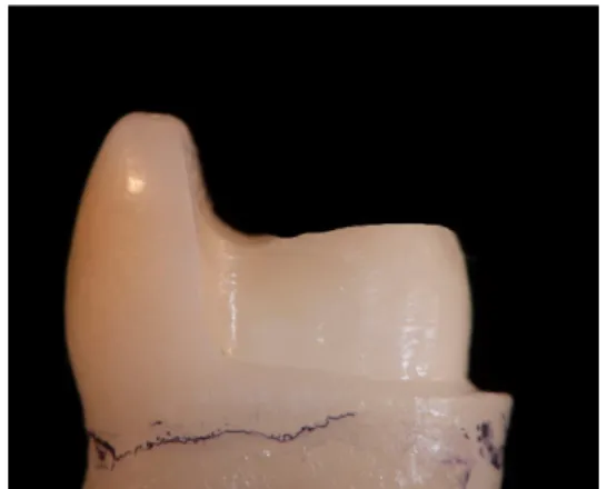
Group 1 (control, full crown preparation): Premolar preparation began with 1.5mm depth cuts on the occlusal table through the occlusal groove, preparation was tapered ∼4-8 degrees and a wide 1.0mm wide rounded shoulder margin was placed 1.0mm above the CEJ. All internal line angles were rounded, and all surfaces were smoothed. (Figure 2.1 A)
Group 2 MODL onlay preparation: Premolar preparation began with creation of 2mm depth cut mesiodistally through the central groove and extended laterally to completely include the nonfunctional cusp and preserve 2mm of the buccal-lingual width of the functional cusp. Occlusal reduction was then completed. Mesial, lingual and distal axial wall preparation resulted in a 2mm pulpal depth from the occlusal cavo-surface margin of functional cusp and axial wall height of approximate 3mm. A wide 1mm wide rounded shoulder margin was placed above the CEJ. All the internal preparation angles were rounded, and all surfaces were smoothed. (Figure 2.1 B)
1mm above the CEJ. All the internal preparation angles were rounded, and all surfaces were smoothed. (Figure 2.1 C)
Group 4 MODFL onlay veneer: Premolar preparation began with creation of 1mm depth cuts so as to allow uniform removal of tooth structure following the occlusal anatomy. A wide 1mm wide rounded shoulder margin was placed with axial height of 1mm inclined towards the occlusal surface. All the internal preparation angles were rounded, and all surfaces were smoothed. (Figure 2.1 D)
B:Group 2: Preserving 2mm non-functional cusp
A:Group 1: Full crown( control)
C:Group 3: Preserving 2mm
functional cusp D;Group 4: Overlay/Tabletop
2.3.2 Fabrication of the restorations
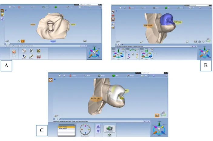
CAD/CAM blocks of 3Y-Monolithic zirconia (ZirCad, A1 LT Ivoclar Vivadent) were used. (Table 2.1). An intraoral scanner, Planmeca Emerald (Planmeca, Finland)) were used to scan the preparations and restorations were designed using software (Romexis E4D, Texas, USA) (figure 2.2). Minimal occlusal thickness of the onlays was 1mm, minimal axial thickness was 1mm were milled in PlanMill 40S (Planmeca, Finland). Preset tolerances within the software programing ensured minimal occlusal thickness of 1.5mm and minimal axial thickness of 1mm for all restorations. The milled restorations were 20-25% enlarged to compensate for shrinkage after the sintering process. Restorations were crystallized in speed sintering oven Programat CS4
(Ivoclar)
Figure 2.2.Planmeca Emerald scan for restoration fabrication. A, Initial scan of prepared tooth. B, Onlay restoration proposal. C, Onlay restoration design prepared for mi
A
C
2.3.3 Cementation of restorations:
Restorations were bonded according to APC concept:
A: Air particle abrasion with aluminum oxide particles 50µ particle size, low pressure (2 bar) for 15 seconds at the distance of 10mm and internal surfaces were thoroughly rinsed the with air/water spray and dried.
P: Primer Monobond Plus (Ivoclar Vivadent: Schaan, Liechtenstein) was applied to abraded surfaces for 60 seconds and dispersed it with a strong stream of air per manufacturer’s instructions. (Table 2.1)
Multilink Primer (Ivoclar Vivadent: Schaan, Liechtenstein) was applied to the tooth preparations per manufacturer’s instructions. In brief, primer A and B were mixed in a 1:1 ratio (1 drop of Primer A and 1 drop of Primer B). The self-etching and self-curing mixture were then applied with a scrubbing action for 30 seconds to the entire preparation using a micro brush, starting with the enamel surface.
C: Composite resin: Cementation of restorations was accomplished using the Multilink automix (Ivoclar Vivadent, Schaan, Liechtenstein) per manufacturer’s instructions. (Table 2.1)
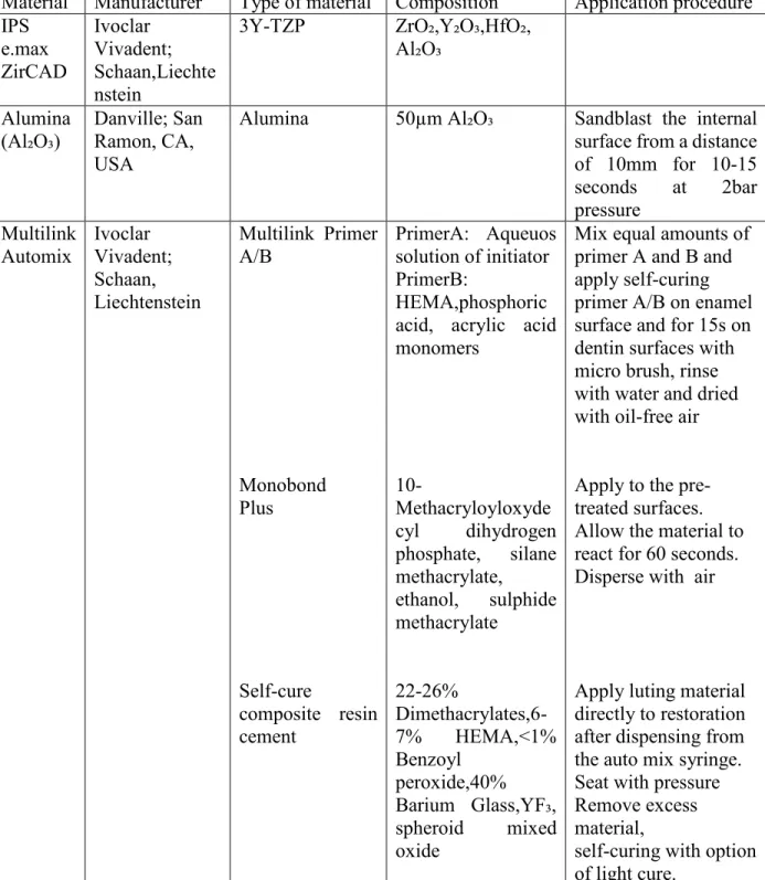
Table 2.1. Materials Used
Material Manufacturer Type of material Composition Application procedure IPS e.max ZirCAD Ivoclar Vivadent; Schaan,Liechte nstein
3Y-TZP ZrO₂,Y₂O₃,HfO₂, Al₂O₃
Alumina
(Al₂O₃) Danville; San Ramon, CA, USA
Alumina 50µm Al₂O₃ Sandblast the internal surface from a distance of 10mm for 10-15 seconds at 2bar pressure
Multilink
Automix Ivoclar Vivadent; Schaan, Liechtenstein Multilink Primer A/B Monobond Plus Self-cure composite resin cement PrimerA: Aqueuos solution of initiator PrimerB:
HEMA,phosphoric acid, acrylic acid monomers 10-Methacryloyloxyde cyl dihydrogen phosphate, silane methacrylate, ethanol, sulphide methacrylate 22-26% Dimethacrylates,6-7% HEMA,<1% Benzoyl peroxide,40% Barium Glass,YF₃, spheroid mixed oxide
Mix equal amounts of primer A and B and apply self-curing primer A/B on enamel surface and for 15s on dentin surfaces with micro brush, rinse with water and dried with oil-free air
Apply to the pre-treated surfaces. Allow the material to react for 60 seconds. Disperse with air
Apply luting material directly to restoration after dispensing from the auto mix syringe. Seat with pressure Remove excess material,
2.3.4 Fatigue testing
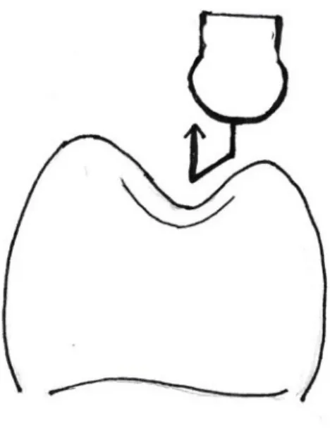
Specimens in each group were exposed to dynamic loading of 70 N at a frequency of 1.4 Hz for 1.2 million cycles with simultaneous thermocycling (10,000 cycles, 5-55˚C, 30s dwell time/cycle) in a computer controlled multifunctional mastication simulator (SD Mechatronik, Fedkirchen- Westerham, Germany) to accomplish fatiguing. This protocol of thermomechanical fatigue application of 1.2 million cycles were equivalent to approximately 5 years of clinical performance.79
Figure 2.3. Schematic diagram of mechanical loading pattern
2.3.5 Evaluating failure
Wear detectors mounted in each chamber monitored the specimens and recorded the cycle number during which specimens’ failure may have occurred. All surviving specimens were examined under 4.5X magnification for visible damage in the restoration or tooth following fatigue testing. Failure was defined as restoration or tooth fracture development and restoration debonding.
2.3.6 Scanning electron imaging:
coating with a layer of 10nm Gold-Palladium (AuPd). SEM operating conditions were as follows: Normal mode, 2kV accelerating voltage, 10microA beam current, and a working distance of close to 12mm was maintained throughout the analysis.
Also, CPD (Critical point drying) was performed on Tousimis Aotosamdri-931 CPD system.
Statistical analysis: The failure rate for each group was determined by visual inspection of each test sample after testing. Restoration-antagonist contact areas will be evaluated by Scanning Electron Microscopy for evidence of material fatigue. A test of Proportions statistical analysis will be conducted to identify survival rates.
2.4 Results:
2.4.1 Fatigue testing:
All specimens in Group 1, 3 and 4 survived the simulated aging process. There were no macroscopically visible cracks or fractures within the tooth structure and ceramic restorations. One Group 2 restoration debonded. All samples in Group 2 showed minimal ceramic cohesive fracture (chippings) at the occlusal margin in the area of the antagonist initial contact.
2.4.2 Scanning electron imaging:
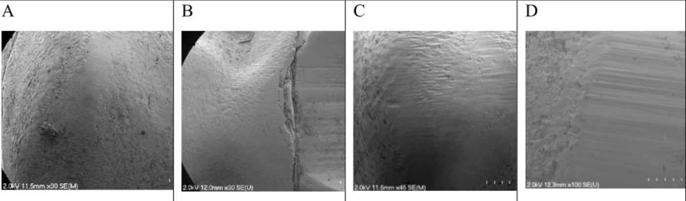
The findings from the visual inspection were verified by SEM and failure modes as observed with SEM were similar in group 2. The crack origin was located at the margin during the cyclic loading.
restorations in Group 1, 3, 4 had chipping of the restoration margins. Further assessment of the occlusal surfaces of all restorations at 100X and 150X magnification revealed no evidence of ceramic fracture development.
A B C D
Figure 2.4. SEM images. A: Group 1. B: Group 2. C: Group 3. D: Group 4
Table 2.2. Findings of SEM Observation:
Margin Crack or Fracture
30X 100X 150X
Groups Yes Problem Yes Problem Yes Problem
1 0 0 0
2 7 0 0
3 0 0 0
4 0 0 0
Statistical analysis results:
Although there was 1 failure in group 2, this failure rate was not statistically significantly different from 0 observed in the other group(P=.30)
For SEM analysis at 30X all margin of 7 samples in group 2 showed a marginal integrity issue. This rate was statistically significantly different from the rate of 0 in the other groups(P=0.0001)
2.6 Discussion:
This in vitro research study exposed adhesively bonded partial coverage zirconia restorations to simultaneous thermo-cycling and mechanical testing in an attempt to more closely simulate an environment in which dental ceramics are normally fatigued.24
Extracted human premolar teeth for restoration abutments were used in this study because of their bonding characteristics, thermal conductivity, modulus of elasticity and strength more closely represent actual clinical conditions as compared with other types of abutments.17,91 Other research studies used a cyclic loading of 49N.92,93,94Research has shown that occlusal forces in the posterior dentition may exceed functional loading forces of 49N during mastication or swallowing so, therefore a cycle loading force of 70N was chosen.95
The first null hypothesis was accepted as none of the tested samples fractured. The high survival rate in this study is comparable to two in-vitro studies.96, 97 Differences in test parameters limit direct comparison of specific results with these studies. To the best of the author’s knowledge, no study has investigated the effect of thermomechanical cyclic loading of monolithic zirconia partial coverage restorations on the bonding efficiency of restoration to tooth structure.
manufacturer’s instructions regardless of the preparation design specifics. This is in agreement of Sasse and Kern studies on single-retainer zirconia-based resin bonding fixed denture prosthesis.98, 99 High 5-year survival rates were demonstrated in these studies when zirconia restorations were adhesively bonded with either 10 MDP- containing composite resin or an adhesive bonding system with 10 MDP. Another clinical study by Kern et al, demonstrated excellent clinical outcomes of cantilever single-retainer zirconia-based restorations (with no retentive features) that had been adhesively bonded with MDP containing composite resin.100
The single bond failure in Group 2 (1 out of 8, a 12.5% failure rate) was not found to be statistically significant (P=.30). Visual examination revealed that the antagonist made contact on the natural tooth and slide over the margin of Group 2 specimens. It may be that antagonist contact with the restoration margin resulted in tensile forces that stressed the adhesive bond of the ceramic onlay to the tooth. It may be that the failure rate in this group may have increased if the number of cycles had been increased.
The steel ball contacting the tooth surface and sliding over the margins results into chipping of ceramic from Hertzian contact stresses. Marginal design of partial coverage zirconia restorations should be carefully and precisely determined so as to avoid direct margin loading, as this has critical effect on the local marginal integrity longevity of the zirconia restoration.
The results from this study add to the existing body of evidence that 3Y zirconia is durable and able to withstand the normal forces of mastication. This is likely because of its excellent flexural strength plus the ability of 3Y zirconia to undergo phase transformation so as to limit defect propagation. Groups 1, 3 and 4 survived extensive thermo-mechanical fatigue testing with no debonding or chipping. Samples in these groups received primarily compressive forces, while samples in Group 2 received a combination of compressive and tensile force. Bulk fracture was not detected in any of the restoration configurations when using SEM assessment at 30,100 and 150X magnification.
the effect of minimizing tensile forces on the adhesive interface as well as protect restoration/tooth interface from direct occlusal contact.
Control groups to assess the role of each variable of the APC technique were not included in this study. A control group to compare the APC technique with conventional cementation, that may typically be used with zirconia preparation designs such as the one utilized in Group 1, was not included in this research study. Therefore, no definitive statements can be made relative to the roles of the APC technique or retention/resistance form in the clinical longevity of partial coverage 2nd generation zirconia restorations. Further research studies should be designed to assess these issues.
The results indicate that it is possible to bond successfully to zirconia using a form of the APC technique. Whether the achieved bond is sufficiently strong and durable to retain non-retentive restorations like veneers and table tops has yet to be determined. The tooth preparations for groups 2, 3 and 4 had less resistance and retention form than a complete crown preparation, but all the tooth preparations used in this study had both retention and resistance form. Future studies should include tooth preparations with no resistance and retention form.
2.7 Limitations of the study:
materials.105 Further research of various zirconia formulations is warranted. The findings of this study may not be predictive of the performance of other zirconia formulations.
This research was also limited by the use of water during testing rather than artificial saliva. It may be that lubricating effects of salivary components are greater than that of water. Further studies should seek to assess the potential difference. This may improve the clinical simulation of this laboratory testing process of zirconia.
2.8 Conclusions:
Within the limitations of this in vitro research study:
1. Due to its fracture resistance, 3Y zirconia may be considered as desirable material for partial coverage restorations
2. Following a strict bonding protocol, bonding zirconia seems to be promising.
REFERENCES
1. Dalli M, Çolak H, Hamidi M. Minimal Intervention Concept: A New Paradigm for Operative Dentistry. Journal of Investigative and Clinical Dentistry. 2012; 3(3):167-75. 2. Kois DE, Isvilanonda V, Chaiyabutr Y, Kois JC. Evaluation of fracture resistance and
failure risks of posterior partial coverage restorations. J Esthetic and Restorative Dentistry.2013;25(2):110-22
3. Terry Donovan, R.J. Simonsen,G. Guertin,R.V. Tucker, Retrospective Clinical evaluation of 1,314 Cast Gold Restorations in Service From 1 To 52 Years J Esthet Restor Dent2004;16(3):194-204
4. Guess PC,Schultheis S, Wolkewitz M, Zhang Y, strub JR. Influence of preparation design and ceramic thickness on fracture resistance and failure modes of premolar partial coverage restorations. J Prosthet Dent 2013;110(4):264-73
5. Van Dijken JWV, Hasselrot L, Örmin A, Olofsson AL. Restorations with extensive dentin/enamel-bonded ceramic coverage. A 5-year follow-up. Eur J Oral Sci. 2001;109(4):222–29
6. Christensen GJ. Porcelain-fused-to metal verses zirconia based ceramic restorations. J Am Dent Assoc.2009;140(8):1036-9
7. Langelnd,K, Langland,LK: Pulpal reaction to crown preparation, temporary crown fixation, and permanenet cementation.J Prosthet Dent. 1965;15:129-43
8. Heymann HO,Jr.EJ, Ritter AV. Sturdevant’s Art and Science of operative Dentistry. Elsevier Health Sciences;2014
9. Zach L, Cohen G. Pulp response to externally applied heat. Oral Surg Oral Med Oral Pathol. 1965;19:515-30
10. Oztürk B, Ușümez A, Oztürk AN, Ozer F. In vitro assessment of temperature change in
the pulp chamber during cavity preparation. J Prosthet Dent. 2004;91(5):436-40
11. Felton D, Madison S, Kanoy M, Maryniuk G. Long term effects of crown preparation on pulp vitality. J Dent Res 1989;68,1009
12. Jackson CR, Skidmore AE, Rice RT. Pulpal evaluation of teeth restored with fixed prosthesis. J Prosthet Dent 1992; 67(3):323-5
14. Van Dijken, Siostrom S. Development of gingivitis around aged restorations of resin- modified glass ionomer cement, polyacid-modified resin composite (compomer) and resin composite. Clin Oral Investig 1998:2(4)180-3
15. Felden A, Schmalz G, Federlin M, Hiller KA. 1998. Retrospective clinical investigation and survival analysis on ceramic inlays and partial ceramic crowns: results up to 7 years. Clin Oral Investig. 2(4):161–67
16. Fuzzi M, Rappelli G. 1998. Survival rate of ceramic inlays. J Dent. 26(7):623–626., Schulz P, Johansson A, Arvidson K. 2003. A retrospective study of Mirage ceramic inlays over up to 9 years. Int. J Prosthodont. 16(5):510–14
17. Stappert CF, Att W, Gerds T, Strub JR. Fracture resistance of different partial-coverage ceramic molar restorations: An in vitro investigation. J Am Dent Assoc.2006;137(4):514-22
18. Lange RT, Pfeiffer P. Clinical evaluation of ceramic inlays compared to compositerestorations. Oper Dent. 2009; 34(3):263-72.
19. Jung H, Friedl KH, Hiller KA, Haller A, Schmalz G. Curing efficiency of different polymerization methods through ceramic restorations. Clin Oral Investig 2001;5(3):156-61
20. Derand T. Stress analysis of cemented or resin-bonded porcelain inlays. Dent. Mater 1991; 7(1):21-4.
21. Naeselius K, Arnelund CF, Molin MK. Clinical evaluation of all-ceramic onlays: a 4-year retrospective study. Int J Prosthodont. 2008; 21(1):40–4.
22. Arnelund CF, Johansson A, Ericson M, Hager P, Fyrberg KA. Five-year evaluation of two resin-retained ceramic systems: a retrospective study in a general practice setting. Int J Prosthodont. 2004; 17(3):302–6.
23. Morimoto S, F.B.W. Rebello de Sampaio, M.M. Braga, N. Sesma,and M. Özcan Survival Rate of Resin and Ceramic Inlays, Onlays, and Overlays:A Systematic Review and Meta-analysis J Dent ResearchVolume: 95 issue: 9, page(s): 985-994
24. Kelly JR. Clinically relevant approach to failure testing of all ceramic restorations. J Prosthet Dent 1999;81(6):652-61
25. Smales RJ, Etemadi S. Survival of ceramic onlays placed with and without metal reinforcement. J Prosthet Dent. 2004; 91:548–53
27. Beier US, Kapferer I, Burtscher D, Dumfahrt H. Clinical performance of porcelain laminate veneers for up to 20 years. International Journal of Prosthodontics 2012; 25:79– 85.
28. Murgueitio R, Bernal G. Three-Year Clinical Follow-Up of Posterior Teeth Restored with Leucite-Reinforced IPS Empress Onlays and Partial Veneer Crowns. J Prosthodont. 2012; 21:340–5.
29. Federlin M, Krifka S, Herpich M, Hiller KA, Schmalz G. Partial ceramic crowns: influence of ceramic thickness, preparation design and luting material on fracture resistance and marginal integrity in vitro. Operative Dentistry 2007;32:251–60
30. Magne P, Schlichting LH, Maia HP, Baratieri LN. In vitro fatigue resistance of CAD/CAM composite resin and ceramic posterior occlusal veneers. Journal of Prosthetic Dentistry 2010;104:149–57
31. Li RW, Chow TW, Matinlinna JP. Ceramic dental biomaterials and CAD-CAM technology: state of the art. J Prosthodont Res 2014;58(4):208-16
32. Wendler M, Belli R, Petschelt A.Mevec D,Harrer W, Lube T, Danzer R, Lohbauer U. Chairside CAD/CAM materials. Part2: Flexural strength testing. Dent Mater 2017;33(1):99-109.
33. Al-Akhali M, Kern M, Elsayed A, Samran A, Chaar MS. Influence of thermomechanical fatigue on the fracture strength of CAD-CAM-fabricated occlusal veneers. J Prosthet Dent. 2019;121(4):644-650.
34. Sen N, Us YO. Mechanical and optical properties of monolithic CAD-CAM restorative materials. J Prosthet Dent 2018;119(4):593-599.
35. Clausen JO, Abou Tara M, Kern M. Dynamic fatigue and fracture resistance of non-retentive all-ceramic full-coverage molar restorations. Influence of ceramic material and preparation design. Dent Mater. 2010; 26(6):533–8.
36. Denry I, Kelly JR. State of the art of zirconia for dental applications. Dental Materials 2008;24(3):299–307.
37. Homaei E, Farhangdoost K, Tsoi JKH, Matinlinna JP, Pow EHN. Static and fatigue mechanical behavior of three dental CAD/CAM ceramics. J Mech Behav Biomed Mater 2016; 59:304-313.
38. Ferrari M, Vichi A, Zarone F. Zirconia abutments and restorations: from laboratory to clinical investigations. Dent. Mater 2015 31(3): e63-76.\
40. A.J. Raigrodski, G.J. Chiche, N. Potiket, J.L. Hochstedler, S.E. Mohamed, S. Billiot, D.E. MercanteThe efficacy of posterior three-unit zirconium-oxide-based ceramic fixed partial dental prostheses: a prospective clinical pilot study. J Prosthet Dent 2006;96 (4):237-244 41. Stawarczyk B, Ozcan M, Roos M, Trottmann A, Sailer I, Hämmerle CH. Load-bearing
capacity and failure types of anterior zirconia crowns veneered with overpressing and layering techniques.Dent. Mater.2011;27(10):1045-1053
42. Chen YW, Moussi J, Drury JL, Wataha JC. Zirconia in biomedical applications. Expert Rev Med Devices 2016;13(10):945-963
43. Denry I, Kelly JR. Emerging ceramic based materials for dentistry. J Dent Res 2014;93(12):1235-42
44. Garvie RC, Hannink RH, Pascoe RT. Ceramic steel? Nature 1975; 258: 703-704
45. Garvie RC, Nicholson PS. Phase analysis in zirconia systems. J Am Ceram Soc 1972; 55(6): 303-305
46. Heuer AH, Lange FF, Swain MV, Evans AG. Transformation toughening: An overview. J Am Ceram Soc 1986;69(3):1151-2916
47. Alraheam IA, Donovan T, Boushell L, Cook R, Ritter AV, Sulaiman TA. Fracture load of two thicknesses of different zirconia types after fatiguing and thermocycling. P Prosthet Dent.2019 (In press).
48. Alraheam IA, Donovan T, Rodgers B, Boushell L, Sulaiman TA. Effect of masticatory simulation on the translucency of different types of dental zirconia. J Prosthet. Dent 2019;122: 404-9.
49. Zhang Y, Lawn BR, Rekow ED, ThompsonVP. Effect of sandblasting on the long-term performance of dental ceramics. J. Biomed. Mater Res B Appl. Biomater. 2004; 71(2):381– 386.
50. Kern M, Wegner SM. Bonding to zirconia ceramic: Adhesion methods and durability. Dent. Mater 1998;14(1):64-71.
51. Bottino MA, Valandro LF, Scotti R, Buso L. Effect of surface treatments on the resin bond to zirconium based ceramic. Int J Prosthodont 2005;18(1):60-5
52. Wegner SM, Gerdes W, Kern M. Effect of different artificial aging conditions on ceramic-composite bond strength. Int J Prosthodont 2002;15(3):267-72
54. Yagawa S, Komine F, Fushiki R, Kubochi K, Kimura F, Matsumura H. Effect of priming agents on shear bond strengths of resin-based luting agents to a translucent zirconia material. J. Prosthodont. Res. 2018;62(2):204–209.
55. Noda Y, Nakajima M, Takahashi M, Mamanee T,Hosaka K, Takagaki T, Ikeda M, Foxton RM, Tagami J. The effect of five kinds of surface treatment agents on the bond strength to various ceramics with thermocycle aging. Dent. Mater. J. 2017;36(6): 755–761
56. Lopes GC, Spohr AM, De Souza GM. Different Strategies to Bond Bis-GMA-based Resin Cement to Zirconia. J. Adhes Dent. 2016:18(3): 239–246.
57. Ahn J.S, Yi Y.A, Lee Y, Seo DG. Shear Bond Strength of MDP-Containing Self-Adhesive Resin Cement and Y-TZP Ceramics: Effect of Phosphate Monomer-Containing Primers. Biomed. Res. Int. 2015.
58. Liu D, Pow EHN, Tsoi J.K, Matinlinna J.P. Evaluation of four surface coating treatments for resin to zirconia bonding. J. Mech. Behav. Biomed. Mater. 2014;32: 300–309.
59. Yenisey M, Dede DO, Rona N. Effect of surface treatments on the bond strength between resin cement and differently sintered zirconium-oxide ceramics. J. Prosthodont. Res. 2016; 60(1): 36–46.
60. Cheung GJ, Botelho MG. Zirconia Surface Treatments for Resin Bonding. J. Adhes Dent. 2015; 17(6): 551–558.
61. Bomicke W, Schurz A, Krisam J, Rammelsberg P, Rues S. Durability of Resin-Zirconia Bonds Produced Using Methods Available in Dental Practice. J. Adhes Dent. 2018; 18(1): 17–27.
62. Shin YJ, Shin Y, Yi YA, Kim J, Lee IB, Cho BH, Son HH, Seo DG. Evaluation of the shear bond strength of resin cement to Y-TZP ceramic after different surface treatments. Scanning 2014; 36(5): 479–486.
63. Tzanakakis EG, Tzoutzas IG, Koidis PT. Is there a potential for durable adhesion to zirconia restorations? A systematic review J Prosthet Dent 2016;115(1):9-19.
64. Derand P, Derand T.Bond strength of luting cements to zirconium oxide ceramics. Int J Prosthodont 2000;13(2):131-5
65. Wegner SM, Kern M. Long term resin bond strength to zirconia ceramic. J Adhes Dent Int J Prosthodont.2000; 2(2):139-47.
67. Inokoshi M, De Munck, Minakuchi S, VanMeerbeek B. Meta-analysis of Bonding Effectiveness to Zirconia Ceramics, J Dent Res.2014; 93(4):329-334.
68. Thammajark P, Inokoshi M, Chong S, Guazato M. Bonding of composite cements to zirconia: A systematic review and meta-analysis of in vitro studies. J Mech Behav Biomed Mater.2018; 258-268.
69. Blatz MB, Alvarez M, Sawyer K, Brindis M. How to Bond Zirconia: The APC Concept. Compend Contin Educ Dent.2016; 37(9):611-616.
70. Özcan M. Air Abrasion of Zirconia Resin-bonded Fixed Dental Prostheses Prior to Adhesive Cementation:Why and How? J. Adhes Dent. 2013;15(4):394.
71. Yang L, Chen B, Xie H, Chen Y, Chen Y, Chen C. Durability of Resin Bonding to Zirconia Using Products Containing 10-Methacryloyloxydecyl Dihydrogen Phosphate. J. Adhes Dent. 2018;20(4): 279–287.
72. Steiner M, Mitasias ME, Ludwig K, Kern M. In vitro evaluation of a mechanical testing chewing simulator. Dent Mater 2009;25(4):494-499
73. Donovan T.E, Alraheam I.A, Sulaiman T.A. An evidence- based evaluation ofcontemporary dental ceramics. Dental updates.2018; 45(6):541-546.
74. Hedegard B. Need for correlation between laboratory testing and clinical research. Inn National bureau of standards special Publication 354. Dental material research proceedings of the 50th anniversary symposium, October 6-8, 1968, U.S Department of commerce; 1972: 187-99.
75. DeLong R, Douglas WH. An artificial oral environment for testing dental materials IEEE Trans Biomed Eng, 1991; 38(4):339-345.
76. Al-Akhali M, Kern M, Elsayed A, Samran A, Chaar MS. Influence of thermomechanical fatigue on the fracture strength of CAD-CAM-fabricated occlusal veneers. J Prosthet Dent. 2019; 121(4):644-650.
77. De Boever JA, McCall WD Jr, Holden S, Ash MM Jr. Functional occlusal forces: an investigation by telemetry. J Prosthet Dent 1978;40(3):326-33.
78. Gibbs CH, Mahan PE, Lundeen HC, Brehnan K, Walsh EK, Holbrook WB. Occlusal forces during chewing and swallowing as measured by sound transmission. J Prosthet Dent.1981; 46(4):443-9.
80. Nawafleh N, Hatamleh M, Elshisyab S, Mack F. Lithium Disilcate Retorations Fatigue Testing Parameters: A Systematic Review. J Prosthodont.2016:25(2):116-26
81. Gillings BRD, Graham CH, Duckmanton NA: Jaw movements in young adults men during chewing. J Prosthet Dent 1973;29:616-627
82. Hiiemae K, Heath MR, Heath G,et al: Natural bites, food consistency and feeding behavior in man. Arch Oral Biol1996;41:175-189
83. Woda A, Mishellany A, Peyron MA: The regulation of masticatory function and food bolus formation. J oral Rehabil.2006;33:840-849
84. Kim B, Zang Y,Pines M et al: Fracture of Porcelain Veneered structures in Fatigue. J Dent Res 2007;86:142-146
85. Rosentritt M, Behr M, Gebhard R & Handel G: Influence of stress simulation parameters on the fracture strength of all-ceramic fixed-partial dentures. Dental Materials 2006; 22: 176-182.
86. Behr M, Hindelang U, Rosentritt M, Lang R, Handel G. Comparison of failure rates of adhesive-fixed partial dentures for in vivo and in vitro studies Clinical Oral Investigations.2000; 4: 25-30
87. de Gee A, Pallav P, Davidson CL. Effect of abrasion medium on wear of stress-bearing composites and amalgam in vitro Journal of Dental Research.1986;65: 654-658
88. Ma L, Guess PC, Zhang Y. Load bearing properties of minimal-invasive monolithic lithium disilicate and zirconia occlusal onlays: Finite element and theoretical analysis. Journal of Dental Material 2013;29(7):742-751
89. Harsha MS, Praffulla M, Babu MR, Leneena G, Krishna TS, Divya G. The Effect of Cavity Design on Fracture Resistance and Failure Pattern in Monolithic Zirconia Partial Coverage Restorations - An In vitro Study. J clin Diagn Res 2017;11(5):ZC45-ZC48;
90. Oyar P, Durkan R. Effect of cavity design on the fracture resistance of zirconia onlay ceramics. Niger J clin Pract 2018; 21(6):687-91.
91. Homaei E, Farhangdoost K, Pow E, Matinlinna J, Akbari M, Tsoi J. Fatigue resistance of monolithic CAD/CAM ceramic crowns on human premolars. Ceramics Int 2016;42(14):15709-15717
93. Krejci I, Reich T, Lutz F, Albertoni M. An in vitro test procedure for evaluating dental restoration systems.1. A computer-controlled mastication simulator [In German]. Schweiz Monatsschr Zahnmed 1990;100(8):953-60
94. Kern M, Strub JR, Lu XY. Wear of composite resin veneering materials in a dual-axis chewing simulator. J Oral Rehabil 1999; 26(5):372-8.)
95. Gibbs CH, Mahan PE, Mauderli A, Lundeen HC, Walsh EK. Limits of human bite strength. J Prosthet Dent 1986; 56(2):226-9.
96. Krejci I, Lutz F, Reimer M, Heinzmann JL. Wear of ceramic inlays, their enamel antagonists, and luting cements. J Prosthet Dent 1993;69(4):425-30
97. Mehl A, Pfeiffer A, Kremers L, Hickel R. Marginal accuracy of Cerec 2 inlays with extended cavities and weakened cusps [in German]. Dtsch Zahnarztl Z 1998; 53:57-60. 98. Sasse M, Kern M. CAD/CAM fabricated single retainer zirconia ceramic resin-bonded
fixed dental prostheses: clinical outcome after 5 years. International J comput Dentist.2013;16(2):109-18.
99. Sasse M, Kern M. Survival of anterior cantilevered all- ceramic resin-bonded fixed dental prostheses made from zirconia ceramic. J Dent. 2014; 42(6):660-663.
100. Kern M, Passia N, Sasse M, Yazigi C. Ten-year outcome of zirconia ceramic cantilever resin-bonded fixed dental prostheses and the influence of the reasons for missing incisors. J Dent. 2017;65:51-55
101. Lawn BR, Deng Y, Thompson VP. Use of contact testing in the characterization and design of all-ceramic crown like layer structures: a review. J Prosthet Dent 2001;86(5):495-510 102. Bhowmick S, Zhang Y, Lawn BR. Competing fracture modes in brittle materials subject
to concentrated cyclic loading in liquid environments: Bilayer structures. J Mater Res. 2005; 20(10):2792-800
103. Jung YG, Wuttiphan S, Peterson IM, Lawn BR. Damage modes in dental layer structures. J Dent Res 1999; 78(4):887-97