THE IMPACT OF THE MICROBIOTA ON TRANSCRIPTIONAL REGULATION
IN THE VERTEBRATE INTESTINE
J. Grayson Camp
A dissertation submitted to the faculty of the University of North Carolina at
Chapel Hill in partial fulfillment of the requirements for the degree of Doctor of
Philosophy in the Curriculum of Genetics and Molecular Biology
Chapel Hill
2012
Approved By:
ABSTRACT
J. GRAYSON CAMP: The Impact of the microbiota on transcriptional regulation in the vertebrate intestine
(Under the direction of John F. Rawls)
Animals evolved in a world pre-dominated by microscopic organisms.
Colonization of intestinal tracts at birth by microbes initiates the next generation of an
ancient symbiosis that profoundly impacts our physiology and pathophysiology. A record
of this symbiosis is encoded in our genomes. In this dissertation, I explore how
regulatory regions embedded in non-genic DNA mediate transcriptional responses to the
intestinal microbiota. Extensive research has demonstrated that the complex community
of microbes residing within our intestine (the gut microbiota) contributes biochemistries
that enhance nutrient digestion, metabolize xenobiotics, and collectively function as an
important environmental factor that modulates host energy balance and immunity.
However, the mechanisms that host cells use to perceive and respond to these microbial
activities are not well understood. I used the zebrafish and mouse gnotobiotic models to
define mechanisms by which the microbiota regulates host transcription in the intestinal
epithelium at the single gene and genome-wide scales. The intestinal microbiota
enhances dietary energy harvest leading to increased lipid storage in peripheral tissues.
This effect is caused in part by the microbial suppression of intestinal expression of a
circulating inhibitor of Lipoprotein lipase called Angiopoietin-like 4 (Angptl4/Fiaf). I
utilized the zebrafish in which host regulatory DNA can be rapidly analyzed in a live,
that confers intestine-specific transcription and microbial suppression of angptl4. I used comparative sequence analysis from 12 fish species, functional mapping, and
mutagenesis to define the minimal set of regulatory sequences required for activity of the
angptl4 intestinal CRM. I applied computational prediction and DNA affinity
chromatography to discern candidate transcription factors regulating angptl4 intestinal expression. At the genomic level, I employed DNase-seq and FAIRE-seq in the intestine
of germ-free and conventionally-raised mice and zebrafish to facilitate the discovery of
CRMs meditating host responses to the microbiota genome-wide. This work provides a
novel paradigm for understanding how microbial signals interact with tissue-specific
regulatory networks to control host gene expression and elucidates mechanisms
mediating over 500 million years of co-existence and co-evolution of vertebrate hosts
ACKNOWLEDGEMENTS
Foremost, I would like to thank my mentor, John Rawls, for all of his guidance and patience during my graduate training. His curiosity, keen sense for the proper experiment, and remarkable ability to see the bigger picture has been a rich source of inspiration for me and I hope to continue to learn from him as I mature as a scientist. His steadfast support and appreciation for both the scientific and personal well being of the individuals in his lab has made this journey a great experience. I also thank him for turning me on to Parasite Rex and my interview with him for IBMS, as this marked a node in my life.
I would like to thank everyone in the Rawls Lab, past and present: Michelle Kanther, Ivana Semova, Ed Flynn, James Minchin, Jordan Cocchiaro, Sandi Wong, Jim Davison, Amy Jazwa, Chad Trent, Lantz Mackey, Laura Mackey, Linh Pham, Jessica Russell. These people have contributed to my life in different ways and I appreciate the
friendships and common experiences we’ve had together over the past 5 years. Michelle Kanther was a model for her organizational skills and did her best to try to keep me in
line. Ivana Semova was a thought provoking lab mate and friend and we had many great times together in and out of lab. Jordan Cocchiaro was a conscientious lab mom and
attitude. Amy Jazwa was my first mentee and I learned many valuable lessons from this experience.
The research environment at UNC has been an open, inspiring, and collaborative place to do science. My committee members provided direction and much appreciated advice on my projects. I was lucky enough to collaborate with the labs of Jason Lieb and Greg Crawford and appreciate especially the invaluable technical assistance of Jeremy Simon, Chris Frank, and Yoichiro Shibata. Special thanks to Sausyty Hermreck and Cara Marlow for their immense support, and to Bob Duronio and Pat Brennwald for their leadership. I was positively impacted by many more fellow scientists and research staff, too many to mention by name, but I am grateful to all of them.
The support of my friends and family has been very important to me during my time at UNC, and it is difficult to express how much I am thankful for them all. Maggie McCormick is like a sister and will understand why I don’t write more about her
contribution to my well being. Agos Santoro is a great friend and being around her lively personality has made me happy over the past couple of years. Doug McIntyre was an ideal roommate. I heartily enjoyed listening to his ponderings and appreciate his sense of Walker Texas Ranger-like ideals. Anne-Marie Neiser exemplified honesty, moralty, and optimism and helped me grow up. Special thoughts go out to Robert Sons, Bryan Richardson, Chris Schmidt, Meghan Morgan, all of the fellow IBMS students, and my
intelligence, and exuberance. It would be difficult to imagine my life without her to help me travel through it.
At the end of each research chapter I have acknowledged the specific contributions that other people have made to the work presented in that chapter. The results
presented in Chapter 3 of this dissertation derive from a previously published article:
J. Gray Camp, Amy L. Jazwa, Chad M. Trent, and John F. Rawls. Intronic cis-regulatory modules mediate tissue-specific and microbial suppression of Angptl4/Fiaf transcription. PLoS Genetics 8: e1002585.
In Chapter 5, Chris Frank generated the ileum libraries and helped with the computational analysis of the data. Yoichiro Shibata aligned the DNase-seq reads to the mouse genome and helped with peak calling. Jeremy Simon performed the initial
processing of the zebrafish FAIRE-seq reads, aligned them to the zebrafish genome, and performed the peak calling. The liver and kidney DNase datasets are unpublished and were generously provided by the Crawford lab. John Mayfield dissected the liver and kidney tissue from the mice and Alexias Safi made the DNase libraries.
PREFACE
The Waking By Theodore Roethke
I wake to sleep, and take my waking slow. I feel my fate in what I cannot fear. I learn by going where I have to go. We think by feeling. What is there to know?
I hear my being dance from ear to ear. I wake to sleep, and take my waking slow. Of those so close beside me, which are you? God bless the Ground! I shall walk softly there,
And learn by going where I have to go. Light takes the Tree; but who can tell us how?
The lowly worm climbs up a winding stair; I wake to sleep, and take my waking slow.
Great Nature has another thing to do To you and me, so take the lively air, And, lovely, learn by going where to go. This shaking keeps me steady. I should know.
What falls away is always. And is near. I wake to sleep, and take my waking slow.
TABLE OF CONTENTS
Abstract……….ii
Acknowledgment ………..iv
Preface ………vii
List of Figures ………..xii
List of Tables………....xiv
CHAPTER 1 Introduction …..….………..………...………..1
2 Host-Microbiota Symbiosis and Transcription in the Vertebrate Intestine ...3
2.1 Overview ………...3
2.2 Introduction ...4
2.3 The Microbiota Impacts Intestinal Physiology ...6
2.3.1 An evolutionarily conserved developmental step ……….6
2.3.2 Ontogeny of the intestinal microbiota ……….8
2.3.3 Gross anatomy of the vertebrate GI tract ………..9
2.3.4 Cellular anatomy of the vertebrate midgut ………..11
2.3.5 Genetic repertoire of the intestinal microbiota ………12
2.3.6 Gnotobiotic vertebrate models: the power of comparison …………13
2.4 Transcriptonal Regulation in the Intestine ...15
2.4.1 Molecular anatomy of transcriptional regulation ……….15
!
!
2.4.3 Transcription factors mediating intestinal transcription ...192.5 Genomics Approaches to Discover CRMs ………...23
2.5.1 Genomes as reagents for CRM discovery ...23
2.5.2 The role of chromatin in gene regulation ……….24
2.5.3 Transcriptional genomics in the intestine ………27
2.5.4 Model organisms are in vivo assay systems ………..28
2.5.5 CRMs as mediators of host-microbe symbiosis ………..31
3 Intronic Cis-Regulatory Modules Mediate Tissue-Specific and Microbial Control of Angptl4/Fiaf Transcription ...32
3.1 Overview ...32
3.2 Introduction ...33
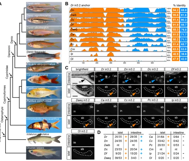
3.3 Results ...37
3.3.1 Tissue-specific expression of zebrafish angptl4 ...37
3.3.2 Conservation in DNA sequence guides CRM discovery …………...37
3.3.3 The angptl4 proximal promoter does not recapitulate mRNA expression patterns ………..40
3.3.4 Angptl4 intronic CRMs confer tissue-specific transcription ………..41
3.3.5 Evolution of the islet and intestinal regulatory modules ………45
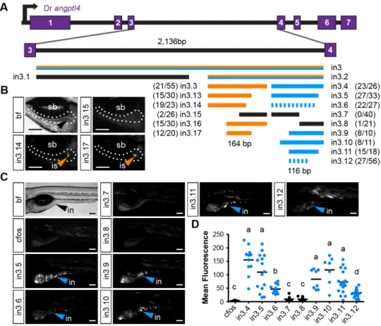
3.3.6 Truncation mapping of CRMs ………48
3.3.7 Site-directed mutagenesis of CRMs ……….49
3.3.8 The in3.4 module recapitulates angptl4 suppression by the microbiota ………..51
3.4 Discussion ……….53
3.4.1 Non-overlapping CRMs confer cell-type specific transcription of angptl4 ………53
3.4.2 The nature of microbial signals regulating intestinal transcription of angptl4 ………57
transcription of angptl4 ………...59
3.5 Materials and Methods ………60
3.6 Acknowledgements ………..67
3.7 Supporting Information ……….68
4 Towards Identification of Transcription Factors Regulating Intestinal Expression of Angptl4/Fiaf………82
4.1 Overview……….82
4.2 Introduction ………83
4.3 Results ………88
4.3.1 Computational prediction of transcription factors ………...88
4.3.2 Substiution of the GATA factor binding motif ………..88
4.3.3 Generation of an in vitro binding assay ………90
4.3.4 DNA-affinity chromatography and mass spectrometry to identify transcription factors ………..92
4.4 Discussion ………101
4.4.1 Potential transcription factors regulating intestinal transcription of angptl4 ……….101
4.4.2 Optimization of methods for the unbiased discovery of transcription factors ...102
4.5 Materials and Methods ………..107
4.6 Acknowledgements ………119
5 Pilot Atlas of Open Chromatin in the Intestinal Epithelium of Mouse and Zebrafish ………...115
5.1 Overview………...115
5.2 Introduction ………..………...116
5.3 Results ……….………..……….118
5.3.1 Strategy to discover microbially-responsive CRMs genome-wide………..118
intestinal epithelium………...122
5.3.3 Establishing DNase-seq in the zebrafish intestinal epithelium ………...125
5.3.4 Establishing FAIRE-seq in the zebrafish intestinal epithelium………...126
5.3.5 Preliminary analysis of pilot DNase-seq and FAIRE-seq CONV-R datasets ……….129
5.3.6 General features of DNase-seq in ileal mIECs ………129
5.3.7 DNase-seq elucidates putative cell-type specific CRMs ………….132
5.3.8 DNase-seq predicts transcription factors regulating intestinal gene expression ……….134
5.3.9 DNase-seq predicts transcription factors regulating microbial response ……….136
5.3.10 FAIRE-seq uncovers ancient CRMs in the zebrafish ………139
5.5 Discussion ………142
5.5.1 Genomic atlas of open chromatin in the vertebrate intestinal epithelium ……….142
5.5.2 Open chromatin maps to predict transcription factors ……….143
5.5.3 Integrating mouse and zebrafish open chromatin maps ………….146
5.5.4 Microbial impact on cis-regulatory function and evolution ………..149
5.6 Materials and Methods ………..151
5.7 Acknowledgements ………157
6 Future Prospectus ………..159
6.1 Overview ………..160
6.2 Zebrafish and Transcriptional Regulation Analysis ………...162
6.3 Host-Microbe Symbiosis and Adaptive Evolution ……….164
6.4 Host-Microbe Symbiosis and Genome Sandboxes ………..166
6.5 Concluding Remarks ……….168
List of Figures
2.1 Colonization by microorganisms is an evolutionarily
conserved developmental step ………..7
2.2 General features of the zebrafish model ………11
2.3 Non-coding DNA in gene regulation ………...18
2.4 Microbial suppression of intestinal expression of angptl4 ………..22
2.5 Methods for genome-wide discovery of open chromatin ………26
3.1 Tissue-specific expression of zebrafish angptl4 mRNA ………..39
3.2 Multiple-species alignments reveal conservation in angptl4 gene structure and location of conserved non-coding regions ……….41
3.3 Non-overlapping regulatory modules within angptl4 intron 3 confer liver, islet, and enterocyte-specific reporter expression ………..44
3.4 Functional evolution of the islet and intestinal regulatory modules in 12 fish species ……….47
3.5 Truncation mapping of the islet and intestinal regulatory module ……….50
3.6 Site-directed mutagenesis defines DNA motifs required for intestinal expression ………52
3.7 Summary of functional conservation and mapping of islet and intestinal regulatory information ………..54
3.8 The intestinal module in3.4 recapitulates microbial suppression of angptl4 ………57
3.S1 Phylogeny of Angptl4 and Angptl3 proteins from multiple vertebrate species ………68
3.S2 Alignment of Angptl4 proteins from multiple vertebrate species ……….69
3.S4 The zebrafish angptl4 in3.4 intestinal module exhibits
hallmarks of a classical enhancer ……….71
3.S5 Multiple-species sequence alignment of teleost angptl4 in3.3 modules …………72
3.S6 Multiple-species sequence alignment of teleost angptl4 in3.4 modules …………73
3.S7 The intronic module in3.2 recapitulates microbial suppression of angptl4………74
3.S8 Mouse Angptl4 intron 3 drives expression in circulating blood cells but not in the zebrafish liver, islet, or intestine ………..75
4.1 Mutation of a predicted GATA factor-binding site abolishes intestinal expression ………89
4.2 Factors in zebrafish IEC nuclear extracts bind the in3.4-CR regulatory region ……….………91
4.3 DNA affinity pull-down using wild type and subGATA probes ………93
4.4 DNA affinity pull-down using wild type and scrambled probes ………..95
4.5 DNA affinity pull-down using wild type and scrambled competitors ………..97
5.1 Experimental strategy to discover microbiota regulated CRMs ………...120
5.2 Experimental strategy and description of zebrafish IEC datasets ………..121
5.3 Establishment of DNase-seq in mouse IECs ………..124
5.4 Establishment of DNase-seq in zebrafish IECs ………..126
5.5 Establishment of FAIRE-seq in zebrafish 6dpf GI tracts and adult IECs …………128
5.6 General features of DNase-seq open chromatin sites ………..131
5.7 DNase-seq distinguishes cell-type specific open chromatin in the ileum ………...133
5.8 Ileum DNase-seq predicts motifs regulating intestinal gene expression ………….135
5.9 Motif prediction using DH sites near microbiota regulated genes ………137
5.10 FAIRE-seq in zebrafish IECs uncovers ancient CRMs ………...141
List of Tables!
2.1 Common terminology in gnotobiotic research ………...12
2.2 Unsolved mysteries in host-microbe symbiosis ……….20
3.S1 Angiopoietin-like protein sequences used for inferring phylogeny ………...76
3.S2 Primer sequences used in this study ………...80
3.S3 Allele designations for stable lines created in this study ………...81
4.1 Mass spectrometry results from wild type and scrambled pull-downs using IPI database ……….99
4.2 Mass spectrometry results from wild type and scrambled pull-downs using UniProt database ………..100
4.3 Primers and oligo sequences used in this study ………113
5.1 Motif prediction using ileum-specific DH sites ………136
5.2 Summary of motif prediction using DH sites near microbiota regulated genes ….138 5.3 Summary of the intersection of FAIRE-seq peaks with zCNEs ………142
CHAPTER 1
Introduction
The body surfaces of humans and other animals are colonized at birth by
microorganisms. The majority of microbial residents on the human body exist within
gastrointestinal tract (GI) communities, where they engage in symbiosis with host cells.
The host genome encodes the ability to respond to microbial activities and therefore
constitutes a nexus and historical record for this ancient symbiosis. Gene-specific and
genome-wide profiling of host gene expression has provided an important window into
the microbial impact on host physiology and pathobiology, however the mechanisms
underlying host transcriptional responses to the microbiota are poorly understood.
Recent advances in high-throughput sequencing have expanded our ability to perceive
the membership and physiologic traits of microbial communities along the GI tract.
These same tools have in parallel dramatically expanded the functional understanding of
vertebrate genomes in the fields of comparative genomics, transcriptional regulation,
and chromatin biology. I propose in this dissertation that it is time to merge microbiota
research with these fields in order to gain new mechanistic insights into how microbial
symbiosis can impact transcriptional regulation and its evolution on a genomic scale.
The following chapters will describe our current understanding of the microbial impact on
transcriptional regulation in the intestinal epithelium, the contributions this dissertation
provides to advancing that knowledge, and strategies to further probe the ancient
relationship between our own cells and those of our microbial counterparts. Chapter 2
methods to study transcriptional regulation and genomics in the vertebrate intestinal
epithelium. Chapter 3 is a gene-centric analysis of the mechanisms that control
transcription of angiopoietin-like 4 (angptl4) a gene that is expressed in the intestinal
epithelium, functions in systemic lipid metabolism, and is dynamically regulated by the
microbiota. I utilized the unique features of the zebrafish model to elucidate and
characterize the in vivo activity of multiple DNA cis-regulatory modules (CRMs) that
confer tissue-specific expression and microbial control of angptl4. The functional
counterparts to CRMs are protein factors that bind DNA in trans to specify a genomic
locus for transcriptional activation or repression. In Chapter 4, I discuss my efforts
towards discovering transcription factors that regulate intestinal expression of zebrafish
angptl4. This focused analysis on a single gene fostered an in depth appreciation of the
intricacies that underlie gene expression programs and also established a set of
methods to functionally assay CRMs in vivo in the presence and absence of a
microbiota. Further, this work highlighted that sequence alignment alone limits the
discovery of active regulatory regions in the zebrafish and other genomes. My ultimate
goal is to understand how host-microbiota symbiosis impacts the evolution of non-coding
cis-regulatory DNA. I therefore expanded my efforts to the genomic scale. In Chapter 5, I
elucidated the genome-wide regulatory map of open chromatin in mouse and zebrafish
intestinal epithelial cells and set forward plans to probe the impact of the microbiota on
this chromatin landscape. Finally, in Chapter 6, I discuss future research initiatives at the
interface of host-microbiota symbioses and transcriptional genomics. Cumulatively, this
body of work vertically advanced the field of lipid metabolism by providing novel
molecular mechanisms for tissue-specific and microbial regulation of angptl4 expression,
and provides a powerful new platform for genome-wide discovery and characterization of
CHAPTER 2
Host-Microbiota Symbiosis and Transcription in the Vertebrate Intestine
2.1 Overview
Vertebrate gastro-intestinal (GI) tracts are home to a vast community of
microorganisms that are integral for the development and health of the host animal. This
chapter introduces salient features of host-microbe symbiosis highlighting the broad
impact of the microbiota on intestinal epithelial cell biology in the small intestine. I
provide an overview of current knowledge of transcriptional regulation in the intestinal
epithelium and discuss how existing genomic approaches can be applied to elucidate
regulatory DNA and transcription factors mediating intestine-specific responses to the
microbiota. I discuss ways in which integrating genomic views of transcription with
microbiota research will yield novel insight into the impact of environmental factors on
transcription regulatory programs and genome evolution. Finally, I reinforce the
importance of further developing gnotobiotic model systems to explore and functionally
2.2 Introduction
Multicellular animals (metazoans) evolved in a world that was predominated for
billions of years by single-celled organisms. The advent of multicellularity [1] in the
pre-metazoan lineages approximately 700-800 million years ago [2] led to a remarkable set
of evolutionary innovations allowing for increased cellular specialization and organismal
growth. This growth created new physical habitats and metabolic niches for intrepid
microorganisms to colonize, and gave rise to anatomically distinct symbiotic
relationships between present day animals and associated microbes. The
gastrointestinal (GI) tract of vertebrates and other animals is one of the most diverse
habitats colonized by microbes (known as the intestinal or gut microbiota or flora).
Microbes reach high densities within the GI tract of animals where they impact various
aspects of host biology including nutrient digestion [3], xenobiotic metabolism [4],
epithelial barrier function [5], immune homeostasis [6,7] and collectively function as an
important environmental factor that modulates host energy storage [8-11]. It is no
surprise then that the intestinal microbiota has now been implicated in many human
pathologies including metabolic syndrome [12,13], inflammatory bowel disease [14,15],
cardiovascular disease [16] and others [17]. A more complete understanding of the
mechanisms mediating host response to microbial activity within the GI tract is needed if
therapeutic manipulations of the microbiota and the host responses they evoke are to be
achieved.
The ancient symbiotic relationships between the host and microbial species
within the GI tract are complex and human attempts to describe them use words such as
mutualism, commensalism, parasitism, amensalism, and other more specialized
terminology. It is becoming clear that host-microbe engagement is context dependent
where environmental variables corroborate with microbial and host genetics to manage a
of these interactions is, to a large extent, recorded in our genomes. These
information-coding systems are both elegant and cryptic. Genomic deoxyribonucleic acid (DNA) is
composed of aperiodic sequences of nucleotides that encode the instructions to develop
and replicate both uni- and multi-cellular life forms. We are in the midst of a revolution in
the biological sciences. The advent of “next-generation” DNA sequencing technologies
[22,23] and their creative application to biological problems has profoundly altered our
view of nature. High-throughput sequencing has made transformative impact in the
scientific analysis of intestinal microbiota and vertebrate transcriptional genomics.
However, the overlap between these two fields is fledgling at best. As DNA sequencing
technologies continue to rapidly evolve, as do their democratization, the types of
questions [24] open to interrogation co-evolve with them. With this perspective, I seek to
highlight opportunities where advances in our understanding of the functional regions
within metazoan genomes can be used to uncover the impact and history of
host-microbe symbioses on human biology. I will first describe in broad detail the ontogeny of
the intestinal microbiota and provide illustrative examples of the microbial contribution to
nutrient metabolism and absorption in the small intestine. I then highlight how
genome-wide profiling of gene expression has been an important window into the microbial
impact on host physiology and pathobiology, and argue that the mechanisms underlying
host transcriptional responses are poorly understood. Finally, I distill recent advances in
the fields of chromatin biology and transcriptional regulation, and describe how these
advances can be used to unravel microbial effects on gene regulation in the small
intestinal epithelium, where I feel there is dramatic potential for knowledge gain.
In principle, my core points can be applied to the colonic epithelium as well as
any cell within the animal body and in various symbiotic contexts. Inquiry into the effects
of the intestinal microbiota on host biology confronts a deep and complex world of
anatomy and physiology, and immunology, the majority of which was outside the scope
of my thesis work. I touch on some of these topics and provide primary references where
appropriate and comprehensive reviews where needed. The microbiota exerts a
profound effect on immune system development, homeostasis, and evolution and
application of transcriptional genomics to these areas will yield insight, but I have opted
to focus mostly on the role of the microbiota and host transcription in the realm of
nutrient metabolism where significant gaps in our knowledge exist. Throughout this
Chapter and the subsequent dissertation, I attempt to highlight how viewing
host-microbe symbiosis and transcriptional regulation through the lens of comparative
evolution can shed light on human biology. I focus predominately on what has been
learned through experimentation on mice and zebrafish as these are genetic model
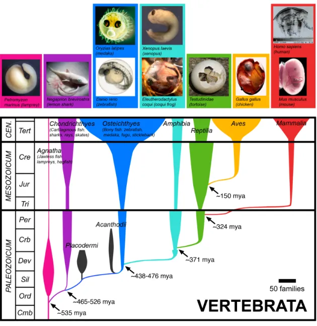
organisms amenable to gnotobiotics and represent approximately 450 million years of
divergent co-evolution with their constituent microbiota.
2.3 The Microbiota Impacts Intestinal Physiology
2.3.1 An evolutionarily conserved developmental step
The developing vertebrate embryo is encased within the confines of a structure
that creates a barrier (though not always impenetrable [25]) between self and the
external environment, a fascinating feature common to nearly all metazoans. Whether a
shell or womb, this structure ensures that emergence of the offspring into the outside
world occurs at a defined point in development. Colonization of the vertebrate intestine
by environmental microorganisms begins at the moment of emergence from this
chorionic structure and a life-long interaction between host and microbe ensues. This
developmental step has occurred for every generation of offspring in nature since at
2.3.2 Ontogeny of the intestinal microbiota
Microorganisms (including virus, phage, Achaea, Bacteria, protozoa, and Fungi)
assemble into diverse and dynamic communities within the gastrointestinal tracts of all
animals. The extent of this diversity has only been uncovered in the past decade through
the application of high-throughput sequencing to gene sequences derived from small
subunit ribosomal RNA (SSU rRNA; 16s rRNA in Bacteria and Achaea, and 18S rRNA in
Eukarya) and using these sequences to infer phylogenetic relationships between
microbes within complex communities [27-29]. This technique has spawned hundreds of
studies cataloging the microbial community composition in feces and different
anatomical locations of various organisms [30-32] and large-scale collaborative efforts
are in progress to define the dynamics of community organization in human populations
[33,34]. One of the novel outcomes has been the realization that microbial community
composition (~ 160 bacterial “species” in the human gut) is governed by ecological
principles derived from macro-scale ecology such as dispersal, diversification, niche
construction, environmental selection, and drift [18-21,35]. These principles, along with
the selective effects of host diet, lifestyle, age, and genetics, shape local microbial
assemblage within the geographical context of a living multicellular host organism
sensitive and responsive to its microbial habitants [9,36]. Once assembled, the intestinal
microbiota and their collective genomes (microbiome [37] or metagenome [38]) function
as a non-self metabolic organ dramatically affecting the metabolic potential for dietary
nutrient extraction and de novo synthesis of essential nutrients.
Of particular interest is the finding that fecal microbiota from adult human
monozygotic twins show no more similarity to each other than adult dizygotic twins
suggesting the heritability of the microbiota, at least in humans, is low [30,39]. Fecal
microbiota from biological mothers of teenage American twins showed no more similarity
co-habitating mothers and fathers had very similar microbiotas [39]. This data suggests that
the long-term effects on microbial community composition of vertical transmission of an
initial inoculum of microbes from mother to offspring, as well as host genetics, are
apparently not as significant as continuous environmental exposures, lifestyle, and diet
in humans. Indeed, diet explains much of the variance between microbial communities
when comparing across mammals [31,40] and controlled experiments have revealed diet
to be a major determinate of microbial community composition [30]. For example,
changing mice from a normal diet to a high fat diet results in dramatic shifts in microbial
community composition within 1 day of diet change [41]. Most comprehensive studies to
date have focused primarily on the low hanging fruit of the fecal microbiota from humans
or mice and it should be noted that the anatomical location within the GI tract also has
distinct microbial communities. Furthermore, humans and laboratory mice are
anomalous in their lifestyles with respect to the rest of the natural animal kingdom
whereby genetics and vertical transmission may play stronger roles in other vertebrates.
Our increased knowledge of the variables controlling assembly and homeostasis of this
metabolic organ illustrates a growing need to understand the functional impact of the
observed microbial diversity and dynamics on host cell biology.
2.3.3 Gross anatomy of the vertebrate GI tract
The major purpose of the digestive system is to convert exogenously acquired
foodstuffs into nutrients and energy required for maintenance, growth and reproduction
of the animal. The source of and relative reliance on the major classes of exogenous
nutrient substrates such as carbohydrates, proteins, lipids, and vitamins widely varies
across animal lineages. For example, protein requirements for fish (44-60% in most
Danio rerio diets) are much higher than those of laboratory mice (~18-20% for Mus
ontogeny and particular genetics of the individual organism [42,44]. Naturally, distinct
evolutionary histories involving diet choice and substrate reliance have shaped the
morphological and functional anatomy of vertebrate GI tracts [45]. The vertebrate GI
tract has distinct functional regions along the proximal-distal axis and broad comparisons
between vertebrates can best be made using nomenclature such as headgut, foregut,
pancreas and biliary system, midgut, and hindgut. Compared with herbivorous
ruminants, the foregut (esophagus, stomach), midgut (small intestine: duodenum,
jejunum, ileum), and hindgut (cecum, proximal colon, distal colon) of omnivorous
mammals such as mice and humans are anatomically similar [45]. In humans, nutrient
processing and absorption has distinct anatomical hotspots along the length of the
midgut. For example, carbohydrates, proteins, and lipids are absorbed in each section,
but to the greatest extent in the duodenum. Conversely, bile acids and some vitamins
are absorbed mostly in the ileum [46]. The zebrafish GI tract is also functionally distinct
along the proximal-distal axis (foregut or anterior intestine or segment 1, midgut or
middle intestine or segment 2, hindgut or posterior intestine or segment 3) however in
contrast to mammals, teleost fish do not have a stomach (Figure 2.2). Similar to
mammals, the majority of nutrient absorption and digestion in the zebrafish most
probably occurs in the anterior intestine, whereas the zebrafish hindgut (which lacks a
cecum) appears morphologically similar to the mammalian colon [47]. The extent to the
functional similarity of nutrient metabolism between zebrafish and human is not entirely
known and represents an interesting direction for comparative physiology. Nonetheless,
it is believed that vertebrates use similar cellular pathways and molecular machines
Figure 2.2: General features of the zebrafish model.
(i) Zebrafish undergo rapid and external development with a functioning GI tract at approximately 5 days post fertilization (dpf). (ii) The zebrafish digestive tract includes a liver (li), exocrine pancreas (pa), endocrine pancreatic islet (is), functionally segmented intestine (segment 1/anterior/foregut, segment 2/middle/midgut, segment 3/posterior/hindgut). Muscle (mu) and swim bladder (sb) are colored in gray. (ii) The intestinal epithelium is composed of absorptive enterocytes, goblet cells, and enteroendocrine cells. The anterior (A) and posterior (P) axes are denoted. Adult zebrafish maintain a functionally segmented intestine and have intestinal folds/villi similar to mice, but crypts are absent. (iii) Zebrafish are optically transparent and amenable to transgenesis. The image shows a 6 dpf double transgenic larvae expressing a reporter driven by an intestine specific promoter (red, Tg(ifabp:DsRed)) and a neutrophil specific promoter (green, Tg(mpo:egfp)). (iv) Zebrafish are amenable to gnotobiotic rearing techniques. At 0 dpf embryos within their protective chorions can be derived germ-free (GF) by surface sterilizing the chorion with solutions of iodine and bleach. (iv) GF animals can then be reared in sterile chambers such as cell culture flasks and fed specialized sterile diets.
2.3.4 Cellular anatomy of the vertebrate midgut
The vertebrate midgut is lined with a layer of rapidly self-renewing epithelial cells
that provide the cellular interface between the host organism and the intestinal
microbiota. The primary function of the midgut epithelium is to digest and absorb
nutrients, provide a physical barrier against microbial infiltration to the interior of the
body, and signal dietary information to other organ systems. In both fish and mammals,
three major differentiated cell types (absorptive enterocytes, mucous-secreting goblet
cells, and hormone-secreting enteroendocrine cells) largely perform these roles. Mice
and other mammals have a fourth cell type (paneth cells) located at the base of the villi
in the crypts of Lieberkühn in the small intestine, which have roles in innate immunity
seem to be paneth cells or crypts in the zebrafish intestine [47], however bactericidal
proteins such as defensins are conserved and expressed in the zebrafish intestinal
epithelium [49]. Absorptive enterocytes comprise 80-90% of the midgut epithelium
whereas up to 15 enteroendocrine cell subtypes are scattered throughout the mucosa
comprising ~1% of epithelial cells [50]. Generally, paneth cells are enriched in the
murine ileum and absent from the colon, and goblet cell number increases distally along
the longitudinal axis reaching maximum numbers in the colon (~4-16%) [48]. In the
zebrafish, enteroendocrine cells are observed only in the anterior intestine (segment 1),
goblet cells are located in all regions, and distinct populations of enterocytes constitute
the anterior versus mid/posterior intestine [47].
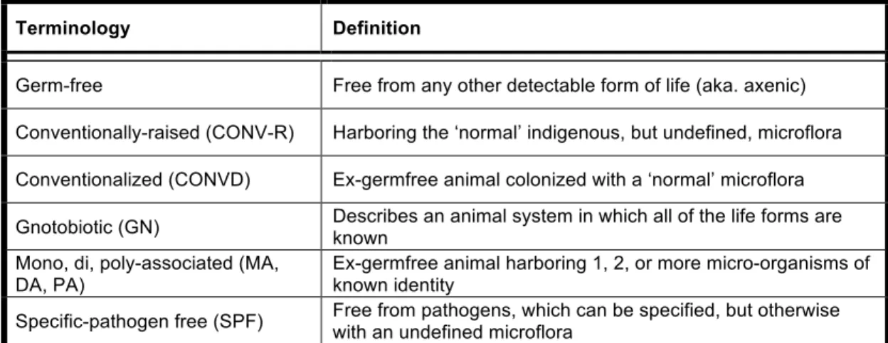
Terminology Definition
Germ-free Free from any other detectable form of life (aka. axenic)
Conventionally-raised (CONV-R) Harboring the ‘normal’ indigenous, but undefined, microflora
Conventionalized (CONVD) Ex-germfree animal colonized with a ‘normal’ microflora
Gnotobiotic (GN) Describes an animal system in which all of the life forms are known
Mono, di, poly-associated (MA,
DA, PA) Ex-germfree animal harboring 1, 2, or more micro-organisms of known identity
Specific-pathogen free (SPF) Free from pathogens, which can be specified, but otherwise with an undefined microflora
Table 2.1: Common terminology used in gnotobiotic research
2.3.5 Genetic repertoire of the intestinal microbiota
The gut microbiome (the cumulative genomes of the gut microbiota) encodes
enzymes that aid in digestion of macromolecules and pathways that contribute
metabolites distinct from the host repertoire [30,51-53]. In a pivotal study, Qin et al. used
metagenomic sequencing to describe the microbial genes prevalent in fecal samples
human gene counterpart and revealed a number of functional complementarities
between the host genome and the microbial metagenome. For example, gut bacteria
genomes are enriched for genes involved in fermentation of polysaccharides to generate
energy and in the process releasing short-chain fatty acids (such as acetate, propionate,
and butyrate) as by products, which are used by the host also as an energy source [3].
Microbial metagenomes are further enriched for genes involved in amino acid and lipid
biosynthesis, and have capabilities for xenobiotic metabolism such as degradation of the
common food supplement benzoate into pimeloyl-coenzyme-A, a precursor to biotin
synthesis [52]. Intriguingly, this study also showed that only about 38% of prevalent
microbial genes are common to all of the human-associated fecal microbiotas. It will be
interesting to see how this trend varies along the length of the intestinal tract and in other
host species. As the frontline, the intestinal epithelium senses these microbial-derived
products and responds to them. Comparisons in animals with and without a microbiota
are crucial to elucidate these responses.
2.3.6 Gnotobiotic vertebrate models: the power of comparison
Because evolution selected for general sterility within the chorion of the
developing vertebrate embryo (Figure 2.1), researchers are able to derive animals
microbe-free (germ-free, GF; Table 2.1) by sterilizing the external surface of the womb
or egg and rearing the animals in an environment impervious to microorganisms. The
first germ-free animal (a guinea pig) was obtained in 1895 by Nuttal and Thierfeld [54]
and later entire colonies of germ-free rodents could be established [55]. Since that time
germ-free derivation and rearing procedures [56] have been established for many other
vertebrates including chickens, rabbits, gerbils, pigs, dogs, and zebrafish [57]. As
expected, significant hurdles were encountered early on due to difficulties in maintaining
successful reproduction. This is still an obstacle with the zebrafish and we have not yet
generated sterile zebrafish colonies that can be propagated through successive
generations. However, there are numerous technical difficulties associated with deriving
and rearing rodents GF, and therefore requires the generation and maintenance of
stable GF colonies. This provides a hurdle to using genetics to study host responses to
the microbiota. In contrast, the relative ease of GF zebrafish derivation, rapid and
external development, optical transparency, and genetic tractability make it an efficient
system to explore host mechanisms mediating microbial responses (Figure 2.2).
Despite initial concerns about the capacity for axenic life, GF mice tend to be
leaner and longer-lived than conventionally-raised (CONV-R) mice when fed appropriate
diets [58]. Germ-free mice or zebrafish can be colonized with single microorganisms
(mono-association, MA) or consortia of microorganisms (conventionalized, CONVD),
and the microbial impact on various biological processes can be monitored. This has
provided decades of phenotypic knowledge concerning the impact of the microbiota on
the physiologic environment encountered by intestinal epithelial cells [3,58]. For
example, the presence of a microbiota deconjugates and dehydoxylates bile acids [59],
metabolizes bilirubin [60], degrades glycoproteins produced by goblet cells [61], and
increases epithelial cell turnover [62]. Application of metabolomics in GF versus
CONV-R/CONVD have described widespread microbial impact on the nutrient environment [53],
consistent with the functional complementarities of host and microbial genomes revealed
by metagenomics [52,63]. One consequence of the interplay between bacteria and host
diet is an apparent increase in lipid absorption by enterocytes, which also led to
increased lipid deposition in extra-intestinal tissues such as liver hepatocytes [64]. These
studies highlight that the metabolic landscape encountered by intestinal epithelial cells is
epithelial responses to the microbiota and their evolution have not been studied with
modern tools.
2.4 Transcriptional Regulation in the Intestine
An obvious yet fascinating feature that distinguishes multicellular life from
unicellular counterparts is the use of a single genome to build, connect, and maintain a
myriad of specialized cell types. The faithful execution of these processes requires
precise spatiotemporal orchestration of gene expression. Vertebrate genomes contain
billions of nucleotides (1-2 meters in length) that are assembled into linear chromosomes
and packaged as chromatin into tiny spaces (10µm3) within the cell. Protein-coding
genes comprise only a small percentage (less than 5%) of nucleotides in vertebrate
genomes, and the function of the remaining non-coding regions is of considerable
interest [65,66]. It is becoming apparent that much of the non-coding DNA regions
function to regulate cell-type specific deployment of proteins and RNAs at the level of
transcription [67-70]. In this section, I review salient features of eukaryotic transcriptional
regulation and discuss how sequencing based technologies are well suited for
elucidating regulatory mechanisms governing host-microbe symbioses.
2.4.1 Molecular anatomy of transcriptional regulation
There has been significant progress in the field of transcriptional regulation since
the discovery of RNA polymerase [71], regulatory DNA [72], and summarization of the
central dogma of molecular biology [73]. A plethora of general transcription factors and
mediator co-activator subunits that enable RNA polymerase assembly and recruitment to
the transcription start site of genes are known [74]. Known also are checkpoints and
polymerase can be paused or poised [76] and that chromatin and its remodelers play
active roles throughout gene regulation [77,78]. We now observe that the genome is
pervasively transcribed and that RNA can regulate its own expression [79,80]. We are
even beginning to understand transcriptional events in single cells at single molecule
resolution [81,82]. Of specific interest to this thesis, concerns the knowledge that
specification and tuning of transcriptional activity proceeds through coordinate
interactions between sequence specific transcription factors (also called transcriptional
activators/repressors or transcriptional regulators) and cis-acting non-coding DNA
(called cis-regulatory module, CRM or cis-regulatory element, CRE or cis-regulatory
region). CRMs can be classified into two broad categories: (i) a promoter, composed of a
core RNA polymerase binding sites and proximal regulatory sequences and (ii) distal
regulatory regions, such as enhancers, repressors, insulators or locus control regions
[83] (Figure 2.3). Both classes engage in regulatory functions, but it is the second class
that appears to exhibit the majority of cell type and environment-specific regulatory
control. Distal CRMs are often functionally autonomous [68] harboring binding sites for
many transcription factors [84], and can be located anywhere (near the transcription start
site, within introns, within exons, tens of thousands of base-pairs up or downstream,
even on different chromosomes) (Figure 2.3). Chromatin looping plays a role in directly
linking a distal CRM with the target gene promoter, however alternative indirect
mechanisms such as place holding, spreading, and non-coding RNA intermediaries
appear to better explain some experimental observations [85]. Recent work showed
convincingly that intragenic CRMs could function both as an enhancer, and also as a
promoter upon deletion of the proximal promoter of the target gene [86]. Identifying
CRMs, discovering the transcription factors they bind, and deciphering the logic
2.4.2 The microbiota modulates host transcription in the intestine
Gene-specific and genome-wide profiling of gene expression has been an
important window into the impact of the microbiota on host biology. In a recent
comprehensive survey in the duodenum, jejunum, ileum, and colon, it was shown that
the microbiota differentially regulates gene expression across the length of the GI tract
(5663 cumulatively in the small intestine) [87-89]. The most highly enriched gene
categories in the small intestine were associated with innate and adaptive immunity and
nutrient metabolism, suggesting that many of the observed microbiota-related
phenotypes have a transcriptional component. Similar results in genome-wide surveys of
differentially expressed genes in GF versus CONV-R or CONVD zebrafish established a
set of evolutionarily conserved transcriptional responses to the microbiota [90]. Notably,
host cell response depends on the particular composition of microbes within
communities in the intestine. For example, colonization of GF mice with a zebrafish
microbiota elicited both overlapping and distinct transcriptome responses compared to
colonization with a mouse microbiota. In fact, a microbiota transplanted from rat to
mouse did not stimulate gene expression changes required for proper immune system
development exemplifying the specificity of the co-evolved symbiosis between host
species and their microbiota [91]. Though gene expression changes are often governed
at the level of transcription, post-transcriptional regulation through alterations in mRNA
splicing, stability, or translation may also be influenced by microbial activities.
It should be noted that genes do not have to be differentially regulated by the
microbiota to be important for host-microbe symbiosis. This was observed for Toll-like
receptor (TLR) 5, a transmembrane protein that recognizes bacterial flagellin and
mediates the homeostatic balance between infection and inflammation. TLR5 was not
differentially transcribed in response to the microbiota [87] yet TLR5 deficient mice have
Figure 2.3: Non-coding DNA in gene regulation.
such cases where the expressed gene mediates host-microbe symbiosis and
transcription takes place without effect from the microbiota, then one might expect
relatively high expression in the intestinal epithelium or associated lymphoid tissues
when compared to other tissues or cell types. In this way, transcription can be regulated
by mechanisms controlling cell type specificity and/or environmental response. It would
be tedious to catalog the thousands of genes and their functions that are either highly
expressed in the intestinal epithelium or differentially regulated by the microbiota.
Instead, I highlight 10 genes that have a defined role in mediating host-microbe
symbiosis (Table 2.2) on which new genomics methodologies should be able to
comprehensively address their regulatory mechanics. Notably, Angiopoietin-like 4
(Angptl4), a central regulator of lipid metabolism and fat storage, is suppressed
specifically in the intestinal epithelium by the microbiota [8]. Angptl4 suppression leads
to increased fat storage in CONV-R and CONVD mice in comparison to GF counterparts
due to its role as a direct inhibitor of Lipoprotein lipase (LPL) (Figure 2.4). This
suppression is conserved in the zebrafish and therefore represents an evolutionarily
ancient regulatory event. The mechanisms governing Angptl4 intestinal transcription and
microbial suppression are unknown, as is the case for the majority of genes mediating
host-microbe interactions in the intestinal epithelium. In Chapter 2, I harness the unique
attributes of the zebrafish (Figure 2.2) to understand the mechanisms controlling Angptl4
suppression by the microbiota.
2.4.3 Transcription factors mediating intestinal epithelial gene expression
Sequence specific DNA-binding factors regulate gene expression at the level of
transcription by selecting a gene locus for activation or repression. There are many
transcription factors that are known to regulate gene expression in the intestinal
Table 2.2: Unsolved mysteries in host-microbe symbioses
homeobox factors (Cdx), Krüpple-like factors (KLF), Hepatic nuclear factors (HNFs),
Peroxisome proliferator-activated receptors (PPARs), GATA binding factors (GATA),
Nuclear Factor kappa B (NFκB), Signal transducer and activator of transcription
(STATs), and Suppressor of cytokine signaling (SOCS), any of which could mediate
multiple aspects of host-microbe symbiosis. NFκB has been extensively studied as a
central regulator of inflammatory responses to the microbiota in mammals [92,93] and
zebrafish [94], and functions as an inducible transcription factor in diverse cell lineages
[95]. GATA 4,5,6 are all expressed in the intestinal epithelium in mouse [96-98] and
zebrafish [99,100] functioning to modulate the expression of genes involved in epithelial
cell differentiation, nutrient metabolism [101] and immune responses [102]. Nuclear
receptors such as PPARs [103], HNFs [104], Liver X receptor (LXR) [105,106], Vitamin Gene Regulation
Tissue/Cell-type Species Function REF
Angptl4 Suppressed by
microbiota
Midgut
(ileum)/IEC Dr, Mm Mediates microbiota-associated obesity Bäckhed, 2004; Rawls, 2004 Alpi Induced by
microbiota
Midgut/IEC Dr, Mm Promotes mucosal tolerance to
the microbiota Cheesman, 2007 T1r3, Slglt-1, αGus Induced by
microbiota Midgut/IEC Mm Increased sucrose intake in GF mice Swartz, 2012
RegIIIg Induced by microbiota
Midgut/PC Mm Bactericidal c-type lectin
expressed by paneth cells
Hooper, 2006 Mucin1-4 Differentially regulated by microbiota Midgut, Hindgut/IEC
Mm Mucins maintain barrier function
and are substrates for microbial symbionts
Wei, 2012; Comelli, 2007
Ang-1 Induced by microbiota
Midgut/IEC Mm Promotes microbiota induced
vascular remodeling via tissue factor (TF) glycosylation
Reinhardt, 2012
α1,2-FT Induced by microbiota
Midgut (Ileum)/IEC
Mm One of many
fucosyltransferases regulated by Bacteroides thetaiotaomicron
Bry, 1996
Crt1 Induced by
microbiota Midgut/IEC Mm Mediates absorption of heavy metals in intestinal epithelium perhaps through competition with microbiota
Hooper, 2002
Tlr-5 unchanged Midgut/PC Mm Absence of expression results
in microbiota-induced metabolic syndrome
D Receptor (VDR) [107], Farnesoid X receptor/Bile acid receptor (FXR/BAR) [108],
represent an important class of transcription factors that could have direct roles in
host-microbe symbiosis by sensing microbial metabolites such as short-chain fatty acids and
bile acid derivatives. Wnt signaling pathway transcription factors such as T cell factor
(TCF) [109], Cdx1/2 [110] and Sox9 [111], and Notch signaling pathway transcription
factors Hes1, Math1 [112] and Krüppel-like factors 4/5 [113] have highly conserved roles
in intestinal cell-fate specification and differentiation. The Transforming growth
factor-beta (TGF-ß) signaling pathway and associated transcription factors from the Smad
family [114] as well as the Jak/Stat pathway and associated Stat and Socs [115]
transcription factors perform integral regulatory functions in epithelial tissues to maintain
mucosal integrity, renewal, and repair.
Each of these transcription factor families and associated signaling pathways are
integral to intestinal epithelial cell physiology. However, most of this knowledge has been
interpreted with a focus on stem cell biology, immune response, or diseases such as
cancer. There is much less knowledge available concerning the role of these factors in
the context of host-microbe symbiosis. As an example, searching Pubmed with the
terms Wnt AND intestine gives 521 publications, whereas searching with Wnt AND
microbiota gives 5 (521:5). The trend is similar for NFkB (1065:25), PPAR (391:11), TGF
Beta (1249:25), Jak/stat (42:2, both are Drosophila papers), nuclear receptor (2542:34).
Varying the terms or using ISI web of knowledge gives a similar story. Furthermore,
searching for microbiota OR gut flora (4552) AND transcription factor gives only 136
references compared to intestine AND transcription factor (7525). Even less is known
about the cis-regulatory modules that interpret transcription factor activity in this context.
With new tools and powerful model systems in place, it is an appropriate time to fully
Figure 2.4: Microbial suppression of intestinal expression of Angptl4
2.5 Genomic Approaches to Discover CRMs
2.5.1 Genomes as reagents for CRM discovery
Prior to the genomics age, practical discovery and functional characterization of
regulatory regions was limited to DNA in close proximity to the transcription start site of
single genes. The same technological advances in sequencing that have reignited
interest in the intestinal microbiota have been driving the discovery of the features and
functions of non-coding DNA [23,68,116]. As of writing this thesis, there are currently
thirty-four publically available sequenced vertebrate genomes and soon there will be
thousands more [117,118]. The diversity of sequenced vertebrates allows one to view
genomes through the powerful lens of evolutionary time. Sequence alignment has
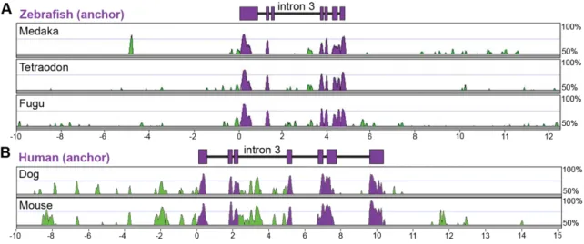
revealed conservation in distal CRMs [119-121] with constraint far above what is
expected for neutrally evolving DNA [122], thus implying function. Sets of conserved
non-coding elements (CNEs) are enriched for known regulatory regions (enhancers,
insulators, suppressors, LCRs) and many have been tested for functional enhancer
activity in vivo [123,124]. A recent study showed that the extent to conservation (or the
appearance of a conserved regulatory region in branches of the phylogeny) is
dependent on the type of gene regulated [120]. This work discovered that regulatory
innovations probably occurred in waves, where CNEs near genes involved in
transcription factor activity or development were ancient and CNEs near genes involved
in signaling pathways or post-translational protein modifications were more recent. This
confirmed previous studies revealing that non-coding DNA conservation between fish
and mammals is low compared to protein coding regions, and that CNEs shared
between fish and mammals are enriched near developmental transcription factors [124].
There are therefore limitations to using sequence conservation as the only metric for
discovering cis-regulatory modules. First, a conservation approach requires both distant
sequence alignment algorithms may not detect all conservation. Third, one interpretation
from the Lowe et al. study could be that regulatory modules involved in certain biological
processes evolve differentially. Fourth, the rules governing constraint and evolvability of
cis-regulatory modules are not well understood. Fifth, and most importantly,
conservation does not predict cell type and environment-specific activity. Therefore,
other methods can be used to distinguish active non-genic DNA.
2.5.2 The role of chromatin in gene regulation
Eukaryotic genomic DNA is bundled into nucleosomes consisting of
approximately 147 base pairs of DNA wrapped around 8 histone protein cores (two
copies each of H2A, H2B, H3, H4) [125]. Nucleosome spacing and posttranslational
modification status can have a profound effect on gene regulation [126]. Spacing
proceeds through a combination of statistical positioning based on intrinsic DNA
sequence affinity of the histone octamer, competition with other proteins for DNA
binding, and active positioning by chromatin modifying factors [127]. Promoter regions
and CRMs have the unique feature of being “open” or nucleosome depleted, allowing
transcription factors and polymerases to access regulatory DNA regions. Histones are
highly conserved proteins and each core histone contains a conserved unstructured tail.
Histone tails can be modified post-translationally at specific residues by covalent
methylation, acetylation, phosphorylation, ubiquitination, and many others [128]. A
technique called chromatin immunoprecipitation (ChIP) has been applied extensively to
investigate where histones with different modifications are located on DNA [129]. Briefly,
in this method (I) DNA-protein complexes are cross-linked in vivo, (ii) cells are lysed and
chromatin sheared, (ii) complexes are immunoprecipitated with an antibody targeting the
protein, (iii) crosslinks are reversed, (iv) and the DNA sequence determined using PCR,
specific histone tail residues are differentially modified dependent on the location of that
nucleosome in relation to functional elements within a genome (so called histone marks)
[68,130]. Importantly, these marks are also associated with the activity of the genomic
region (activation, repression, pausing, poised). The histone marks H3K4me1
(active/poised) and H3K27ac (active) associate with enhancer regions and can
distinguish cell type specific regulatory activity [131]. Furthermore, a number of
antibodies used in mammals can function well in the zebrafish [132,133]. The same
technique, ChIP-seq, can also be used to define target sites of transcription factors
across the genome, though binding doesn’t necessarily lead to functional output [134]. It
should be noted that a new version of ChIP-seq, dubbed ChIP-exo [135], is perhaps
superior to classical ChIP because it localizes the DNA binding protein to single
base-pair resolution.
Two complementary genome-wide methods, DNase-seq [136,137] and
FAIRE-seq [138,139], take advantage of the observation that eviction or destabilization of
nucleosomes from chromatin is a characteristic feature of functional CRMs in eukaryotic
genomes (Figure 2.5). DNase-seq is the genome-wide extension of the classical DNase
I footprinting assay [140]. DNase I footprinting harnesses the feature that protein factors
binding naked DNA block DNase I mediated enzymatic cleavage of underlying
nucleotides, thus giving a quantitative footprint of the DNA binding factor. In the context
of chromatin, the vast majority of DNA is protected from digestion by nucleosomes
whereas regions adjacent to transcription factor binding are accessible or hypersensitive
to DNase I cleavage (Figure 2.5A). This allows identification of “open” chromatin regions,
which have very strong correlations with a variety of other markers (transcription factor
binding, histone marks) of active non-coding regulatory function [136]. Within the “open”
region defined by increased DNase I sensitivity there is often a discernible footprint of
Figure 2.5: Methods for genome-wide discovery of open chromatin
local decrease in DNase I sensitivity [142]. Combined with a high signal-to-noise ratio,
DNase-seq offers a powerful and validated method to discern nucleosome depleted
regions as well as transcription factor-DNA interactions across the genome.
Formaldehyde-Assisted-Isolation-of-Regulatory-Elements (FAIRE) is an
alternative approach to discover “open” chromatin based on differences in cross-linking
efficiencies between DNA and nucleosomes compared to DNA and sequence-specific
DNA-binding proteins (Figure 2.5B). In this assay, cells are covalently cross-linked
briefly with formaldehyde, lysed and sonicated, and sheared chromatin is extracted with
phenol/chloroform. Extraction enriches unbound DNA into the aqueous phase and
protein-bound DNA is trapped to the organic/aqueous phase interface. Unbound DNA is
isolated and assayed for locus-specific (via quantitative PCR) or genome-wide (via
microarray, high-throughput sequencing) enrichment patterns. The signal-to-noise ratio
for FAIRE-seq is not as high on average as DNase-seq, and it has yet to be proven as a
method for elucidating transcription factor footprints (Figure 2.5C). However, FAIRE
does not require nuclei isolation so samples do not need to be in single cell
suspensions, and other experimental practicalities [139] position FAIRE-seq to be
amenable to higher-throughput capabilities. Both genomics tools, DNase-seq and
FAIRE-seq, can uncover a range of cell-type specific elements (promoters, enhancers,
silencers, insulators, locus control regions) and do not require an antibody [141]. The
impact of environmental factors, such as changes in microbial community composition
and diet, on open chromatin dynamics is not well understood, and neither assay has
been applied to primary intestinal epithelial cells in mouse or zebrafish.
2.5.3 Transcriptional genomics in the intestine
These studies and many others have illuminated the regulatory landscape of
datasets from the small intestinal epithelium are lacking. Recently, ChIP-seq
experiments targeting GATA6, CDX2, HNF4α were performed in Caco-2 cells (a
pseudo-intestinal epithelial cell line) [143,144], and a dataset for FXR in primary small
intestinal epithelial cells from mouse is also available [145]. A recent study analyzed the
genome-wide chromatin landscape in primary crypts and cancerous crypts from the
human colon and identified enhancer activity that was lost or gained in cancerous crypts,
thus explaining a majority of transcriptional changes observed in cancerous versus
non-cancerous cells [146]. I suspect that similar changes to the chromatin landscape in
response to microbial activity in the intestine could drive specific transcriptional
programs that mediate host-microbe symbiosis. To date, there has been little effort to
define the histone modification, transcription factor binding, or chromatin accessible
landscape in any cell type in an axenic animal.
2.5.4 Model organisms are in vivo assay systems
Consortiums such as the Encyclopedia of DNA elements projects (ENCODE)
and modENCODE projects have launched large-scale efforts aimed at identifying all
functional elements within the genome of H. sapiens, D. melanogaster, and C. elegans.
These ambitious initiatives will greatly enhance our current knowledge base for a defined
set of species, tissues, and ontogenies. However, genomic regulatory responses to
environmental change are fundamentally unknown, yet extremely important in all areas
of biological and biomedical research. Furthermore many of the defined efforts in
vertebrates will be performed in cell lines which no doubt have historic value in
generating our current understanding of transcriptional regulation, but carry many
caveats. It is my feeling that no existing computational, cell-culture, or in vitro system
can fully reproduce the complexity associated with cellular, tissue, and
Therefore, to understand cis-regulatory function and evolution, there is a need to
perform discovery of regulatory regions in primary cells and functional characterization in
vivo [123,147]. In this light, the intestinal epithelium has a number of features that
distinguish it as an ideal genomics system to study the evolution of cis/trans regulatory
programs mediating host-microbe symbiosis. First, the cells are abundant, accessible,
and can be collected with relative ease. Second, the fundamental role of the intestinal
epithelium is similar in all metazoans providing many model systems for cross-species
comparisons. Third, the intestinal epithelium marks the primary interface with the
microbiota and intestinal epithelial cells experience dynamic environmental factors.
Fourth, despite extensive physiologic, cellular, and molecular knowledge, there is a
striking paucity of information regarding genome regulation in this vital organ.
Probiotic, prebiotic, antibiotic, pharmaceuticals, strange foods and exotic
beverages are consumed daily with limited knowledge of their impact on the microbial
and host cells with which these foreign substance directly interface. An increased
understanding of the mechanisms mediating the general impact of the microbiota on
enterocyte, enteroendocrine, goblet, and paneth cell gene expression in a healthy
context would pave the way for interrogation of these mechanisms in disease states.
Once a set of conserved microbial response mechanisms are defined, then we can
create reporter systems to determine the specific microbial signals and host signaling
pathways that host cells use to perceive and respond to microbial activities. The
zebrafish will be particularly useful for systematic genetic screening of microbial mutant
strains [148] and chemical libraries [149,150]. A major route towards infection by
pathogens is through the intestinal epithelium, and pathogens can subvert mechanisms
that have evolved to maintain homeostasis between host cells and commensal microbes
[151]. Furthermore, symbiosis is a sliding scale where one-time mutualists can become