Middle East Respiratory Syndrome Coronavirus NS4b Protein Inhibits
Host RNase L Activation
Joshua M. Thornbrough,a*Babal K. Jha,b*Boyd Yount,cStephen A. Goldstein,aYize Li,aRuth Elliott,aAmy C. Sims,cRalph S. Baric,c,d Robert H. Silverman,b Susan R. Weissa
Department of Microbiology, Perelman School of Medicine at the University of Pennsylvania, Philadelphia, Pennsylvania, USAa; Department of Cancer Biology, Lerner Research Institute, Cleveland Clinic, Cleveland, Ohio, USAb; Department of Epidemiologycand Department of Microbiology and Immunology,dUniversity of North Carolina at Chapel Hill, Chapel Hill, North Carolina, USA
*Present address: Joshua M. Thornbrough, Adelphi Research Global, Doylestown, Pennsylvania, USA; Babal K. Jha, Translational Hematology & Oncology Research, Taussig Cancer Research Institute, Cleveland Clinic, Cleveland, Ohio, USA.
ABSTRACT
Middle East respiratory syndrome coronavirus (MERS-CoV) is the first highly pathogenic human coronavirus to
emerge since severe acute respiratory syndrome coronavirus (SARS-CoV) in 2002. Like many coronaviruses, MERS-CoV carries
genes that encode multiple accessory proteins that are not required for replication of the genome but are likely involved in
pathogenesis. Evasion of host innate immunity through interferon (IFN) antagonism is a critical component of viral
pathogene-sis. The IFN-inducible oligoadenylate synthetase (OAS)-RNase L pathway activates upon sensing of viral double-stranded RNA
(dsRNA). Activated RNase L cleaves viral and host single-stranded RNA (ssRNA), which leads to translational arrest and
subse-quent cell death, preventing viral replication and spread. Here we report that MERS-CoV, a lineage C
Betacoronavirus
, and
re-lated bat CoV NS4b accessory proteins have phosphodiesterase (PDE) activity and antagonize OAS-RNase L by enzymatically
degrading 2=,5=-oligoadenylate (2-5A), activators of RNase L. This is a novel function for NS4b, which has previously been
re-ported to antagonize IFN signaling. NS4b proteins are distinct from lineage A
Betacoronavirus
PDEs and rotavirus gene-encoded
PDEs, in having an amino-terminal nuclear localization signal (NLS) and are localized mostly to the nucleus. However, the
ex-pression level of cytoplasmic MERS-CoV NS4b protein is sufficient to prevent activation of RNase L. Finally, this is the first
re-port of an RNase L antagonist expressed by a human or bat coronavirus and provides a specific mechanism by which this occurs.
Our findings provide a potential mechanism for evasion of innate immunity by MERS-CoV while also identifying a potential
target for therapeutic intervention.
IMPORTANCE
Middle East respiratory syndrome coronavirus (MERS-CoV) is the first highly pathogenic human coronavirus to
emerge since severe acute respiratory syndrome coronavirus (SARS-CoV). MERS-CoV, like other coronaviruses, carries genes
that encode accessory proteins that antagonize the host antiviral response, often the type I interferon response, and contribute to
virulence. We found that MERS-CoV NS4b and homologs from related lineage C bat betacoronaviruses BtCoV-SC2013 (SC2013)
and BtCoV-HKU5 (HKU5) are members of the 2H-phosphoesterase (2H-PE) enzyme family with phosphodiesterase (PDE)
ac-tivity. Like murine coronavirus NS2, a previously characterized PDE, MERS NS4b, can antagonize activation of the OAS-RNase L
pathway, an interferon-induced potent antiviral activity. Furthermore, MERS-CoV mutants with deletion of genes encoding
ac-cessory proteins NS3 to NS5 or NS4b alone or inactivation of the PDE can activate RNase L during infection of Calu-3 cells. Our
report may offer a potential target for therapeutic intervention if NS4b proves to be critical to pathogenesis in
in vivo
models of
MERS-CoV infection.
Received13 February 2016Accepted25 February 2016 Published29 March 2016
CitationThornbrough JM, Jha BK, Yount B, Goldstein SA, Li Y, Elliott R, Sims AC, Baric RS, Silverman RH, Weiss SR. 2016. Middle East respiratory syndrome coronavirus NS4b protein inhibits host RNase L activation. mBio 7(2):e00258-16. doi:10.1128/mBio.00258-16.
EditorPeter Palese, Icahn School of Medicine at Mount Sinai
Copyright© 2016 Thornbrough et al. This is an open-access article distributed under the terms of theCreative Commons Attribution 4.0 International license.
Address correspondence to Susan R. Weiss, [email protected].
This article is a direct contribution from a Fellow of the American Academy of Microbiology. External solicited reviewers: Stanley Perlman, University of Iowa; Brenda G. Hogue, Arizona State University.
M
iddle East respiratory syndrome coronavirus (MERS-CoV)
infections range from mild upper respiratory infections to
severe acute respiratory distress syndrome, with a global case
fa-tality rate of 36% (1, 2). MERS-CoV has predominantly affected
the Kingdom of Saudi Arabia and neighboring countries with
spo-radic cases arising in Europe and North America as the result of
travel to and from the Middle East (3). A recent outbreak of
MERS-CoV in South Korea has raised the specter that
unrecog-nized infections combined with potential superspreaders may
pose a much greater risk of significant travel-associated outbreaks
of MERS-CoV than previously suspected, particularly in health
care settings (4, 5). The lethality of MERS-CoV and the ease of
global travel necessitate further study and understanding of the
mechanisms of MERS-CoV pathogenesis.
RESEARCH ARTICLE
MERS-CoV, a lineage C
Betacoronavirus
, has a 30-kb
positive-sense single-stranded RNA (ssRNA) genome. As with all CoVs,
the first 5
=
two-thirds of the genome consists of the replicase
en-coded in open reading frame 1a (ORF1a) and ORF1b. The
re-maining 3
=
one-third encodes the structural proteins spike (S),
envelope (E), membrane (M), and nucleocapsid (N) as well as
accessory proteins (encoded in ORF3 to ORF5 and ORF8b) that
are not required for genome replication but likely act as immune
antagonists that may be critical for pathogenesis (6–8). Indeed,
MERS-CoV and other CoVs induce type I interferon (IFN) only
weakly and late in infection (9–15), suggesting that coronaviruses
have evolved mechanisms of immune evasion. Furthermore, there
are reports that MERS-CoV accessory protein NS4a (encoded by
ORF4a), NS4b (encoded by ORF4b), and NS5 (encoded by
ORF5), when overexpressed from plasmids, antagonize type I IFN
induction and signaling at various points in the pathways (16–19).
NS4a is a double-stranded RNA (dsRNA) binding protein (17).
NS4b is detected primarily in the nucleus (16–19), making NS4b
and the severe acute respiratory syndrome coronavirus
(SARS-CoV) ORF3a-encoded proteins (20, 21) the only known
corona-virus proteins thus far to be detected in the nucleus (20, 21). A
MERS-CoV mutant with deletions of ORFs 3 to 5 is attenuated for
replication in human airway-derived Calu-3 cells (22), and a
mu-tant with deletions of ORFs 4a and 4b is attenuated for replication
in hepatic carcinoma-derived Huh-7 cells (23).
The type I IFN response is an essential component of antiviral
innate immunity and is initiated when host sensors initiate
signal-ing pathways resultsignal-ing in transcription of type I IFN genes (9, 10,
24). IFN activates transcription of numerous IFN-stimulated
genes (ISG); among these and most relevant to this report are the
OAS
genes (25). Oligoadenylate synthetase (OAS), upon
detec-tion and binding of dsRNA, synthesizes 2
=
,5
=
-oligoadenylate
(2-5A) [p
x5
=
A(2
=
p5
=
A)
n;
x
⫽
1 to 3;
n
ⱖ
2] from intracellular ATP
that induces the homodimerization of latent RNase L, leading to
its subsequent activation (24, 26, 27). Activated RNase L cleaves
both viral and host ssRNA preferentially at UU and UA
dinucle-otide sequences, leading to translational arrest and apoptosis, and
limits viral replication and spread
in vitro
and
in vivo
(24, 28, 29).
In addition, RNA cleavage products can be recognized by RNA
sensors, leading to further augmentation of IFN production and
signaling (30).
We have shown previously that lineage A
Betacoronavirus
mouse
hepatitis virus (MHV) NS2 is a determinant of cellular and organ
tropism. MHV NS2 is a 2
=
,5
=
-phosphodiesterase (PDE) that
antago-nizes the type I IFN response by blocking activation of the
OAS-RNase L pathway and is a critical determinant of MHV
hepatoviru-lence (7, 29). Here we report that by structural homology,
biochemistry, and biological measures, MERS-CoV NS4b and
ho-mologs encoded by related bat lineage C
Betacoronavirus
,
BtCoV-SC2013 (BtCoV-SC2013) and BtCoV-HKU5 (HKU5), are also 2
=
,5
=
-PDEs
that can antagonize the IFN-inducible OAS-RNase L pathway.
RESULTS
Alignment and modeling of NS4b proteins.
To elucidate the
function of MERS-CoV NS4b, we queried the primary amino acid
sequence with the National Center for Bioinformatics (NCBI)
Ba-sic Local Alignment Search Tool (BLAST). MERS-CoV NS4b was
found to have no primary amino acid sequence homology outside
of lineage C
Betacoronavirus
. To broaden the search for
homolo-gous proteins, we used tertiary structural prediction and
homol-ogy search by Phyre
2(31) and I-TASSER (32, 33). The top scoring
structure identified by both servers was the central domain of
Rattus norvegicus
A kinase anchoring protein 7 isoform gamma or
delta (AKAP7
␥
/
␦
) (PDB:
2VFK
), a 2H-phosphoesterase (2H-PE)
superfamily member with 2
=
,5
=
-PDE activity (34) (Fig. 1A). These
enzymes are characterized by two H-
⌽
-[ST]-
⌽
motifs (where
⌽
is
a hydrophobic residue) separated by an average of 80 residues
(35). To generate a more accurate predicted structure, the 2H-PE
domain of MERS-CoV NS4b was modeled directly on AKAP7
using one-to-one threading on Phyre
2followed by loop and side
chain refinement in Modeller (Fig. 1B) (29, 31). For comparison,
the recently solved structure of lineage A
Betacoronavirus
mouse
hepatitis virus (MHV) NS2, a 2H-PE with 2
=
,5
=
-PDE activity, is
also shown (Fig. 1C) (36).
MERS-CoV NS4b shares significant homology with closely
re-lated bat coronavirus (BtCoV) proteins encoded by SC2013 and
HKU5, 47% and 31% amino acid sequence homology, respectively.
While both BtCoV homologs have the 2 [H-
⌽
-(S/T)-
⌽
] predicted
catalytic motifs, SC2013 NS4b (previously referred to as NS3c [37])
had low-confidence structural homology (
⬍
45%) with AKAP7, and
HKU5 NS4b had no significant structural homology with any
struc-tures in the Phyre
2database (38). However, alignment of the regions
surrounding the H-
⌽
-(S/T)-
⌽
motifs with cellular AKAP7 from rat,
mouse (
Mus musculus
,
NP_061217.3
), and virus-encoded 2
=
,5
=
-PDEs illustrates the conservation of these two motifs (Fig. 2A). We
have previously shown that the central domain of the
M. musculus
AKAP7
␥
and simian rotavirus A (RVA), strain SA11, VP3 protein
carboxy-terminal domain (CTD) are catalytically active 2
=
,5
=
-PDEs (28, 34, 35, 39). Additionally, MERS-CoV NS4b and BtCoV
homologs, like the rat and mouse AKAP7 but unlike any of the
known viral PDEs, including MHV NS2 or rotavirus VP3 CTD,
contain an amino-terminal nuclear localization signal (NLS)
(Fig. 2B) (16, 18, 19). In addition to a PDE domain, AKAP7
␥
also
contains a carboxy-terminal binding domain for the regulatory
subunit II (RII) of cyclic AMP (cAMP)-dependent protein kinase
A (PKA) (PKA-RII-
␣
-BD) (39), and VP3 contains
amino-terminal guanylyltransferase (Gtase) and methyltransferase
(Mtase) domains (E) (28), neither of which are found adjacent to
coronavirus PDE domains.
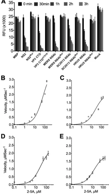
NS4b from MERS-CoV and both BtCoVs are enzymatically
active 2=,5=-phosphodiesterases that cleave 2-5A.
To determine
whether MERS-CoV, BtCoV-SC2013, and BtCoV-HKU5 NS4b
encode enzymatically active PDEs, NS4b genes were expressed in
Escherichia coli
as maltose binding protein (MBP) fusion proteins
and purified by affinity chromatography followed by ion exchange
and size exclusion chromatography. Purified proteins were
incu-bated with various 2-5A substrate concentrations and
subse-quently used to assess enzyme kinetics via a fluorescence
reso-nance energy transfer (FRET)-based RNase L activation assay
(29). Substrate steady-state kinetic studies were performed (see
Materials and Methods for details), and the quantity of cleaved
2-5A was determined as a function of time from 180-min progress
curves conducted in triplicate. The best-fit parameter estimates
are shown in Table 1. All three proteins were capable of cleaving
2-5A and preventing the activation of RNase L with similar kinetic
parameters to MHV NS2. As expected, mutant proteins
containg a His-to-Arg mutation in the second catalytic motif were
in-capable of cleaving 2-5A (Fig. 3A). Although the activities of all
three NS4b proteins were similar to that of MHV NS2 (Fig. 3B),
velocities plateaued at lower concentrations of 2-5A compared with
MHV NS2, suggesting that the lineage C proteins become saturated
more quickly at higher 2-5A concentrations (Fig. 3C to E).
Expression of MERS-CoV NS4b and BtCoV homologs from
chimeric MHV.
To investigate OAS-RNase L antagonism in cell
FIG 2 Cellular and viral 2=,5=-phosphodiesterase (PDE) domains. (A) Alignment of known and predicted cellular and viral 2=,5=-PDE sequences. Catalytic motifs [H-⌽-(S/T)-⌽] are indicated by the red and blue boxes. Rat (Rattus norvegicus[Rn]) and mouse (Mus musculus[Mm]) AKAP sequences and human (H) and bat (Bt) coronavirus sequences are shown. (B) Comparison of known features of full-lengthM. musculusAKAP7␥, MHV NS2, RVA VP3 CTD and lineage C NS4b proteins including nuclear localization sequence (NLS) and PDE domains. PKA-RII-␣-BD, a binding domain for regulatory subunit (RII) of cAMP-dependent protein kinase A (37), guanylyltransferase (Gtase), and methyltransferase (Mtase) domains are also indicated (32).
culture, using the backbone of catalytically inactive NS2 with the
H126R substitution (NS2
H126R) (MHV
Mut), we constructed
chi-meric MHVs that express one of the NS4b proteins, and for each
chimeric MHV, we constructed a corresponding catalytically
in-active mutant, each with a carboxy-terminal Flag tag (18, 34). In
addition, we constructed chimeric MHVs expressing MERS-NS4b
with a deletion of the amino-terminal 52 amino acids including
the NLS (MHV-MERS
⌬
1-52
WT) as well as its catalytically inactive
mutant MHV-MERS
⌬
1-52
Mut. Thus, wild-type (WT) or mutant
NS4b genes were cloned into the MHV
Mutgenome in place of
MHV ORFs 4a and 4b which encode protein(s) with no known
function in MHV replication or pathogenesis (40) (Fig. 4). To
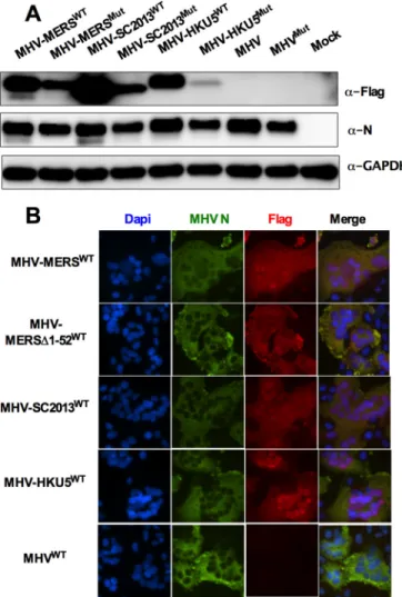
assess expression of NS4b proteins from chimeric viruses, bone
marrow-derived macrophages (BMM) derived from mice
genet-ically deficient in RNase L (RNase L
⫺/⫺) were infected with each
of the chimeric viruses in addition to WT MHV and MHV
Mutexpressing catalytically inactive mutant NS2. (RNase L
⫺/⫺BMM
were used rather than B6 BMM because an active PDE is required
for robust replication in the latter [29].) BMM were lysed at 12 h
postinfection, and protein lysates were analyzed by Western
im-munoblotting with antibodies directed against Flag to detect
NS4b proteins, antibodies against viral nucleocapsid to assess viral
replication, and antibodies against glyceraldehyde-3-phosphate
dehydrogenase (GAPDH) as a loading and transfer control. All
NS4b proteins were expressed in RNase L
⫺/⫺BMM but to
differ-ent extdiffer-ents, and mutant NS4b proteins were expressed at lower
levels than WT NS4b proteins were, possibly reflecting reduced
protein stability as a result of the mutations (Fig. 5A). Similar
findings were obtained with chimeric virus infection of 17Cl-1
cells (data not shown).
All three NS4b proteins have an amino-terminal tripartite
NLS; previous studies reported that MERS-CoV NS4b is
predom-inantly detected in the nucleus (41) and that overexpressed
BtCoV-HKU5 NS4b is solely detected in the nucleus (16).
Previ-ously, we found that murine AKAP7 was exclusively nuclear and
that it could not act as a PDE to rescue replication of chimeric
mutant MHV unless the NLS was removed and cytoplasmic
ex-pression was achieved (34), so it was important to determine the
subcellular localization of NS4b protein during chimeric virus
in-fection. Thus, immunofluorescence staining was carried out to
assess expression and subcellular localization of expression of each
NS4b protein. Murine L2 cells were infected with chimeric viruses
expressing both MERS-CoV NS4b and corresponding CoV
ho-mologs, fixed at 8 h postinfection, and stained with anti-Flag
an-tibodies (NS4b) and anan-tibodies against the MHV nucleocapsid
protein, which is expressed in the cytoplasm (Fig. 5B).
Interest-ingly, MERS-CoV and BtCoV-SC2013 NS4b proteins localized to
the nucleus with expression in the cytoplasm as well, while the
BtCoV-HKU5 NS4b homolog was almost completely nuclear,
similar to reports of overexpressed NS4b in the literature (16–18).
As expected, MHV-MERS
⌬
1-52
WTexpressing an NS4b with
de-letion of the NLS was localized to the cytoplasm.
TABLE 1 Enzyme kinetic parametersa
Enzyme
ppp5=A2=p5=A2=p5=A (2-5A substrate)
kcat(s⫺1) Km(M) kcat/Km(M⫺1s⫺1)
MHV NS2 3.9⫾0.4 6.7⫾1.6 0.6 MERS-CoV NS4b 2.1⫾0.2 2.6⫾0.6 0.8 SC2013 NS4b 2.4⫾0.1 2.9⫾0.6 0.8 HKU5 NS4b 1.9⫾0.1 1.3⫾0.3 1.4
aThe enzyme kinetic parameters were determined by GraphPad Prism by the equation
Y⫽Et⫻kcat⫻[X/(Km⫹X)], whereYis the velocity of reaction in micromolar per
second,Xis the substrate concentration in micromolar, and Et is the concentration of enzyme catalytic sites. The kinetics were determined in triplicate reactions, and data are expressed as means⫾standard errors of the means.
NS4b proteins rescue replication of MHV
Mutin B6 BMM by
antagonizing RNase L.
NS2 expression is necessary for robust
replication of MHV in BMM derived from B6 BMM (WT) but is
not required in RNase L
⫺/⫺BMM (7, 29). To elucidate the roles of
the NS4b proteins in the context of viral infection, we inoculated
B6 BMM or RNase L
⫺/⫺BMM with wild-type MHV (MHV
WT) or
MHV expressing a catalytically inactive NS2 (MHV
Mut) as well as
chimeric MHV
Mutexpressing either WT or mutant MERS-CoV
(both full-length and with amino acids 1 to 52 deleted) and
BtCoV-SC2013 NS4b proteins (see Fig. 4 for schematics of the
chimeric viruses). All three WT NS4b proteins, but not mutant
NS4b proteins, were able to rescue MHV
Mutreplication in WT
BMM (Fig. 6A to D). Similar results were obtained for the pair of
viruses expressing BtCoV HKU5 NS4b proteins (data not shown).
MHV expressing MERS-CoV WT NS4b (MHV-MERS
WT)
repli-cated to a titer equivalent to that of WT MHV. Viruses expressing
the BtCoV homologs replicated well in B6 BMM but not quite to
the same extent as WT MHV or MHV-MERS
WT, similar to
find-ings with the chimeric viruses expressing PDE of RVA VP3 CTD
and cellular AKAP7 protein (28, 34) (Fig. 6D and data not shown).
As expected, viruses expressing mutant as well as WT NS4b
pro-teins replicated to high titer in RNase L
⫺/⫺BMM. Although
lo-calization of NS4b proteins was primarily nuclear, these results
demonstrate that the level of expression of NS4b PDE at low levels
in the cytoplasm was sufficient to rescue replication in
MHV-MERS- and MHV-SC2013-infected B6 BMM.
rRNA degradation during infection is indicative of RNase L
activation, and we have previously used this read out as an indirect
measure of RNase L activation (29). In addition, we previously
demonstrated that enzymatically active PDEs of RVA VP3 and
AKAP7, but not catalytically inactive mutant proteins, were
capa-ble of preventing rRNA degradation in B6 BMM during MHV
chimeric virus infection as determined by analyzing total RNA
levels postinfection on an Agilent bioanalyzer. Similarly,
MHV-MERS
WT, both full-length and MHV-MERS
⌬
1-52
WTmutant and
MHV-SC2013
WTclearly prevented RNase L-mediated RNA
deg-radation, while viruses expressing the inactive mutant proteins
activated the OAS-RNase L pathway. Surprisingly,
BtCOV-HKU5
WT(expressed during MHV-HKU5
WTinfection) was
un-able to efficiently prevent rRNA degradation despite the fact that it
did confer efficient replication in B6 BMM (data not shown). This
may be due to the mostly nuclear localization of MHV-HKU5
WT,
which is discussed further below.
MERS-CoV NS4b rescues replication of MHV
Mutin vivo
in
B6 mice.
To determine whether MERS-CoV NS4b was capable of
rescuing replication of MHV
Mutin vivo
, we infected B6 and RNase
L
⫺/⫺mice with chimeric viruses expressing either
MHV-MERS
WTor enzymatically inactive virus (MHV-MERS
Mut) and
compared replication to MHV
WT. NS4b, but not NS4b
Mut,
res-cued MHV
Mutreplication to MHV
WTlevels in B6 mice (Fig. 7).
Additionally, gross pathology was restored for MHV-MERS
WT,
which is typically fully abrogated without an enzymatically active
NS2, but not for MHV-MERS
Mut(data not shown). This confirms
that MERS-CoV NS4b can act as an RNase L antagonist and
con-tribute to pathogenesis
in vivo
.
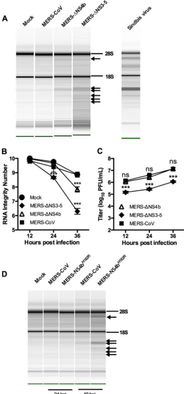
MERS-CoV inhibits rRNA degradation in a human airway
cell line.
We constructed a MERS-CoV NS4b deletion mutant
(MERS-
⌬
NS4b) and mock inoculated or inoculated a human
air-way epithelial cell line, Calu-3, with MERS-CoV, MERS-
⌬
NS4b,
or MERS-CoV lacking NS3 to NS5 (MERS-
⌬
NS3-5) (22). We
measured replication and rRNA degradation through the course
of a one-step growth curve. The characteristic signature of RNase
L activation in human cells is present during MERS-
⌬
NS4b and
MERS-
⌬
NS3-5 infections at late time points (Fig. 8A) and can be
FIG 4 Construction of NS4b-expressing recombinant MHV. (A) Diagrammatic representation of MHVMutgenome (expressing catalytically inactive NS2H126R). The letters designate genes encoding structural proteins, and the numbers designate ORFs encoding nonstructural proteins. ORFs 1a and 1b together comprise 20 kb and are not to scale. (B) Wild-type or catalytic mutant NS4b genes are lined up with the MHV ORF4a or ORF4b insertion site to construct chimeric viruses. Nuclear localization signals (NLSs) and phosphodiesterase (PDE) domains are indicated.
quantified by Agilent RNA integrity number (RIN) and plotted
over time (Fig. 8B). The characteristic rRNA degradation
signa-ture is not present during MERS-CoV infection or mock
infec-tion. We observed no reduction in titer for MERS-
⌬
NS4b but
nearly a 10-fold reduction in titer for MERS-
⌬
NS3-5 compared
with wild-type MERS-CoV infection (Fig. 8C). Thus, while
MERS-CoV deletion mutants activate RNase L in Calu-3 cells,
rRNA degradation was not very extensive compared to that
ob-served in Sindbis virus infection of human A549 cells.
The more robust rRNA degradation seen with MERS-
⌬
NS3-5
than with MERS-
⌬
NS4b suggested that there may be additional
mechanisms of RNase L antagonism by MERS-CoV. In fact, NS4a
is a dsRNA binding protein that may help sequester viral dsRNA
and prevent stimulation of OAS and subsequent activation of
RNase L (17, 18). In addition, since NS4a and NS4b are expressed
from the same mRNA by an unknown mechanism of initiation of
translation, it is possible that deletion of ORF4b could result in
increased expression of the upstream ORF encoding NS4a and
increased dsRNA binding and inhibition of RNase L, negating the
need for a PDE to antagonize RNase L. Thus, we constructed
MERS-NS4b
H182R, expressing NS4b with an inactive PDE, and
assessed its replication and ability to active RNase L in Calu-3 cells.
While MERS-NS4b
H182Rreplicated similarly to WT MERS (data
not shown), it did promote RNase L degradation and like the
deletion mutants only at late times postinfection (Fig. 8D).
Thus, MERS-CoV NS4b and BtCoV homologs have 2
=
,5
=
-PDE
activity that antagonizes RNase L activation
in vitro
and is
depen-dent on a catalytic His residue. In addition, like MHV NS2, lineage
C
Betacoronavirus
NS4b activity is crucial for protection of rRNA
integrity and for efficient replication of chimeric MHV in cell
culture and in the case of MERS-CoV NS4b, in mice. While RNase
L activation in Calu-3 cells is observed with both MERS-CoV
de-letion mutants, it is clearly less extensive than observed in the
chimeric MHV infections of BMM and has less effect on viral
replication as discussed below.
DISCUSSION
Here we demonstrate that MERS-CoV and related BtCoV NS4b
pro-teins inhibit the OAS-RNase L pathway by enzymatic cleavage of
2-5A, the activator of RNase L, potentially antagonizing IFN
signal-ing. The type I IFN response is an essential component of the antiviral
innate immune response and is activated when host sensors detect
viral dsRNA, leading to production of type I IFN (9, 10, 24). IFN
activates transcription of IFN-stimulated genes (ISG), leading to the
development of an antiviral state in the infected cells and in
neigh-boring cells, thereby restricting viral spread. Based on overexpression
experiments, it was previously reported that MERS-CoV NS4b and
HKU5 NS4b proteins antagonize type I IFN signaling by several
dif-ferent methods, including interaction with Tank binding kinase 1
(TBK1)/I
B kinase
(IKK
), leading to inhibition of IFN regulatory
factor 3 (IRF3) phosphorylation, nuclear localization, and
subse-quent beta interferon (IFN-

) transcription (16, 18, 19). It was
re-cently shown by overexpression of NS4b and an IFN-

promoter
reporter plasmid that MERS-CoV NS4b interacts with as yet
un-known nuclear targets inhibiting IRF3- and IRF7-induced IFN-

transcription (19). It is important to note that there is little if any
information on IFN antagonism during MERS-CoV infection, and
none of these reports recognized the enzymatic activity of NS4b
pro-teins. Nevertheless, the previous publications in combination with
our current study of the enzymatic activity of NS4b proteins suggest
that NS4b proteins are multifunctional with activities in both the
cytoplasm and nucleus, consistent with its localization in both
cellu-lar compartments.
Searches for sequence homology between MERS-CoV NS4b in
protein databases were unsuccessful. However, NS4b was
pre-dicted by tertiary structural modeling to be a 2H-PE and the fact
that it is conserved in all sequenced lineage C
Betacoronavirus
genomes suggested that NS4b is an enzymatically active 2
=
,5
=
-PDE
(29), as confirmed in this study. Furthermore, NS4b protein
an-tagonizes the OAS-RNase L pathway by cleaving 2-5A and
block-ing the subsequent activation of RNase L (7, 29). Additionally,
closely related lineage C
Betacoronavirus
BtCoV-SC2013 and
BtCoV-HKU5 NS4b proteins are enzymatically active PDEs with
similar activity to MERS-CoV NS4b. We have demonstrated that
these lineage C proteins can cleave 2-5A
in vitro
and that their
kinetic parameters are similar to those of NS2.
Limitation of RNase L activation can have a profound effect on
enhancing virus replication and spread as well as leading to
de-creased IFN production in myeloid cells (7, 29, 42). Indeed, we
previously found that the 2
=
,5
=
-PDE of MHV NS2 is a determinant
of myeloid cell tropism and a critical liver virulence factor in mice
(29). Thus, to initially assess the effectiveness of NS4b proteins, we
utilized the chimeric MHV NS2 mutant virus system that was
previously used to demonstrate activity for RVA and AKAP PDEs
FIG 6 Lineage C PDE NS4b proteins functionally replace MHV NS2 in primary BMM. (A to D) Representative one-step growth curve of WT and mutant (Mut) MHV (A) and chimeric viruses expressing WT and mutant NS4b proteins (diagrammed in Fig. 4B), WT and mutant MHV-MERS (B), WT and mutant MHV-MERS⌬1-52 (C), and WT and mutant MHV-SC2013 (D) in B6 and RNase L⫺/⫺BMM (MOI of 1 PFU;n⫽3). Statistical significance was determined by two-way analysis of variance (ANOVA) with Sidak’s multiple comparisons and is indicated as follows: *,Pvalue of⬍0.05; **,P⬍0.01; ***,P⬍0.001. Values that are not significantly different (ns) are indicated. Values are means⫾SEM (error bars). These data are from one representative experiment of three experiments. (E) rRNA degradation pattern 15 h postinfection in B6 BMM. Data are from one representative experiment of three (MOI of 1 PFU;n⫽3). The positions of 28S and 18S rRNAs are shown to the right of the gel.
(28, 34). Lineage C PDEs were expressed from chimeric MHV
with an NS2
Mutbackground, encoding an inactive PDE.
MERS-CoV NS4b and BtMERS-CoV homologs were detected by
immunoblot-ting to different extents, as normalized to the amount of MHV
nucleocapsid protein detected, with the mutant proteins less well
expressed compared to WT proteins. The different levels of
ex-pression of WT proteins and the more efficient exex-pression of the
WT PDEs compared to the corresponding mutant proteins was
observed in both BMM and murine 17Cl-1 fibroblasts (Fig. 5 and
data not shown), suggesting that there may be differences in
sta-bility in the WT proteins and that the mutant proteins may be less
stable. Nevertheless, we were able to rescue replication in murine
BMM with wild-type MHV-MERS, wild-type BtCoV-2013
(Fig. 6), and wild-type BtCoV-HKU5 (data not shown) but not
with viruses expressing corresponding mutant PDEs and in the
case of MHV-MERS
WT, but not MHV-MERS
Mut,
in vivo
in the
mouse liver as well. Since the NS4b catalytic mutants used here are
poorly expressed, they do not provide the ideal control and we
cannot rule out the possibility that either one or more would
res-cue at a higher expression level or that factors other than the PDE
activity contribute to rescue. This, however, is highly unlikely, as
we know from several previous studies (28, 29, 34) that efficient
expression of catalytic mutant PDE does not rescue replication of
MHV NS2 mutant virus, while supplying with heterologous PDE
does (28, 29, 34).
We have previously demonstrated that cytoplasmic
localiza-tion of PDE activity is necessary to antagonize the OAS-RNase L
pathway. Murine AKAP7, which contains an NLS, can rescue only
chimeric PDE-deficient MHV in the absence of the NLS (34).
Interestingly, it is unnecessary to remove the NLS from the MERS
and SC2013 NS4b proteins to achieve rescue of chimeric MHV,
and in fact, removal of the N-terminal 52 amino acids, including
the NLS, of MERS-CoV NS4b confers no replication
enhance-ment (Fig. 6), suggesting that the NLS neither hinders nor
im-proves OAS-RNase L antagonism. Recombinant HKU5 NS4b
clearly has 2
=
,5
=
-PDE activity (Fig. 3), and MHV-HKU5
WT(but
FIG 7 Catalytically active MERS-CoV NS4b rescues replication of MHVMut in the livers of B6 mice. Liver titers (n⫽19 or 20) day 5 postinfection (MOI of 2,000 PFU) of MHVWTand MHVMut(A) or MHV-MERSWTand MHV-MERSMut(B). Statistical significance was determined by2test with Yates’ correction and is indicated as follows: *,P value of⬍0.05; ***,P⬍0.001. Values are means⫾SEM (error bars). Values that are not significantly differ-ent (ns) are indicated. These data are pooled from four experimdiffer-entsnot MHV-HKU5
Mut) clearly replicates efficiently in B6 BMM
(data not shown), demonstrating that HKU5 NS4b can replace the
function of NS2 in the MHV chimeric background. However,
there was still detectable RNA cleavage in BMM infected with both
MHV-HKU5
WTand MHV-HKU5
Mut(data not shown). The
most likely explanation is that BtCoV-HKU5 NS4b does not
an-tagonize RNase L activation as well as MERS-CoV or BtCoV
SC2013 NS4b does, because it is more localized to the nucleus,
consistent with our findings with the PDE of AKAP7 (34). It is also
possible the rRNA is more susceptible to cleavage than viral RNA;
therefore, the less effective BtCoV-HKU5 NS4b cannot
antago-nize RNase L effectively enough to spare rRNA while still
protect-ing viral and/or cellular mRNA.
We have demonstrated that MERS-CoV antagonizes
OAS-RNase L activation in Calu-3 cells and that mutant viruses with
deletions of either NS3 to NS5 or NS4b alone as well as the
cata-lytic mutant MERS-NS4b
H182Ractivate this pathway. Although all
mutants induced rRNA degradation, only MERS-
⌬
NS3-5 had a
reduction in viral titer by 24 h postinfection. Thus, the lack of
effect on replication of MERS-
⌬
NS4 or MERS-NS4b
H182Rcorre-lates with the weak rRNA degradation observed and indicates a
lack of biological effect in this cell type of deleting or inactivating
the NS4b PDE. This may be due in part to the very late times
postinfection (24 to 48 h) that we observe rRNA degradation;
virus replication is already quite robust by that time, and it may be
too late for restriction of replication to occur.
We have found previously that cell type is critical for robust
acti-vation of RNase L and reduction in viral titers during infection of
murine cells and have observed activation of RNase L by MHV
Mutonly in myeloid cells (43) and endothelial cells (unpublished data).
The use of Calu-3 epithelial airway cells may explain the lack of
re-striction of MERS-
⌬
NS4b replication and only 10-fold reduction for
MERS-
⌬
NS3-5, when in murine BMM, abrogation of RNase L
an-tagonism by MHV, which is observed by 9 h postinfection, leads to
100- to 1,000-fold reduction in viral titers. Indeed, RNase L is not
readily activated in Calu-3 cells compared to other cell types; the
amount of rRNA degradation observed in Sindbis virus-infected
A549 cells is clearly more than in Calu-3 cells (Fig. 8A), and we did not
observe robust activation of RNase L Calu-3 cells even when
trans-fected with the dsRNA surrogate poly(I · C) (data not shown).
Fur-ther studies are being directed at identifying a cell type in which PDE
activity enhances viral replication.
MATERIALS AND METHODS
Cell lines and mice.Murine L2 (L2) and baby hamster kidney cells ex-pressing MHV receptor (BHK-MHVR) were cultured as described previ-ously (7, 44). Calu-3 clone 2B4 cells (a kind gift from Chien-Te Tseng, University of Texas Medical Branch, Galveston, TX) were cultured with Gibco minimum essential medium (MEM) (Thermo Fisher Scientific, Grand Island, NY) supplemented with 20% fetal bovine serum (FBS) (HyClone, GE Healthcare Bio-Sciences, Pittsburg, PA), penicillin-streptomycin (Gibco), and amphotericin B (Fungizone) (Gibco). Vero CCL-81 cells (ATCC, Manassas, VA) were cultured, and the MERS-CoV plaque assay was performed as previously described (22). Human A549 cells were cultured in RPMI 1640 medium (Gibco) supplemented with 10% FBS, 100 U/ml of penicillin, and 100g/ml streptomycin. All cells were maintained at 37°C and 5% CO2. B6 mice and RNase L⫺/⫺mice were bred and maintained in the University of Pennsylvania animal facility, and the protocols were approved by the Institutional Animal Care and Use Committee at the University of Pennsylvania. Primary BMM were derived from bone marrow harvested from the hind limbs (tibia and femur) of
4-to 6-week-old B6 or RNase L⫺/⫺mice and described previously (29, 45).
Cells were cultured in Dulbecco modified Eagle medium (DMEM) (Gibco) supplemented with 10% FBS (HyClone) and 20% L929 cell-conditioned media for 6 days before infection.
Plasmids and recombinant viruses.NS4b sequences for MERS-CoV (GenBank accession no.AFS88939.1), SC2013 (GenBank accession no. AHY61340.1), and HKU5 (accession no.YP_001039965.1) were obtained from NCBI. NS4b cDNA sequences were synthesized with the addition of a 5= SalI site and 3=Flag epitope and NotI site and then cloned into pUC57 (Bio Basic, Markham, Ontario, Canada). Site-directed mutagenesis was per-formed on each NS4b plasmid to generate point mutants (CAC to CGC) containing histidine-to-arginine mutations within the second catalytic motif yielding H182R, H186R, and H183R for MERS-CoV, SC2013, and HKU5 NS4b proteins, respectively, and subsequently sequence verified. Each NS4b gene was then PCR amplified and subcloned in frame with maltose binding protein into pMAL protein expression vector. MHV NS2 and NS2H126Rwere previously cloned into pMAL parallel-2 vector (29).
MHVWT, MHVMut, and all chimeric viruses were constructed with the MHV strain A59 infectious clone (44). Each WT or mutant NS4b cDNA was subcloned into open reading frame 4 (ORF4) of the infectious clone plasmid GEGFP(EGFP stands for enhanced green fluorescent protein), and sequences were confirmed as described previously (28). The full-length cDNAs were assembled with A to E (MHVWT), F (MHVMut), and G (NS4b) fragments, and RNA wasin vitrotranscribed and recovered as previously described (44). Briefly, infectious clone fragments were excised from their respective plasmids by restriction enzyme digestion. The frag-ments were gel purified and ligatedin vitroto create a full-length cDNA. cDNA wasin vitrotranscribed with mMessage mMachine T7 transcrip-tion kit (Ambion; Thermo Fisher, Grand Island, NY), generating full-length genomic RNA. Each genomic RNA transcript was split into two distinct pools and electroporated into BHK-MHVR along with N-protein transcript using Gene Pulser II (Bio-Rad, Hercules, CA). Electroporated cells were incubated until cytopathology was evident throughout, freeze thawed three times, and plaque purified, and one plaque from each pool was utilized in parallel for all assays.
MERS-⌬NS4b was constructed using the MERS-CoV infectious clone (22). To delete NS4b, sequence was synthesized (Bio Basic) in which muta-tions were made in the overlap region between NS4a and NS4b to abrogate putative start codons (T2C and T17C; all positions relative to the NS4b start) and to insert premature stop codons (A91T, C97A, and C102G). Addition-ally, residues 106 to 669 were deleted to remove the majority of NS4b but leave in place transcriptional regulatory sequence 5. The truncated NS4b cassette and MERS-CoV infectious clone F plasmid were digested with PacI and SanDI, and truncated NS4b was ligated into the MERS-CoV F plasmid. To construct the MERS-NS4bH182Rmutant, two PCRs were performed using the F plasmid as the template to synthesize overlapping DNA fragments and introducing an H182R substitution. These two templates were then joined in an overlapping extension PCR. The resultant product was digested with PacI and SanDI and cloned into the MERS F plasmid. Assembly of the infectious clone and recovery of infectious virus were performed as described previously (22). The potential gain-of-function (GOF) concerns of MERS-⌬NS4b and MERS-NS4bH182R were evaluated and reviewed by NIH under grant U19AI107810 and approved for continued study. The MERS-⌬NS3-5 mu-tant (22) was generated prior to GOF regulations. The MERS-⌬NS3-5 and catalytic mutant display attenuated growth on select interferon-competent human cell lines.
SC2013, and HKU5 or their catalytically inactive mutants described in the previous section) in 150l of assay buffer (20 mM HEPES [pH 7.2], 10 mM MgCl2, 1 mM dithiothreitol) were incubated at 30°C with various concentra-tions of (2=-5=)p3A3. Aliquots of 20l were withdrawn at different times, and the reactions were stopped by heat inactivation at 95°C for 3 min followed by 30-min centrifugation at 20,000⫻g(4°C) and supernatants were carefully removed. A fluorescence resonance energy transfer (FRET)-based RNase L activation assay was used to determine enzyme kinetics by measuring the uncleaved, intact (2=,5=)p3A3as previously described (47). The amounts of 2-5A cleaved were determined by the relative fluorescence units (RFU) using standard curves with different concentrations of authentic 2-5A. The reaction velocities were determined by nonlinear fit of the data. ThekcatandKmwere determined in GraphPad Prism 5.0 for Windows by plotting velocity against substrate concentrations, and data were analyzed by the following equation: Y⫽Et⫻kcat⫻[X/(Km⫹X)], whereYis the velocity of the reaction in micromolar per second,Xis the substrate concentration in micromolar, and Et is the concentration of enzyme catalytic sites. The kinetics were determined in triplicate reactions.
In vitroinfections. (i) Chimeric MHV infections.BMM were mock inoculated or inoculated with MHVWT, MHVMut, MHV-MERSWT, MHV-MERSMut, MHV-SC2013WT, MHV-SC2013Mut, MHV-HKU5WT, or MHV-HKU5Mut. Virus was added to cells at a multiplicity of infection (MOI) of 1 PFU/cell and allowed to adsorb for 1 h at 37°C. Cultures were washed with phosphate-buffered saline (PBS) (three times) and fed with medium. At the times indicated in the figures, cells were fixed and ana-lyzed for protein expression by immunofluorescent staining, lysed, and analyzed for protein expression by immunoblotting or analyzed for deg-radation of RNA, or supernatants were harvested for quantification of viral titers by plaque assay on L2 cells (48).
(ii) MERS-CoV infections of Calu-3 cells.Prior to infection, Calu-3 cells were washed once with PBS (Gibco) to remove residual fetal bovine serum (FBS). Cells were inoculated under biosafety level 3 (BSL3) condi-tions with MERS-CoV, MERS-⌬NS4b, MERS-NS4bH182R, or
MERS-⌬NS3-5 at an MOI of 1 PFU/cell and allowed to absorb for 40 min at 37°C. The inoculum was then removed, and the cells were washed twice with PBS prior to adding media containing low FBS (4%). At the time points indicated in the figures, 100l of medium was collected and subsequently analyzed by plaque assay in Vero CCL-81 cells as previously described (22), and total RNA was harvested in 1 ml Trizol.
Immunoblotting.At 12 h postinfection, cells were lysed in Nonidet P-40 (NP-40) buffer (1% NP-40, 2 mM EDTA, 10% glycerol, 150 mM NaCl, and 50 mM Tris [pH 8.0]) containing protease inhibitors (Roche). Protein concentrations were measured using a DC protein assay kit (Bio-Rad). Supernatants were mixed 3:1 with 4⫻SDS-PAGE sample buffer. Samples were boiled, separated by 4 to 15% SDS-PAGE, and transferred to polyvinylidene difluoride (PVDF) membranes. Blots were blocked with 5% nonfat milk and probed with the following antibodies: anti-Flag M2 mouse monoclonal antibody (Agilent) (1:1,000), anti-N mouse monoclo-nal antibody (a gift from Julian Leibowitz) (1:400), and anti-GAPDH mouse monoclonal antibody (Thermo Fisher Scientific) (1:1,000). Anti-mouse secondary antibodies labeled with horseradish peroxidase (HRP) (Santa Cruz; 1:5,000) were used to detect the primary antibodies. The blots were visualized using SuperSignal West Dura extended-duration substrate (Thermo Scientific). Blots were probed sequentially with anti-bodies with the blots stripped between antibody treatments.
Indirect immunofluorescence assay.L2 cells were infected at an MOI of 1 PFU/cell for 8 h with MHVWT, MHV-MERSWT, MHV-SC2013WT, or MHV-HKU5WT. Cells were washed with Dulbecco’s phosphate-buffered saline (DPBS) (Gibco) and then fixed with 4% paraformaldehyde in DPBS (Gibco). After fixation, cells were permeabilized with 0.1% Triton in DPBS (Gibco) and then blocked with bovine serum albumin. Cells were then stained with mouse monoclonal anti-nucleocapsid (1:200) and rab-bit anti-Flag polyclonal (Sigma) (1:2,000) antibodies. After the cells were washed, secondary staining with Alexa Fluor 488-labeled goat anti-mouse (1:400) and Alexa Fluor 594-labeled goat anti-rabbit (1:1,000) antibodies
was performed. The cells were washed once more, and the nuclei were then stained with 4=,6=-diamidino-2-phenylindole (DAPI). Stained cells were imaged at a magnification of⫻400 on an Eclipse TE2000-U fluores-cence microscope (Nikon Instruments, Inc.). Images were acquired using NIS-Elements Basic Research microscope imaging software (Nikon In-struments, Inc.). This was performed at least twice for all viruses.
Qualitative and quantitative analyses of RNase L-mediated rRNA degradation. RNA from B6 BMM infected with MHVWT, MHVMut, MHV-MERSWT, MHV-MERSMut, MHV-SC2013WT, MHV-SC2013Mut, MHV-HKU5WT, or MHV-HKU5Mutwas harvested with an RNeasy kit (Qiagen, Valencia, CA) 15 h postinoculation. Alternatively, Calu-3 cells infected with MERS-CoV, MERS-⌬NS4b, MERS-⌬NS3-5, or MERS-NS4bH182R, and at the time points indicated in the figures, RNA was harvested with Trizol (Sigma Aldrich, St. Louis, MO), extracted with chlo-roform (Sigma), and precipitated with isopropanol (Sigma). RNA was then separated and analyzed on RNA nanochips with an Agilent Bioana-lyzer 2100 (Agilent Technologies, Santa Clara, CA), and the pseudogels were normalized for concentration (49). RNA integrity numbers (referred to as RIN values) (50) are measurements of RNA integrity produced by the Agilent Bioanalyzer. RNA from A549 cells infected with Sindbis virus (51) was used as a marker for the pattern of human RNase L-dependent rRNA cleavage products.
Inoculation of mice.Four-week-old B6 or RNase L⫺/⫺mice were
anesthetized with isoflurane (Abbott Laboratories, Chicago, IL) and inoc-ulated intrahepatically with MHVWT, MHVMut, MHV-MERSWT, or MHV-MERSMutin 50l of DPBS (Gibco) containing 0.75% bovine se-rum albumin (Sigma). Mice were humanely euthanized with CO2and perfused with DPBS (Gibco), and their livers were harvested at day 5 postinoculation. Viral titers were determined by plaque assay of lysates (48). This study was carried out in strict accordance with theGuide for the Care and Use of Laboratory Animals(41) and the Public Health Service Policy on Humane Care and Use of Laboratory Animals (52).
ACKNOWLEDGMENTS
Research reported in this publication was supported by the National In-stitute of Allergy and Infectious Disease of the National InIn-stitutes of Health (NIH) under award F32AI114143 to J.M.T., R21AI114920 to S.R.W., R01AI104887 to S.R.W. and R.H.S, and U19AI107810 to R.S.B.
The content is solely the responsibility of the authors and does not necessarily represent the official views of the NIH. The potential gain of function (GOF) concerns of MERS-⌬NS4b and MERS-NS4bH182Rwere evaluated and reviewed by NIH under grant U19AI107810 and approved for continued study. The MERS-⌬NS3-5 mutant was generated prior to GOF regulations. The funders had no role in study design, data collection and interpretation, or the decision to submit the work for publication.
FUNDING INFORMATION
This work, including the efforts of Joshua M. Thornbrough, was funded by HHS | NIH | National Institute of Allergy and Infectious Diseases (NIAID) (F32AI114143). This work, including the efforts of Joshua M. Thornbrough, Babal Kant Jha, Stephen A. Goldstein, Ruth Elliott, Robert H. Silverman, and Susan R. Weiss, was funded by HHS | NIH | National Institute of Allergy and Infectious Diseases (NIAID) (R21AI114920). This work, including the efforts of Joshua M. Thornbrough, Babal Kant Jha, Stephen A. Goldstein, Yize Li, Ruth Elliott, Robert H. Silverman, and Susan R. Weiss, was funded by HHS | NIH | National Institute of Allergy and Infectious Diseases (NIAID) (R01AI104887). This work, including the efforts of Boyd Yount, Amy C. Sims, and Ralph S. Baric, was funded by HHS | NIH | National Institute of Allergy and Infectious Diseases (NIAID) (U19AI107810).
REFERENCES
syndrome coronavirus disease from Saudi Arabia: a descriptive study. Lancet Infect Dis 13:752–761. http://dx.doi.org/10.1016/S1473 -3099(13)70204-4.
2.World Health Organization. 2015. Middle East respiratory syndrome coronavirus (MERS-CoV)⫺Saudi Arabia. Disease outbreak news, 24 July 2015. World Health Organization, Geneva, Switzerland.
3.Bialek SR, Allen D, Alvarado-Ramy F, Arthur R, Balajee A, Bell D, Best S, Blackmore C, Breakwell L, Cannons A, Brown C, Cetron M, Chea N, Chommanard C, Cohen N, Conover C, Crespo A, Creviston J, Curns AT, Dahl R, Dearth S, DeMaria A, Echols F, Erdman DD, Feikin D, Frias M, Gerber SI, Gulati R, Hale C, Haynes LM, Heberlein-Larson L, Holton K, Ijaz K, Kapoor M, Kohl K, Kuhar DT, Kumar AM, Kundich M, Lippold S, Liu L, Lovchik JC, Madoff L, Martell S, Matthews S, Moore J, Murray LR, Onofrey S, Pallansch MA, Pesik N, Pham H, et al.
2014. First confirmed cases of Middle East respiratory syndrome corona-virus (MERS-CoV) infection in the United States, updated information on the epidemiology of MERS-CoV infection, and guidance for the public, clinicians, and public health authorities - May 2014. MMWR Morb Mor-tal Wkly Rep63:431– 436.
4.Kucharski AJ, Althaus CL. 2015. The role of superspreading in Middle East respiratory syndrome coronavirus (MERS-CoV) transmission. Euro S u r v e i l l 2 0 :1 4 – 1 8 . h t t p : / / d x . d o i . o r g / 1 0 . 2 8 0 7 / 1 5 6 0 -7917.ES2015.20.25.21167.
5.Nishiura H, Miyamatsu Y, Chowell G, Saitoh M. 2015. Assessing the risk of observing multiple generations of Middle East respiratory syndrome (MERS) cases given an imported case. Euro Surveill20(27):pii⫽21181. http://dx.doi.org/10.2807/1560-7917.ES2015.20.27.21181.
6.Cotten M, Lam TT, Watson SJ, Palser AL, Petrova V, Grant P, Pybus OG, Rambaut A, Guan Y, Pillay D, Kellam P, Nastouli E. 2013. Full-genome deep sequencing and phylogenetic analysis of novel human beta-coronavirus. Emerg Infect Dis19:736 –742.http://dx.doi.org/10.3201/ eid1905.130057.
7.Roth-Cross JK, Stokes H, Chang G, Chua MM, Thiel V, Weiss SR, Gorbalenya AE, Siddell SG. 2009. Organ-specific attenuation of murine hepatitis virus strain A59 by replacement of catalytic residues in the puta-tive viral cyclic phosphodiesterase ns2. J Virol83:3743–3753.http:// dx.doi.org/10.1128/JVI.02203-08.
8.Weiss SR, Navas-Martin S. 2005. Coronavirus pathogenesis and the emerging pathogen severe acute respiratory syndrome coronavirus. Mi-crobiol Mol Biol Rev 69:635– 664. http://dx.doi.org/10.1128/ MMBR.69.4.635-664.2005.
9.Roth-Cross JK, Bender SJ, Weiss SR. 2008. Murine coronavirus mouse hepatitis virus is recognized by MDA5 and induces type I interferon in brain macrophages/microglia. J Virol82:9829 –9838.http://dx.doi.org/ 10.1128/JVI.01199-08.
10. Kint J, Fernandez-Gutierrez M, Maier HJ, Britton P, Langereis MA, Koumans J, Wiegertjes GF, Forlenza M. 2015. Activation of the chicken type I interferon response by infectious bronchitis coronavirus. J Virol
89:1156 –1167.http://dx.doi.org/10.1128/JVI.02671-14.
11. Zhou H, Zhao J, Perlman S. 2010. Autocrine interferon priming in macrophages but not dendritic cells results in enhanced cytokine and chemokine production after coronavirus infection. mBio1:e00219-10. http://dx.doi.org/10.1128/mBio.00219-10.
12. Versteeg GA, Bredenbeek PJ, van den Worm SH, Spaan WJ. 2007. Group 2 coronaviruses prevent immediate early interferon induction by protection of viral RNA from host cell recognition. Virology361:18 –26. http://dx.doi.org/10.1016/j.virol.2007.01.020.
13. Lau SK, Lau CC, Chan KH, Li CP, Chen H, Jin DY, Chan JF, Woo PC, Yuen KY. 2013. Delayed induction of proinflammatory cytokines and suppression of innate antiviral response by the novel Middle East respira-tory syndrome coronavirus: implications for pathogenesis and treatment. J Gen Virol94:2679 –2690.http://dx.doi.org/10.1099/vir.0.055533-0. 14. Zielecki F, Weber M, Eickmann M, Spiegelberg L, Zaki AM,
Matroso-vich M, Becker S, Weber F. 2013. Human cell tropism and innate im-mune system interactions of human respiratory coronavirus EMC com-pared to those of severe acute respiratory syndrome coronavirus. J Virol
87:5300 –5304.http://dx.doi.org/10.1128/JVI.03496-12.
15. Zhou J, Chu H, Li C, Wong BH, Cheng ZS, Poon VK, Sun T, Lau CC, Wong KK, Chan JY, Chan JF, To KK, Chan KH, Zheng BJ, Yuen KY. 2014. Active replication of Middle East respiratory syndrome coronavirus and aberrant induction of inflammatory cytokines and chemokines in human macrophages: implications for pathogenesis. J Infect Dis209:
1331–1342.http://dx.doi.org/10.1093/infdis/jit504.
16. Matthews KL, Coleman CM, van der Meer Y, Snijder EJ, Frieman MB. 2014. The ORF4b-encoded accessory proteins of Middle East respiratory syndrome coronavirus and two related bat coronaviruses localize to the nucleus and inhibit innate immune signalling. J Gen Virol95:874 – 882. http://dx.doi.org/10.1099/vir.0.062059-0.
17. Niemeyer D, Zillinger T, Muth D, Zielecki F, Horvath G, Suliman T, Barchet W, Weber F, Drosten C, Müller MA. 2013. Middle East respi-ratory syndrome coronavirus accessory protein 4a is a type I interferon antagonist. J Virol87:12489 –12495.http://dx.doi.org/10.1128/JVI.01845 -13.
18. Yang Y, Zhang L, Geng H, Deng Y, Huang B, Guo Y, Zhao Z, Tan W. 2013. The structural and accessory proteins M, ORF 4a, ORF 4b, and ORF 5 of Middle East respiratory syndrome coronavirus (MERS-CoV) are po-tent interferon antagonists. Protein Cell4:951–961.http://dx.doi.org/ 10.1007/s13238-013-3096-8.
19. Yang Y, Ye F, Zhu N, Wang W, Deng Y, Zhao Z, Tan W. 2015. Middle East respiratory syndrome coronavirus ORF4b protein inhibits type I in-terferon production through both cytoplasmic and nuclear targets. Sci Rep5:17554.http://dx.doi.org/10.1038/srep17554.
20. Freundt EC, Yu L, Park E, Lenardo MJ, Xu XN. 2009. Molecular determinants for subcellular localization of the severe acute respiratory syndrome coronavirus open reading frame 3b protein. J Virol83:
6631– 6640.http://dx.doi.org/10.1128/JVI.00367-09.
21. Pewe L, Zhou H, Netland J, Tangudu C, Olivares H, Shi L, Look D, Gallagher T, Perlman S. 2005. A severe acute respiratory syndrome-associated coronavirus-specific protein enhances virulence of an attenu-ated murine coronavirus. J Virol79:11335–11342. http://dx.doi.org/ 10.1128/JVI.79.17.11335-11342.2005.
22. Scobey T, Yount BL, Sims AC, Donaldson EF, Agnihothram SS, Men-achery VD, Graham RL, Swanstrom J, Bove PF, Kim JD, Grego S, Randell SH, Baric RS. 2013. Reverse genetics with a full-length infectious cDNA of the Middle East respiratory syndrome coronavirus. Proc Natl Acad Sci U S A 110:16157–16162. http://dx.doi.org/10.1073/ pnas.1311542110.
23. Almazán F, DeDiego ML, Sola I, Zuñiga S, Nieto-Torres JL, Marquez-Jurado S, Andrés G, Enjuanes L. 2013. Engineering a replication-competent, propagation-defective Middle East respiratory syndrome coronavirus as a vaccine candidate. mBio4:e00650-13.http://dx.doi.org/ 10.1128/mBio.00650-13.
24. Silverman RH. 2007. Viral encounters with 2=,5=-oligoadenylate synthe-tase and RNase L during the interferon antiviral response. J Virol81:
12720 –12729.http://dx.doi.org/10.1128/JVI.01471-07.
25. Der SD, Zhou A, Williams BR, Silverman RH. 1998. Identification of genes differentially regulated by interferon alpha, beta, or gamma using oligonucleotide arrays. Proc Natl Acad Sci U S A95:15623–15628.http:// dx.doi.org/10.1073/pnas.95.26.15623.
26. Cooper DA, Jha BK, Silverman RH, Hesselberth JR, Barton DJ. 2014. Ribonuclease L and metal-ion-independent endoribonuclease cleavage sites in host and viral RNAs. Nucleic Acids Res42:5202–5216.http:// dx.doi.org/10.1093/nar/gku118.
27. Huang H, Zeqiraj E, Dong B, Jha BK, Duffy NM, Orlicky S, Thevaku-maran N, Talukdar M, Pillon MC, Ceccarelli DF, Wan LC, Juang YC, Mao DY, Gaughan C, Brinton MA, Perelygin AA, Kourinov I, Guarné A, Silverman RH, Sicheri F. 2014. Dimeric structure of pseudokinase RNase L bound to 2-5A reveals a basis for interferon-induced antiviral a c t i v i t y . M o l C e l l 5 3 :2 2 1 – 2 3 4 . h t t p : / / d x . d o i . o r g / 1 0 . 1 0 1 6 / j.molcel.2013.12.025.
28. Zhang R, Jha BK, Ogden KM, Dong B, Zhao L, Elliott R, Patton JT, Silverman RH, Weiss SR. 2013. Homologous 2=,5=-phosphodiesterases from disparate RNA viruses antagonize antiviral innate immunity. Proc Natl Acad Sci U S A110:13114 –13119.http://dx.doi.org/10.1073/ pnas.1306917110.
29. Zhao L, Jha BK, Wu A, Elliott R, Ziebuhr J, Gorbalenya AE, Silverman RH, Weiss SR. 2012. Antagonism of the interferon-induced OAS-RNase L pathway by murine coronavirus ns2 protein is required for virus replica-tion and liver pathology. Cell Host Microbe11:607– 616. http:// dx.doi.org/10.1016/j.chom.2012.04.011.
30. Malathi K, Dong B, Gale M, Jr, Silverman RH. 2007. Small self-RNA generated by RNase L amplifies antiviral innate immunity. Nature448:
816 – 819.http://dx.doi.org/10.1038/nature06042.
31. Kelley LA, Sternberg MJ. 2009. Protein structure prediction on the Web: a case study using the Phyre server. Nat Protoc4:363–371.http:// dx.doi.org/10.1038/nprot.2009.2.
32. Roy A, Kucukural A, Zhang Y. 2010. I-TASSER: a unified platform for automated protein structure and function prediction. Nat Protoc
5:725–738.http://dx.doi.org/10.1038/nprot.2010.5.
33. Zhang Y. 2008. I-TASSER server for protein 3D structure prediction. BMC Bioinformatics9:40.http://dx.doi.org/10.1186/1471-2105-9-40. 34. Gusho E, Zhang R, Jha BK, Thornbrough JM, Dong B, Gaughan C,
Elliott R, Weiss SR, Silverman RH. 2014. Murine AKAP7 has a 2=,5= -phosphodiesterase domain that can complement an inactive murine coro-navirus ns2 gene. mBio 5:e01312-14. http://dx.doi.org/10.1128/ mBio.01312-14.
35. Mazumder R, Iyer LM, Vasudevan S, Aravind L. 2002. Detection of novel members, structure-function analysis and evolutionary classifica-tion of the 2H phosphoesterase superfamily. Nucleic Acids Res 30:
5229 –5243.http://dx.doi.org/10.1093/nar/gkf645.
36. Sui B, Huang J, Jha BK, Yin P, Zhou M, Fu ZF, Silverman RH, Weiss SR, Peng G, Zhao L. 12 January 2016. Crystal structure of the mouse hepatitis virus ns2 phosphodiesterase domain that antagonizes RNase L activation. J Gen Virolhttp://dx.doi.org/10.1099/jgv.0.000395. 37. Woo PC, Wang M, Lau SK, Xu H, Poon RW, Guo R, Wong BH, Gao
K, Tsoi HW, Huang Y, Li KS, Lam CS, Chan KH, Zheng BJ, Yuen KY. 2007. Comparative analysis of twelve genomes of three novel group 2c and group 2d coronaviruses reveals unique group and subgroup features. J Virol81:1574 –1585.http://dx.doi.org/10.1128/JVI.02182-06.
38. De Groot RJ, Baker SC, Baric RS, Brown CS, Drosten C, Enjuanes L, Fouchier RAM, Galiano M, Gorbalenya AE, Memish ZA, Perlman S, Poon LLM, Snijder EJ, Stephens GM, Woo PCY, Zaki AM, Zambon M, Ziebuhr J. 2013. Middle East respiratory syndrome coronavirus (MERS-CoV): announcement of the Coronavirus Study Group. J Virol 87:
7790 –7792.http://dx.doi.org/10.1128/JVI.01244-13.
39. Gold MG, Smith FD, Scott JD, Barford D. 2008. AKAP18 contains a phosphoesterase domain that binds AMP. J Mol Biol375:1329 –1343. http://dx.doi.org/10.1016/j.jmb.2007.11.037.
40. Ontiveros E, Kuo L, Masters PS, Perlman S. 2001. Inactivation of ex-pression of gene 4 of mouse hepatitis virus strain JHM does not affect virulence in the murine CNS. Virology289:230 –238.http://dx.doi.org/ 10.1006/viro.2001.1167.
41. National Research Council. 2011. Guide for the care and use of laboratory animals, 8th ed. National Academies Press, Washington, DC.
42. Banerjee S, Chakrabarti A, Jha BK, Weiss SR, Silverman RH. 2014.
Cell-type-specific effects of RNase L on viral induction of beta interferon. mBio5:e00856-14.http://dx.doi.org/10.1128/mBio.00856-14.
43. Zhao L, Birdwell LD, Wu A, Elliott R, Rose KM, Phillips JM, Li Y, Grinspan J, Silverman RH, Weiss SR. 2013. Cell-type-specific activation of the oligoadenylate synthetase-RNase L pathway by a murine coronavi-rus. J Virol87:8408 – 8418.http://dx.doi.org/10.1128/JVI.00769-13. 44. Yount B, Denison MR, Weiss SR, Baric RS. 2002. Systematic assembly of
a full-length infectious cDNA of mouse hepatitis virus strain A59. J Virol
76:11065–11078.http://dx.doi.org/10.1128/JVI.76.21.11065-11078.2002. 45. Caamaño J, Alexander J, Craig L, Bravo R, Hunter CA. 1999. The NF-kappa B family member RelB is required for innate and adaptive im-munity toToxoplasma gondii. J Immunol163:4453– 4461.
46. Sheffield P, Garrard S, Derewenda Z. 1999. Overcoming expression and purification problems of RhoGDI using a family of “parallel” expression vectors. Protein Expr Purif 15:34 –39.http://dx.doi.org/10.1006/ prep.1998.1003.
47. Thakur CS, Xu Z, Wang Z, Novince Z, Silverman RH. 2005. A conve-nient and sensitive fluorescence resonance energy transfer assay for RNase L and 2=,5=oligoadenylates. Methods Mol Med116:103–113.http:// dx.doi.org/10.1385/1-59259-939-7:103.
48. Gombold JL, Hingley ST, Weiss SR. 1993. Fusion-defective mutants of mouse hepatitis virus A59 contain a mutation in the spike protein cleavage signal. J Virol67:4504 – 4512.
49. Xiang Y, Wang Z, Murakami J, Plummer S, Klein EA, Carpten JD, Trent JM, Isaacs WB, Casey G, Silverman RH. 2003. Effects of RNase L mutations associated with prostate cancer on apoptosis induced by 2=,5= -oligoadenylates. Cancer Res63:6795– 6801.
50. Schroeder A, Mueller O, Stocker S, Salowsky R, Leiber M, Gassmann M, Lightfoot S, Menzel W, Granzow M, Ragg T. 2006. The RIN: an RNA integrity number for assigning integrity values to RNA measurements. BMC Mol Biol7:3.http://dx.doi.org/10.1186/1471-2199-7-3.
51. Frolova EI, Fayzulin RZ, Cook SH, Griffin DE, Rice CM, Frolov I. 2002. Roles of nonstructural protein nsP2 and alpha/beta interferons in deter-mining the outcome of Sindbis virus infection. J Virol76:11254 –11264. http://dx.doi.org/10.1128/JVI.76.22.11254-11264.2002.