THE ROLE OF TUMOR DESMOPLASIA IN NANOPARTICLE DELIVERY OF DRUGS AND GENES
Lei Miao
A dissertation submitted to the faculty at the University of North Carolina at Chapel Hill in partial fulfillment of the requirements for the degree of Doctor of Philosophy in Division of Molecular
Pharmaceutics in the Eshelman School of Pharmacy
Chapel Hill 2016
Approved by: Leaf Huang Philip C. Smith Elena V. Batrakova Xiao Xiao
ii © 2016 Lei Miao
iii ABSTRACT
Lei Miao: The Role of Tumor Desmoplasia in Nanoparticle Delivery of Drugs and Genes (Under the direction of Leaf Huang)
In desmoplastic tumors, stroma cells capture nanoparticles (NPs), preventing them from reaching tumor cells, resulting in compromised anti-tumor efficacy. This dissertation focuses on understanding the basis role of tumor associated fibroblasts (TAFs), one of the major stroma cells constituting desmoplasia, in NP delivery and tumor resistance, as well as proposing strategies to overcome the TAF-elicited barriers and improve efficacy.
While the capture of therapeutic NPs in TAFs interferes tumor-stroma crosstalk and inhibits tumor progression, we found that the chronic exposure of NPs paradoxically induced the secretion of survival factors (e.g., Wnt16) from the damaged TAFs, facilitating tumor proliferation and metastasis. Therefore, we proposed the delivery of siRNA against Wnt16 to TAFs via the off-target capture, to downregulate this survival factor. The priming of damaged fibroblasts could synergize with a nanoformulation of cisplatin, and benefit the treatment of a desmoplastic bladder cancer xenograft (UMUC3/3T3). Since the off-target delivery of NPs have been verified, we further utilized the same rationale to generate a group of tumor-suppressive TAFs through transfecting TAFs with a plasmid encoding highly secretable TNF-related apoptosis-inducing ligand (sTRAIL). The production of sTRAIL from TAFs bypassed the stroma barrier and resulted in efficient killing of tumor cells.
Furthermore, we also proposed a stroma depletion method via combination therapy of cisplatin NPs and gemcitabine NPs. This combination was not only detrimental to tumor cells, but induced superior apoptosis in TAFs of the UMUC3/3T3 model. To ensure the sufficient synergy, we further designed a nano-formulation with ratiometric co-loading and co-delivery of these two
iv
into nano-cores with similar hydrophobic surface and particle size, allows for their simultaneously and ratiometric loading in a single PLGA NPs. This combinatory NPs showed potent anti-cancer efficacy compared to each regimens in separate NPs.
v
vi
ACKNOWLEDGEMENTS
First of all, I would like to express my utmost gratitude and appreciation to my supervisor, Dr. Leaf Huang who offered me this great educational opportunity in his lab as a graduate student. In the past few years, he provided me constant support, priceless guidance and extraordinary wisdom throughout my PhD journey. He is a remarkable and exceptional mentor I am so blessed to have. I will be forever thankful to his mentorship, inspiration and sense of humor. I would also like to thank my committee members, Dr. Elena Batrakova, Dr. Xiao Xiao, Dr. William Kim and Dr. Philip Smith for their precious guidance throughout the years. They advised me through the obstacles in
completion of my research work and reviewed all my progress and dissertation. In addition, I would like to extend my special appreciation to Dr. Gregory Forest, Dr. Samuel Lai, Dr. Shawn Hingtgen, Dr. Shutao Guo, Dr. Yuhua Wang and Dr. Jing Zhang for their collaboration and as my external advisors.
vii
viii
TABLE OF CONTENTS
LIST OF TABLES ... xi
LIST OF FIGURES ... xii
LIST OF ABBREVATIONS AND SYMBOLS ... xvi
CHAPTER 1: STROMAL BARRIERS AND STRATEGIES FOR THE DELIVERY OF NANOMEDICINE TO DESMOPLASTIC TUMORS ... 1
1.1 Summary ... 1
1.2 Introduction ... 2
1.3 Mathematical Modeling and In Vitro Models of NP Intratumoral Distribution ... 3
1.4 Enhanced Permeability and Retention Effect and Anti-cancer NPs in the Clinical Trials .... 5
1.5 Tumor Microenvironment Barriers for Intratumoral NPs Diffusion and Distribution ... 6
1.6 Physicochemical Properties of NPs influences NPs transport in Stroma-rich Tumors ... 14
1.7 Strategies to Improve NPs extravasation and Penetration ... 17
1.8 Conclusions and Future Perspectives ... 26
CHAPTER 2: THE BINDING SITE BARRIER ELICITED BY THE TUMOR ASSOCIATED FIBROBLASTS INTERFERES DISPOSITION OF NANOPARTICLES IN THE STROMA-VESSEL TYPE DESMOPLASTIC ... 33
2.1 Summary ... 33
2.2 Introduction ... 34
2.3 Methods and Materials... 35
2.4 Results... 47
ix
CHAPTER 3: PRIMING OF THE DAMAGED TUMOR ASSOCIATED FIBROBLASTS ENHANCES THERAPEUTIC EFFICACY OF CISPLATIN
NANOPARTICLES FOR DESMOPLASTIC BLADDER CANCER TREATMENT ... 77
3.1 Summary ... 77
3.2 Introduction ... 78
3.3 Materials and Methods... 80
3.4 Results... 91
3.5 Discussion and Conclusion ... 102
CHAPTER 4: IN SITU GENERATION OF TUMOR-SUPPRESSIVE FIBROBLASTS BY HARNESSING OFF-TARGET DISPOSITIONS OF NANOPARTICLES IN TUMOR ASSOCIATED FIBROBLASTS ... 124
4.1 Summary ... 124
4.2 Introduction ... 125
4.3 Materials and Methods... 127
4.4 Results... 135
4.5 Discussion and Conclusion ... 144
CHAPTER 5: SYNERGISTIC DEPLETION OF TUMOR ASSOCIATED FIBROBLASTS VIA COMBINED GEMCITABINE AND CISPLATIN NANOPARTICLES IMPROVES DESMOPLASTIC BLADDER CANCER TREATMENT ... 166
5.1 Summary ... 166
5.2 Introduction ... 167
5.3 Material and Methods ... 169
5.4 Results and Discussion ... 176
x
CHAPTER 6: NANOPARTICLES WITH PRECISE RATIOMETRIC CO-LOADING AND CO-DELIVERY OF GEMCITABINE AND CISPLATIN FOR
TREATMENT OF DESMOPLASTIC BLADDER CANCER ... 193
6.1 Summary ... 193
6.2 Introduction ... 194
6.3 Materials and Methods... 196
6.4 Results... 203
6.5 Discussion and Conclusion ... 213
CHAPTER 7: SUMMARY AND FUTURE STUDIES ... 226
7.1 Summary of Current Work ... 226
7.2 Significance and Novelty of Current Studies... 228
7.3 Future Expectations ... 229
APPENDIX I TABLE OF ANTIBODIES USED IN THE STUDY ... 231
APPENDIX II PRIMERS USED IN THE STUDY ... 233
xi
LIST OF TABLES
Table 1.1 Summary of stromal barriers and strategies ... 28
Table 1.2 Design of ECM targeted NP ... 30
Table 2.1 LCP NPs characterization ... 60
Table 2.2 Parameters in the mathematical modeling ... 61
Table 3.1 Effect of different treatments on serum ALT, AST, BUN and creatinine levels ... 104
Table 4.1 Characterization of LPD NPs ... 147
Table 4.2 Blood chemistry after treatments ... 147
Table 6.1 Characteristic features of the optimized single drug PLGA NP and dual Drug PLGA Combo NP ... 215
xii
LIST OF FIGURES
Figure 1.1 Scheme of desmoplastic tumors ... 32
Figure 2.1 Characterization of LCP NPs. ... 62
Figure 2.2 Fluorescence Intensity of DiI/Texas Red Oligo labeled NPs, in comparison with only DiI labeled NPs and only Texas Red Oligo labeled NPs. ... 63
Figure 2.3 Stroma-vessel architecture affects the intratumoral distribution of non-targeted NPs... 64
Figure 2.4 UMUC3/3T3 recapitulates NPs distribution pattern in the desmoplastic tumors. ... 65
Figure 2.5 Time dependent association of non-targeted and targeted NPs in fibroblasts and other cells (tumor cells). ... 66
Figure 2.6 Sigma Receptor is expressed in αSMA positive TAFs. ... 67
Figure 2.7 The correlation between Sigma R level and distribution of targeted NP. ... 68
Figure 2.8 Flow Cytometry analysis of Sigma R ... 69
Figure 2.9 Binding affinity and uptake rate of LCP NPs (S/L, +/-AA) in UMUC3 and activated fibroblasts. ... 70
Figure 2.10 Establishment and Characterization of the “Core-Shell” exvivo tumor spheroid model. . 71
Figure 2.11 Penetration, binding and internalization kinetics of LCP NP (L/S, +/- AA) in a” Core-Shell” ex-vivo spheroid model. ... 72
Figure 2.12 Time-dependent penetration of DiI labeled S-LCP NPs (+/-AA) in UMUC3 only spheroid. ... 73
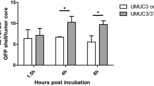
Figure 2.13 The ratio of mean fluorescence intensity (MFI) between the GFP shell and tumor core at different time points. ... 74
Figure 2.14 Mathematical modeling of S-LCP NPs (+/-AA) distribution in the core-shell spheroid and predictions of cellular uptake of S-LCP NPs (+/-AA) ... 75
Figure 2.15 Off-target distribution of NPs in infiltrating leucocytes of the desmoplastic UMUC3 xenografts. ... 76
Figure 3.1 Illustration of the Non-contact Co-culture Model for mechanistic study ... 105
Figure 3.2 Cisplatin NPs induced UMUC3/3T3 tumor resistance through elevated expression of Wnt16. ... 106
Figure 3.3 Quantification of in vivo Wnt16 protein level. ... 107
xiii
Figure 3.5 Wnt16 level in different tumor cell lines treated with cisplatin ... 109
Figure 3.6 Cisplatin NPs were delivered off-target to TAFs in the UMUC3/GFP-3T3 tumors after intravenous injection. ... 110
Figure 3.7 Pathology and treatment of PDX model ... 111
Figure 3.8 In vitro gene transfection of siWnt NPs. ... 112
Figure 3.9 Mechanistic study of Wnt16 on neighboring tumor cells. ... 113
Figure 3.10 In vitro mechanistic study of Wnt16 on neighboring stromal cells. ... 114
Figure 3.11 Dynamic tumor microenvironment remodeling by cisplatin NPs and siWnt NPs. ... 116
Figure 3.12 Blood vessel remodeling by cisplatin NPs and siWnt NPs after multiple doses. ... 118
Figure 3.14 IV injection of siWnt NPs with cisplatin NPs inhibited UMUC3/3T3 tumor growth. ... 119
Figure 3.15 IV injection of siWnt NPs (0.6 mg/kg) with cisplatin NPs (1 mg/kg) led to tumor regression when the tumor is big (volume ~700 mm3). ... 120
Figure 3.16 HE staining of major organs from 5 injections of PBS, siCont NPs, cisplatin NPs, siWnt NPs and siWnt NPs/cisplatin NPs. ... 121
Figure 3.17 Hematological test of whole blood collected from healthy nude mice treated with 5 doses of different treatments as indicated. ... 122
Figure 3.18 Diagram of the proposed mechanism ... 123
Figure 4.1 Characterization of LPD. ... 148
Figure 4.2 Cell populations that take up LPD in the stroma-vessel type tumors. ... 149
Figure 4.3 Calculation of DiI positive cells in each cell population by flow cytometry (n = 4). ... 150
Figure 4.4 Secretable TRAIL produced by fibroblasts induces apoptosis of neighboring UMUC3 tumor cells. ... 151
Figure 4.5 In vitro transfection efficiency of LPD. ... 152
Figure 4.6 Intravenous administration of sTRAIL LPD inhibited stroma-vessel UMUC3/3T3 tumor growth. ... 153
Figure 4.7 Expression of TRAIL (or fusion GFP) in the fibroblasts in situ. ... 154
Figure 4.8 Fibroblasts (in situ) that secreted TRAIL induced the apoptosis of neighboring tumor cells. ... 155
xiv
Figure 4.10 sTRAIL LPD induces the reprogramming of residual
fibroblasts and remodeling of TME. ... 157
Figure 4.11 TGF-β pathway was downregulated after sTRAIL LPD treatment ... 158
Figure 4.12 sTRAIL LPD induces normalization of blood vessel ... 159
Figure 4.13 Remodeling of TME facilitates the delivery and antitumor effect of a second-wave nanoformulated cisplatin. ... 160
Figure 4.14 Histology of orthotopic BXPC3-Luc2 xenograft. ... 161
Figure 4.15 Intravenous administration of sTRAIL LPD inhibited the orthotopic desmoplastic BXPC3 tumor growth. ... 162
Figure 4.16 Expression of GFP within different cell populations of BXPC3-Luc2 tumors after 3 doses of NPs. ... 162
Figure 4.17 Tumor environment changes after treatments in BXPC3 model. ... 163
Figure 4.18 Cytotoxicity of LPD. ... 164
Figure 4.19 Diagram of proposed mechanism... 165
Figure 5.1 Histopathology of bladder cancer ... 184
Figure 5.2 Effects of different treatments on the inhibition of fibroblast growth ... 185
Figure 5.3 Stroma depletion after single dose of combo NPs. ... 186
Figure 5.4 In vitro sensitivities of UMUC3 and NIH3T3 to GMP and cisplatin ... 187
Figure 5.5 Pharmacokinetics profiles of GMP (free or NPs) and cisplatin (free or NPs). ... 188
Figure 5.6 Tumor growth inhibition effects of different formulations on desmoplastic bladder cancer model (UMUC3/3T3) ... 189
Figure 5.7 DNA-Platinum adduct formation after combo NPs treatment ... 190
Figure 5.8 Evaluation of the tumor vessel leakiness ... 191
Figure 5.9 Effects of the combination of GMP NPs and Cisplatin NPs on VEGF expression in UMUC3/3T3 bearing mice ... 192
Figure 6.1 Diagram of PLGA NPs and mechanism of combination therapy. ... 216
Figure 6.2 TEM image of GMP cores (A) and CP cores (B). ... 217
xv
Figure 6.4 Dual-drug ratiometric loading in Combo NPs. ... 218
Figure 6.5 EE and LD of NPs ... 219
Figure 6.6 Size of PDI of NPs ... 219
Figure 6.7 Ratiometric cellular uptake and release of dual drugs from Combo NP. ... 220
Figure 6.8 Tumor inhibition effects of free drugs, Combo free, cisplatin NPs, GMP NPs, Sepa NPs and Combo NPs on a desmoplastic bladder cancer xenograft (UMUC3/3T3)... 221
Figure 6.9 Mechanistic studies of the combination therapy. ... 222
Figure 6.10 Western blot of PARP, cleaved PARP, caspase-3 and GAPDH in the tumor lysates after 3-dose treatment. ... 223
Figure 6.11 Biodistribution of Combo NPs, Sepa NPs, and Combo free in major organs 10 h post intravenous injection into desmoplastic UMUC3/3T3 bearing nude mice. ... 223
Figure 6.12 HE staining of major drug accumulating organs after three injections of treatments. .... 224
xvi
LIST OF ABBREVATIONS AND SYMBOLS
AA anisamide
ALT alanine aminotransferase αSMA alpha smooth muscle actin
AST aspartate aminotransferase
BM basement membrane
BSB Binding Site Barrier BUN blood urea nitrogen Deff diffusion coefficients
DMMA 2,3-dimethylmaleic anhydride DOPA dioleoylphosphatydic acid
DOTAP 1,2-dioleoyl-3-trimethylammonium-propane chloride salt
DOX doxorubicin
DSPE-PEG 1,2-distearoryl-sn-glycero-3-phosphoethanolamine-N-[methoxy (polyethyleneglycol-2000) ammonium salt
ECM extracellular matrix
EDS energy dispersive microscopy EPR enhanced permeation and retention FAPα fibroblasts activation protein alpha
FRET intermolecular Főrster resonance energy transfer GAG glycosaminoglycans
GFP green fluorescence protein
HA hyaluronan
HCT hematocrit
xvii
HGB hemoglobin
HSV herpes simplex virus
ICP-MS inductively coupled plasma mass spectrometry IF immunofluorescent staining
IFP interstitial fluid pressure IHC immunohistochemistry staining IRES internal ribosomal entry site ITZ isoleucine zipper
LCP NPs lipid coated calcium phosphate nanoparticles LOX lysyl oxidase
LPD lipid-coated polycation DNA complexes NER nucleotide excision repair
NPs nanoparticles
NSCLC non-small cell lung cancer
PAI-1 plasminogen activator inhibitor type 1 PDAC pancreatic ductal adenocarcinoma PSC pancreatic stellate cells
PNIPAM poly(N-isopropylacrylamide) PVC poly(vinyl chloride)
SHH sonic hedgehog Sigma R sigma receptor
TAFs tumor associated fibroblasts TAMs tumor associated macrophages
xviii TME tumor microenvironment
RBC red blood cell
RFP red fluorescence protein
sTRAIL secretable TNF-related apoptosis inducing ligand TGF-β transforming growth factor beta
TNFα tumor necrosis factor alpha VDR vitamin D receptor
VEGF vascular endothelial growth factor WBC white blood cell
1
CHAPTER 1: STROMAL BARRIERS AND STRATEGIES FOR THE DELIVERY OF
NANOMEDICINE TO DESMOPLASTIC TUMORS1
1.1 Summary
Nanoparticles (NPs) based delivery formulations have become a leading delivery strategy for cancer imaging and therapy. The success of NP-based therapy relies heavily on their ability to utilize the enhanced permeability and retention (EPR) effect and active targeting moieties to their advantage. However, these methods often fail to enable a uniform NP distribution across the tumor, and lead to insufficient local concentrations of drugs. Oftentimes, this heterogeneous drug distribution is one of the primary reasons for suboptimal treatment efficacy in NPs delivery platforms. Herein, we seek to examine the biophysical causes of heterogeneous NPs distribution in stroma-rich desmoplastic tumors; namely the abnormal tumor vasculature, deregulated extracellular matrix and high interstitial hypertension associated with these tumors, and also the off-target depletion of NPs in non-tumor stroma cells. It is suggested that these factors help explain the discrepancy between promising outlooks for many NPs formulations in preclinical studies, but suboptimal clinical outcomes for most FDA approved nanoformulations. Furthermore, examination into the role of the physicochemical properties of NPs on successful drug delivery was conducted in this chapter. In light of the many formidable barriers against successful NP drug delivery, we provided possible approaches to mitigate delivery issues from the perspective of stromal remodeling and NPs design.
1This chapter previously appeared as a review article in Journal of Controlled Release. The original
2
In all, this chapter seeks to provide guidelines for optimizing nanoparticle-based cancer drug delivery through both modified nanoparticle design and alleviation of biological barriers to successful therapy.
1.2 Introduction
3
both the trans-vascular and interstitial transport of NPs. Furthermore, the high level of extracellular molecules, increased solid stress and high interstitial pressure act as another set of barriers to successful NPs extravasation. Consequently, the limited NPs perfusion directly inhibits the therapeutic efficacy of the nanocarriers. The heterogeneity of the tumor stroma microenvironment underlines the importance of intratumoral off-target distribution of NPs to non-tumor stroma cells, since their disparate response towards treatment may be another major mechanisms behind chemotherapeutic resistance and compromised clinical outcomes [4].
Herein, we first present mathematical models for intratumoral NPs transport to achieve a better understanding of this complex process. We then discuss the key physiological barriers for NPs transport, and analyze the design of NPs for enhanced intratumoral transport. Finally, we summarize the strategies to overcome delivery barriers through remodeling the tumor microenvironment and designing the tumor microenvironment-responsive NPs.
1.3 Mathematical Modeling and In Vitro Models of NP Intratumoral Distribution
The intratumoral delivery of macromolecules and NPs requires several steps in transport, including vascular transport, transvascular transport, interstitial transport, cellular binding, internalization and metabolism (Figure 1.1) [1]. All these steps are generally limited by pathophysiology of tumors. To better understand the biophysical underpinnings of these transport barriers, Jain and his colleagues have developed several mathematical models to simulate the intratumoral behaviors of NPs [5-7].
4
proportional to hydraulic conductivity of the vessel wall, the surface area of the vessel and also the influence of interstitial fluid pressure. All parameters can be measured using standard intravital microscopy, multiphoton microscopy and optical frequency domain imaging [1, 9-12]. This modeling formula emphasizes the potential influence of blood vessel area, pore size and interstitial fluid pressure (IFP) on NPs transport.
Interstitial transport, mainly indicating the diffusion of NPs through the extracellular matrix (ECM) toward tumor cell targets, is another significant step in determining NPs diffusion and penetration. Interstitial transport follows the Darcy’s theory, which requires calculation of the diffusion coefficients (Deff) of NPs in the ECM [8, 13]. Diffusivity of NPs in the ECM was modeled in vitro using matrigel or collagen confined diffusion chamber models. Diffusion coefficients were determined using these in vitro ECM models by non-linear fits of intensity gradients to a diffusion model (e.g. Fickian model) [3, 13, 14]. Diffusion coefficients of macromolecules and liposomes can also be quantified in vivo using either single-photon fluorescence recovery after photobleaching or two photon fluorescence correlation microscopy. These measurements are more clinically relevant, but limited by equipment requirements and cost [14, 15].
5
fluorescein from NPs were further included into the differential equation. However, these mathematical equations are all confined to in vitro 3D tumor models or intratumoral injection of NPs, and do not consider the effect of vascular transport and plasma clearance. In another study, Schmidt et al. modeled the cellular binding affinity of the targeted molecules along with the dynamic plasma clearance of macromolecules into the mechanistic compartmental model, and avidly studied the effect of molecular size and binding affinity on tumor targeting [19].
Although, most of the mathematical and in vitro models are based on assumptions and limitations, the overall modeling of intratumoral transport of NPs still provides a semi-quantitative method for the extrapolation of parameters such as NPs physicochemical properties and tumoral barriers on NPs transport. This can then be extended so as to predict dynamics of NPs transport and the therapeutic outcomes of NPs delivering chemotherapy and gene therapy.
1.4 Enhanced Permeability and Retention Effect and Anti-cancer NPs in the Clinical Trials
6
1.5 Tumor Microenvironment Barriers for Intratumoral NPs Diffusion and Distribution
Ironically enough, the mechanistic basis for the EPR effect also comprises one of the primary barriers to NPs delivery. Namely, the elevated IFP [23] and increased solid stress act to inhibit successful NPs extravasation into the tumor. This paradoxical observation explains the discrepancy between promising preclinical research and the subpar clinical outcomes for NPs application. Therefore, successful NPs drug delivery relies heavily on the balance of these two competing aims (Table 1.1).
1.5.1 Abnormal Tumor Vasculature Plays Paradoxical Roles in NPs-based Delivery
When compared with non-cancerous tissues, tumor vessels are known to be heterogeneous, leaky and dilated, leaving avascular spaces of various sizes. In addition, abnormal vessel-wall structures with heterogeneous basement membranes, wide inter-endothelial junctions and large pore sizes contribute to the irregularity of the tumor vasculature [2, 24-27]. These factors therefore compromise NPs transport and undermines the efficacy of therapeutic agents.
7
velocity in tumors is independent of vessel diameter and unevenly distributed. The poorly perfused or even unperfused blood supply leads to hypoxia and acidic conditions, which bolsters drug resistance and further limits NPs diffusion [2]. Aside from blood vessel constriction, the lymphatic vessels in the tumor are also compressed by proliferating cancer cells, causing collapse. The inefficient drainage of fluid from the tumor center coupled with fluid leakage from tumor vessels contributes to interstitial hypertension, which further limits NPs perfusion deep into the tumor core [4].
1.5.2 Acidic and Hypoxia Limit Nanotherapeutic Approaches to Necrotic Areas
8
tumor drug target. Specifically, some treatments use the acidic environment to induce drug release from polymeric nanoparticles [37].
1.5.3 High Interstitial Fluid Pressure Limits NPs Convection and Accumulation
Another notable contradiction that arises when discussing the mechanism behind the EPR effect is the elevated IFP. The balance between elevated IFP and the increased NPs uptake via EPR effect influences successful NPs delivery. High IFP is known to be the result of a variety of factors. Firstly, the dense surrounding collagen matrix of the tumor microenvironment is rich in tumor associated fibroblasts (TAFs), which contract and tighten the collagen network by secreting ECM associated molecules and integrin dependent binding [23, 38]. This first barrier physically limits the expansion of the tumor cavity in response to growth. Continuous unregulated tumor proliferation in this enclosed space then compresses blood and lymphatic vessels due to growth induced solid stress, which in turn prevents the efficient discharge both NPs and interstitial fluid from the tumor [20, 39, 40]. An elevated IFP is known to be particularly detrimental for large molecule/nanoparticle delivery, which rely primarily on convection for their extravasation [26]. High IFP acts against convection and force NPs to enter via passive diffusion, a kinetically slower process. Furthermore, IFP induced vessel constriction has been shown to cause tumor hypoxia, where the increased precedent of angiogenic and growth factors contribute to lymph node metastasis and drug resistance. These secreted factors are then relocated from the tumor periphery toward the outer invasive front due to the IFP gradient where they communicate with fibroblasts to induce resistance and metastasis [41-43]. A high IFP therefore obstructs the therapeutic efficacy of NPs and leads to heterogeneous drug distribution in the tumor stroma.
1.5.4 Abnormal Extracellular Matrix Interferes with NP-based Drug Delivery
9
compressed network in turn results in the accumulation of solid stress and dictates interstitial transport [44]. At the cellular level, ECM is localized at two different intratumoral sites, the basement membrane (BM) and the interstitial matrix.
10
collagen IV provides a transient niche with leaky tumor vasculature, low and thin BM, a very beneficial window for NPs delivery. Besides the collagen meshwork, some earlier works also proposed that the extensively charged heparin sulfate chain, which is attached to the laminin/collagen IV network, is essential for the microscopic filtering of positive charged particles. Simultaneously, the nidogen molecules and the protein core of the perlecan complex geometrically hinder the negatively charged particles [51]. Overall, BM characteristics such as the matrix density, presence of proteoglycans and angiogenesis mediated BM remodeling limit the extravasation of NPs from blood vessels into the interstitium of tumors.
11
but also [54]narrows the inter-fiber spacing, retarding the movement of particles [55]. The collagen organization pattern also causes disparate NPs diffusion. Crosslinking of collagen via lysyl oxidase (LOX), regulated by fibronectin and organized by SPARC (secreted protein acidic and rich in cysteine), resulted in the stiffness of collagen fibers [55, 56]. These molecules are therefore used as interesting target candidates to inhibit stromal stiffness. With this aim, Kanapathipillai et al. designed a PLGA loaded LOX inhibitory antibodies to decrease collagen crosslinking and improve therapy [57].
The contribution of GAGs toward macromolecular diffusion is controversial. As one of the major non-sulfated GAG, hyaluronan is a linear polysaccharide with repeating disaccharide units of β-d-glucuronic acid and N-acetyl-β-D-glucosamine [54]. In most cases, the elimination of hyaluronan negatively affected nanoparticle transport, opposite to the effect of eliminating collagen [15]. The polymerization of HA has been shown to partition the collagen matrix into aqueous and viscous compartments. Hyaluronidase treatment increased the proportion of slow-diffusing compartments and shifted the slow diffusion coefficient to smaller values [58, 59]. On the other hand, for certain tumors such as pancreatic cancer, in which more than 70% ECM consists of HA, the degradation of HA resulted in increased drug diffusion [60]. The sulfated glycosaminoglycan, similar to that in the BM, carries a highly negative charge, which can inhibit the transport of macromolecules or NPs by forming aggregates [2, 61].
1.5.5 Stromal Cells Regulate the Interstitial Distribution of NPs
12
NPs, which extravasate from adjacent microvessels, compromising the internalization of therapeutic NPs in cancer cells and in turn, the overall therapeutic effect.
13
14
induced the formation of resistant phenotypes of prostate cancer [70]. Overall, the off-target delivery of NPs in TAF severely hinders NPs penetration and induced convoluted anti-tumor efficacy.
Immune cells, including B cells, T cells, granulocytes, dendritic cells, myeloid derived suppressor cells and macrophages, are indispensable constituents of the tumor microenvironment that modulate the intra-tumoral immune response [75, 76]. Owing to the partial peri-vasculature localization of infiltrated immune cells (other immune cells are likely to distribute in inflammatory hypoxia and necrosis area) [77] and phagocytic properties of some of the immune cells (e.g. macrophages) [78], off-target internalization of NPs is inevitable. Using an orthotopic model of melanoma and fluorescently labeled PRINT nanoparticles, Roode et al demonstrated that association between tumor-associated macrophages (TAM) and NPs were 4-fold greater than that of cancer cells despite TAM constituting only 1% of all cells in tumors [79]. In another study, the correlation between increased delivery and release of CKD-602 from S-CKD602 liposomes, and increased expression of CD11c-positive dendritic cells in a SKOV3 ovarian xenograft suggested that the NPs disposition may be associated with the phagocytic cells (i.e. DC and TAM) [80, 81]. Although, the off-target association of NPs in leukocytes and the general immune compartment may modulate the immune pathway (i.e. Stat-3, ERK) and modify the suppressive tumor microenvironment to synergistically improve cancer vaccines (data not shown), direct phagocytosis of NPs by phagocytic cells may deplete the NPs and limits accumulation into the tumor. Overall, the cellular components of the tumor stroma deplete NPs through multiple mechanisms and interfere with the therapeutic outcome of anti-cancer agents.
1.6 Physicochemical Properties of NPs influences NPs transport in Stroma-rich Tumors
15
better than the larger NPs (> 50 nm) [52]. The inverse relationship between diffusion rate and NPs size was also observed in vitro on the multicellular spheroid models [82]. One potential explanation is that the vascular pore size and the cross-linked collagen fiber mesh form pores that are in between the size of the large and small particles. Therefore, both transvascular and interstitial transport of smaller NPs occur rapidly [83]. However, one should note that NPs smaller than 10 nm are likely to be excreted from the kidney, at least partially. Therefore, NPs below the 10 nm limit exhibit compromised pharmacokinetic profiles and an increase in collateral damage toward normal organs. Increasing the size of NPs will provide selectivity, but at the cost of limiting extravasation and diffusion.
Aside from particle size, surface charge also affects intratumoral transport by regulating NPs’ diffusive mobility in the ECM. Both PEGylated NPs and neutrally charged liposomes exhibit quasi-free diffusive motion in ECM hydrogel and have the advantage of deep penetration into tumors. Cationic NPs (e.g. DOTAP liposomes), on the other hand, were entrapped in the hydrogel [51]. However, cationic NPs have been shown to exhibit optimized transvascular transport by preferential targeting to the tumor endothelial cells and electrostatic attraction with the negatively-charged vessel pores [84, 85].Furthermore, positively charged NPs are more likely to be taken up by proliferating cells (e.g. tumor cells) compared to neutral and negatively charged NPs, which is an additional advantage for effective drug delivery [18].
As far as the shape of NPs is concerned, research has shown that NPs or macromolecules with linear, rod-like semi-flexible configurations diffuse and penetrate more efficiently into the interstitial matrix compared with solid spherical particles of similar size. The shape of therapeutic NPs also affects their circulation time in the blood stream. For example, rod-shaped micelles have a circulation lifetime ten times longer than their spherical counterparts [2, 5, 86].
16
binding affinity may elicit a binding site barrier. This regards a phenomenon where NPs binding to target cells paradoxically reduces diffusion deep into tumors. The binding site barrier was first observed during antibody delivery into tumors and later found to be present for NPs based delivery as well. High avidity may also comprise the selectivity, since particles may also inadvertently bind to non-tumor cells expressing low levels of tumor specific determinant and depleted accordingly. After all, targeting ligands exclusive to tumor cells are unlikely to exist [87, 88].
17
NPs interact with stroma, or whether the covered biomolecules will exchange with stroma and dynamically reform a new bio-corona are also influential for NPs disposition and need more detailed investigation.
Overall, the physiological properties of NPs greatly influence the pharmacokinetics, transvascular transport, intratumoral penetration and cellular internalization in paradoxical manners. This helps to explain the inconsistency between preclinical animal studies and clinical outcomes and therefore, physicochemical properties of NPs need to be optimized for each tumor.
1.7 Strategies to Improve NPs extravasation and Penetration
Examination of the barriers that hinder NPs delivery has opened doors for new treatment regimens that seek to mediate these factors. Generally, these approaches involve restoring the abnormal tumor vasculature and interstitial stress towards that of normal tissue, and modifying NPs with environmentally responsive modifications to enhance delivery (Table 1.1).
1.7.1 Normalization of Tumor Vasculature Benefits NPs Extravasation
18
variety of monoclonal antibodies (mAb) against VEGF and other angiogenic signaling factors have been designed. For example, bevacizumab (Avastin), the first approved anti-angiogenic mAb, and its derivative, ranibizumab have been applied in the treatment of metastatic colorectal cancer. Furthermore, the inhibition of heparanase, which plays a major role in angiogenesis, has also been considered as a promising tumor priming strategy [90]. The resulting modifications reduced size of pores in the vessel walls and decreased IFP, allowing NPs extravasation to occur through convection rather than diffusion, a much faster process for free molecules and small NPs (<12 nm) [8].
19
vascular permeability. Transiently raising the systemic pressure by infusing vasoconstrictors (e.g., angiotensin II) can also increase the vascular permeability and consequently increase NPs extravasation. A combined treatment might also beneficial for hypovasculature model, with one treatment to alleviate solid stress through depletion of stromal cells or extracellular matrix, and a subsequent or concurrent vascular normalization treatment to improve perfusion [93]. Overall, the approaches for remodeling tumor vasculatures to improve NPs delivery vary with regards to vasculature contents and abnormalities, as well as the size of the therapeutic NPs. One should be cautious when choosing strategies and agents since the efficacy of treatment depends largely on the nature of each specific tumor [67].
1.7.2 Normalization of the Extracellular Matrix Improves NPs Penetration
20
stimulate collagenase synthesis and down-regulate collagen production [99, 100]. Additionally, relaxin is safer compared with bacterial collagenase for in vivo application and proposed for long term use [20].
Similar to collagen, GAG are also key matrix element that induces vascular collapse; among which, hyaluronan (HA) is a major component [96]. HA polymerizes into cage-like structures, partitioning the interstitial space into aqueous and viscous compartments as previously investigated. The use of hyaluronidase to improve the tumor permeability is controversial. High doses of hyaluronidase collapse the HA-based water swelling cage, increase the ECM viscosity and thereby reduces the diffusion coefficient of NPs [101]. Notably, elevated expression of tumor-derived hyaluronidase has been used as a diagnostic cue for high-grade bladder cancer and limited perfusion, suggesting the negative outcome of combining hyaluronidase with therapeutic NPs [59, 102]. However, in pancreatic ductal carcinoma (PDAC), with HA overpowering collagen and constituting 70% of the ECM, the opposite is observed [54]. In a genetically engineered mouse model of PDAC, PEG-PH20, a PEGylated recombinant human PH20 hyaluronidase, can effectively improve vascular perfusion of doxorubicin and gemcitabine [54]. Intratumoral administration of bovine hyaluronidases has also shown promise in several xenograft models [103, 104]. A recent study in a human osteosarcoma xenograft model indicated that hyaluronidase induced a 4-fold increase in the distribution of liposomal doxorubicin [60]. Therefore, the application of HA is not limited to small molecules and can also be a promising combinatory component to improve the delivery of NPs with larger particle size [54].
21
normal stealth particles [106]. The major concern for enzyme coated NPs is maintaining the enzymatic activity during, and after conjugation with the NPs. Also, the pharmacokinetic profile of the coated NPs proves important as well. Recent work by Ji et al examined these major concerns with enzyme coated NPs platforms and confirmed the applicability of the design for improved NPs diffusion [108].
1.7.3 Disruption of Stromal Cells Improves the Intratumoral NPs Delivery
22
23
neighboring tumor cells. Fibroblasts can be engineered into macrophage like cells, providing us hope for in situ engineering using NPs delivered agents to convert fibroblasts into natural killers [120].
1.7.4 Design of Extracellular Matrix Targeted NPs
24
than the peripheral tissues, suggesting that temperature is not only applied as an external force but also an internal physicochemical feature of the ECM [125]. Abnormal temperature gradients were also observed later in brain tumors and melanoma [126, 127]. It is suspected that the elevated temperature in tumor regions is due to glycolysis degradation and reaction, energy exhaustion [128]. Temperature dependent response is usually governed by a sharp nonlinear change in the conformation and physicochemical properties of at least one component of the nanoformulations across their phase transition temperature. The sharp response triggers the release of free drugs from the cargos. Ideally, materials exhibiting relatively sharp thermal phase transitions around body temperature would be utilized to form the thermo-response NPs [123]. The commonly used chemicals are summarized in figure, among which the thermo-responsive polymer, including poly(N-isopropylacrylamide) (PNIPAM), poly(vinyl chloride) (PVC) and their derivatives were most widely investigated. Thermo-responsive NPs were designed according to the aforementioned two strategies. Besides polymer micelles, liposome and peptide conjugates were also designed with thermal responsive components [129]. For liposomes, temperature responsiveness usually arises from a phase transition of the constituent lipids and the associated conformational variations in the lipid bilayers [123]. However, since the tumor temperature is hard to detect, most of the studies in this field focus on the in vitro characterization.
pH-25
sensitive chemical bonds (e.g., imidazole functionality of histidine, 2,3-dimethylmaleic anhydride (DMMA) modified amine moieties) [121]. Accordingly, designs of nanoformulations can be classified into two categories. One is the use of polyacids or polybase with ionizable groups that undergo conformational changes in response to environmental pH variations; the other is the design of polymeric systems with acid-sensitive bounds whose cleavage enables the release of molecules anchored at the polymer backbone. For example, a series of micellar NPs possessing ultrasensitive pH-activatable fluorescence emission were formulated by Gao’s group [132, 133]. The pH-activatable nano-probe was composed of a PEG conjugated poly(2-(hexamethyleneimino)ethyl methacrylate) copolymer and showed a micellization-disintegration transition within a narrow pH range of 0.25 units. The release of fluorescent dye from micelles in response to extracellular pH can nonlinearly amplify tumor microenvironment specific signals for imaging purposes [132].
26
ECM. A recent study by Wong et al. indicated that a multistage NPs composed of 100 nm gelatin core covered with 10 nm quantum dots showed deep tumor diffusion. The triggered release of smaller NPs upon MMP degradation lowered the diffusional hindrance in the interstitial matrix compared to larger NPs. In the meantime, the lymphatic clearance rate for 10 nm NPs was significantly lower in comparison with free small molecular drugs [3, 16].
1.8 Conclusions and Future Perspectives
27
preclinical studies but later failure in clinical trials. Further, we hope to provide guidance for the possibility of individual therapy.
Thirdly, in addition to delivering therapeutic and diagnostic NPs to solid tumors, the delivery of NPs to metastatic sites for cancer therapy is even more difficult considering the difference in TME between primary tumors and the metastatic niche. Recently, Swami et al. approached this challenge by formulating NPs to target myeloma and the bone metastatic microenvironments [139]. Furthermore, a recent study on the relationship of melanoma-derived exosomes, which induce vascular leakiness at pre-metastatic sites, may provide a means of passively targeting NPs to metastatic sites [140]. More systemic cancer treatments may require the combination of immune therapy, and other methods.
28 Table 1.1 Summary of stromal barriers and strategies
Major Barriers
Type Barriers
Constituent Barriers Mechanism Strategy Mechanisms Strategy Agents Applied Tumor Models Ref Vascula ture Angiogenesis ; Tortuosity; Low blood flow; High viscosity IFP increase; Solid stress Increase; EPR effect decrease Normalizatio n DC101 (VEGF-R mAb); bevacizumab, ranibizumab (VEGF mAb); SST0001 (Heparanase inhibitor); Trastuzumab (HER2-R mAb) Hypervasc ulature: colon carcinoma, myeloma, melanoma [2, 20, 67, 90] Leakiness Improvement Imatinib (PDGF antagonist); LY364947 (TGF-β inhibitor); Thrombin (vasoactive agents) Angiotensin II (transient vessels pressure raising agent) Hypovascu lature: pancreatic, lung and breast carcinoma [52, 67, 68]
Stroma BM Collagen IV Thickness,
mesh size, orientation, and density limit NP penetration
Degradation Collagenase;
MMPs
Lung and breast carcinoma
[48]
Nidogen Crosslinkin
g Collagen Network, and hinder the anionic NP
- Modulating the
surface charge and antifouling effect of NP
- [2,
51, 58] Perlecan
GAG Trap the
cationic NP
- -
ECM Collagen I Same as
Collagen IV
Degradation Collagenase;
Relaxin Lung carcinoma, melanoma [97-99]
HA Partitioning
collagen matrix into aqueous and viscous compartme nts Degradation (Paradox) Hyaluronidase; PEGPH20 Osteosarco ma, PDAC [54, 60] Stromal Cells
TAFs Secrete
29 Off target depletion NP TAF Depletion (Paradox)
30 Table 1.2 Design of ECM targeted NP2
Stimuli Factors Major Stimuli Structure Formulati on
Materials Stimuli Criteria
Disease Model (Cell line)
Drug In
Vitro& In Vivo Ref . Thermo Sensitiv e PNIPAA m Polymeric Micelle PNIPAAm-b-PLA LCST 36°~40°
- Dox in vitro [14
1] P(NIPAAm
-co - NDAPM)-b-PCL
LCST 36.5°
- Prednisone
Acetate
in vitro [14
2]
PNAS-b - PNIPAAm-b-PCL
LCST 36.5°
Hela Dox in vitro [14
3]
DHBCS, PEG-b -PNIPAM
LCST 32.0°
PKH26 Dox in vitro [12
1]
PNVCL Chitosan-g
-PNVCL
LCST 38.0°
L929, MCF7, PC3, KB
Curcumin in vitro [14
4, 145 ] P(mNVCL) - co-PNVCL LCST1 20~24°; LCST2 30~42° B16-F10 melanoma
Dox in vitro [14
6]
DPPC, DSPC
Liposome DPPC,
DSPC, CHOL, DSPE-PEG2000
Tm 41°
SK-BR- 3,MDA-MB-435 breast cancer; U87-MG glioma; B16F10
Dox in vivo
& in vitro (externa l) [14 7, 148 ] Leucine Zipper Peptide Lipid-peptide NP Leucine zipper peptide;DP PC, DSPC,MSP C, HSPC
Tm 40° B16F10,
SW480
Dox in vivo
& in vitro [14 9] Elastin & Elastin mimetic
NP Elastin/DN
A
aggregation
Tm 50-60°
- - - [15
0]
pH Sensitiv
e
PHis Micelle PEG-PHis-PLL or PLL-b -PEG and PHis-b -PEG Mixed pKa ~6.5; dissemb ling 4T1 breast cancer
Dox in vivo [14
2, 151 , 152 ]
PHEMA-b- HCT116 Dox in vivo [15
2PNIPAAm, poly(N-isopropylacrylamide); NDAPM, N-(3-(dimethylamino)propyl)methacrylamide; PNAS, poly(N-acroyloxysuccinimide); PNVCL, poly(N-vinylcaprolactam); DPPC, 1,2-dipalmitoyl-sn-glycero-3-phosphocholine; DSPC, 1. 1,2-distearoyl-1,2-dipalmitoyl-sn-glycero-3-phosphocholine; PAE,
31
PHis human colon & in
vitro
3]
PAE PEG-b
-(PLA-co -PAE)
pKa ~7.0
HepG2 Dox in vitro [15
4]
PEG-PAE BT-20,
B16F10 melanoma
Paclitaxel in vitro [15
5]
PC7A
PEG-b-PC7A
pKa ~6.9
A549 Fluorescen
ce
in vivo [13
2, 133 , 156 ]
PSD Polyplex
PEG-PSD/PEI NP pKa ~7.0 A2780, human ovarian carcinoma
Gene in vitro [15
7]
Chitosan NP Chitosan-silica nanosphere s pKa~6. 3; swelling MCF-7 Breast cancer
TNFα in vitro
& in vivo [15 8] Enzyme Sensitiv e MMP2- cleavabl e octapept ide
liposome PEG-MMP
cleavable peptide-PE (Gly-Pro- Leu-Gly- Ile-Ala-Gly-Gln)
MMP 4T1 breast
cancer Fluorescent probes in vitro & in vivo [15 9]
gelatin NP gelatin-gold/quant um dots fabricated multistage NP C6 glioma cells Gold NP, quantum dots in vitro & in vivo [3, 160 ] GPA peptide sequence Drug Conjugate Peptide-FAP cleavable substrate-promelittin protoxin
FAP MCF7 breast
33
CHAPTER 2: THE BINDING SITE BARRIER ELICITED BY THE TUMOR ASSOCIATED FIBROBLASTS INTERFERES DISPOSITION OF NANOPARTICLES
IN THE STROMA-VESSEL TYPE DESMOPLASTIC3
2.1 Summary
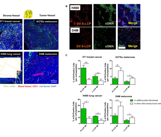
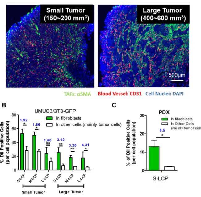
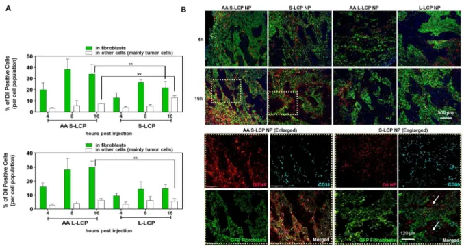
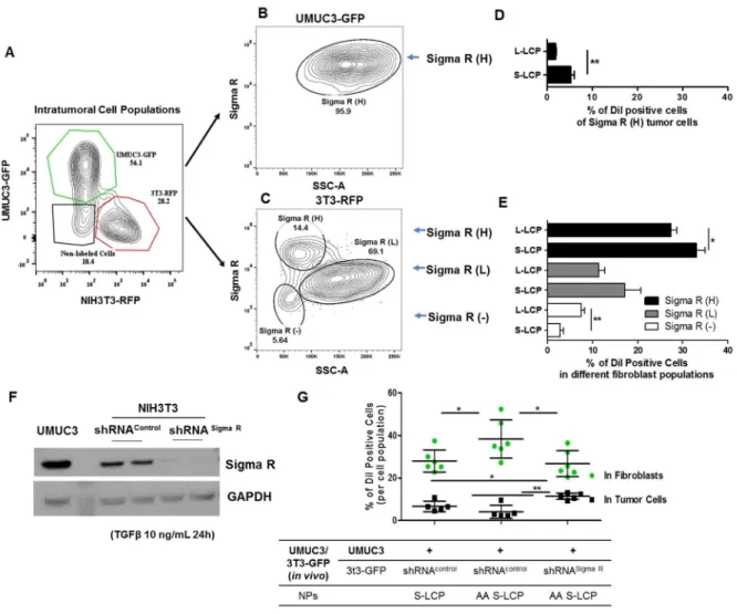
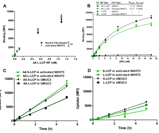
The binding site barrier (BSB) was originally described decades ago that binding of antibody to cells near the vessel prevents it from deep penetration into the tumor. It is revisited herein with respect to the intratumoral cellular disposition of nanoparticles (NPs). Specifically, BSB limits NP diffusion and results in unintended internalizations of NPs by stroma cells localized near the blood vessels. This not only limits the therapeutic outcome, but also poses the danger of off-target adverse effects. In the current study, we showed that tumor associated fibroblasts (TAFs) are the major component of the BSB, particularly in desmoplastic tumors with a stroma-vessel architecture where TAFs align with the blood vessels. Specifically, TAFs’ distance to blood vessels, expression of receptor proteins, and binding affinity affect the intensity of BSB. The physical barrier elicited by extracellular matrix also prolongs the retention of NPs in stroma, potentially contributing to BSB. The influence of particle sizes on the BSB was also investigated. The highest BSB was found with small (~18 nm) NPs targeted with the anisamide ligand. The uptake of these NPs by TAFs was about 7-fold higher than that of the other cells 16 h post intravenous injection. This was because TAFs also expressed the sigma receptor under the influence of TGF-β secreted by the tumor cells. Overall, the current study underscores the importance of BSB in the delivery of nanotherapeutics and provide rationale of exploiting BSB to target TAFs.
34 2.2 Introduction
The BSB hypothesis was originally proposed by Weinstein to explain the non-uniform distribution of monoclonal antibodies in tumor nodules [161, 162]. Specifically, he proposed that cell populations localized near the blood vessels with high antigen density and binding affinity are likely to elicit strong BSB [161, 162]. Beyond affecting antibodies, the BSB may be extended to the intratumoral dispositions of NPs [87]. Unintended binding of NPs to cells in vicinity of the blood vessels may significantly decrease the number NPs available for penetration into the tumor matrix [73]. Considering the heterogeneous stromal cell populations around the blood vessels, unintended uptake of NPs into these cells constitutes the basis of the off-target effect [81, 87].
35
Herein, the function of fibroblasts as BSB for NPs uptake was quantitatively investigated using lipid coated calcium phosphate nanoparticles (LCP NPs). LCP NPs possessed a steric surface with brush PEG coating and cationic lipid shell for enhanced uptake and release. In addition, LCP NPs demonstrated stable delivery of several modalities including macro-biomolecules and small phosphorylated drugs [170, 171]. Therefore, LCP NPs were used to evaluate tumor perfusion and predict therapeutic outcomes. By tuning the surfactant ratio, LCP NPs could also be prepared with variable sizes [172]. The influence of particle sizes on the BSB uptake was also investigated. Anisamide (AA), a model targeting ligand, was added on the surface of LCP NPs to evaluate the role of targeting ligands in dictating intratumoral cellular association of NPs [173]. To investigate fibroblasts’ role on BSB, their distance to blood vessels, expression of receptor proteins, and binding affinity were quantified. An in vitro tumor spheroid model was also established to evaluate the BSB. Based on the spheroid model, a mathematical model was created to assay the influence of each independent parameter. Overall, this study emphasizes the role of the BSB in dictating NPs delivery. Considering the large population of stromal cells, this study also investigates a platform to quantitatively evaluate the effect of the BSB on other stromal cell populations, providing guidance for NP-mediated treatment of desmoplastic tumors.
2.3 Methods and Materials
2.3.1 Materials
36
oligo DNA (sense sequence, 5’-CAAGGGACTGGAAGGCTGGG-3’) and Texas-Red labeled sense-strand oligo DNA (sequence: 5’-[TxRd]CAAGGGACTGGAAGGCTGGG-3’) were both synthesized by Sigma Aldrich (St. Louis, MO). Cholesterol, 4-methoxybenzoyl chloride, 2-bromoethylamine hydrobromide, N,N-diisopropylethylamine (DIPEA) , dichloromethane, triton X-100, Igepal CO-520 and cyclohexane were purchased from Sigma-Aldrich (St Louis, MO) without further purification.
2.3.2 Cell Culture and Animals
The human bladder transitional cell line (UMUC3) was provided by Dr. William Kim (University of North Carolina at Chapel Hill, NC). The mouse melanoma cell line D4M was a gift from Dr. Constance E. Brinckerhoff (Geisel School of Medicine at Dartmouth, NH). The mouse embryonic fibroblast cell line (NIH3T3), mouse breast cancer 4T1, human melanoma cell line A375lu, and human non-small cell lung cancer H460 were obtained from UNC Tissue Culture Facility. These cell lines were cultured in Dulbecco’s Modified Eagle’s Media (DMEM) (Invitrogen, Carlsbad, CA) (for UMUC3, A375lu, H460), Advanced MEM Media (For D4M), RMPI1640 (For 4T1) supplemented with streptomycin (100 μg/ mL) (Invitrogen), penicillin (100 U/mL), and 10% fetal bovine serum (Sigma, St. Louis, MO) respectively. NIH3T3 was cultured in DMEM supplemented with 10% Bovine calf serum (Hyclone, Logan, Utah). Cells were cultured in a humidified incubator at 37 °C with 5% CO2.
37 2.3.3 Synthesis of DSPE-PEG2000-AA
The synthesis was performed according to the previous synthetic protocol with some modifications [173]. Firstly, an aqueous solution of 2-bromoethylamine hydrobromide (1.32g, 6.4 mmol) was mixed with 4-methyoxybenzoyl chloride (1g, 5.8 mmol) in 50 mL of warm benzene to synthesize the N-(2-bromoethyl)-4-methoxy-benzamide. The mixture was shaken and cooled under running water. At the meantime, a 5% aqueous solution of sodium hydroxide was added dropwise to the mixed emulsion. The precipitate was solidified out of the reaction mixture within a few minutes to form an amorphous mass. The mixture was continued stirred for 1h. And then, the sold amide was filtered with suction and washed once with benzene and air dried for 2-3h [174]. Then, the synthesized N-(2-bromoethyl)-4-methoxy-benzamide (100 mg, 0.4 mmol) was reacted with DSPE-PEG-NH2 (100 mg, 23.3 μmol) in acetonitrile (5 mL) in the presence of DIPEA (30 μL, 0.2 mmol) at 65-70 °C for 16h. After removing the solvent by rotary evaporation, 5 mL of methanol was added to dissolve the pellet followed by precipitating with excess ether (50 ml). The mixture was then kept at -80 °C overnight. Afterwards, the precipitate was collected by centrifugation and recrystallized twice. The overall yield was 70%. The product was characterized by NMR and TLC as reported elsewhere [173].
2.3.4 Preparation of LCP NPs with Tunable Size
38
the same method as mentioned above. To tune the LCP core size, a Trition surfactant system (cyclohexane/hexanol/Trion X-100=75/0/15, v/v/v) was mixed with the original Igepal surfactant system. As the portion of the Triton surfactant system increased (from 1:0, to 1:7, as Igepal:Triton surfactant), the size of LCP core could be enlarged from 8~10 nm to ~50 nm. To create the outer leaflet coating, 100 μL of 20 mM DOTAP, 100 μL of 20 mM cholesterol and 50 μL of 20 mM of DSPE-PEG2000 (all in chloroform) were mixed with the LCP cores. After the removal of chloroform, the precipitate was suspended in a small volume of pre-warmed THF and ethanol, and then dispersed in water. As expected, when the cores size increased to 50nm, the final LCP enlarged to around 65nm in diameter. To create the AA targeted LCP NPs, 20 μL of 20mM DSPE-PEG2000-AA was mixed with 30 μL of 20mM DSPE-PEG2000 and added to the outer leaflet lipid mixture followed by the same preparation method.
Particle size and zeta potential of purified LCP NPs were measured using a Malvern ZetaSizer Nano series (Westborough, MA). The morphology of LCP cores and LCP NPs were determined by TEM microscopy (JEOL 100 CS II). The final LCP NPs were negatively stained with 2% uranyl acetate.
2.3.5 Preparation of Fluorescence-labeled LCP NPs
39
2.3.6 GFP/RFP Lentivirus Transduction in NIH3T3 Fibroblast and UMUC3
Lentiviral agents with RFP and GFP were generously provided by Dr Shawn Hingtgen (University of North Carolina at Chapel Hill, NC). The virus particles were assembled via transfecting 293FT packaging cells. The virus-containing culture medium was harvested and filtered through 0.45-μm syringe filters 48 to 96 hours post-transfection. NIH3T3 fibroblasts and UMUC3 cells were then infected by exposure to virus-containing medium for 48 hours, maintained in the complete medium, followed by selection in puromycin.
2.3.7 Sigma Receptor Silencing with Small Hairpin RNA (shRNA) in NIH3T3 Cell Lines
Sigma R shRNA lentivirus particles and control shRNA lentiviral particles were purchased from Santa Cruz. The transduction process were performed followed the protocols provided by the manufacturer. In brief, the NIH3T3 and GFP transfected NIH3T3 cells were infected by exposure to virus-containing medium for 12 to 16h, maintained in the NIH3T3 complete medium, followed by selection in puromycin. The interference of Sigma R was confirmed by western blot with pretreating the cells with TGFβ (10 ng/mL, 24h).
2.3.8 Preparation of Tumor Models
40 2.3.9 Plasma Clearance
Tumors were allowed to grow until reaching a diameter of 4–8 mm (2–3 weeks post-inoculation) for in vivo pharmacokinetics and biodistribution studies. Mice were injected intravenously via the tail vein with 3H labeled LCP NPs with similar 3H levels. At 5 min, 15 min, 30 min, 1, 2, 4, 8, 12, 24, 48, and 72 h post-injection, blood samples were collected from the saphenous vein using a heparinized capillary tube. The amounts of radioactivity in the blood and tissue samples were determined by liquid scintillation.
2.3.10 Intratumoral Distribution of LCP NPs.
3H (3H-chesterolester) -labeled LCP NPs were intravenously administered into nude mice bearing UMUC3/3T3 GFP (300 mm3) (n = 4). At determined time points, mice were euthanized. Tumor tissues were transferred to a dish and dissociated with tumor cell digestion solution (1 mg/ml collagenase A, 0.1 mg/mL HAse, 0.2 mg/mL DNAse), followed by incubation at 37°C for 40 min. Dissociated tumor cells were then collected by centrifugation. Extracellular radioactivity was collected from the supernatant. This method was validated previously to produce highly pure cytoplasmic fractions. Dissociation of the tissue samples likely leads to a certain degree of cell rupture: therefore, the amount of 3H-LCP NPs measured in the intracellular fractions may be an underestimate. However, it should be noted that all tumors harvested from each treatment groups were homogenized in the same manner. In this way, the data afforded relative comparisons with no preferential bias towards mice treated with either method.
2.3.11 Measurement of Binding Affinity of L-LCP NPs for UMUC3 and Activated NIH3T3.
41
properties of LCP to liposomes, the concentration of LCP NPs was roughly converted from the concentration of phospholipid on the basis of the approximate number of phospholipid molecules per 120 nm liposome (80,000). Then the reaction was stopped by washing twice with FACs buffer. Afterwards, the amount of cell-bound NPs was quantified by flow cytometry in BD FACs Machine. Kd values were determined by the following equation:
MFI = MFImin + MFImax( ),
where MFI=mean fluorescence intensity, MFImin=background fluorescence, MFImax was calculated from the plot. The term [NP] has been substituted for [NP-NPbound], the unbound NP concentration, based on the assumption that the concentration of bound NPs is much less than total NPs.
2.3.12 Flow Cytometry Analysis.
42
Fluor 647 conjugated anti-rabbit secondary (Abcam, Cambridge, MA) staining. All staining procedures were carefully compensated by flow cytometry. To analyze the expression of Sigma R in tumor cells and fibroblasts, and to evaluate how the expression levels correlated with the DiI-LCP NPs’ distribution, cells were stained with an anti-Sigma R antibody (Santa Cruz biotechnology, Inc.) following the same staining protocol. The UMUC3 cells were pre-transfected with red fluorescence protein (RFP) and fibroblasts were pre-transfected with green fluorescence protein (GFP) to define the cell populations (n=3~6). The flow cytometry data were analyzed using FlowJo 7.6.1 (FLOWJO, Ashland, OR). The % of DiI-positive cells per cell population was calculated according to the following equation:
% of DiI positive cells per cell population
=% of ("#$ %&'' ()('*'+,#)- + "#$% of ("#$ %&'' ()(*'+,#)- ) of the parent cells/%&'' ()(*'+,#)-∓)of the parent cells
Cell population indicates either GFP-3T3, RFP-UMUC3, Sigma R+ or CD45+ cell populations.
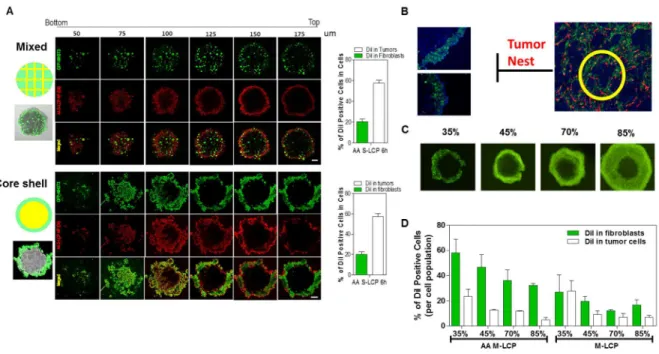
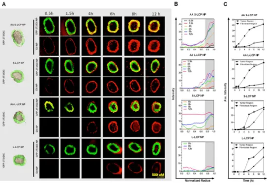
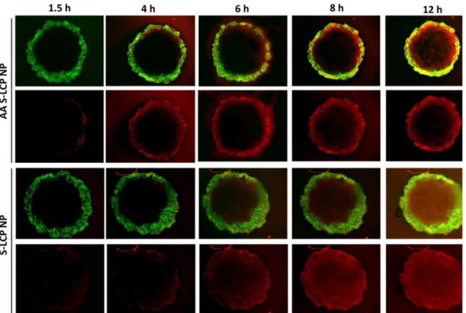
2.3.13 In vitro NP Penetration in a Core-Shell Tumor Spheroid Model.
43
and the total scanning was 90 mm in depth. Only the ones with uniform and compact core-shell structure were selected for the follow-up studies (approximatedly 20 out of the 96 well-plate). For the time-lapse assay, fluorescent images were acquired at fixed focus at determined time points. Radial fluorescence intensity profiles in the spheroids were generated using a customized script in ImageJ. Average fluorescence intensity were determined for two annular regions; the tumor region (non-fluorescence) and the fibroblast region (fluorescence region). The treated spheroids were also washed with PBS, and dissociated with collagenase and trpsinase. The DiI positive cell populations in both fibroblasts and tumor cells were quantified by Flow Cytometry.
2.3.14 Mathematical Modeling of the 3D spheroid
We model the tumor spheroids as perfect spheres (radius 1 = 50045) with radially symmetric NP concentrations (parameters shown in Table 2.2). The Stokes-Einstein diffusion coefficient in water for a particle of radius 18-5 is "8= 4.9 × 10=45>/hour. We expect the effective diffusion coefficient of NPs within the spheroid to be less than the Stokes-Einstein diffusion coefficient due to the porous ECM. We also consider that the diffusion coefficient within the fibroblast layer may be less than regions of the spheroid containing tumor cells. The equation for the extracellular concentration of NPs within the spheroid is
@A
@, = "B>A(C, ,) − FA(C, ,), 0 < C < 1,
where F is the NP absorption rate. Initially, there are no NPs within the spheroid, so we set
A(C, 0) = 0, 0 < C < 1.
Each spheroid is in a solution with a fixed concentration %IJ of NPs. Covering each sphere is a permeable barrier, possibly due to ECM, restricting the diffusive flux of NPs into the spheroid. This is modeled by imposing a boundary condition at the spheroid edge:
44
where K is the rate at which NPs enter the spheroid. Based on NP distribution within the spheroids, we find that %IJK = 2.34 ± 0.097, which did not vary significantly between spheroids or between targeted and non-targeted NPs. The averaged concentration absorbed into cells is
%PQRSTQUV(,) =3F1WX C>
Y
Z A(C, ,)[C.
The total concentration of NPs (both absorbed and extracellular) within the spheroid is
A\(C, ,) = A(C, ,) + F X A(C, ])[].
^
Z
To determine the diffusivity and the NP absorption rate, we first examine the data set for the S-LCP spheroid. We define the diffusion time _`= 1>/(4") as the time it takes for a concentration starting at the spheroid edge to diffuse inward so that the concentration at the center is roughly 80% of the value at the edge. Because the concentration is distributed relatively evenly within the tumor, we can conclude that NPs have time to diffuse throughout the spheroid before they get absorbed; in other words _`< 1/F . We find that " = 3.1 × 10=45>/hour and F = 0.25(hour/a) provides a reasonable match between the model and the data.
We next examine how the NP distribution within the UMUC3-only spheroid changes for AA S-LCP NPs. Because the AA S-LCP NPs have roughly the same hydrodynamic radius as the S-LCP NPs, we assume they have the same diffusion coefficient. The AA S-LCP NPs have a higher uptake rate than the S-LCP NPs. The distribution of NPs within the UMUC3-only spheroid is shifted toward the edge because more NPs are absorbed before they have time to diffuse very far into the spheroid. We find that a four-fold increase in the uptake rate for AA-S LCP provides a reasonable match between our model and the data.
45
stroma layer is reduced. The model prediction of the NP distribution with a 65% reduction of the diffusion coefficient in the stroma matches well with experimental data.
The mathematical model provides qualitative guidance and reasonable quantitative accuracy. Improved accuracy can be achieved by expanding the model to account for non spherical shape of tumor spheroids, heterogeneity within the spheroid, and variations in the diffusivity due to ECM and cell density. However, based on the qualitative behavior and observed timescales of absorption and diffusion, we conclude that an increased absorption rate acts as a barrier to NP penetration into the spheroid center when the average absorption time becomes less than the diffusion time (i.e., 1/F <
_`), and that this effect is amplified when the diffusion coefficient is reduced in the fibroblast layer at
the edge of the tumor.
2.3.15 Western-blot Analysis