Expanded Glucose Import Capability Affords
Staphylococcus aureus
Optimized Glycolytic Flux during Infection
Nicholas P. Vitko, Melinda R. Grosser, Dal Khatri, Thurlow R. Lance, Anthony R. Richardson
Department of Microbiology and Immunology, University of North Carolina at Chapel Hill, Chapel Hill, North Carolina, USA
N.P.V. and M.R.G. contributed equally to this report.
ABSTRACT Acquisition of numerous virulence determinants affordsStaphylococcus aureusgreater pathogenicity than other skin-colonizing staphylococci in humans. Additionally, the metabolic adaptation ofS. aureusto nonrespiratory conditions en-countered during infection (e.g., hypoxia, nitric oxide, iron chelation) has been implicated as contributing toS. aureus viru-lence. Specifically,S. aureushas been shown to ferment glycolytic substrates in nonrespiratory environments encountered within the host. Here, we show thatS. aureushas acquired unique carbohydrate transporters that facilitate the maximal uptake of host sugars and serve to support nonrespiratory growth in inflamed tissue. The carbohydrate substrates of 11S. aureus trans-porters were identified, and at least four of their genes encodeS. aureusglucose transporters (glcA,glcB,glcC, andglcU). More-over, two transporter genes (glcAandglcC) are unique toS. aureusand contribute disproportionately to the nonrespiratory growth ofS. aureuson glucose. Targeted inactivation of sugar transporters reduced glucose uptake and attenuatedS. aureusin a murine model of skin and soft tissue infections. These data expand the evidence for metabolic adaptation ofS. aureusto invasive infection and demonstrate the specific requirement for the fermentation of glucose over all other available carbohydrates. Ulti-mately, acquisition of foreign genes allowsS. aureusto adopt a metabolic strategy resembling that of infiltrating host immune cells: high glycolytic flux coupled to lactate excretion.
IMPORTANCE The bacterial pathogenStaphylococcus aureuscauses a wide range of human infections that are costly and diffi-cult to treat.S. aureusdiffers from closely related commensal staphylococci in its ability to flourish following the invasion of deeper tissue from the skin surface. There,S. aureusprimarily uses glucose to grow under respiration-limiting conditions im-posed by the immune system. It was previously unclear howS. aureusthrives in this environment when otherStaphylococcus species cannot. Our results provide evidence thatS. aureushas acquired an expanded repertoire of carbohydrate transporters. In particular, four glucose transporters contribute to efficientS. aureusgrowth during infection. Thus,S. aureushas evolved to maximize its glucose uptake abilities for enhanced glycolytic flux during tissue invasion. This dependence on glucose acquisition forS. aureusvirulence may also explain links between serious infectious complications associated with diabetic patients exhibit-ing elevated blood glucose levels.
Received23 February 2016Accepted12 May 2016Published21 June 2016
CitationVitko NP, Grosser MR, Khatri D, Lance TR, Richardson AR. 2016. Expanded glucose import capability affordsStaphylococcus aureusoptimized glycolytic flux during infection. mBio 7(3):e00296-16. doi:10.1128/mBio.00296-16.
EditorMichele S. Swanson, University of Michigan
Copyright© 2016 Vitko et al. This is an open-access article distributed under the terms of theCreative Commons Attribution 4.0 International license. Address correspondence to Anthony Richardson, [email protected].
S
taphylococcus aureus is a Gram-positive coccus that asymp-tomatically colonizes healthy human skin (1, 2). However, a compromised skin barrier or mucous membrane can lead to se-vereS. aureusinfections, including: skin and soft tissue infections (SSTIs), bacteremia, osteomyelitis, pneumonia, and toxic shock syndrome (3–5). Many other species of staphylococci (e.g.,S. epi-dermidis,S. haemolyticus,S. saprophyticus, etc.) also colonize hu-man skin but cause disease far less frequently and with less severity thanS. aureus(6). This difference has been extensively studied and is generally attributed to the combined presence of numerous unique virulence factors in theS. aureusgenome, such as toxins, adhesins, antiphagocytic factors, and protein A (7–9). Absent from this explanation is the contribution of metabolic adaptation. TheS. aureuslife cycle can plausibly be described as low-level growth on the skin surface with periodic penetration of deeper tissue environments marking a phase of enhanced growth andincreased incidence of transmission. Major physiological differ-ences between the skin surface and underlying tissue include ox-ygen concentrations, micronutrient availability, nitrogen sources, carbon sources, and pH (10–12). In general, the skin surface has lower levels of carbohydrates and peptides, relatively high levels of oxygen, and an acidic pH. Sterile tissue, on the other hand, con-tains an abundance of carbohydrates and peptides, lower levels of free oxygen, and a more neutral pH. However, invasion of sterile tissue byS. aureusleads to the activation of several innate immune responses that combine to limit bacterial respiration (e.g., iron chelation, nitric oxide [NO] production, and robust oxygen con-sumption by innate immune cells) (13–17). Thus, natural selec-tion would dictate thatS. aureushas adapted to take advantage of the unique metabolites present within sterile tissue (e.g., peptides and carbohydrates) in a manner compatible with increased resis-tance to host inflammation (i.e., respiration inhibition).
Recently, we demonstrated thatS. aureusrequires both glyco-lysis and lactate fermentation for SSTIs and bloodstream infec-tions and that only carbohydrates support the growth ofS. aureus under both high NO stress and anaerobiosis (i.e., nonrespiratory conditions) (13, 16). Additionally, the lack of abundant iron during infection limits respiration and necessitates high glyco-lytic flux coupled to lactate excretion (15, 17). This metabolic strategy, which is similar to that of activated immune cells, allows for the generation of ATP in a redox-balanced, respiration-independent manner. However, aside from the presence of a unique lactate dehydrogenase gene (ldh1) in theS. aureus ge-nome that promotes enhanced redox balancing during respi-ration inhibition, there is a lack of molecular evidence support-ing a contribution of metabolic adaptation to infection as a distinguishing characteristic ofS. aureus(13). Given the high evolutionary conservation of glycolysis among the kingdoms of life, we postulated that metabolic adaptation to promote high glycolytic flux would most easily be achieved by the acquisition of additional carbohydrate importers (18).
Bacterial carbohydrate transporters can be divided into those that modify the sugar during transport (i.e., phosphotransferase system [PTS] transporters) and those that do not (i.e., primary and secondary active transporters) (19–21). PTS transport pro-ceeds via a phosphorelay system that transfers the phosphoryl group of phosphoenolpyruvate (PEP) through a series of carrier proteins (EI and HPr) to a transporter (EII) and then on to the sugar as it is imported. PTS sugar transporters are composed of at least three subunits: EIIA, EIIB, and EIIC. The EIIA and EIIB subunits transfer the phosphoryl group from HPr to the sugar, while the EIIC subunit acts as a sugar-specific transmembrane receptor. Interestingly, the EII subunits may be encoded as indi-vidual polypeptides or fused into multisubunit proteins. PTS-dependent carbohydrate transport is unique to bacteria and is the predominant form of sugar uptake. PTS-dependent transport is also functionally linked to the transcriptional regulation of cellu-lar metabolism via carbon catabolite repression (mediated by CcpA in Gram-positive bacteria), which further contributes to overall metabolic efficiency.
In this report, we show thatS. aureusexhibits better nonrespi-ratory growth than other skin-dwelling staphylococci and par-tially attribute this phenomenon to an increased capacity for car-bohydrate import. More specifically, we identify the carcar-bohydrate substrates for 11 putative sugar transporters and demonstrate that S. aureusexhibits preferential uptake of glucose during infection as a result of the combined activities of at least four glucose trans-porters, two of which are newly acquired and therefore unique to S. aureus.
RESULTS
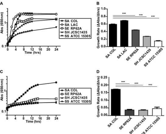
S. aureusexhibits better anaerobic growth than other staphylo-cocci.S. aureusgrows in the presence of NO levels that inhibit both respiration and the growth of other staphylococci (13). To test whether this enhanced growth behavior occurs under other nonrespiratory conditions, we compared the anaerobic growth of S. aureusstrains COL and LAC to that ofS. epidermidisRP62A, S. haemolyticusATCC 29970, andS. saprophyticusATCC 15305 in a rich medium. Both strains ofS. aureusexhibited better growth than the otherStaphylococcusspecies, as evidenced by significantly greater growth rates and terminal optical densities (ODs) (Fig. 1A and B). Next, we compared the anaerobic growth ofS. aureusCOL
to that of the other staphylococcal species in chemically defined medium (CDM) with glucose as the primary carbon source. Once again,S. aureusexhibited better anaerobic growth than the other Staphylococcusspecies (Fig. 1C and D). These data suggest that unique glycolytic and/or fermentative capabilities account for the enhanced growth ofS. aureusunder nonrespiratory conditions.
S. aureusencodes an expanded repertoire of predicted car-bohydrate transporters. One explanation for the increased growth rate ofS. aureusunder nonrespiratory conditions could be an increased capacity to import fermentable carbohydrates. To test this hypothesis, we performed a comparative genome analysis of putative carbohydrate transporters encoded byS. aureusCOL, S. aureusLAC,S. epidermidisRP62A,S. haemolyticusJCSC1435, andS. saprophyticusATCC 15305. We found thatS. aureus en-codes the largest total number of carbohydrate transporters (22), as well as the most unique carbohydrate transporters (10) (Fig. 2; see Table S1 in the supplemental material). Interestingly, 4 of the 10 uniqueS. aureusalleles are predicted to encode glucose trans-porters: SAUSA300_0191 (glcA), SAUSA300_0194, SAUSA300_ 0236 (glcC), and SAUSA300_0259 (see Table S1). Importantly, glucose is (i) largely absent from the skin surface, (ii) the most abundant free carbohydrate in human serum, and (iii) used by activated innate immune cells to both produce and resist inflam-matory radicals.
Carbohydrate uptake inS. aureusis mostly PTS dependent and contributes disproportionately to nonrespiratory growth. The majority (21/29) of putativeS. aureuscarbohydrate transport proteins are predicted to be PTS proteins. Thus, we decided to test the contribution of PTS-dependent carbohydrate transport to the nonrespiratory growth ofS. aureusby using a PTS-deficient stain ofS. aureus(ptsH-H15A). The H15A substitution in PtsH pre-vents the transfer of the phosphoryl group from EI to PtsH, thereby inhibiting PTS-dependent sugar uptake but not directly affecting interactions with CcpA, the master regulator of carbon catabolite repression (21). To confirm the efficacy of this muta-tion and identify PTS-dependent substrates, we compared the aer-obic growth of wild-type (WT) andptsH-H15A mutantS. aureus on 16 different carbohydrates. Previous studies demonstrated PTS-dependent utilization of at least seven carbohydrates (lac-tose, fruc(lac-tose, galac(lac-tose, mal(lac-tose, sucrose, glucose, and mannitol) byS. aureus(23). In line with these observations, we found that loss of PTS-dependent sugar uptake preventedS. aureusgrowth on 10 carbohydrates (mannose, fructose, galactose, mannitol, N-acetylglucosamine, N-acetylmannosamine, maltose, sucrose, trehalose, lactose, and turanose) and reduced its growth on two carbohydrates (glucose and maltotriose) but did not affect its growth on ribose (Table 1). These data show thatS. aureus carbo-hydrate utilization is largely PTS dependent.
and under metal-limited conditions (Fig. 3E). Similar results were obtained with S. aureusCOL (data not shown). Thus, the in-creased reliance ofS. aureus on carbohydrate transport during nonrespiratory growth implicates carbohydrate transporter
ac-quisition as a possible mechanism of metabolic adaption ofS. au-reusto infection.
Substrate identification for individualS. aureus PTS pro-teins.To identify the sugar specificity of individualS. aureusPTS FIG 1 S. aureusdisplays better anaerobic growth than CoNS. Anaerobic growth ofS. aureus(SA) COL and LAC,S. epidermidis(SE) RP62A,S. haemolyticus(SH) ATCC 29970, andS. saprophyticus(SS) ATCC 15305 in TSB (A) and CDM plus 25 mM glucose (C) (n⫽3). Corresponding average growth rates for TSB and CDM plus glucose are displayed in panels B and D, respectively (n⫽3; error bars show the pooled standard error of the mean). Growth rates were calculated from 2 to 4 h (S. aureusLAC) and 3 to 5 h (S. aureusCOL,S. epidermidis,S. haemolyticus, andS. saprophyticus) in TSB and from 2 to 8 h in CDM plus 25 mM glucose. Statistical significance was calculated with a Student two-sidedttest (***,Pⱕ0.001). Abs, absorbance.
FIG 2 S. aureusEncodes enhanced carbohydrate transport capability. Shown is a Venn diagram depicting the presence and conservation of putative carbohy-drate transport proteins in the genomes ofS. aureusCOL,S. epidermidisRP62A,S. haemolyticusJCSC1435, andS. saprophyticusATCC 15305.S. aureusencodes more overall transporters (n⫽22) and the highest number of unique transporters not found in any of the other species (n⫽10).
proteins, we screened mutants with insertions in all of the pre-dicted PTS protein-encoding genes for aerobic growth defects on selected carbohydrates. Previous studies identified fruA and mtlFAas encoding fructose and mannitol importers, respectively (24, 25). In support of these observations, we found that themtlF andmtlAmutants were unable to grow on mannitol, whileS. au-reusCOL, a naturalfruAmutant, exhibited poorer growth than S. aureusJE2 on fructose. Additionally, we identified PTS trans-port proteins contributing to the uptake of nine other carbohy-drates. Overall, our results link individual PTS transport proteins to the uptake of 11 of the 12 PTS-dependent sugars (Table 2). All phenotypes were confirmed in a secondS. aureusstrain (COL) background following transduction (data not shown). Consistent with the growth phenotypes of theptsH-deficient mutant, not a singleS. aureusPTS transposon mutant displayed a growth defect on glucose, suggesting that glucose uptake (i) is genetically redun-dant and (ii) likely requires both PTS and non-PTS-dependent transporters.
S. aureusglucose transport is highly redundant.To identify theS. aureusglucose transporters, four different candidate genes were mutated via allelic replacement (three PTS transporters [glcA,glcB, and SAUSA300_0236] and one non-PTS transporter [glcU]) and then combined into all possible double, triple, and quadruple mutants. The deleted genes were chosen on the basis of a combination of sequence similarity to known glucose transport-ers and high expression levels during aerobic growth on glucose (data not shown). We found that only theS. aureusquadruple mutant (⌬glcA⌬glcB⌬glcU⌬SAUSA300_0236) exhibited a sub-stantial aerobic growth defect on glucose (the quadruple mutant is referred to here asS. aureus⌬G4, and the SAUSA300_0236 gene is referred to asglcC) (Fig. 4A). Additionally, each putative glucose transporter was able to independently complement the aerobic growth defect of theS. aureus⌬G4 mutant on glucose (Fig. 4B). Lastly, we found that attenuation of⌬G4 mutant growth was fairly specific to glucose (see Table S2 in the supplemental material)
(ManNAc and GlcNAc were not tested, as GlcA and GlcC were previously implicated in their transport; see Table S1 in the sup-plemental material).
To confirm thatglcA,glcB,glcC, andglcUencode glucose trans-porters, we performed radiolabeled-glucose uptake assays with WTS. aureus, the⌬G4 mutant, and the⌬G4 mutant containing plasmids constitutively expressing each of the four glucose trans-porters. The⌬G4 mutant exhibited significantly less glucose up-take than WTS. aureus(45% of the WT level) (Fig. 4C). Comple-mentation of the ⌬G4 mutant with glcA, glcB, glcC, or glcU increased its glucose uptake to 98, 110, 105, or 56% of the WT level, respectively. The lack of significant uptake complementa-tion byglcUcould be explained by the fact thatglcUencodes a member of the glucose/ribose porter family, a family of secondary active transporters that rely on proton motive force (PMF) for energy (26, 27). The dense cell pellet conditions required to per-form these uptake assays likely have a negative impact on PMF. This would specifically decrease the activity of PMF-dependent GlcU more than that of the PEP-dependent PTS transporters GlcA, GlcB, and GlcC. Importantly, theS. aureus⌬G4 mutant is still capable of importing glucose and exhibits residual aerobic growth on glucose. These data indicate the presence of an addi-tional glucose transporter(s).
To rule out the contribution of other PTS-dependent trans-porters to S. aureusglucose uptake, we compared the aerobic growth of WT and⌬G4,ptsH-H15A⌬glcU, andptsH-H15A⌬glk mutantS. aureusCOL. Glucose kinase, encoded byglk, is respon-sible for phosphorylating intracellular glucose taken up by non-PTS transporters. Thus, withoutglk, intracellular glucose cannot be catabolized unless transported via PTS proteins. The growth of theS. aureus⌬G4 andptsH-H15A⌬glcUmutants was indistin-guishable, whereas theS. aureusptsH-H15A⌬glkmutant exhib-ited no residual growth in CDM with glucose as the primary car-bon source (see Fig. S1 in the supplemental material). These data indicate that one or several unidentified non-PTS-dependent transporters are responsible for the remainingS. aureus⌬G4 glu-cose uptake observed.
GlcA and GlcC contribute disproportionately to the non-respiratory growth ofS. aureuson glucose.Next, we compared the aerobic, anaerobic, and NO-exposed growth of the various S. aureusglucose transporter mutants in CDM with glucose as the primary carbon source. The two triple mutants lacking both unique glucose transporters (GlcA and GlcC), and thus only ex-pressing GlcB or GlcU, grew significantly more poorly than the other triple mutants, in which either GlcA or GlcC remained func-tional (Fig. 4D and E). This suggests that the unique glucose trans-porters GlcA and GlcC contribute disproportionately toS. aureus growth on glucose under nonrespiratory conditions. The ability of either GlcA or GlcC alone to individually maintain WT growth under nonrespiratory conditions cannot be explained by its ex-pression level. TheglcAtranscript levels were commensurate with those ofglcBandglcU(see Fig. S2 in the supplemental material). Moreover,glcCtranscription was less robust under all of the con-ditions tested. Furthermore, none of the glucose transporter genes responded to the presence or absence of glucose or to respiratory inhibition, with the exception ofglcC, which showed modest in-duction under anaerobiosis. Thus, other factors must explain the ability of GlcA or GlcC to fully restore growth by itself, such as translation efficiency, protein stability, and/or affinity for glucose. TABLE 1 Identification of PTS-dependent carbohydrates that support
growth ofS. aureus
Sugar
Growth of:
COL LAC
WT ptsH-H15A mutant WT ptsH-H15A mutant
Glucose ⫹⫹⫹a ⫹⫹ ⫹⫹⫹ ⫹⫹
Mannose ⫹⫹⫹ ⫹⫹⫹
Fructose ⫹ ⫹⫹⫹
Galactose ⫹ ⫹
Ascorbate
Mannitol ⫹⫹⫹ ⫹⫹⫹
Sorbitol
GlcNAc ⫹⫹ ⫹⫹
ManNAc ⫹ ⫹
Ribose ⫹⫹ ⫹⫹ ⫹ ⫹⫹
Maltose ⫹⫹⫹ ⫹⫹⫹
Sucrose ⫹⫹⫹ ⫹⫹⫹
Trehalose ⫹⫹ ⫹⫹
Lactose ⫹⫹ ⫹⫹
Turanose ⫹ ⫹
Maltotriose ⫹⫹⫹ ⫹ ⫹⫹⫹ ⫹⫹
a⫹⫹⫹, grows as well as when cultured with glucose;⫹⫹, grows to same terminal
Rich medium provides alternative carbohydrates to support nonrespiratory growth ofS. aureus.We hypothesized that the acquisition of additional glucose transporters might partially ex-plain the enhanced nonrespiratory growth phenotypes exhibited byS. aureusin both CDM and rich medium. Thus, we compared the growth of WTS. aureus(normal transport) with that of the ⌬G4 (significantly reduced glucose uptake) andptsH-H15A⌬glcU (severe defect in all carbohydrate import)S. aureus (COL and LAC) mutants in Bacto tryptic soy broth (TSB; BD; catalog no. 211825) under respiratory and nonrespiratory conditions. We ob-served almost no growth defect in theS. aureus ⌬G4 mutants under aerobic or nonrespiratory conditions in TSB (including NO stress, anaerobiosis, and metal chelation) (see Fig. S3 in the sup-plemental material). This suggests that glucose transport is non-essential for growth under nutrient-rich conditions, perhaps be-cause of the presence or uptake of other carbohydrates. In line
with this hypothesis, we observed an additive effect of the⌬glcU andptsH-H15A mutations under aerobic conditions (see Fig. S3). However, theptsH-H15A⌬glcUmutant exhibited drastic growth rate reductions under nonrespiratory conditions, including an-aerobiosis, NO·exposure, and metal chelation (see Fig. S3). These data indicate that carbon is not a limiting factor forS. aureusin TSB and that uptake of other carbohydrates can compensate for a reduction inS. aureusglucose uptake even when high glycolytic flux is required upon respiration inhibition.
Glucose uptake contributes toS. aureusvirulence in a mu-rine SSTI model.To investigate the relative contributions of glu-cose and other carbohydrates toS. aureusgrowth or survival dur-ing infection, C57BL/6 mice were subcutaneously infected with 1 ⫻107 CFU of WT or⌬G4,ptsH-H15A, ptsH-H15A⌬glcU, or
⌬glcUmutantS. aureusLAC. At 5 days postinfection, theS. aureus ⌬G4 mutant was significantly attenuated by the abscess burden FIG 3 PTS-dependent carbohydrate uptake contributes disproportionately to the nonrespiratory growth ofS. aureus. (A) Glucose yield (milligrams of glucose consumed per milligram [dry weight] of biomass) ofS. aureusCOL under respiratory and nonrespiratory (anaerobic and NO-stressed) conditions (n⫽3; error bars show the standard error of the mean).S. aureusconsumes ~3-fold more glucose per cell under nonrespiratory conditions. Statistical significance was calculated with a Student two-sidedttest (*,Pⱕ0.01). (B to E) Growth curves of WT and PTS-deficient (ptsH-H15A)S. aureusLAC in TSB under aerobic (B), anaerobic (C), NO-stressed (D), and metal-limiting (E) conditions (n⫽3). Abs, absorbance.
(~1 log), while theptsH-H15A mutant, despite losing the func-tions of three of the four glucose transporters and all other PTS carbohydrate transporters, was not significantly attenuated (Fig. 5A). Combining the⌬glcUandptsH-H15A mutations re-sulted in greater attenuation thanS. aureus⌬G4 alone (~2-log difference from the WT). This difference was not just due to mu-tation of theglcUallele, as the⌬glcUsingle mutant was also not significantly attenuated in comparison with the WT. Importantly, we observed no reversion of theptsH-H15A mutation over the course of infection.
Interestingly, when we infected mice with a dose (1⫻108CFU) that results in measurable skin lesions, theS. aureus⌬G4 mutant produced almost no abscess formation, unlike the WT (Fig. 5B). However, when grown on blood agar plates, theS. aureus⌬G4 mutant exhibited no obvious hemolysis defects (see Fig. S4 in the supplemental material). Altogether, these data suggest that glu-cose is the primary carbohydrate utilized byS. aureusduring skin infections and that other carbohydrates contribute minimally to the growth or survival ofS. aureus. This conclusion is supported by our observation that the elimination of all PTS carbohydrate transport only attenuatesS. aureuswhen glucose transport is also limited (Fig. 5A). Factors that necessitate the fermentation of glu-cose during infection include iron chelation and NO production (13, 15, 16). Additionally, as the abscess progresses, oxygen be-comes scarce, further limiting the efficiency of respiration. This becomes apparent with Hypoxyprobe staining as early as day 7 but is overwhelmingly measurable by day 12 (Fig. 5C). Thus, the com-bination of iron chelation, NO production, and hypoxia within S. aureusskin abscesses necessitates fermentative metabolism and the robust import of glucose by the bacterium.
DISCUSSION
Many variables within host tissue necessitate nonrespiratory growth, including the production of immune radicals, the
seques-tration of iron, and the inevitable hypoxia that arises at sites of inflammation because of the rapid consumption of oxygen by active immune cells.S. aureushas evolved to thrive under all of these stresses provided it has a rich source of carbohydrates, par-ticularly glucose. Under aerobic conditions, respiration contrib-utes directly to PMF, which in turn is used to generate ATP. In the absence of respiration, the only source of ATP is substrate level phosphorylation. Moreover, PMF has to be adequately main-tained by consumption of ATP and reversal of the F1F0-ATPase. Therefore, under nonrespiratory conditions,S. aureus requires enhanced glycolytic flux, as demonstrated by a⬎3-fold increase in glucose consumption (Fig. 3A).
In order to accommodate an elevated level of glycolytic flux, S. aureus must efficiently acquire host carbohydrates. Impor-tantly, glucose is the most abundant free carbohydrate in the hu-man body, and elevated host glucose levels are associated with greaterS. aureusdisease (11, 22, 28–30). However, efficient glu-cose uptake is likely difficult in inflamed tissue spaces, given that infiltrating phagocytes rapidly consume tissue glucose by running a metabolic scheme not unlike Warburg metabolism (i.e., robust glucose oxidation combined with extensive lactate secretion) (31). Infiltrating neutrophils rely very little on the trichloroacetic acid (TCA) cycle or mitochondrial respiration, likely because these energy-efficient pathways are susceptible to the reactive immune radicals produced by these immune cells. By acquiring additional glucose uptake capabilities, as well as a highly active lactate dehy-drogenase, S. aureus has distinguished itself from other skin-dwelling staphylococci and evolved to “mimic” the metabolic state of the host at sites of inflammation.
It should be noted that glucose is not the only substrate for the S. aureus-specific GlcA and GlcC transporters. We found that GlcA and GlcC are solely responsible for the uptake of GlcNAc and ManNAc, respectively, which may be indicative of a role in pepti-doglycan homeostasis. However, the selective pressure for these TABLE 2 Individual PTS Tn insertions tested for growth on a subset of utilizable carbon sources
SAUSA300 Tn insertion locus
Gene name
EII
subunit(s) Family
Phenotype on:
Glucose Mannose Fructose Galactose Mannitol GlcNAc ManNAc Maltose Sucrose Trehalose Lactose Maltotriose
None
0191 ptsG/glcAABC PTS-Glc PLa CLa
0194 BC PTS-Glc
0236 BC PTS-Glc CLa
0239 A PTS-Gat 0240 B PTS-Gat 0241 C PTS-Gat 0259 A PTS-Glc 0332 C PTS-Asc
0448 BC PTS-Glc CLa PLa
1315 ABC PTS-Fru PLa
1672 BC PTS-Glc
2105 mtlA BC PTS-Fru CLa
2107 mtlF A PTS-Fru CLa
2150 lacE BC PTS-Lac CLa CLa
2151 lacF A PTS-Lac CL CL
2270 BC PTS-Glc PLa PLa
2324 BC PTS-Glc PLa
2476 glcB ABC PTS-Glc
2576 ABC PTS-Fru PL
transporters during infection is likely their affinity for glucose. This conclusion is drawn from the fact that the⌬glcA⌬glcC mu-tant, which is completely devoid of GlcNAc or ManNAc import, is fully virulent (data not shown). Thus, a role in cell wall homeo-stasis cannot explain the maintenance of these two genes. Rather, attenuation in the animal model of skin infection requires loss of either all carbohydrate transport (ptsH-H15A⌬glcU) or specific loss of glucose transport (⌬G4) (Fig. 5A). The fact that theptsH -H15A mutant alone (unable to utilize almost all carbohydrates, with the exception of glucose) is fully virulent implies that all other carbohydrates found within the host environment are incapable of sustainingS. aureusin vivo.
In addition to meeting the energy needs of the cell, the effect of glucose onS. aureusvirulence factor regulation in the context of
infection should not be ignored. Specifically,in vitroglucose in-duces the expression ofS. aureusbiofilm-related genes (cidAand icaA) and modulates the expression of the genes for a master vir-ulence regulator (agr/RNAIII), toxins (hla,sec, andtst), and pro-tein A (spa) (32–36). This may explain the complete loss of lesion formation in mice infected with the⌬G4 mutant despite only a modest reduction in the viable CFU count (Fig. 5). Although we did not observe a loss of hemolytic activity inS. aureus⌬G4 grown in vitroon blood agar plates (see Fig. S4 in the supplemental ma-terial), this experiment is not quantitative and does not rule out a difference in the kinetics or cumulative levels of toxin production. Similarly, we found that theS. aureusptsH-H15A⌬glcUmutant exhibited normal hemolysis but displayed reduced pigment for-mation. This defect may be explained as follows: (i) slow growth of FIG 4 Contributions of the identified glucose transporters to the nonrespiratory growth ofS. aureus. (A) Aerobic growth of WT and selected double, triple, and quadrupleS. aureusCOL glucose transporter mutants in CDM plus 25 mM glucose (n⫽3). (B) Representative aerobic growth curve demonstrating comple-mentation ofS. aureusCOL⌬G4 mutant growth in CDM plus 25 mM glucose by each individual glucose transporter (n⫽3). (C) Percent [U-14C]glucose uptake
byS. aureusCOL⌬G4 relative to that of the WT, as well as⌬G4 complemented with each individual glucose transporter gene. Uptake by each strain was measured following 12 min of incubation with radiolabeled substrate and then normalized to that of the WT (n⫽4; error bars show the standard error of the mean). Statistical significance was calculated with a Student two-sidedttest (*,Pⱕ0.05; **,Pⱕ0.01; ***,Pⱕ0.001). (D and E) Nonrespiratory growth rate ofS. aureus
COL⌬G4, relative to that of the WT, compared to that of mutants expressing individual transporter genes from their native promoters. Strains were cultured anaerobically (D) or under NO stress (10 mM NOC-12–1 mM DEA-NO) (E) (n⫽3; error bars show the pooled standard error of the mean). Statistical significance was calculated with a Student two-sidedttest (**,Pⱕ0.01; ***,Pⱕ0.001). Abs, absorbance.
the mutant in TSB may delaysigBactivation of thecrtOPQMN operon, and/or (ii) reduced carbohydrate uptake may limit the intracellular availability of glucose, a required substrate for staphyloxanthin production (37, 38). Regardless, our data indi-cate that carbohydrate uptake may also contribute toS. aureus infection via regulation of virulence factor production.
To contextualize ourin vivofindings, one must also consider that respiration, iron acquisition, and the TCA cycle have all been shown to contribute toS. aureusvirulence (39–42). Thus, we can-not accurately state that inflamed tissue spaces are strictly non-respiratory. However, it is clear from our work thatS. aureus dis-plays enhanced nonrespiratory growth phenotypes and that glycolysis-based fermentation is equally required for infection. These seemingly paradoxical findings can be reconciled by con-sidering the temporal and spatial aspects of infection. For in-stance, NO production and oxygen availability are temporally regulated during S. aureus abscess development (14). Skin abscess-inducible NO synthase activity is highest 1 to 7 days after S. aureusinjection and then wanes as the infection clears. How-ever, over time, the abscesses become hypoxic (Fig. 5C). These data suggest thatS. aureususes carbohydrate-based fermentative metabolism to overcome instances of high NO exposure encoun-tered early during infection and instances of hypoxia later in in-fection. Moreover, bacteria within murine renal abscess have been shown to be relatively starved of iron (43). Thus, until bacterial numbers are reached such that efficient hemolysis releases hemo-globin into the tissue, allowingS. aureussufficient iron to respire, S. aureusmay rely on nonrespiratory metabolism to thrive, neces-sitating rapid import of glucose.
Regardless of when or whyS. aureus glycolytic and glucose transporter-deficient mutants are attenuated during infection, the fact that they exhibit any attenuation at all emphasizes the impor-tance of glucose toS. aureusdisease. In particular, this observation may partially explain the unique susceptibility of uncontrolled diabetics toS. aureusinfections (30). Diabetes is an important risk factor forS. aureusdisease, with diabetic individuals exhibiting an increased incidence and severity ofS. aureusSSTIs, bloodstream infections, and endocarditis (22, 28, 29, 44–46). If the susceptibil-ity of diabetics toS. aureusinfection is, in fact, augmented by the enhanced propensity ofS. aureusto acquire and ferment glucose, then the development of novelS. aureusglycolysis inhibitors by Kumar et al. may constitute a particularly effective treatment for diabetics withS. aureusinfections, one that both limitsS. aureus growth and reduces its destructive capacity during infection (47).
MATERIALS AND METHODS
Bacterial strains and medium.All staphylococci were cultivated in TSB or CDM, wherein the primary carbon source could be modified (48). Individual carbohydrates added to CDM were carbon balanced to 25 mM glucose for all experiments, except the NO growth assay (see explanation below). Casamino Acids were added to the CDM at 0.5%. Chloramphen-icol was added to TSB (10g/ml) and CDM (2.5g/ml) during the growth of plasmid-containing strains. All of the strains utilized in this study are listed in Table S3 in the supplemental material. All mutant strains, except the PTS insertion mutants, were generated via allelic re-placement withEscherichia coli-S. aureusshuttle vectors pBT2ts, pBTK, pBTE, and pBTS and the new vector pBTT as previously described (49). pBTT was constructed by amplifying thetetKallele fromS. aureusCOL plasmid pT181 (tet.3A and tet.3B) and then cloning it into the XmaI site of FIG 5 S. aureusglucose transporter mutants show attenuated virulence in a murine SSTI model. (A) Abscess burdens on day 5 following the subcutaneous injection of 1⫻107CFU ofS. aureusLAC (5ⱕnⱕ17; error bars show the standard error of the mean). Statistical significance was determined by analysis of
variance with multiple comparisons (*,Pⱕ0.05). (B) Lesion sizes following the subcutaneous injection of 1⫻108CFU ofS. aureusLAC (5ⱕnⱕ10; error bars
pBT2ts. The PTS insertion mutants were ordered from the Nebraska Mu-tant Transposon Library (Network on Antimicrobial Resistance in Staph-ylococcus aureus) and verified by PCR upon arrival (24). For the plasmids and primers used for mutant construction, verification, and complemen-tation, see Table S3. Importantly, all of the mutants used for virulence studies were fully transduced, with the exception of theptsH-H15A and
ptsH-H15A⌬glcUmutants. Since theptsH-H15A mutation is markerless, we constructed and verified three independentptsH-H15A mutants in the LAC background. We then separately transduced the⌬glcUmutation into each of these three mutants and verified that each mutant grew identically under aerobic conditions in glucose, Casamino acids, and TSB.
Growth curves.Staphylococcuscultures were grown overnight in TSB at 37°C with shaking at 250 rpm. For aerobic, metal-restricted, and NO-treated bacterial growth curves, overnight cultures ofS. aureus were washed twice with phosphate-buffered saline (PBS) and diluted into TSB with or without 2,2-dipyridyl (1 mM) or into CDM with or without car-bon to an initial OD at 600 nm (OD660) of 0.04. Diluted cultures were then
aliquoted into a 96-well plate (200l/well) and incubated in a Tecan Infinite M200 microplate reader set to 37°C with 1-mm orbital shaking. Growth was monitored via absorbance at 650 nm every 15 min for 24 h. For NO growth curves, 10 mM NOC-12 (Santa Cruz Biotechnology; cat-alog no. 202246) and 1 mM DEA NONOate (A. G. Scientific; catcat-alog no. D-1013) were added to the cultures at an OD650of 0.15. To extend the fermentative phase ofS. aureusNO-resistant growth, an additional, iden-tical, dose of NO donors was added to each well 1.5 h later. To ensure continued substrate availability during such prolonged NO exposure (S. aureusutilizes carbon inefficiently during NO-induced fermentation), we used 50 mM glucose for these experiments. For anaerobic growth curves, the overnight cultures were washed twice with PBS and diluted into 5 ml of prewarmed (37°C) TSB or CDM with or without carbon with or without 50 mM potassium nitrate to an OD660of 0.08. Cultures were
prepared in duplicate in 16- by 150-mm glass tubes containing 1-mm stir bars. Following dilution, cultures were immediately transferred into a Coy anaerobic chamber and grown at 37°C with stirring. Growth was moni-tored hourly by reading absorbance at 650 nm.
Growth rate and lag analysis.Growth rates were calculated with the formula⫽ ⌬ln(A650)⌬time (hours). The time intervals used for growth rate analysis are experiment specific and thus are provided in the figure legends. Lag time was calculated as the time (hours) until cultures reached an OD650of 0.2.
Glucose yield calculation.Glucose yield was measured in milligrams of glucose consumed per milligram (dry weight) of biomass forS. aureus
COL, our primary laboratory strain. Glucose consumption was moni-tored by enzymatically (R-Biopharm) determining glucose in 200-l cultures of CDM plus 25 mM glucose over a 4-h period following NO exposure or during a 4-h period during aerobic or anaerobic growth at mid-exponential phase. Dry-weight biomass was determined by vacuum filtering 100 ml of mid-exponential-phaseS. aureusCOL culture (OD660
of 1) in triplicate through a 10-cm Millipore 0.45-m-pore-size filter. The filter was then baked overnight at 65°C. Weights were averaged, and the weights of baked sterile filters were subtracted to yield an average dry weight of anS. aureuscell of ~2.8⫻10⫺13g. WhileS. aureusLAC (used for
animal experiments) exhibits similar elevated glucose consumption un-der nonrespiratory conditions, the dry-weight biomass of this strain per OD unit was not directly determined, but it is not expect to differ signif-icantly from that of COL.
Bioinformatic analysis of carbohydrate transporters. First, we searched the NCBI gene/protein and UniProt databaseS. aureusCOL,
S. aureusLAC, S. epidermidis RP62A,S. haemolyticus JCSC1435, and
S. saprophyticusATCC 15305 genomes with the keywords PTS, sugar transporter, sugar permease, carbohydrate transporter, carbohydrate per-mease, glucose, fructose, mannose, mannitol, sucrose, galactose, ascor-bate, sorbitol,N-acetylglucosamine,N-acetylmannosamine, ribose, malt-ose, trehalmalt-ose, lactmalt-ose, maltotrimalt-ose, trisaccharides, disaccharides, and monosaccharides. All of the putative carbohydrate transporters
discov-ered in this manner were then entdiscov-ered as queries in BLASTP searches against all five of the genomes mentioned above.
All of the candidate sugar transporters from this expanded search were then compiled into a list. Next, we performed forward and reciprocal BLASTP searches for each predicted protein on this list against all Staph-ylococcus genomes, as well as the transporter classification database (http://www.tcdb.org). Sequence homology was determined with an E value cutoff of 1e⫺50. Lastly, we used the ortholog predictor provided
through xBASE (http://www.xbase.ac.uk/) and visually inspected/com-pared the genomic context of each gene with MetaCyc ( http://metacy-c.org/). Homology, as depicted in Table S1 in the supplemental material, required sequence homology (i.e., an E value of⬍1e⫺50and a positive
reciprocal BLASTP result), a corresponding result from the xBASE or-tholog predictor, and a visual confirmation of shared genomic context.
Real-time qRT-PCR.S. aureusCOL was grown in 50 ml of CDM plus 25 mM glucose or Casamino Acids (0.5%) in 250-ml flasks at 37°C with shaking at 250 rpm. At an OD660of 0.5, 25 ml of each culture was added to
an equal volume of ice-cold ethanol-acetone (1:1) and frozen at⫺80°C (aerobic cultures). To assess gene expression during NO exposure, a sep-arate set of cultures (CDM plus glucose) was treated with 5 mM DETA-NONOate (Cayman Chemical; catalog no. 82120) for 1 h, quenched, and then frozen. Lastly,S. aureusCOL was grown in 50 ml of CDM plus glucose in the anaerobic chamber at 37°C with stirring. At an OD660of 0.5, 25 ml of culture was removed from the chamber in a 50-ml conical tube devoid of oxygen, immediately quenched, and then frozen. RNA was then harvested, and gene expression was analyzed as previously described (49). Transcript levels of selected genes were normalized torpoD transcript levels, which deviated very little across our experimental conditions. For the primers used for quantitative reverse transcriptase PCR (qRT-PCR) analysis, see Table S3 in the supplemental material.
Radiolabeled-glucose uptake assays. S. aureus COL strains were grown in TSB in 50-ml culture volumes to late exponential phase (OD660 of 1 to 1.2). Cells were centrifuged for 10 min at 5,000⫻gand then immediately resuspended to an OD660of 20 in warm CDM (37°C). Att0,
a mixture of glucose and [14C]glucose was added to a 1-ml aliquot of each
culture to reach final concentrations of 2 mM glucose and 100M [14
C]g-lucose. Cells were incubated in a 37°C heat block. At 12 min following glucose addition, 150l of culture was removed and immediately diluted into 900l of CDM containing 20 mM unlabeled glucose. The diluted cells were pelleted, washed once with 500l of CDM (20 mM glucose), and then resuspended in 150l of CDM (20 mM glucose). The resus-pended cells were added to a scintillation vial containing 4 ml of EcoScint A scintillation fluid (National Diagnostics). To determine the level of radioactivity in each sample, a Beckman LS6500 Multi-Purpose Scintilla-tion Counter was used to measure counts per minute.
Hemolysis assays.To detect hemolysis activity,S. aureusLAC strains (WT,⌬G4,ptsH-H15A [isolates 1 to 3], andptsH-H15A⌬glcU [isolates 1 to 3]) were streaked onto blood agar (Remel; tryptic soy agar [TSA] with sheep blood; catalog no. R01200) from freezer stocks and incubated at 37°C for 36 h. Plates were subsequently incubated at 4°C for 12 h and then imaged with a digital microscope.
Virulence assays.For virulence assessment, 6- to 8-week-old female C57BL/6 mice from The Jackson Laboratory (Bar Harbor, ME) were anes-thetized with tribromoethanol (Avertin, 0.08 mg/kg; Acros Organics; cat-alog no. 421430100) shaved (on the flank), and injected subcutaneously (on the flank) with 1⫻107CFU of WT or⌬G4,⌬glcU,ptsH-H15A, or ptsH-H15A⌬glcUmutantS. aureusLAC in 20l of sterile PBS. Impor-tantly, two separate isolates of theptsH-H15A andptsH-H15A⌬glcU mu-tants were used for infection of at least five mice apiece. On day 5, mice were euthanized and the abscesses were removed, homogenized in 500l of PBS, and dilution plated on TSA to enumerate CFU. To control for reversion of theptsH-H15A mutation during infection, WT (positive con-trol) andptsH-H15A andptsH-H15A⌬glcU mutant abscesses were plated on CDM agar plus sucrose (25 mM), incubated at 37°C for 48 h, and then inspected for colonies.
Fluorescence immunohistochemistry.The Hypoxyprobe-1 Omni kit (Hypoxyprobe Inc., Burlington, MA) was used for immunochemical detection of tissue hypoxia. Briefly, mice were injected intraperitoneally with 60 mg/kg pimonidazole HCl 30 min prior to euthanasia. Following euthanasia, infected tissues were fixed in 10% formalin, paraffin embed-ded, and sectioned (5m). Unstained sections were deparaffinized with a series of xylene and ethanol washes, followed by antigen retrieval in boil-ing 10 mM sodium citrate buffer (pH 6). Tissues were blocked with 10% donkey serum (Jackson ImmunoResearch, West Grove, PA) and subse-quently incubated with anti-Hypoxyprobe PAb2627AP (Hypoxyprobe Inc.). The primary antibody was detected by incubation with a biotinyl-ated donkey anti-rabbit antibody, followed by incubation with streptavidin-conjugated Dylight 594 (Jackson ImmunoResearch). Tissues were mounted with ProLong antifade gold containing 4=,6-diamidino-2-phenylindole (Invitrogen, Grand Island, NY) and imaged on an Olympus BX60 fluorescence microscope with iVision software v.4.0.0 (BioVision Technologies, New Minas, Nova Scotia, Canada).
SUPPLEMENTAL MATERIAL
Supplemental material for this article may be found athttp://mbio.asm.org/ lookup/suppl/doi:10.1128/mBio.00296-16/-/DCSupplemental.
Figure S1, TIF file, 1.4 MB. Figure S2, TIF file, 1.4 MB. Figure S3, TIF file, 1.4 MB. Figure S4, TIF file, 1.4 MB. Table S1, DOCX file, 0.2 MB. Table S2, DOCX file, 0.1 MB. Table S3, DOCX file, 0.2 MB.
ACKNOWLEDGMENTS
This work was supported by NIH grants from the Institute of Allergy and Infectious Diseases (R01-AI093613 and R21-AI111707), a Pew Biomedi-cal Scholars award (A12-0105), and two American Heart Association pre-doctoral fellowships (13PRE15200002 to N.P.V. and 13PRE19830003 to M.R.G.).
Mutants defective in each PTS system were obtained from the Ne-braska Transposon Mutant Library, Omaha, NE.
FUNDING INFORMATION
This work, including the efforts of Anthony R. Richardson, was funded by HHS | NIH | National Institute of Allergy and Infectious Diseases (NIAID) (R01-AI093613 and R21-AI111707). This work, including the efforts of Nicholas P. Vitko and Melinda Rose Grosser, was funded by American Heart Association (AHA) (13PRE15200002 and 13PRE19830003).
The funders had no role in study design, data collection and interpreta-tion, or the decision to submit the work for publication.
REFERENCES
1.Graham PL, Lin SX, Larson EL. 2006. A U.S. population-based survey of Staphylococcus aureus colonization. Ann Intern Med144:318 –325.
http://dx.doi.org/10.7326/0003-4819-144-5-200603070-00006. 2.Schechter-Perkins EM, Mitchell PM, Murray KA, Rubin-Smith JE,
Weir S, Gupta K. 2011. Prevalence and predictors of nasal and extranasal staphylococcal colonization in patients presenting to the emergency de-partment. Ann Emerg Med 57:492– 499.http://dx.doi.org/10.1016/ j.annemergmed.2010.11.024.
3.Naimi TS, LeDell KH, Como-Sabetti K, Borchardt SM, Boxrud DJ, Etienne J, Johnson SK, Vandenesch F, Fridkin S, O’Boyle C, Danila RN, Lynfield R. 2003. Comparison of community- and health care-associated methicillin-resistant Staphylococcus aureus infection. JAMA 290: 2976 –2984.http://dx.doi.org/10.1001/jama.290.22.2976.
4.Moore CL, Hingwe A, Donabedian SM, Perri MB, Davis SL, Haque NZ, Reyes K, Vager D, Zervos MJ. 2009. Comparative evaluation of epide-miology and outcomes of methicillin-resistant Staphylococcus aureus (MRSA) USA300 infections causing community- and healthcare-associated infections. Int J Antimicrob Agents34:148 –155.http:// dx.doi.org/10.1016/j.ijantimicag.2009.03.004.
5.Schlievert PM, Shands KN, Dan BB, Schmid GP, Nishimura RD. 1981. Identification and characterization of an exotoxin from Staphylococcus aureus associated with toxic-shock syndrome. J Infect Dis143:509 –516.
http://dx.doi.org/10.1093/infdis/143.4.509.
6.Becker K, Heilmann C, Peters G. 2014. Coagulase-negative staphylo-cocci. Clin Microbiol Rev27:870 –926.http://dx.doi.org/10.1128/ CMR.00109-13.
7.Peacock SJ, Moore CE, Justice A, Kantzanou M, Story L, Mackie K, O’Neill G, Day NP. 2002. Virulent combinations of adhesin and toxin genes in natural populations ofStaphylococcus aureus. Infect Immun70: 4987– 4996.http://dx.doi.org/10.1128/IAI.70.9.4987-4996.2002. 8.Foster TJ, Höök M. 1998. Surface protein adhesins of Staphylococcus
aureus. Trends Microbiol6:484 – 488.http://dx.doi.org/10.1016/S0966 -842X(98)01400-0.
9.Otto M. 2014. Science directStaphylococcus aureustoxins. Curr Opin Microbiol17:32–37.http://dx.doi.org/10.1016/j.mib.2013.11.004. 10. Harvey CJ, LeBouf RF, Stefaniak AB. 2010. Formulation and stability of
a novel artificial human sweat under conditions of storage and use. Toxi-col In Vitro24:1790 –1796.http://dx.doi.org/10.1016/j.tiv.2010.06.016. 11. Psychogios N, Hau DD, Peng J, Guo AC, Mandal R, Bouatra S,
Sinelnikov I, Krishnamurthy R, Eisner R, Gautam B, Young N, Xia J, Knox C, Dong E, Huang P, Hollander Z, Pedersen TL, Smith SR, Bamforth F, Greiner R, McManus B, Newman JW, Goodfriend T, Wishart DS. 2011. The human serum metabolome. PLoS One6:e16957.
http://dx.doi.org/10.1371/journal.pone.0016957.
12. Kutyshenko VP, Molchanov M, Beskaravayny P, Uversky VN, Tim-chenko MA. 2011. Analyzing and mapping sweat metabolomics by high-resolution NMR spectroscopy. PLoS One6:e28824.http://dx.doi.org/ 10.1371/journal.pone.0028824.
13. Richardson AR, Libby SJ, Fang FC. 2008. A nitric oxide-inducible lactate dehydrogenase enables Staphylococcus aureus to resist innate immunity. Science319:1672–1676.http://dx.doi.org/10.1126/science.1155207. 14. Thurlow LR, Joshi GS, Clark JR, Spontak JS, Neely CJ, Maile R,
Richardson AR. 2013. Functional modularity of the arginine catabolic mobile Element contributes to the success of USA300 methicillin-resistant Staphylococcus aureus. Cell Host Microbe13:100 –107.http://dx.doi.org/ 10.1016/j.chom.2012.11.012.
15. Friedman DB, Stauff DL, Pishchany G, Whitwell CW, Torres VJ, Skaar EP. 2006. Staphylococcus aureus redirects central metabolism to increase iron availability. PLoS Pathog 2:e87. http://dx.doi.org/10.1371/ journal.ppat.0020087.
16. Vitko NP, Spahich NA, Richardson AR. 2015. Glycolytic dependency of high-level nitric oxide resistance and virulence inStaphylococcus aureus. mBio6:e00045-15.http://dx.doi.org/10.1128/mBio.00045-15.
17. Hammer ND, Skaar EP. 2011. Molecular mechanisms of Staphylococcus aureus iron acquisition. Annu Rev Microbiol 65:129 –147.http:// dx.doi.org/10.1146/annurev-micro-090110-102851.
18. Peregrín-Alvarez JM, Sanford C, Parkinson J. 2009. The conservation and evolutionary modularity of metabolism. Genome Biol10:R63.http:// dx.doi.org/10.1186/gb-2009-10-6-r63.
19. Dills SS, Apperson A, Schmidt MR, Saier MH. 1980. Carbohydrate transport in bacteria. Microbiol Res44:385– 418.
20. Postma PW, Lengeler JW, Jacobson GR. 1993. Phosphoenolpyruvate: carbohydrate phosphotransferase systems of bacteria. Microbiol Rev57: 543–594.
21. Kotrba P, Inui M, Yukawa H. 2001. Bacterial phosphotransferase system (PTS) in carbohydrate uptake and control of carbon metabolism. J Biosci Bioeng92:502–517.http://dx.doi.org/10.1016/S1389-1723(01)80308-X. 22. Muller LM, Gorter KJ, Hak E, Goudzwaard WL, Schellevis FG,
Hoe-pelman AI, Rutten GE. 2005. Increased risk of common infections in patients with type 1 and type 2 diabetes mellitus. Clin Infect Dis41: 281–288.http://dx.doi.org/10.1086/431587.
23. Egan JB, Morse ML. 1965. Carbohydrate transport in Staphylococcus aureusI. Genetic and biochemical analysis of a pleiotropic transport mu-tant. Biochim Biophys Acta97:310 –319.http://dx.doi.org/10.1016/0304 -4165(65)90096-6.
24. Fey PD, Endres JL, Yajjala VK, Widhelm TJ, Boissy RJ, Bose JL, Bayles KW. 2013. A genetic resource for rapid and comprehensive phenotype screening of nonessentialStaphylococcus aureusgenes. mBio4:e00537–12.
http://dx.doi.org/10.1128/mBio.00537-12.
sugar-driven bacterial growth in airway surface liquid. Cell Mol Life Sci71: 4665– 4673.http://dx.doi.org/10.1007/s00018-014-1635-y.
26. Castro R, Neves AR, Fonseca LL, Pool WA, Kok J, Kuipers OP, Santos H. 2009. Characterization of the individual glucose uptake systems of
Lactococcus lactis: mannose-PTS, cellobiose-PTS and the novel GlcU per-mease. Mol Microbiol71:795– 806.http://dx.doi.org/10.1111/j.1365 -2958.2008.06564.x.
27. Jack DL, Yang NM, Saier MH, Jr.2001. The drug/metabolite transporter superfamily. Eur J Biochem268:3620 –3639.http://dx.doi.org/10.1046/ j.1432-1327.2001.02265.x.
28. Fowler VG, Miro JM, Hoen B, Cabell CH, Abrutyn E, Rubinstein E, Corey GR, Spelman D, Bradley SF, Barsic B, Pappas PA, Anstrom KJ, Wray D, Fortes CQ, Anguera I, Athan E, Jones P, van der Meer JTM, Elliott TSJ, Levine DP, Bayer AS, ICE MD Investigators. 2005. Staphy-lococcus aureusendocarditis: a consequence of medical progress. JAMA 293:3012–3021.http://dx.doi.org/10.1001/jama.293.24.3012.
29. Movahed MR, Hashemzadeh M, Jamal MM. 2007. Increased prevalence of infectious endocarditis in patients with type II diabetes mellitus. J Dia-betes Complications 21:403– 406. http://dx.doi.org/10.1016/ j.jdiacomp.2007.07.003.
30. Kourany WM, Miro JM, Moreno A, Corey GR, Pappas P, Abrutyn E, Hoen B, Habib G, Fowler V, Jr., Sexton DJ, Olaison L, Cabell CH, ICE MD Investigators. 2006. Influence of diabetes mellitus on the clinical manifestations and prognosis of infective endocarditis: a report from the International Collaboration on Endocarditis-Merged Database. Scand J Infect Dis38:613– 619.http://dx.doi.org/10.1080/00365540600617017. 31. Cheng S-C, Quintin J, Cramer RA, Shepardson KM, Saeed S, Kumar V,
Giamarellos-Bourboulis EJ, Martens JH, Rao NA, Aghajanirefah A, Manjeri GR, Li Y, Ifrim DC, Arts RJW, van der Veer BM, Deen PMT, Logie C, O’Neill LA, Willems P, van de Veerdonk FL, van der Meer JWM, Ng A, Joosten LAB, Wijmenga C, Stunnenberg HG, Xavier RJ, Netea MG. 2014. mTOR- and HIF-1-mediated aerobic glycolysis as met-abolic basis for trained immunity. Science345:1250684.http://dx.doi.org/ 10.1126/science.1250684.
32. Seidl K, Müller S, François P, Kriebitzsch C, Schrenzel J, Engelmann S, Bischoff M, Berger-Bächi B. 2009. Effect of a glucose impulse on the CcpA regulon in Staphylococcus aureus. BMC Microbiol 9:95.http:// dx.doi.org/10.1186/1471-2180-9-95.
33. Seidl K, Goerke C, Wolz C, Mack D, Berger-Bächi B, Bischoff M. 2008.
Staphylococcus aureusCcpA affects biofilm formation. Infect Immun76: 2044 –2050.http://dx.doi.org/10.1128/IAI.00035-08.
34. Seidl K, Bischoff M, Berger-Bächi B. 2008. CcpA mediates the catabolite repression oftstinStaphylococcus aureus. Infect Immun76:5093–5099.
http://dx.doi.org/10.1128/IAI.00724-08.
35. Duncan JL, Cho GJ. 1972. Production of staphylococcal alpha toxin. II. Glucose repression of toxin formation. Infect Immun6:689 – 694. 36. Regassa LB, Novick RP, Betley MJ. 1992. Glucose and nonmaintained
pH decrease expression of the accessory gene regulator (agr) in Staphylo-coccus aureus. Infect Immun60:3381–3388.
37. Pelz A, Wieland K-P, Putzbach K, Hentschel P, Albert K, Götz F. 2005. Structure and biosynthesis of staphyloxanthin fromStaphylococcus
au-reus. J Biol Chem280:32493–32498. http://dx.doi.org/10.1074/ jbc.M505070200.
38. Katzif S, Lee EH, Law AB, Tzeng YL, Shafer WM. 2005. CspA regulates pigment production inStaphylococcus aureusthrough a SigB-dependent mechanism. J Bacteriol187:8181– 8184.http://dx.doi.org/10.1128/ JB.187.23.8181-8184.2005.
39. Hammer ND, Reniere ML, Cassat JE, Zhang Y, Hirsch AO, Indriati Hood M, Skaar EP. 2013. Two heme-dependent terminal oxidases power
Staphylococcus aureusorgan-specific colonization of the vertebrate host. mBio4:e00241-13.http://dx.doi.org/10.1128/mBio.00241-13.
40. Pishchany G, McCoy AL, Torres VJ, Krause JC, Crowe JE, Jr., Fabry ME, Skaar EP. 2010. Specificity for human hemoglobin enhances Staph-ylococcus aureusinfection. Cell Host Microbe 8:544 –550. http:// dx.doi.org/10.1016/j.chom.2010.11.002.
41. Spahich NA, Vitko NP, Thurlow LR, Temple B, Richardson AR. 5 February 2016.Staphylococcus aureuslactate- and malate-quinone oxi-doreductases contribute to nitric oxide resistance and virulence. Mol Mi-crobiol.http://dx.doi.org/10.1111/mmi.13347.
42. Kinkel TL, Roux CM, Dunman PM, Fang FC. 2013. TheStaphylococcus aureusSrrAB two-component system promotes resistance to nitrosative stress and hypoxia. mBio4:e00696 –13.http://dx.doi.org/10.1128/ mBio.00696-13.
43. Reniere ML, Skaar EP. 2008. Staphylococcus aureus haem oxygenases are differentially regulated by iron and haem. Mol Microbiol69:1304 –1315.
http://dx.doi.org/10.1111/j.1365-2958.2008.06363.x.
44. Chu VH, Cabell CH, Benjamin DK, Kuniholm EF, Fowler VG, Enge-mann J, Sexton DJ, Corey GR, Wang A. 2004. Early predictors of in-Hospital death in infective endocarditis. Circulation109:1745–1749.
http://dx.doi.org/10.1161/01.CIR.0000124719.61827.7F.
45. Lipsky BA, Tabak YP, Johannes RS, Vo L, Hyde L, Weigelt JA. 2010. Skin and soft tissue infections in hospitalised patients with diabetes: cul-ture isolates and risk factors associated with mortality, length of stay and cost. Diabetologia53:914 –923.http://dx.doi.org/10.1007/s00125-010 -1672-5.
46. Federspiel JJ, Stearns SC, Peppercorn AF, Chu VH, Fowler VG. 2012. Increasing US rates of endocarditis with Staphylococcus aureus: 1999 –2008. Arch Intern Med172:363–365.http://dx.doi.org/10.1001/ archinternmed.2011.1027.
47. Kumar NS, Dullaghan EM, Finlay BB, Gong H, Reiner NE, Jon Paul Selvam J, Thorson LM, Campbell S, Vitko N, Richardson AR, Zoraghi R, Young RN. 2014. Discovery and optimization of a new class of pyruvate kinase inhibitors as potential therapeutics for the treatment of methicillin-resistant Staphylococcus aureus infections. Bioorg Med Chem 22: 1708 –1725.http://dx.doi.org/10.1016/j.bmc.2014.01.020.
48. Vitko NP, Richardson AR. 2013. Laboratory maintenance of methicillin-resistant Staphylococcus aureus (MRSA). Curr Protoc Microbiol Chapter 9:Unit 9C.2.http://dx.doi.org/10.1002/9780471729259.mc09c02s28. 49. Crooke AK, Fuller JR, Obrist MW, Tomkovich SE, Vitko NP,
Richard-son AR. 2013. CcpA-independent glucose regulation of lactate dehydro-genase 1 inStaphylococcus aureus. PLoS One8:e54293.http://dx.doi.org/ 10.1371/journal.pone.0054293.