Vpr Enhances HIV-1 Env Processing and Virion Infectivity in
Macrophages by Modulating TET2-Dependent IFITM3
Expression
Qi Wang,
a,bLishan Su
a,baLineberger Comprehensive Cancer Center, University of North Carolina at Chapel Hill, Chapel Hill, North Carolina, USA bDepartment of Microbiology & Immunology, The University of North Carolina at Chapel Hill, Chapel Hill, North Carolina, USA
ABSTRACT
HIV-1 Vpr enhances viral replication in human macrophages via
multi-ple mechanisms that are not clearly defined. It does not affect HIV-1 virion
produc-tion during the first round of infecproduc-tion. We have recently discovered that Vpr targets
the DNA demethylase TET2 for degradation, which leads to sustained interleukin-6
(IL-6) expression and elevated HIV-1 replication. We report here that Vpr enhanced
Env processing in infected macrophages, associated with increased Env
incorpora-tion into virions with higher infectivity. Interestingly, IFITM3 was constitutively
ex-pressed in macrophages in a TET2-dependent fashion. We showed that
Vpr-enhanced Env processing depended genetically on TET2 and IFITM3. We further
showed that Vpr reduced IFITM3 expression by reducing demethylation of the
IF-ITM3 promoter in macrophages, associated with degradation of TET2 and reduced
TET2 binding to the IFITIM3 promoter. Our findings indicate that the Vpr-TET2 axis
enhances HIV-1 replication in macrophages via two independent mechanisms:
re-duced IFTIM3 expression to enhance Env processing and virion infectivity and
sus-tained IL-6 expression to increase HIV-1 replication. The Vpr-TET2 axis may provide a
novel target to develop therapeutics to inhibit HIV-1 infection and pathogenesis.
IMPORTANCE
How Vpr enhances HIV-1 replication in macrophages is still unclear.
We report here that Vpr enhanced HIV-1 Env processing during the first round of
HIV-1 replication, resulting in virions with higher Env incorporation and viral
infectiv-ity. These higher-quality viral particles contributed to elevated infection during the
second round and spreading infection in macrophages and other HIV-1 target cells.
We have recently discovered that TET2 is a novel host factor degraded by Vpr,
which leads to sustained IL-6 expression in macrophages. Interestingly,
Vpr-enhanced HIV-1 Env processing depended on both the IFITIM3 and TET2 genes. The
constitutive expression of IFITIM3 expression in macrophages was maintained by
TET2, which demethylated the IFITIM3 promoter. We conclude that the Vpr degrades
TET2 to enhance HIV-1 replication in macrophages by reducing IFITIM3 expression to
increase viral Env processing, virion incorporation, and infectivity and by sustaining
IL-6 expression to increase HIV-1 gene expression. The Vpr-TET2 axis may serve as a
novel target to develop anti-HIV drugs to inhibit HIV-1 infection and pathogenesis.
KEYWORDS
demethylation, Env processing, HIV-1 infectivity, IFITM3, TET2, Vpr
H
IV-1 infection of macrophages contributes to its persistent infection in HIV-1
patients with highly active antiretroviral therapy (HAART), bringing complexity to
viral eradication for HIV-1 cure (1). Although macrophages are relatively resistant to
HIV-1 infection compared to activated CD4
⫹T cells (2), HIV-1 has evolved to efficiently
infect macrophages, which is enhanced by HIV-1 viral proteins such as viral protein R
(Vpr) to counteract macrophage-specific restrictions (3, 4). Vpr, a small 14-kDa protein,
CitationWang Q, Su L. 2019. Vpr enhances HIV-1 Env processing and virion infectivity in macrophages by modulating TET2-dependent IFITM3 expression. mBio 10:e01344-19.https:// doi.org/10.1128/mBio.01344-19.
Invited EditorChristopher Aiken, Vanderbilt University Medical Center
EditorTerence S. Dermody, University of Pittsburgh School of Medicine
Copyright© 2019 Wang and Su. This is an open-access article distributed under the terms of theCreative Commons Attribution 4.0 International license.
Address correspondence to Lishan Su, [email protected].
Received29 May 2019
Accepted24 July 2019
Published
RESEARCH ARTICLE
Host-Microbe Biology
is functionally critical for HIV-1 pathogenicity
in vivo
(5). Vpr has been reported to
promote infection of nondividing cells, especially macrophages (6). However, when
virus entry, reverse transcription (RT), integration, gene expression, virion assembly
and budding, or total virions are measured, Vpr does not affect the first-round
replication of HIV-1 in macrophages. Multiple mechanisms seem to contribute to
Vpr-enhanced HIV-1 replication, including more infectious virions and
cytokine-dependent effects (7, 8).
The exact mechanism of Vpr-enhanced HIV-1 replication in macrophages, however,
remains elusive. Vpr is believed to facilitate HIV-1 replication through its interaction
with VprBP (9), a most abundant CUL4 binding partner first discovered by its binding
with Vpr (also known as DCAF1) (10, 11). Several host proteins have been reported to
be targeted by Vpr for ubiquitylation by the CRL4
VprBPE3 ligase, including MCM10 (12),
UNG2 (13), and MUS81 (14). Recently, we discovered that Vpr targets the DNA
demeth-ylase TET2, which functions as a repressor to resolve induction of the interleukin-6 (IL-6)
gene in HIV-1-infected macrophages (15). TET2 deactivates gene expression through
recruitment of the histone deacetylase (HDAC) complex to promoter DNA (16). In
macrophages, Vpr-induced TET2 depletion prevents efficient resolution of IL-6
induc-tion during HIV-1 infecinduc-tion, which enhances HIV-1 infecinduc-tion in macrophages.
Interest-ingly, the TET2 dioxygenase activity is not required for the suppression of IL-6 gene
expression during its resolution phase (16).
In mammalian cells, the majority of CpG dinucleotides outside the CpG islands (CGIs)
are methylated at the C-5 position of cytosine (5mC) throughout the genome to stably
maintain intergenic and heterochromatic regions in a transcriptionally inert chromatin
state. CGIs, on the other hand, are associated with many (
⬃
70%) promoters (17) and,
when methylated, are associated with gene silencing. TET methylcytosine dioxygenases
(TET1, -2, and -3 in mammalian cells [18, 19]) catalyze three steps of iterative oxidation,
first converting 5mC to 5-hydroxymethyl cytosine (5hmC), then 5hmC to 5-formyl
cytosine (5fC), and finally 5fC to 5-carboxy cytosine (5caC). 5caC can be removed by
DNA glycosylase TDG, resulting in 5-unmodified cytosine (20, 21). TET2 is thus a
dioxygenase that catalyzes oxidative decarboxylation of
␣
-KG, creating a highly reactive
intermediate that converts 5mC to 5hmC (22) and activates gene expression through
promotion of DNA demethylation of their promoters (23).
We report here that Vpr enhanced Env processing, associated with increased HIV-1
infectivity during the first round of infection in macrophages. Vpr-enhanced Env
processing depended genetically on TET2 and IFITM3, which is constitutively expressed
in macrophages in a TET2-dependent fashion. We further showed that Vpr reduced
IFITM3 expression by degrading TET2 in macrophages, associated with reduced
de-methylation of the IFITM3 promoter. We demonstrate that the Vpr-TET2 axis enhanced
HIV-1 replication in macrophages via two independent mechanisms: (i) reduced IFTIM3
expression to enhance Env processing and virion infectivity and (ii) sustained IL-6
expression to increase HIV-1 replication.
RESULTS
Vpr enhances HIV-1 Env processing and virion infectivity during the first round
of replication in macrophages.
We investigated the role of Vpr in enhancing HIV-1
replication in human primary macrophages. As previously reported (6), we observed
that macrophage-tropic Vpr
⫹HIV-1 or Vpr
⫺HIV-1 infected and replicated to similar
levels during the first cycle of infection at 2 days postinfection (dpi) in
monocyte-derived macrophages (MDMs). However, Vpr
⫹HIV-1 showed elevated levels of
repli-cation at 4 dpi as determined by HIV-p24 enzyme-linked immunosorbent assay (ELISA)
(Fig. 1A) or by intracellular HIV-1 p24 staining (see Fig. S1 in the supplemental material).
To confirm that the first cycle of HIV-1 replication was not affected by Vpr, we added
reverse transcriptase inhibitor nevirapine (NVP) at 2 dpi to block second-round HIV-1
infection. We found that Vpr enhanced viral replication at 4 dpi, but failed to do so
when NVP was added at 2 dpi (Fig. 1A and Fig. S1).
To explore the mechanism by which Vpr enhances second-round viral replication,
we examined processing of HIV-1 viral proteins in MDMs infected with Vpr
⫹or Vpr
⫺HIV-1 during first-round infection. As shown in Fig. 1B and C, the ratio of gp120 to
gp160 or gp120/p24 in Vpr
⫹HIV-1 infected MDMs was
⬃
5-fold higher than that of
MDMs infected with Vpr
⫺HIV-1. We confirmed the results with MDMs derived from
three different donors, and the enhanced ratio of gp120 to gp160 or gp120/p24 by Vpr
is summarized in Fig. 1C. Consistently, HIV-1 virions produced from MDM infected with
Vpr
⫹HIV-1 showed 3-fold-higher levels of HIV-1 Env proteins (Fig. 1D and E), as
previously reported (7). When the relative infectivity was measured, HIV-1 virions
produced from MDM infected with Vpr
⫹HIV-1 showed 2.5-fold-higher infectivity by
TZM-bl luciferase indicator assay (Fig. 1F, relative luciferase activity/ng p24), or by the
MAGI (multinuclear activation of galactosidase indicator) infectious unit (IU) assay. We
showed that Vpr
⫹HIV-1 virions contained 4
⫻
10
3IU/ng p24 and Vpr
⫺HIV-1 virions
contained 2
⫻
10
3IU/ng p24 (Fig. 1G, relative IU/ng p24). We further showed that
virions produced from MDMs infected with Vpr
⫹HIV-1 were more resistant to inhibition
by soluble CD4 (sCD4) than Vpr
⫺HIV-1 virions produced from the first round of viral
replication in MDMs. Vpr
⫺HIV-1 virions were
⬃
10
⫻
more sensitive to sCD4 inhibition,
with a 50% inhibitory concentration (IC
50) of 0.034
g/ml, whereas Vpr
⫹HIV-1 virions
were relatively refractory to sCD4 inhibition, with an IC
50of 0.366
g/ml (see Fig. S2 in
the supplemental material). These results indicate that Vpr enhances Env processing
FIG 1 Vpr enhances Env processing and virion infectivity in MDMs. (A) Vpr has no effect on first-round HIV-1 replication in macrophages. MDMs were infected with HIV-1 or HIV-1 ΔVpr viruses (MOI⫽0.1). Levels of p24 in the supernatant were assessed at 2 and 4 dpi. Cells were treated with 2M nevirapine (NVP) at 2 dpi, where indicated, to inhibit the second round of HIV-1 infection. (B) Vpr enhances HIV-1 Env processing in macrophages. Shown are Western blots of cell lysates from MDMs infected with HIV-1 for 4 days, with NVP added at 2 dpi. Env proteins gp160 and gp120 were detected with anti-gp120 antibody, gp41 was detected by anti-gp41 antibody, p24 was detected by anti-p24 antibody, and actin was detected by antiactin antibody. Samples diluted 1⫻or 2⫻were tested as indicated. (C) Vpr-enhanced gp120/gp160 and gp120/p24 ratios in MDMs are summarized with MDMs from three independent experiments with three different donors. (D) Vpr enhances Env incorporation in virions from MDMs. Viral supernatants were concentrated from MDMs infected with HIV-1 or HIV-1 ΔVpr viruses at 2 dpi by ultracentrifugation (over 20% sucrose). Western blotting was performed by anti-gp120, -p24, and -gp41 antibodies. (E) Vpr-enhanced gp120/p24 ratios and gp41/p24 ratios in virions produced from MDMs are summarized from two different donors. (F and G) Vpr enhances specific infectivity of virions from MDMs. (F) Supernatants with equal amounts of p24 from MDMs infected with wild-type HIV-1 or HIV-1 ΔVpr viruses were used to infect TZM-bl indicator cells. Luciferase activity was measured at 48 hpi. Data (luciferase/ng p24) from three wells were used from each donor MDM, and 3 technical repeats were performed with supernatant from each well. MDMs derived from 3 donors (n⫽3) were used to repeat the experiments. Shown are the mean values of MDMs from 3 donors.**,P⬍0.01 by nonparametric one-way analysis of variance (ANOVA). (G) Supernatants with equal amounts of p24 from MDMs infected with wild-type HIV-1 or HIV-1 ΔVpr viruses were used to infect indicator MAGI-CCR5 cells. Infectivity represents infectious units (IU) per ng p24. Values are average IU/p24 from three independent experiments with MDMs from three donors.
during HIV-1 infection in MDM to produce more infectious HIV-1 virions with higher Env
incorporation from MDMs.
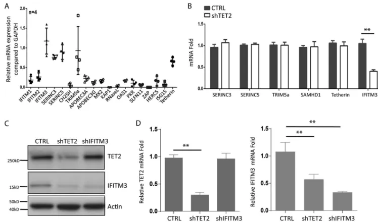
IFITM3 expression in MDMs is dependent on TET2.
To investigate host factors
that may inhibit HIV-1 replication in human MDMs, we measured relative gene
expres-sion of known anti-HIV host factors. Interestingly, 5 of the 19 human genes were
constitutively expressed in resting MDMs (Fig. 2A). We recently reported that Vpr
degrades TET2 through the CRL4
VprbpE3 ligase in macrophages (15). We analyzed the
relative expression of known HIV-1 restriction factors in MDMs transduced with control
or TET2 short hairpin RNA (shRNA). Although 6 genes encoding anti-HIV host factors
were expressed in MDMs (Fig. 2A and B), only IFITIM3 was expressed in a
TET2-dependent fashion at both mRNA and protein levels (Fig. 2B to D).
Vpr enhances Env processing/virion infectivity via TET2- and
IFITM3-dependent mechanisms.
IFITM3 has been reported to inhibit virion assembly of
various viruses, including HIV-1 (24, 25). IFITM3 has been recently reported to inhibit
Env processing in HIV-1-infected target cells (24). This suggests that the Vpr-TET2 axis
may modulate IFITM3 expression to enhance HIV-1 Env processing and virion infectivity
in MDMs. We thus depleted TET2 or IFITM3 through shRNA-mediated knockdown in
MDMs to explore whether Vpr enhanced Env processing through these two proteins
(see Fig. S3A in the supplemental material). The ratio of gp120 to gp160 in control
MDMs infected with Vpr
⫹HIV-1 was 5-fold higher than that of Vpr
⫺HIV-1. In MDMs
with depletion of TET2 or IFITM3, both Vpr
⫹HIV-1 and Vpr
⫺HIV-1 showed a similar
level of Env processing (Fig. 3A). Vpr failed to further enhance the ratio of gp120 to
gp160 in HIV-1-infected MDMs with depletion of TET2 or IFITM3 (Fig. 3A). IFITM3 in
target cells has been reported to inhibit HIV-1 entry (26). Consistently, predepletion of
FIG 2 IFITM3 is constitutively expressed in MDMs via TET2-dependent mechanisms. (A) Relative gene expression of HIV-1 restriction factors in MDMs derived from 4 donors is summarized. (B) MDMs were transduced with lentiviral vectors encoding control shRNA or with TET2 shRNA. Relative expression of restriction factors with detectable expression in MDMs, including Serinc3, Serinc 5, Trim 5a, SAMHD1, tetherin, and IFITM3, was measured. (C) TET2 and IFITM3 were detected with anti-TET2 antibody and anti-IFITM3 antibody by Western blotting with cell lysates from MDMs transduced with control shRNA, TET2 shRNA, and IFITM3 shRNA. (D) TET2 (left panel) and IFITM3 (right panel) mRNAs were detected by RT-qPCR in MDMs transduced with control, TET2, and IFITM3 shRNA. *and**indicateP⬍0.05 andP⬍0.01, respectively.
TET2 or IFITM3 led to elevated entry of both Vpr
⫹HIV-1 and Vpr
⫺HIV-1 (Fig. 3A;
Fig. S3B and C). We further measured Env levels in HIV-1 virions produced from MDMs
with depletion of TET2 and IFITM3. HIV-1 virions produced from control MDMs infected
with Vpr
⫹HIV-1 showed 3-fold-higher levels of Env proteins than Vpr
⫺HIV-1 virions.
Consistent with the elevated Env processing, Vpr
⫹HIV-1 and Vpr
⫺HIV-1 virions from
MDMs with depletion of TET2 or IFITM3 contained equal amounts of Env proteins
(Fig. 3B and C). Moreover, Vpr
⫹or Vpr
⫺HIV-1 virions produced from MDMs with
depletion of TET2 or IFITM3 showed equal infectivity measured by TZM-bl luciferase
assay and by MAGI IU assay (Fig. 3E; Fig. S3D). Our data suggest that the TET2-IFITM3
axis enhances HIV-1 Env processing and virion infectivity in MDMs.
To further confirm that Vpr enhances Env processing/virion infectivity via TET2- and
IFITM3-dependent mechanisms in macrophages, we generated and tested a primary
Vpr
⫹and Vpr
⫺macrophage/CCR5-tropic 1 clone. MDMs were infected with
HIV-Yu-2 and HIV-HIV-Yu-2 ΔVpr viruses. Vpr showed no effect on the first-round HIV-1
repli-cation (Fig. 4A). Vpr similarly enhanced Yu-2 Env processing in MDMs (Fig. 4B and C),
and Env incorporation in virions with elevated infectivity (Fig. 4D to F). As expected, the
Vpr-mediated enhancement of Env processing in MDM cells, Env levels in virions, and
virion infectivity in MDMs depended on TET2 and IFITM3 (Fig. 4G and H). Therefore, Vpr
enhances Env processing/virion infectivity in a TET2- and IFITM3-dependent manner in
macrophages with both CXCR4/CCR5 dualtropic (HIV-R3A) and CCR5-tropic (Yu-2) HIV-1
strains.
Vpr degrades TET2 to prevent demethylation of the IFITM3 promoter in MDMs.
To explore the mechanism of the Vpr-TET2 axis on IFITM3 expression in MDMs, MDMs
were treated with vesicular stomatitis virus G protein (VSV-G) pseudotyped virus-like
particles (VLPs) in the presence or absence of Vpr. As we reported previously (15), Vpr
FIG 3 Vpr enhances Env processing/infectivity in MDMs via TET2- and IFITM3-dependent mechanisms. (A) MDMs transduced with lentiviral vectors with shRNA-mediated knockdown of TET2 and IFITM3 were infected with HIV-1 or HIV-1 ΔVpr viruses as described above. Western blot analysis of cell lysates was performed as in Fig. 1B. The gp120/gp160 ratio of HIV-1 from control MDMs is defined as 100%. (B) MDMs were transduced with lentivirus vectors with control shRNA, shTET2, and shIFITM3. Viral particles were purified by ultracentrifugation (over 20% sucrose) from the aforementioned MDMs infected with HIV-1 or HIV-1 ΔVpr viruses for 4 days, with NVP added at 2 dpi. Five nanograms of p24 per virion for each sample was added per lane for analysis of gp120, p24, and gp41 in virions. Western blotting was performed by using anti-gp120, p24, and gp41 antibodies. (C) The ratios of gp120/p24 and of gp41/p24 are summarized from three different donors. (D) Supernatants from MDMs infected with HIV-1 or HIV-1 ΔVpr viruses were used to infect TZM-bl indicator cells. The relative infectivity is defined as HIV-induced luciferase/ng (n⫽3 donors).
reduced only TET2 protein but not mRNA (Fig. 5A). However, both mRNA and protein
levels of IFITM3 were decreased in MDMs treated with Vpr
⫹VLPs or when TET2 was
depleted (Fig. 5A and B), indicating that TET2 increased IFITM3 expression in MDMs. We
further performed chromatin immunoprecipitation-quantitative PCR (ChIP-qPCR)
ex-periments to measure the binding of TET2 and its demethylated 5hmC level to the
IFITM3 promoter in control MDMs and TET2 knockdown MDMs, after treatment with
Vpr
⫹or Vpr
⫺VLPs. TET2 bound efficiently to the IFITM3 promoter in control or Vpr
⫺VLP-treated MDMs (Fig. 5C). Consistent with TET2 binding to the IFITM3 promoter, an
elevated 5hmC level was enriched at the IFITM3 promoter in MDMs (Fig. 5D). Vpr
⫹VLP
or shTET2 transduction in MDMs abolished TET2 and dramatically reduced 5hmC levels
at the IFITM3 promoter (Fig. 5C and D). We conclude that Vpr reduces constitutive
expression of IFITM3 in MDMs by inhibiting TET2-mediated demethylation of the IFITM3
promoter (Fig. 5E).
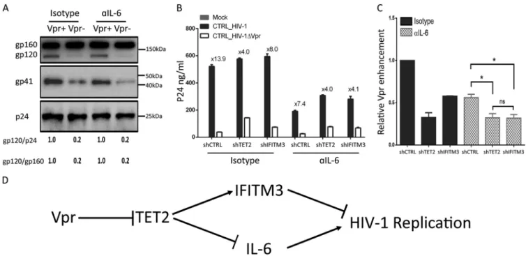
The Vpr-TET2 axis enhances HIV-1 replication in MDMs by reducing IFITM3
expression and by sustaining IL-6 induction after HIV-1 infection.
We recently
reported that Vpr degrades TET2 to prolong IL-6 expression and enhance viral
repli-cation in MDMs (15). To test whether the elevated IL-6 contributes to Vpr-enhanced
HIV-1 Env processing in MDMs, we infected MDMs with Vpr
⫹HIV-1 and Vpr
⫺HIV-1
viruses and treated cells with an IL-6 neutralizing antibody for 4 days, with NVP added
at 2 days postinfection. As shown in Fig. 6A, IL-6 signaling had no effect on
Vpr-enhanced Env processing and no effect on the first round of HIV-1 replication. We
further analyzed spreading HIV-1 infection of Vpr
⫹and Vpr
⫺HIV-1 in MDMs for 6 days
FIG 4 The Vpr-TET2 axis enhances HIV-Yu-2 Env processing and virion infectivity in MDMs. (A) MDMs were infected with Yu-2 or Yu-2 ΔVpr viruses (MOI⫽ 0.1). Levels of p24 in the supernatant were assessed at 2 and 4 dpi. Cells were treated with 2M nevirapine (NVP) at 2 dpi where indicated, to inhibit the second round of HIV-Yu-2 infection. (B) Western blots of cell lysates from MDMs infected with Yu-2 or Yu-2 ΔVpr for 4 days with NVP added at 2 dpi. Env proteins gp160 and gp120 were detected with anti-gp120 antibody, gp41 was detected with anti-gp41 antibody, and p24 was detected with anti-p24 antibody. (C) Vpr-enhanced gp120/p24 and gp120/gp160 ratios in MDMs are summarized with MDMs from with two different donors. (D) Viral supernatants were concentrated from MDMs infected with Yu-2 or Yu-2 ΔVpr viruses at 2 dpi by ultracentrifugation (over 20% sucrose). Western blotting was performed by using anti-gp120, -p24, and -gp41 antibodies. (E) Vpr-enhanced gp120/p24 ratios are summarized from two different donors. (F) Supernatants with equal amounts of p24 from MDMs infected with Yu-2 or Yu-2 ΔVpr viruses were used to infect TZM-bl indicator cells. Luciferase activity was measured at 48 hpi. Data (luciferase/ng p24) from three wells were used from each donor MDM, and 3 technical repeats were performed with supernatant from each well. MDMs derived from 2 donors were used to repeat the experiments. (G) MDMs were transduced with control shRNA, shTET2, and shIFITM3. Viral supernatants were concentrated from the aforementioned MDMs infected with Yu-2 or Yu-2 ΔVpr viruses for 4 days, with NVP added at 2 dpi, by ultracentrifugation (over 20% sucrose). Five nanograms of p24 for each virion was added per lane for analysis of gp120, p24, and gp41 in virions. (H) Supernatants from MDMs infected with Yu-2 or Yu-2 ΔVpr viruses were used to infect TZM-bl indicator cells. The relative infectivity is defined by HIV-induced luciferase activity per ng of p24. Supernatant from control MDMs infected with HIV-YU-2 is defined as 100% (n⫽2 donors).
with depletion of TET2 or IFITM3 treated with IL-6 neutralizing antibody. As we have
previously reported (15), depletion of TET2 in MDMs elevated HIV-1 replication and
reduced the relative activity of Vpr to enhance HIV replication by 70% (from 13.9
⫻
to
4.0
⫻
). Interestingly, either IFITIM3 depletion or IL-6 blockade partly reduced the relative
Vpr activity to enhance HIV-1 replication in MDMs, and the combination of IFITIM3
depletion with IL-6 blockade was similar to TET2 depletion in the reduction of
Vpr-enhanced HIV replication (Fig. 6B and C; see Fig. S4 in the supplemental material). These
results suggest that the Vpr-TET2 axis enhances HIV-1 replication in MDMs both by
reducing IFITIM3 expression and by sustaining IL-6 induction, and the virions with
higher infectivity and elevated IL-6 independently enhance HIV-1 replication after the
first round of infection (Fig. 6D).
DISCUSSION
HIV-1 Vpr preferentially enhances HIV-1 replication after the first round of infection
in human macrophages via multiple mechanisms that are not clearly defined. We have
recently discovered that Vpr targets the DNA demethylase TET2 for degradation, which
leads to sustained IL-6 expression and elevated second-round HIV-1 infection. We
demonstrated here that Vpr enhanced Env processing, associated with increased virion
infectivity during the first round of infection in macrophages. We found IFITIM3 was
constitutively expressed in macrophages in a TET2-dependent fashion. Vpr-enhanced
Env processing depended genetically on TET2 and IFITM3. We further showed that Vpr
suppressed IFITM3 expression by reducing its promoter demethylation in
macro-phages, associated with degradation of TET2 and reduced TET2 binding to the IFITIM3
promoter. We conclude that the Vpr-TET2 axis enhances HIV-1 replication in
macro-phages via two independent mechanisms: reduced IFTIM3 expression to enhance Env
FIG 5 Vpr degrades TET2 to reduce demethylation of the IFITM3 promoter in MDMs. (A and B) TET2 is required for the constitutive expression of IFITM3 in MDMs. (A) Western blot analysis of cell lysates was performed with anti-TET2 or anti-IFITM3 antibody. Relative IFITM3 expression is calculated as IFITM3/actin ratios. (B) MDMs with control or TET2 shRNA were treated with VLPs with or without Vpr for 24 h. TET2 and IFITM3 mRNA levels were detected by RT-qPCR. (C and D) TET2 binds to the IFITM3 promoter to catalyze its demethylation. (C) MDMs transduced with control or TET2 shRNA were treated with VLPs with or without Vpr for 24 h. The ChIP assay was performed with anti-TET2 monoclonal antibody (MAb) to determine TET2 binding to the IFITM3 promoter. (D) ChIP with anti-5hmC MAb was performed to determine demethylation at the IFITM3 promoter. Specific IFITM3 promoter DNA by ChIP is normalized to the DNA ChIP with control IgG. DNA ChIP with control IgG is defined as 1.*and**indicateP⬍0.05 andP⬍0.01, respectively. (E) Schematic diagram of TET2-mediated demethylation of the IFITM3 promoter.
processing and virion infectivity and sustained IL-6 expression to increase HIV-1 gene
expression.
The functional importance of Vpr was first reported in rhesus macaques infected
with Vpr-deficient simian immunodeficiency virus (SIV) (5). However, Vpr-deficient HIV-1
viruses show no significant difference in replication in CD4 T lymphocytes (27–29) or in
SCID-hu Thy/Liv mice (30), suggesting Vpr targets host restrictions in myeloid cells to
enhance HIV-1 replication. In the improved humanized mouse model with both human
T and myeloid cells, Vpr has been reported to enhance early HIV-1 infection in
proliferating T cells (31).
Vpr has two widely studied activities: one is its ability to induce G
2cell cycle arrest,
and the other is to increase HIV-1 replication in macrophages (32, 33). The molecular
mechanism of G
2arrest induction by Vpr has been attributed to a number of host target
proteins (34, 35). In one report, Vpr is found to target MUS81 for degradation through the
CRL4
VprBPE3 ligase, resulting in premature activation of the SLX4 complex. This enhances
the cleavage of DNA by SLX4, leading to activation of the DNA damage response and cell
cycle arrest (14). We have recently reported that Vpr could enhance HIV-1 replication in
MDMs through degradation of TET2 via its activity independent of the G
2arrest activity. We
have shown that blocking IL-6 signaling partially reduces the ability of Vpr-mediated TET2
degradation to enhance HIV-1 replication, which suggests that the Vpr-TET2 axis has an
additional role in enhancing HIV-1 replication in MDMs (15).
In agreement with other studies (6), we found Vpr did not enhance HIV-1 virion
production in the first-round infection in MDMs but enhanced HIV-1 Env processing to
produce more infectious HIV-1 virions for the second-round infection (Fig. 1). During
HIV-1 infection, Env is synthesized as gp160 and is subsequently cleaved in the Golgi
apparatus by cellular proteases into gp120 and gp41. The expression, processing,
plasma membrane localization, and preferential incorporation of trimeric gp12/gp41
FIG 6 The Vpr-TET2 axis independently enhances HIV-1 replication through reduced IFITM3 and sustained IL-6 expression. (A) Vpr-enhanced Env processing is independent of IL-6 signaling. MDMs were infected with HIV-1 or HIV-1 ΔVpr viruses for 4 days, with NVP added at 2 dpi, and treated with the isotype control or anti-IL-6 neutralizing antibody. Western blots were performed with cell lysates. Env proteins gp160 and gp120 were detected with anti-gp120 antibody, gp41 was detected by anti-gp41 antibody, and p24 was detected by anti-p24 antibody. gp120/p24 and gp120/gp160 ratios are shown. (B) MDMs transduced with control shRNA, shTET2, and shIFITM3 were infected with HIV-1 or HIV-1 ΔVpr and treated with the isotype control or anti-IL-6 neutralizing antibody. p24 levels in the supernatant were measured at 6 dpi. (C) Relative ability of Vpr to enhance HIV-1 replication in cells treated with the isotype control and anti-IL-6 neutralizing antibody at day 6. Data from two different donors are summarized by setting Vpr-enhanced replication in isotype control MDMs transduced with control shRNA at 1.0.*indicatesP⬍0.05. (D) The Vpr-TET2 axis enhances HIV-1 replication in macrophages independently by modulating expression of IFITM3 and IL-6 during HIV-1 infection.
Env into HIV-1 virions have been extensively investigated (36). It has been reported that
processed HIV-1 gp120/gp41, but not gp160, is preferentially incorporated into virions
via MA-dependent mechanisms (37, 38). Our current results are consistent with this
model, showing that impaired gp160 cleavage in the absence of Vpr leads to reduced
levels of gp120/gp41 and thus lower levels of gp120/gp41 in the virion (24, 39–42). As
we have previously reported, reduced gp120/gp41 in the virions led to reduced
infectivity of virions (defined as infectious activity per virion particle) (43, 44).
IFITM proteins, especially IFITM2 and IFITM3, have been reported to bind HIV-1 Env
and interfere with its processing (24). We demonstrated that of the 19 reported HIV-1
restriction factors, 6 were expressed efficiently in MDMs, but only IFITM3 was expressed
uniquely via TET2-dependent mechanisms (Fig. 2). By degrading TET2, Vpr enhanced
Env processing through reduction of IFITM3 in macrophages or MDMs (Fig. 3 and 4).
This was confirmed with two macrophage-tropic HIV-1 isolates with CXCR4/CCR5
dualtropic (HIV-R3A [Fig. 1 and 3]) and CCR5-tropic Yu-2 (Fig. 4). Besides inhibiting HIV-1
Env processing, IFITM3 has also been reported to restrict entry of multiple viruses,
including HIV-1 (45). Consistently, stable depletion of TET2 and IFITM3 in macrophages
also led to increased HIV-1 entry, and Vpr had no further effect on Env processing or
infectivity (Fig. 3 and 4; Fig. S3). The TET2-IFITM3 axis in MDMs thus suppresses HIV-1
replication both at the Env processing/virion infectivity step and possibly at the viral
entry step in cells previously exposed to Vpr.
IFITM3 expression is regulated by DNA methylation at the IFITM3 promoter (46). We
found TET2 bound to the IFITM3 promoter and TET2 catalyzed the conversion of
5-methylcytosine (5mC) to 5-hydroxymethylcytosine (5hmC) on the IFITM3 promoter in
MDMs (Fig. 5). Therefore, Vpr-mediated degradation of TET2 in MDMs leads to reduced
IFITM3 expression through reduced promoter demethylation. We have recently
re-ported that Vpr-induced degradation of TET2 in macrophages prevents the resolution
of IL-6 induction and leads to elevated HIV-1 replication (15). We showed here that IL-6
was not involved in Vpr-enhanced Env processing and infectivity. The Vpr-TET2 axis
appears to enhance HIV-1 replication in MDMs independently via reducing IFITM3
expression and sustaining IL-6 expression (Fig. 6). The Vpr-TET2 axis enhances HIV-1
infection in MDMs by reducing IFITM3 expression via a TET2 dioxygenase-dependent
mechanism and by sustaining IL-6 induction via TET2 dioxygenase-independent
mech-anisms. It is likely that Vpr still possesses TET2-indepedent activity that can further
enhance HIV-1 infection in macrophages, as suggested by the partial Vpr activity to
enhance HIV replication in TET2-depleted cells (Fig. 6C). The Vpr-TET2 axis may provide
a novel target to develop anti-HIV drugs to inhibit HIV-1 infection and pathogenesis.
MATERIALS AND METHODS
Cell cultures and shRNA.Human buffy coats were obtained from Gulf Coast Regional Blood Center. Primary human peripheral blood mononuclear cells (PBMCs) were isolated from buffy coats using a Ficoll-Paque gradient. Primary monocytes were isolated from PBMCs using negative magnetic selection (EasySep human monocyte isolation kit 19359). Purified monocytes were seeded at a concentration of 1⫻106
cells/ml in complete RPMI and differentiated into monocyte-derived macrophages (MDMs) using 100 ng/ml granulocyte-macrophage colony-stimulating factor (GM-CSF [Peprotech, Princeton, NJ, USA]) for 6 days. The medium was changed every 2 days throughout 6 days of the culture. TZM-bl was obtained from the NIH AIDS Research and Reference Reagent Program (NIH, Bethesda, MD, USA). TZM-bl was maintained in Dulbecco’s modified Eagle’s medium (DMEM) supplemented with 10% heat-inactivated fetal bovine serum, 100 U/ml penicillin, and 100g/ml streptomycin. MAGI/X4R5-cells are maintained in our lab, and the MAGI assay was carried out as previously reported (47). Briefly, MAGI/X4R5 indicator cells were plated in 6-well plates at 1.6⫻105cells per well in complete DMEM plus G418-hygromycin-puromycin 1 day before HIV-1 infection.
Two days after infection, the infected cells were fixed, 600l of staining solution was added to each well, and the cells were incubated for 50 min at 37°C. The staining was stopped by removing the staining solution and washing the cells thoroughly twice with phosphate-buffered saline (PBS). Positive blue dots were counted to determine HIV-1 infectious units and viral infectivity.
pGIPZ lentiviral vectors expressing control (nontargeting), TET2-targeting shRNA were purchased from Dharmacon, GE Healthcare (Lafayette, CO). shRNA targeting human TET2 has the mature antisense sequence TAAGTAATACAATGTTCTT (clone ID V3LHS_363201). shRNA targeting human IFITM3 has the mature antisense sequence TGAGCATCTCATAGTTGGG (clone ID V3LHS_325105). Lentiviruses expressing shRNA were produced by CaCl2-BES [bis(2-hydroxy)ethylamine] transfection in 293T cells (10-cm plate)
using 15g pGIPZ-shRNA vector, 10g packaging construct ΔNRF, and 5g pCMV-VSV-G. The titer of lentiviruses was calculated on 293T cells. Briefly, the day before transduction, a 96-well tissue culture
plate was seeded with 293T cells at 2.5⫻104cells/well in 100l of DMEM complete growth medium.
Twenty-four hours later, the culture medium was gently removed from each well and a 5-fold serial dilution of viral stock was added to each well with triplicates. This was then spun down for 2 h at 1,500⫻gat 37°C. The viral stock was removed, and DMEM complete medium was added for 3 days. The lentivirus titer was calculated based on the number of green fluorescent protein (GFP)-expressing cells detected by flow cytometry. For transduction of primary MDMs, 50 ng of VLP-Vpx was used to treat 4⫻105MDMs for 6 h before lentivirus transduction, and then the cells were washed once with PBS and
incubated with GIPZ-shRNA lentivirus (100 ng of p24). All lentiviral transductions were performed by adding 8g/ml Polybrene and then spin inoculation of the cells for 2 h at 1,500⫻gat 37°C. Transduced MDM cells after 3 days were selected with 1g/ml puromycin for 3 to 7 days. The transduction efficiency was assessed by GFP expression. All MDMs from different donors presented in the data have achieved
⬎90% transduction efficiency. Knockdown efficiency of TET2 and IFITM3 was subsequently measured by reverse transcription (RT)-qPCR.
HIV-1 proviral constructs and virion stock production.X4/R5 dualtropic HIV-1 R3A was generated by our lab as previously described (48). Vpr mutant R3A was generated by insertion of heat-stable antigen (HSA) reporter into the Vpr gene (15). The proviral infectious clone of the macrophage-tropic virus isolate Yu-2 was from the National Institutes of Health AIDS Research and Reference Reagent Program. The same clone disrupted for the Vpr gene, Yu-2 ΔVpr, was generated by PCR to insert two stop codons within the Vpr gene without altering the Vif gene, using the following set of primers: forward, 5=-GATAGATGGAATAAGCCCCAGAAGACTAAGGGCCACAGAGG-3=; and reverse, 5=-CCTCTGTGGCCCTTAGT CTTCTGGGGCTTATTCCATCTATC-3=(49). HIV-1 virions were produced by CaCl2-BES transfection of
pro-viral plasmids in 293T cells. 293T cells cultured on a 10-cm plate were transfected with 30g DNA of proviral plasmid for production of replication-competent HIV-1 viruses. For the production of virus-like-particles (VLPs) carrying or not carrying Vpr, we transfected 293T cells with 6g p3XFLAG-CMV-Vpr or empty vector, 15g pMDLg/pRRE, 4g pRSV-Rev, and 5g pCMV-VSV-G. For the production of VLP-Vpx particles, we used a Gag/Pol-expressing vector, pMDL-X, containing the Vpx-packaging motifs in the p6 region, as described before (50). VLPs were harvested at 48 h posttransfection and purified by ultracen-trifugation (20,000⫻g, 4°C, 2 h). Concentrations of virion stocks were quantified by p24 ELISA (Frederick National Laboratory for Cancer Research—AIDS and Cancer Virus Program).
HIV-1 infection.HIV-1 infection experiments were typically conducted at a multiplicity of infection (MOI) of 0.1, unless otherwise indicated in the figure legends. All HIV-1 infections were typically performed by spin inoculation for 2 h at 1,500⫻gat 37°C in medium containing 8g/ml Polybrene. Virus input was removed after spin inoculation, and fresh medium was added to infected cells. Nevirapine (5M) was added to the cells as indicated in the figure legends and maintained during infection. MDMs were treated with 500 ng of p24 per amount of VLPs by spin inoculation for 2 h at 1,500⫻gat 37°C in medium containing 8g/ml Polybrene.
ELISAs and antibodies.ELISA kits for human alpha interferon (IFN-␣[Mabtech; 3425-1H-20]) were used according to the manufacturer’s instructions. ELISA kits for p24 (Frederick National Laboratory for Cancer Research—AIDS and Cancer Virus Program) were used according to the instructions of the manufacturer. For intracellular p24 detection, infected cells were first permeabilized using Cytofix/ Cytoperm from BD following the manufacturer’s protocol and then incubated with anti-p24 –fluorescein isothiocyanate (FITC) antibody (1:50 dilution [Beckman Coulter; KC-57]). Anti-TET2 was obtained from Millipore (MABE462). Anti-IFITM3 (11714-1) was obtained from Proteintech. Anti-CD14 was obtained from Abcam (ab181470). Antiactin was obtained from Santa Cruz Biotechnology (sc-47778). Anti-gp120 (catalog no. 288), anti-p24 (catalog no. 6458), anti-gp41 (catalog no. 11557), and sCD4 (catalog no. 4615) were all obtained from the NIH AIDS Research and Reference Reagent Program (NIH, Bethesda, MD, USA). Five hundred nanograms per ml of the isotype control (R&D [MAB004]) or anti-IL-6 neutralizing antibody (R&D [MAB2061R]) was used for the neutralization assay.
ChIP assay.For the ChIP assay, cells were collected in an Eppendorf tube and resuspended in 7.5-ml cold PBS. Cellular proteins and DNA were cross-linked by adding 0.5 ml of 16% formaldehyde into the tube (final concentration, 1%) and incubated at 37°C for 15 min. Cells were centrifuged at 1,000 rpm for 5 min, and cell pellets were resuspended in 5 ml PBS buffer containing 0.125 M glycine and 1⫻protease inhibitor cocktail (PIC). The cells were then incubated at room temperature with shaking for 5 min to quench unreacted formaldehyde. The reaction mixture was centrifuged, and the cell pellets were quickly rinsed with 10 ml of cold PBS and 1⫻PIC. Cells were collected after centrifugation and transferred into a new Eppendorf tube with 1 ml cold PBS with 1⫻PIC, followed by centrifugation at 1,000 rpm for 2 min to pellet the cells. Cell pellets were resuspended in 100l of SDS lysis buffer (1% SDS, 10 mM EDTA, 50 mM Tris, pH 8.1, 1⫻PIC) and incubated on ice for 15 min. DNA was sheared by sonication using a Covaris sonicator (13 min at 4°C). Insoluble material was removed by centrifugation at 12,000⫻gat 4°C for 10 min. The supernatants were transferred to fresh tubes, and 800l ChIP dilution buffer containing 1⫻ PIC, 100l Dynabeads, and 10l antibody was added to each tube. Mixtures were incubated overnight at 4°C with rotation. Beads were precipitated by centrifugation at 2,000 rpm for 2 min at 4°C. The bead-protein-chromatin complexes were washed in 0.5 ml each of the cold buffers in the following order: (i) low-salt wash buffer, (ii) high-salt wash buffer, (iii) LiCl wash buffer, and (iv) TE (Tris-EDTA) buffer (all buffers from Millipore). All washes were performed by incubating complexes for 3 to 5 min with rotation followed by centrifugation at 2,000 rpm for 2 min. One hundred microliters of ChIP dilution buffer and 1l proteinase K were then added into each tube, followed by two incubations—the first at 62°C for 2 h and then a second at 95°C for 10 min— both with constant shaking on an Eppendorf ThermoMixer (700 rpm). Samples were cooled down to room temperature and centrifuged at 2,000 rpm for 2 min at 4°C. Supernatants were carefully transferred to a new tube containing 0.5 ml of binding reagent A. The
sample-binding reagent A mixtures were transferred to the spin filter in the collection tube, centrifuged for 30 s at 12,000⫻g, and decanted. Five hundred microliters of wash reagent B was added to the same spin filter in the collection tube and centrifuged for 30 s at 12,000⫻g. The aqueous portion was carefully discarded, followed by centrifugation for 30 s at 12,000⫻g. The spin filter was put into a new collection tube, and 50l of elution buffer C (Millipore) was added to the center of the spin filter membrane. After centrifugation for 30 s at 12,000⫻g, purified DNA was collected for qPCR analysis. The IFITM3 primers used for qPCR analysis of IFITM3 promoter binding TET2 and 5hmc were as follows: forward, 5=-ATTTG TTCCGCCCTCATCTG-3=; and reverse, 5=-GTTTCGGTTTCTCAACAGTTTCCT-3=. Each ChIP DNA fraction’s threshold cycle (CT) value was normalized to the IgG DNA fraction’sCTvalue (ΔCT) at the same time point. The IgG value is defined as 1.
Statistical analysis. Error bars represent the mean ⫾ standard deviation (SD) from triplicate experiments. All statistical analysis was performed with unpaired Student’sttest, unless otherwise indicated in the figure legends, and results are considered significant when thePvalue is⬍0.05.*,**, and ***indicatePvalues of⬍0.05,⬍0.01, and⬍0.001, respectively.
SUPPLEMENTAL MATERIAL
Supplemental material for this article may be found at
https://doi.org/10.1128/mBio
.01344-19
.
FIG S1, PDF file, 0.5 MB.
FIG S2, PDF file, 0.03 MB.
FIG S3, PDF file, 0.3 MB.
FIG S4, PDF file, 0.3 MB.
ACKNOWLEDGMENTS
We thank Yaxu Wu and Amanda Pons for technical assistance and the University of
North Carolina (UNC) CFAR, Flow Cytometry Core Facility, and the UNC TC Core Facility
for core services. We also thank Yue Xiong, Li-Chung Tsao, Lei Lv, and Yangping Xu for
collaborations on Vpr-TET2-related projects and for discussions. We thank all members
of the Su laboratory for critical reading and/or discussion of the paper. We are grateful
to the NIH AIDS Research and Reference Reagent Program for providing valuable
reagents, including antibodies and HIV-1 clones.
This study was supported in part by grants from the National Institutes of Health
(AI27346 and DK119937 to L.S.). The funders had no role in data collection and analysis,
decision to publish, or preparation of the manuscript.
REFERENCES
1. Sattentau QJ, Stevenson M. 2016. Macrophages and HIV-1: an unhealthy constellation. Cell Host Microbe 19:304 –310.https://doi.org/10.1016/j .chom.2016.02.013.
2. Igarashi T, Brown CR, Endo Y, Buckler-White A, Plishka R, Bischofberger N, Hirsch V, Martin MA. 2001. Macrophage are the principal reservoir and sustain high virus loads in rhesus macaques after the depletion of CD4⫹
T cells by a highly pathogenic simian immunodeficiency virus/HIV type 1 chimera (SHIV): implications for HIV-1 infections of humans. Proc Natl Acad Sci U S A 98:658 – 663.https://doi.org/10.1073/pnas.021551798. 3. Connor RI, Chen BK, Choe S, Landau NR. 1995. Vpr is required for
efficient replication of human immunodeficiency virus type-1 in mono-nuclear phagocytes. Virology 206:935–944.https://doi.org/10.1006/viro .1995.1016.
4. Heinzinger NK, Bukrinsky MI, Haggerty SA, Ragland AM, Kewalramani V, Lee MA, Gendelman HE, Ratner L, Stevenson M, Emerman M. 1994. The Vpr protein of human immunodeficiency virus type 1 influences nuclear localization of viral nucleic acids in nondividing host cells. Proc Natl Acad Sci U S A 91:7311–7315.https://doi.org/10.1073/pnas.91.15.7311. 5. Lang SM, Weeger M, Stahl-Hennig C, Coulibaly C, Hunsmann G, Muller J,
Muller-Hermelink H, Fuchs D, Wachter H, Daniel MM. 1993. Importance of vpr for infection of rhesus monkeys with simian immunodeficiency virus. J Virol 67:902–912.
6. Mashiba M, Collins DR, Terry VH, Collins KL. 2014. Vpr overcomes macrophage-specific restriction of HIV-1 Env expression and virion pro-duction. Cell Host Microbe 16:722–735.https://doi.org/10.1016/j.chom .2014.10.014.
7. Collins DR, Lubow J, Lukic Z, Mashiba M, Collins KL. 2015. Vpr promotes macrophage-dependent HIV-1 infection of CD4⫹T lymphocytes. PLoS
Pathog 11:e1005054.https://doi.org/10.1371/journal.ppat.1005054.
8. Freed EO, Mouland AJ. 2006. The cell biology of HIV-1 and other retroviruses. Retrovirology 3:77.https://doi.org/10.1186/1742-4690-3 -77.
9. Romani B, Cohen EA. 2012. Lentivirus Vpr and Vpx accessory proteins usurp the cullin4-DDB1 (DCAF1) E3 ubiquitin ligase. Curr Opin Virol 2:755–763.https://doi.org/10.1016/j.coviro.2012.09.010.
10. McCall CM, Miliani de Marval PL, Chastain PD, II, Jackson SC, He YJ, Kotake Y, Cook JG, Xiong Y. 2008. Human immunodeficiency virus type 1 Vpr-binding protein VprBP, a WD40 protein associated with the DDB1-CUL4 E3 ubiquitin ligase, is essential for DNA replication and embryonic development. Mol Cell Biol 28:5621–5633.https://doi.org/10.1128/MCB .00232-08.
11. Zhang S, Feng Y, Narayan O, Zhao LJ. 2001. Cytoplasmic retention of HIV-1 regulatory protein Vpr by protein-protein interaction with a novel human cytoplasmic protein VprBP. Gene 263:131–140.https://doi.org/ 10.1016/s0378-1119(00)00583-7.
12. Romani B, Shaykh Baygloo N, Aghasadeghi MR, Allahbakhshi E. 2015. HIV-1 Vpr protein enhances proteasomal degradation of MCM10 DNA replication factor through the Cul4-DDB1[VprBP] E3 ubiquitin ligase to induce G2/M cell cycle arrest. J Biol Chem 290:17380 –17389.https://doi
.org/10.1074/jbc.M115.641522.
13. Ahn J, Vu T, Novince Z, Guerrero-Santoro J, Rapic-Otrin V, Gronenborn AM. 2010. HIV-1 Vpr loads uracil DNA glycosylase-2 onto DCAF1, a substrate recognition subunit of a cullin 4A-ring E3 ubiquitin ligase for proteasome-dependent degradation. J Biol Chem 285:37333–37341.
https://doi.org/10.1074/jbc.M110.133181.
14. Laguette N, Bregnard C, Hue P, Basbous J, Yatim A, Larroque M, Kirchhoff F, Constantinou A, Sobhian B, Benkirane M. 2014. Premature activation of the SLX4 complex by Vpr promotes G2/M arrest and escape from
innate immune sensing. Cell 156:134 –145.https://doi.org/10.1016/j.cell .2013.12.011.
15. Lv L, Wang Q, Xu Y, Tsao LC, Nakagawa T, Guo H, Su L, Xiong Y. 2018. Vpr targets TET2 for degradation by CRL4(VprBP) E3 ligase to sustain IL-6 expression and enhance HIV-1 replication. Mol Cell 70:961–970.e5.
https://doi.org/10.1016/j.molcel.2018.05.007.
16. Zhang Q, Zhao K, Shen Q, Han Y, Gu Y, Li X, Zhao D, Liu Y, Wang C, Zhang X, Su X, Liu J, Ge W, Levine RL, Li N, Cao X. 2015. Tet2 is required to resolve inflammation by recruiting Hdac2 to specifically repress IL-6. Nature 525:389 –393.https://doi.org/10.1038/nature15252.
17. Suzuki MM, Bird A. 2008. DNA methylation landscapes: provocative insights from epigenomics. Nat Rev Genet 9:465– 476.https://doi.org/10 .1038/nrg2341.
18. Kriaucionis S, Heintz N. 2009. The nuclear DNA base 5-hydroxymethyl-cytosine is present in Purkinje neurons and the brain. Science 324: 929 –930.https://doi.org/10.1126/science.1169786.
19. Tahiliani M, Koh KP, Shen Y, Pastor WA, Bandukwala H, Brudno Y, Agarwal S, Iyer LM, Liu DR, Aravind L, Rao A. 2009. Conversion of 5-methylcytosine to 5-hydroxymethylcytosine in mammalian DNA by MLL partner TET1. Science 324:930 –935.https://doi.org/10.1126/science .1170116.
20. Choudhary SK, Archin NM, Cheema M, Dahl NP, Garcia JV, Margolis DM. 2012. Latent HIV-1 infection of resting CD4⫹T cells in the humanized
Rag2⫺/⫺␥c⫺/⫺mouse. J Virol 86:114 –120.https://doi.org/10.1128/JVI
.05590-11.
21. Deeks SG. 2011. HIV infection, inflammation, immunosenescence, and aging. Annu Rev Med 62:141–155.https://doi.org/10.1146/annurev-med -042909-093756.
22. Ito S, Shen L, Dai Q, Wu SC, Collins LB, Swenberg JA, He C, Zhang Y. 2011. Tet proteins can convert 5-methylcytosine to 5-formylcytosine and 5-carboxylcytosine. Science 333:1300 –1303. https://doi.org/10 .1126/science.1210597.
23. Wang Y, Xiao M, Chen X, Chen L, Xu Y, Lv L, Wang P, Yang H, Ma S, Lin H, Jiao B, Ren R, Ye D, Guan KL, Xiong Y. 2015. WT1 recruits TET2 to regulate its target gene expression and suppress leukemia cell prolifer-ation. Mol Cell 57:662– 673.https://doi.org/10.1016/j.molcel.2014.12.023. 24. Yu J, Li M, Wilkins J, Ding S, Swartz TH, Esposito AM, Zheng YM, Freed EO, Liang C, Chen BK, Liu SL. 2015. IFITM proteins restrict HIV-1 infection by antagonizing the envelope glycoprotein. Cell Rep 13:145–156.https:// doi.org/10.1016/j.celrep.2015.08.055.
25. Tartour K, Nguyen XN, Appourchaux R, Assil S, Barateau V, Bloyet LM, Burlaud Gaillard J, Confort MP, Escudero-Perez B, Gruffat H, Hong SS, Moroso M, Reynard O, Reynard S, Decembre E, Ftaich N, Rossi A, Wu N, Arnaud F, Baize S, Dreux M, Gerlier D, Paranhos-Baccala G, Volchkov V, Roingeard P, Cimarelli A. 2017. Interference with the production of infectious viral particles and bimodal inhibition of replication are broadly conserved antiviral properties of IFITMs. PLoS Pathog 13: e1006610.https://doi.org/10.1371/journal.ppat.1006610.
26. Foster TL, Wilson H, Iyer SS, Coss K, Doores K, Smith S, Kellam P, Finzi A, Borrow P, Hahn BH, Neil S. 2016. Resistance of transmitted founder HIV-1 to IFITM-mediated restriction. Cell Host Microbe 20:429 – 442.https://doi .org/10.1016/j.chom.2016.08.006.
27. Dedera D, Hu W, Vander Heyden N, Ratner L. 1989. Viral protein R of human immunodeficiency virus types 1 and 2 is dispensable for repli-cation and cytopathogenicity in lymphoid cells. J Virol 63:3205–3208. 28. Balliet JW, Kolson DL, Eiger G, Kim FM, McGann KA, Srinivasan A, Collman
R. 1994. Distinct effects in primary macrophages and lymphocytes of the human immunodeficiency virus type 1 accessory genes vpr, vpu, and nef: mutational analysis of a primary HIV-1 isolate. Virology 200:623– 631.
https://doi.org/10.1006/viro.1994.1225.
29. Eckstein DA, Sherman MP, Penn ML, Chin PS, De Noronha CM, Greene WC, Goldsmith MA. 2001. HIV-1 Vpr enhances viral burden by facilitating infection of tissue macrophages but not nondividing CD4⫹T cells. J Exp
Med 194:1407–1419.https://doi.org/10.1084/jem.194.10.1407. 30. Aldrovandi GM, Zack JA. 1996. Replication and pathogenicity of human
immunodeficiency virus type 1 accessory gene mutants in SCID-hu mice. J Virol 70:1505–1511.
31. Sato K, Misawa N, Iwami S, Satou Y, Matsuoka M, Ishizaka Y, Ito M, Aihara K, An DS, Koyanagi Y. 2013. HIV-1 Vpr accelerates viral replication during acute infection by exploitation of proliferating CD4⫹T cells in vivo. PLoS
Pathog 9:e1003812.https://doi.org/10.1371/journal.ppat.1003812. 32. Belzile JP, Duisit G, Rougeau N, Mercier J, Finzi A, Cohen EA. 2007. HIV-1
Vpr-mediated G2 arrest involves the DDB1-CUL4AVPRBP E3 ubiquitin
ligase. PLoS Pathog 3:e85.https://doi.org/10.1371/journal.ppat.0030085. 33. Chen R, Le Rouzic E, Kearney JA, Mansky LM, Benichou S. 2004. Vpr-mediated incorporation of UNG2 into HIV-1 particles is required to modulate the virus mutation rate and for replication in macrophages. J Biol Chem 279:28419 –28425.https://doi.org/10.1074/jbc.M403875200. 34. Li G, Bukrinsky M, Zhao RY. 2009. HIV-1 viral protein R (Vpr) and its
interactions with host cell. Curr HIV Res 7:178 –183.https://doi.org/10 .2174/157016209787581436.
35. Cassiday PA, DePaula-Silva AB, Chumley J, Ward J, Barker E, Planelles V. 2015. Understanding the molecular manipulation of DCAF1 by the lentiviral accessory proteins Vpr and Vpx. Virology 476:19 –25.https:// doi.org/10.1016/j.virol.2014.11.024.
36. Beitari S, Wang Y, Liu SL, Liang C. 2019. HIV-1 envelope glycoprotein at the interface of host restriction and virus evasion. Viruses 11:311.https:// doi.org/10.3390/v11040311.
37. Tedbury PR, Freed EO. 2014. The role of matrix in HIV-1 envelope glycoprotein incorporation. Trends Microbiol 22:372–378. https://doi .org/10.1016/j.tim.2014.04.012.
38. Moulard M, Hallenberger S, Garten W, Klenk HD. 1999. Processing and routage of HIV glycoproteins by furin to the cell surface. Virus Res 60:55– 65.https://doi.org/10.1016/S0168-1702(99)00002-7.
39. Misra A, Gleeson E, Wang W, Ye C, Zhou P, Kimata JT. 2018. Glycosyl-phosphatidylinositol-anchored anti-HIV Env single-chain variable frag-ments interfere with HIV-1 Env processing and viral infectivity. J Virol 92:e02080-17.https://doi.org/10.1128/JVI.02080-17.
40. Krapp C, Hotter D, Gawanbacht A, McLaren PJ, Kluge SF, Sturzel CM, Mack K, Reith E, Engelhart S, Ciuffi A, Hornung V, Sauter D, Telenti A, Kirchhoff F. 2016. Guanylate binding protein (GBP) 5 is an interferon-inducible inhibitor of HIV-1 infectivity. Cell Host Microbe 19:504 –514.
https://doi.org/10.1016/j.chom.2016.02.019.
41. Lodermeyer V, Suhr K, Schrott N, Kolbe C, Sturzel CM, Krnavek D, Munch J, Dietz C, Waldmann T, Kirchhoff F, Goffinet C. 2013. 90K, an interferon-stimulated gene product, reduces the infectivity of HIV-1. Retrovirology 10:111.https://doi.org/10.1186/1742-4690-10-111.
42. Caillet M, Janvier K, Pelchen-Matthews A, Delcroix-Genête D, Camus G, Marsh M, Berlioz-Torrent C. 2011. Rab7A is required for efficient produc-tion of infectious HIV-1. PLoS Pathog 7:e1002347. https://doi.org/10 .1371/journal.ppat.1002347.
43. Miller ED, Duus KM, Townsend M, Yi Y, Collman R, Reitz M, Su L. 2001. Human immunodeficiency virus type 1 IIIB selected for replication in vivo exhibits increased envelope glycoproteins in virions without alter-ation in coreceptor usage: separalter-ation of in vivo replicalter-ation from mac-rophage tropism. J Virol 75:8498 – 8506.https://doi.org/10.1128/jvi.75.18 .8498-8506.2001.
44. Kao SM, Miller ED, Su L. 2001. A leucine zipper motif in the cytoplasmic domain of gp41 is required for HIV-1 replication and pathogenesis in vivo. Virology 289:208 –217.https://doi.org/10.1006/viro.2001.1112. 45. Diamond MS, Farzan M. 2013. The broad-spectrum antiviral functions of
IFIT and IFITM proteins. Nat Rev Immunol 13:46 –57.https://doi.org/10 .1038/nri3344.
46. Allen EK, Randolph AG, Bhangale T, Dogra P, Ohlson M, Oshansky CM, Zamora AE, Shannon JP, Finkelstein D, Dressen A, DeVincenzo J, Caniza M, Youngblood B, Rosenberger CM, Thomas PG. 2017. SNP-mediated disruption of CTCF binding at the IFITM3 promoter is associated with risk of severe influenza in humans. Nat Med 23:975–983.https://doi.org/10 .1038/nm.4370.
47. Ye C, Wang W, Cheng L, Li G, Wen M, Wang Q, Zhang Q, Li D, Zhou P, Su L. 2017. Glycosylphosphatidylinositol-anchored anti-HIV scFv effi-ciently protects CD4 T cells from HIV-1 infection and deletion in hu-PBL mice. J Virol 91:e01389-16.https://doi.org/10.1128/JVI.01389-16. 48. Meissner EG, Duus KM, Gao F, Yu XF, Su L. 2004. Characterization of a
thymus-tropic HIV-1 isolate from a rapid progressor: role of the enve-lope. Virology 328:74 – 88.https://doi.org/10.1016/j.virol.2004.07.019. 49. Jacquot G, Le Rouzic E, David A, Mazzolini J, Bouchet J, Bouaziz S,
Niedergang F, Pancino G, Benichou S. 2007. Localization of HIV-1 Vpr to the nuclear envelope: impact on Vpr functions and virus replication in macrophages. Retrovirology 4:84. https://doi.org/10.1186/1742-4690-4 -84.
50. Bobadilla S, Sunseri N, Landau NR. 2013. Efficient transduction of my-eloid cells by an HIV-1-derived lentiviral vector that packages the Vpx accessory protein. Gene Ther 20:514 –520. https://doi.org/10.1038/gt .2012.61.