PREVALENCE OF CAROTID ATHEROMA AND ITS CONFOUNDERS ON CONE BEAM COMPUTED TOMOGRAPHY
Ali Z Syed
A dissertation submitted to the faculty of the University of North Carolina at Chapel Hill in partial fulfillment of the requirements for the Master of Science in Oral and Maxillofacial
Radiology in the Department of Diagnostic Sciences at the School of Dentistry.
Chapel Hill 2014
ii ©2014 Ali Z Syed
iii ABSTRACT
Ali Z Syed: Prevalence of calcified carotid atheroma and its confounders on cone beam computed tomography
(Under the guidance of Donald A. Tyndall)
Purpose: To report the prevalence of calcified carotid atheromas that are seen in CBCT scans and to compare carotid atheroma calcifications with other calcifications that are noted in the neck region and to determine if there is any correlation between these calcifications.
Methods: 636 scans were reviewed out of which 380 CBCT scans met the inclusion and exclusion criteria. The findings were grouped into carotid atheroma calcification, ossification of stylohyoid ligament, triticeous cartilage, tonsillolith, sialolith, calcification of superior cornu of thyroid and calcified lymph node
Results: The most prevalent finding was OSHC followed by TC. Superior cornu of thyroid and triticeous calcifications were found more frequently in female patients. However, no correlation was determined between the different calcifications
iv
ACKNOWLEDGEMENTS
This project would not have been possible without the help and support of many people.
First and foremost, I would wish to acknowledge my mentor Dr. Tyndall for his advice, patience and guidance throughout the project. I would like to thank my committee members Dr. Ludlow and Dr. Samuelson for their immense involvement with my project.
I want to thank my faculty Dr. Andre Mol, Dr. Enrique Platin for sharing knowledge and experience.
Thank you radiology staff: Sharon Green, Venesia Terry, Jane Isley and Chanetha McCabe for your valuable support in the clinic.
A special thanks to Dr. Anne Sanders, who has assisted me with my statistics in this project, without her support probably I would have never finished my statistics.
I would like to offer my special thanks to my colleagues and Friends in the program. Dr. Heidi Kohltfarber, Dr. Marty Evers, Dr. Deeba Kashtwari, Dr. Jake Phillips, Dr. Todd Erickson, Dr. Timothy Robert, Dr. Lawrence Gaalaas and Dr. Jacob Dunn, who has supported me in every step of my journey.
v
TABLE OF CONTENTS
LIST OF TABLES ... viii
LIST OF FIGURES ... ix
LIST OF ABBREVATIONS ... xii
INTRODUCTION ... 1
REVIEW OF LITERATURE ... 2
CAROTID ATHEROMA CALCIFICATION ... 2
SIGNIFICANCE ... 3
ANATOMY ... 3
PATHOPHYSIOLOGY ... 4
RADIOGRAPHIC APPEARANCE………..4
TONSILLOLITHS ... 5
ANATOMY ... 5
PATHOPHYSIOLOGY ... 5
RADIOGRAPHIC APPEARANCE ... 6
TREATMENT ... 6
SIALOLITH ... 7
ANATOMY ... 7
PATHOPHYSIOLOGY ... 7
RADIOGRAPHIC APPEARANCE ... 8
TREATMENT ... 8
vi
ANATOMY & PATHOPHYSIOLOGY ... 9
RADIOGRAPHIC APPEARANCE ... 9
TREATMENT ... 9
OSSIFICATION OF STYLOHYOID COMPLEX ... 10
ANATOMY ... 10
PATHOPHYSIOLOGY ... 10
RADIOGRAPHIC APPEARANCE ... 11
TRITICEOUS CARTILAGE CALCIFICATION ... 12
ANATOMY ... 12
PATHOPHYSIOLOGY ... 12
RADIOGRAPHIC APPEARANCE ... 12
CALCIFICATION OF SUPERIOR CORNU OF THYROID ... 13
ANATOMY ... 13
PATHOPHYSIOLOGY ... 13
RADIOGRAPHIC APPEARANCE ... 13
SPECIFIC AIMS AND HYPOTHESIS ... 14
MATERIALS AND METHODS ... 15
SAMPLE SIZE AND PATIENT POPULATION: ... 15
EXCLUSION CRITERIA ... 15
INCLUSION CRITERIA ... 16
IMAGING DEVICE AND SOFTWARE: ... 16
IMAGE REVIEW ... 17
DATA ANALYSIS ... 17
RESULTS ... 18
DISCUSSION ... 19
vii
CONCLUSIONS ... 22 APPENDIX A: EVALUATION OF SOFT TISSUE CALCIFICATIONS USING
viii
LIST OF TABLES
TABLE 1: MOST COMMONLY NOTED SOFT TISSUE CALCIFICATIONS
IN THE NECK WITH MORPHOLOGICAL FEATURES ON CBCT ... 23
TABLE 2. DESCRIPTIVE ANALYSIS AND FREQUENCY OF SOFT TISSUE
CALCIFICATIONS FROM 380 CBCT SCANS ... 24 TABLE 3. PRESENCE OF CAROTID ATHEROMA CALCIFICATION AND OTHER
CALCIFICATIONS DETECTED ON CBCT SCANS ... 25 TABLE 4. RELATIONSHIP OF AGE AND SOFT TISSUE CALCIFICATIONS
DETECTED ON CONE BEAM COMPUTED TOMOGRAPHY
RADIOGRAPHY SCANS ... 26 TABLE 5. THE RELATIONSHIP OF GENDER AND THE PRESENCE OF
SOFT TISSUE CALCIFICATIONS DETECTED ON CBCT ... 27 TABLE 6. ASSOCIATION OF CAROTID ATHEROMA CALCIFICATION AND
TONSILLOLITH, STRATIFIED BY RIGHT- AND LEFT- SIDES... 28 TABLE 7. ASSOCIATION OF CAROTID ATHEROMA CALCIFICATION AND
OSHC, STRATIFIED BY RIGHT- AND LEFT- SIDES ... 29 TABLE 8. ASSOCIATION OF CAROTID ATHEROMA CALCIFICATION AND
TC, STRATIFIED BY RIGHT- AND LEFT- SIDES ... 30 TABLE 9. ASSOCIATION OF CAROTID ATHEROMA CALICIFICATION AND
SIALOLITH, STRATIFIED BY RIGHT- AND LEFT- SIDES ... 31 TABLE 10. ASSOCIATION OF CAROTID ATHEROMA CALCIFICATION AND
CLN, STRATIFIED BY RIGHT AND LEFT SIDES ... 32 TABLE 11. ASSOCIATION OF CAROTID ATHEROMA CALFIFICATION AND
ix
LIST OF FIGURES
FIGURE.1: THE HISTOGRAM DEPICTING PERCENTAGE OF SUBJECTS
BY AGE IN YEARS, WITH NORMAL CURVE SUPERIMPOSED ... 35
FIGURE.2: PANORAMIC RADIOGRAPH DEMONSTRATING BILATERAL CAROTID ATHEROMA CALCIFICATION ... 36
FIGURE 3: MPR VIEW DEMONSTRATING LINEAR OR CURVILINEAR HYPERDENSE ENTITIES OF CAC ... 37
FIGURE.4: 3D VOLUME RENDERING DEMONSTRATING CAROTID ATHEROMA CALCIFICATION ON THE RIGHT SIDE ... 38
FIGURE 5: PANORAMIC RADIOGRAPH DEMONSTRATING TONSILLOLITH ... 39
FIGURE 6: MPR VIEW DEMONSTRATING TONSILLOLITH ... 40
FIGURE.7: PANORAMIC RADIOGRAPH DEMONSTRATING SIALOLITH ... 41
FIGURE.8: OCCLUSAL RADIOGRAPH DEMONSTRATING SIALOLITH ... 42
FIGURE.9: MPR VIEW DEMONSTRATING SIALOLTH ... 43
FIGURE.10: 3D VOLUME RENDERING DEMONSTRATING SIALOLTH ... 44
FIGURE.11: PANORAMIC RADIOGRAPH DEMONSTRATING CALCIFIED LYMPH NODE ... 45
FIGURE.12: MIP VIEW DEMONSTRATING MULTIPLE CALCIFICATIONS: SIALOLITH, TONSILLOLITH, CSTC ... 46
FIGURE 13: PANORAMIC IMAGE SHOWS THE BILATERAL OSHC WITH PSEUDO ARTICULATION (LEFT SIDE IS PARTIALLY TRUNCATED IN THIS IMAGE) ... 47
FIGURE.14: MIP VIEW DEMONSTRATING OSHC... 48
FIGURE.15: MIP VIEW DEMONSTRATING OSHC WITH MEASUREMENTS ... 49
FIGURE.16: CUSTOM SECTION WITH MPR VIEW DEMONSTRATING OSHC ... 50
FIGURE.17: PANORAMIC RADIOGRAPH DEMONSTRATING TRITICEOUS CARTILAGE (BLACK ARROW) AND CALCIFIED SUPERIOR CORNU OF THYROID (WHITE ARROW) ... 51
FIGURE.18: MPR VIEW DEMONSTRATING TRITICEOUS CARTILAGE………...52
x
FIGURE.20: MPR VIEW DEMONSTRATING CALCIFIED SUPERIOR CORNU
OF THYROID ... 54 FIGURE.21: MIP VIEW DEMONSTRATING CALCIFIED SUPERIOR CORNU
xi
LIST OF ABBREVATIONS
CAC Carotid atheroma calcification
CCA Common carotid artery
CBCT Computed beam cone tomography
CLN Calcified lymph node
CT Computed tomography
ECA External carotid artery
ICA Internal carotid artery
MRI Magnetic resonance imaging
MPR Multi planar reformatting
OSHC Ossification of /Ossified stylohyoid complex
CSCT Calcified superior cornu of thyroid
TC Triticeous cartilage
1 Introduction
The earliest imaging in medicine dates back to 1896 when the discoverer of the x-ray, Wilhelm Roentgen, made a radiograph of his wife’s hand.[1] In dentistry Otto Walkhoff took the first dental radiograph on January 14 1896.[1] Since that time 2-D imaging modalities including intra-oral, and extra oral imaging have been used as an adjunct to clinical assessment. The field of oral and maxillofacial radiology has seen an explosive growth in the use of digital 2D and 3D technologies. Depending on the diagnostic task advanced technologies can benefit various dental patients. The various advanced imaging modalities are such as computed tomography (CT) and cone beam computed tomography (CBCT), magnetic resonance imaging (MRI), sialography and ultrasound (US).
CBCT was introduced to dentistry in 1998[2], this technology provides cross sectional imaging that was previously limited to more expensive medical CT. Dentistry quickly adopted this technology because of relatively low patient doses for dedicated maxillofacial scans and high isotropic spatial resolution of osseous structures. [2, 3] CBCT is being used in various clinical applications in dentistry.[4] Example uses include diagnostic evaluation of implants,[2, 5-7], endodontic applications,[8-11] impacted teeth, orthognathic surgery[12], supernumerary teeth[13], maxillofacial trauma[14], craniofacial analysis[15] and root resorption e.tc.,.[3, 16, 17]
2
may demonstrate structures and pathology outside the region of interest for which the clinician is responsible.
Although physicians are involved in the detection of carotid atheromas, the role of the dentist has increased in identifying these lesions when they are calcified. The presence of calcified carotid atheromas in dental panoramic radiographs or CBCT volumes may indicate an increased risk for stroke.[19-21] It is incumbent on the dentist to review all tissues included in the scan, and identify the different soft tissue calcifications and appropriately refer patients to their physicians when a significant finding is noted.
Review of Literature
In the literature the terms ‘‘calcified’’ and ‘‘ossified’’ are often used synonymously. When cartilage becomes transformed into bone, calcification is followed by ossification.[22] In general calcifications are seen in the skeletal part of the body. Unorganized calcifications noted in the soft tissue are termed heterotopic calcifications. Soft tissue calcifications are classified into three major categories: metastatic, idiopathic and dystrophic calcification.
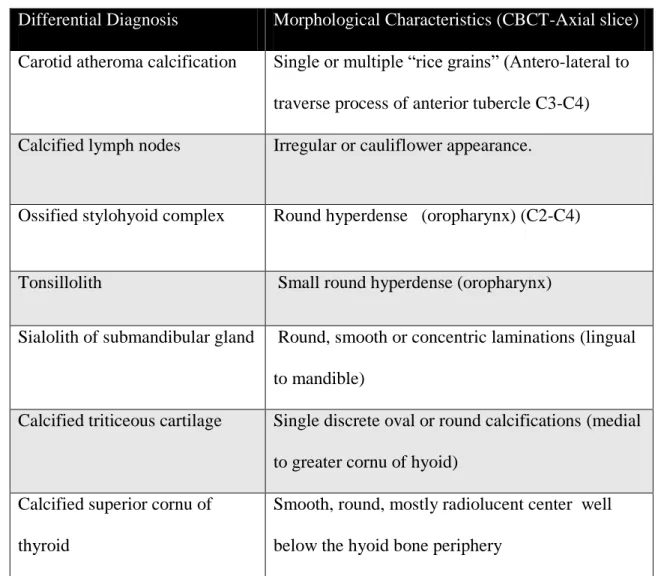
Entities that can appear as hyper-dense in the neck region include salivary calculi, calcified lymphoid tissue, tonsilloliths, ossified stylohyoid complex, and laryngeal cartilage calcifications. It is important to differentiate carotid calcifications from other calcifications in neck to avoid erroneous diagnoses (Table 1).
Carotid atheroma calcification
3
(CBCT) in radiographic images. CAC is pathological in nature and occur in degenerative and dead tissue despite normal serum calcium and phosphate levels.[23]
Extra cranial CAC can occur either in the common carotid artery (CCA) at the junction of the internal carotid artery (ICA) and the external carotid artery. (ECA) located lateral and
inferior to the hyoid bone. [24]
Significance
According to World Health Organization (WHO), ischemic disease and stroke are by far responsible for deaths in comparison with cancers. [25] According to Hubar extra cranial calcifications can cause 60% of thrombo-embolic strokes from plaque buildup at or near the carotid bifurcation. [26] These calcifications can produce ischemic attacks either by occluding the blood flow or by producing emboli. These emboli account for a majority of strokes. [24, 27] In study by Cohen et. al found that the patients experienced end result, such as myocardial infraction (11%), stroke (7%) death (15%) transient ischemic attack (3%) and angina (10%) within an average of 2.7 years of detecting carotid atheroma on panoramic radiographs. [28] Stroke is the fourth leading cause of death in America and also a leading cause of adult disability. [27, 29] In another by Friedlander et al [30] showed that the presence of carotid calcification on panoramic radiograph in women who are over the age of 60 years old may show associated coronary artery calcification this could potentially result in myocardial infraction.
Anatomy
4 Pathophysiology
Atheromas form in the artery’s tunica intima, resulting in atherosclerosis and reduction or occlusion of the lumen, referred to as stenosis. Atherosclerotic lesions that are either stenotic or ulcerated are considered especially unstable, forming intraluminal thrombi. [24, 25]
Radiographic Appearance
CCA may be calcified or non-calcified. The non-calcified versions are not visualized on panoramic radiography. However calcified atheromas may be visualized on the panoramic radiograph as a radiopaque nodular or vertical linear entity inferior to the mandibular angle, adjacent to the cervical vertebrae at the level of the C3-C4 vertebra (Fig 2).[24, 31, 32]
On CBCT in the axial view, most CAC presents as single or multiple “rice grains”. [24, 33] Sometimes CAC can also present as either linear or curvilinear homogeneous calcifications (Fig 3). They are most commonly located in the soft tissue approximately 0 to 10 mm
anterolateral to the anterior tubercle of the transverse process, and either lateral or posterio-lateral to the greater horn of the hyoid bone. In coronal sections, CAC are posterio-lateral to the anterior tubercle of the transverse process of the cervical vertebrae. On sagittal sections, CAC are inferior to the angle of the mandible varying from C3 to C5. [24, 34]
5
stenting) may be recommended if the lesion is occluding more than 60% of the vessel’s lumen.[35]
Tonsilloliths
Lang first described calcification in the oropharynx in 1560.[36] Calcifications that are noted in the enlarged tonsillar crypts are commonly referred to as tonsilloliths. Small
tonsilloliths are common findings, however large tonsilloliths are rare.[37] Anatomy
At the upper end of the pharynx three types of tonsils are noted, they are palatine tonsils, pharyngeal tonsils (adenoids) and lingual tonsils. Palatine tonsils are located on the lateral pharyngeal wall. Pharyngeal tonsils are located in the posterior wall of the pharynx, and lingual tonsils are found at the base of the tongue, the boundaries of “Waldeyer’s ring” of lymphoid tissues are formed by these tonsils. [36] Tonsilloliths are composed primarily of calcium and other minerals such as phosphorus, ammonia, carbonate and magnesium. The calcium salts are calcium hydroxyapatite and calcium carbonate apatite.[38] Histologically, a tonsil consists of a mass of lymphoid tissues that contain follicles with germinal centers. The surface of the tonsil forms crypts lined by stratified squamous epithelium that is less mature than a typical oral mucous membrane.
6 Pathophysiology
Tonsilloliths arise as a result of retained material and bacterial growth in tonsillar or adenoidal crypts, and may be related to chronic recurrent infections. [40] Patient presentation is often asymptomatic. Symptoms when present are varied and can include chronic sore throat and halitosis. The term Halitosis is derived from the Latin halitus (breathed air) and osis (pathology). Halitosis is an unpleasant symptom which may impact the quality of life.[38] Other symptoms that have been reported in the literature include chronic irritating cough, a sense of presence of a foreign body in the throat, dysphagia, and a foul taste.[37] As per Giudice and coauthors
[41]large tonsilloliths can ulcerate either in the supra tonsillar fossa or in the anterior pillar.
Radiographic Appearance
On the panoramic radiograph, tonsilloliths commonly appear as multiple, small, round, ill-defined radiopaque entities and are noted overlapping mid ramus region of the mandible (Fig 5).[36, 39] Superimposition of hard and soft tissue structures on such radiographic images is common in this anatomic region, creating challenges in interpretation.[36, 42]
In CBCT axial images, tonsilloliths appear as multiple small opacifications located anterio-lateral to the oro-pharyngeal airway space immediately medial to the angle of the ramus (Fig 6). Its position can vary with respect to the surface of the airway space. [34]
Treatment
7
laser has been suggested to treat tonsilloliths and may be used to reduce crypt depth to reduce retention of a tonsillolith without compromising the immunological function of the tonsils. [38] Sialolith
Salivary stones, also called sialoliths or calculi, are the calcifications responsible for the obstruction of the secretion of saliva within salivary parenchyma as well as in the duct.[44, 45] Sialolithiasis is a common disease of the salivary glands characterized by obstruction of salivary secretion by a calculus, associated with swelling, pain especially during the meal time, and infection of the affected gland.[46-48].
Anatomy
The submandibular gland is located in the submandibular triangle formed by the anterior and posterior bellies of the digastric muscle and the inferior margin of the mandible. The gland forms a “C” around the anterior margin of the mylohyoid muscle, which divides the
submandibular gland into a superficial and deep lobe. The mandibular branch of seventh cranial nerve courses superficial to the submandibular gland and deep to the platysma.[49]
The submandibular duct (Wharton’s duct), exits the medial surface of the gland and courses between the mylohyoid muscle laterally and hyoglossus muscle medially, and over the genioglossus muscle and finally empties into the anterior aspect of the floor of the mouth. [49] Pathophysiology
8
The submandibular gland may be anatomically predisposed to sialolith development because of a wide lumen and uphill ductal course. An alkaline and viscous character of secretion and reduced salivary flow rate may be reasons for obstruction as well. [49-51]
Submandibular stones are composed of 82% inorganic and 18% organic material. They typically measure between 5 and 10 mm in size, and all stones over 10 mm can be reported as sialoliths of unusual size.[52]
Calculi may be multiple (25%) and may occur within intraglandular ductal tributaries or within the main ducts. When present in the gland itself, the symptoms may be relatively minor, whereas ductal sialoliths usually have a more precipitous presentation as pain and swelling of the involved gland during eating. Sialoliths are prevalent in adults, although they also occur in children as well. [49, 53]
Radiographic Appearance
Although large calculi can be depicted on panoramic radiographs, smaller calculi are more difficult to visualize because of size and if they are non-calcified. [44, 46, 52] According to a study by Soares et al. the salivary calculus develops by continuous deposition at a rate of approximately 1 to 1.5 mm per year. [54] Sialoliths are visualized on panoramic imaging as round, smooth, radiopaque entity, but can be obscured with superimposition of anatomy. (Fig 7).[45] Occlusal radiograph may utilized to visualize sialolith. (Figure 8).
In the axial slice they may appear as single linear or globular homogeneous hyper-dense opacities located medially to the lingual aspect of the mandible (Fig 9). [46, 49]
Treatment
9
location of the calculi. If it's closer to the duct orifice, dilation and catheterization are
recommended. Surgical intervention for gland removal may be needed when the sialolith cannot be retrieved.[50, 52]
Calcified Lymph nodes (CLN)
Lymph nodes are encapsulated structures located throughout the lymphatic system composed of lymphoid cells and supporting tissue. Calcified lymph nodes are a form of dystrophic calcification and are markers of active or latent nodal disease or sequel of prior disease. [55, 56]
Anatomy & Pathophysiology
The most clinically significant lymph nodes in head and neck regions are found in
submandibular, submental, preauricular and cervical regions. [57] Following infectious processes such as sinusitis, tonsillitis, tuberculosis [58] and metastases, the nodes may become fibrous. Consequently, foci of dystrophic calcification may develop. These calcified lymph nodes are generally asymptomatic.[58, 59]
Radiographic Appearance
10 Treatment
Usually the therapeutic treatment is reserved for malignant diseases, where the lymph nodes are excised, treatment is not indicated in the case of a benign process.
Ossification of Stylohyoid Complex
Anatomy
The styloid process is a long, cylindrical, slender, cartilaginous bone extending from the inferior aspect of the petrous part of the temporal bone and located in front of stylomastoid foramen. It lies behind the pharyngeal wall of the palatine fossa, between the internal and external carotid arteries. The stylohyoid ligament is attached to the tip of the styloid process and extends to the lesser cornu of the hyoid bone. The stylohyoid ligament can undergo
mineralization, thus becoming radiographically visible. Some patients with mineralization of the stylohyoid ligament complex can develop symptoms. These symptoms include vague facial pain, throat discomfort, otalgia, and dysphagia. [61]
The stylohyoid and stylomandibular ligaments attach to the styloid process. In addition, the stylohyoid, stylopharyngeus and styloglossus muscles have tendonous attachment to the styloid process.[61-63]
The normal length of the styloid process is approximately 20-30 mm, if it is longer than 30 mm was considered to be styloid process elongation.[61] The prevalence of elongation is 4%-7%.[64]
Pathophysiology
11
tonsillectomy. Other theories that have been proposed include congenital elongation, partial or complete ossification of the stylohyoid ligament, status post-trauma as a result of reactive hyperplasia, and Association with early onset of menopause. [63, 65]
The reported radiographic prevalence of the stylohyoid ossification varies from less than 2% to greater than 30% in the literature. [66] Mostly commonly either in the third or fourth decade the stylohyoid ligament calcification is noted and more frequently in women. Bilateral involvement is common, with or without symptoms. It is important to note that an elongated styloid process does not necessarily signify Eagle’s syndrome, as the majority of individuals exhibiting this anatomical anomaly experience no symptoms.
Radiographic Appearance
On panoramic imaging modality bilaterally styloid process appears as a thin, long, and a tapering radiopaque entity. They extend from the base of the temporal bone inferiorly along the ascending ramus from the styloid process (Fig 13). The radiographic appearance of the
stylohyoid complex may be divided into 3 types. Type 1 (elongated) is characterized by an uninterrupted integrity of the styloid image. In type II (pseudo articulated), the styloid process is apparently joined to the mineralized stylohyoid or stylomandibular ligament mineralized by a single pseudo-articulation, usually located superior the level tangential to the inferior border of the mandible, giving an appearance of an articulated elongated styloid process. Type III
(segmented) consists of either long or short non-continuous portions of the styloid process or interrupted segments which represent ossicles of the mineralized ligament. [67-69]
12 Triticeous Cartilage Calcification
Anatomy
Triticeous cartilages are bilateral ovoid structures that are part of a complex of structures found in the area of the laryngeal skeleton. The word triticeous comes from the Latin word triticeous, meaning resembling a grain of wheat.[72, 73] The triticeous cartilage is located centrally in the lateral thyrohyoid ligament at the level of third and fourth cervical vertebrae (C3-C4). Although clinically, the triticeous cartilage has no known function; it has been suggested that it might help reinforce the lateral thyrohyoid ligament. [72]
Pathophysiology:
Calcification of triticeous cartilages is linked to that of the thyroid cartilage of the larynx. The process of calcification starts at the age of 20, ending by the age of 61. [74]
Radiographic Appearance
On a panoramic radiograph they appear as ovoid radiopacities, measuring approximately 2 to 4 mm wide by 7 to 9 mm in length, and are usually imaged within the pharyngeal air space adjacent to the superior portion of C4 (Fig 17).[72]
13 Calcification of superior cornu of thyroid Anatomy
The framework of the larynx is made up of cartilages. The thyroid cartilage is a major cartilage of the larynx. [22, 73] The skeleton of the larynx consists of 3 unpaired cartilages (thyroid, epiglottis, and cricoid) and 3 smaller paired cartilages (arytenoid, cuneiform, and corniculate cartilages. The thyroid and cricoid cartilages have been found to undergo a greater frequency of calcification in females, but a higher degree of calcification has been noted in male subjects.[22, 75]
Pathophysiology
The exact mechanism of ossification of the thyroid cartilage is not understood very well.[75] Interestingly, it has been noted that ossification begins in the inferior portion and extends superiorly, upon impact these ossified thyroid may be prone to fracture. It was reported that at age 25 thyroid cartilage calcification begins. [22] By age 65-70 most of the cartilage will be ossified; however, in females the calcification has been never complete.
Radiographic Appearance
Mupparapu et al suggested identifying the epiglottis as a landmark initially and following down to the thyroid cartilage will aid in its identification.[22] On a panoramic radiograph the superior cornu of a calcified thyroid cartilage appears as a vertical, smooth, soft tissue
calcification approximately 4 mm wide and 15 mm in length, at the level of C4. Only the
14
In a CBCT axial view the thyroid cartilage is located posterior to the greater cornu of the hyoid bone, inferior to hyoid bone at the level of C4 and appears as a single distinct circular hyper-dense entity. In this projection one can differentiate between superior cornu of thyroid calcification and triticeous cartilage, which is located medial to the posterior extent of the greater cornu of the hyoid ( Fig 20, 21). [34, 71] Treatment is not necessary for calcified superior cornu of thyroid.[76]
Specific Aims and Hypothesis
A review of literature showed no studies which investigated the prevalence of soft tissue calcifications and simultaneously evaluated to demonstrate if there is a correlation between these different soft tissue calcifications
The aims of this research are:
1. To provide a review of literature regarding the various soft tissue calcifications that are visible on radiographs in the neck region
2. To evaluate and report the type of calcifications and their prevalence and
3. To investigate possible correlations between CCA and other calcifications of the neck region.
We hypothesize that;
1. The presence of calcifications in the neck region is unrelated to age 2. The presence of calcifications is unrelated to gender.
15 Materials and Methods
Sample size and patient population
A convenience sample of patients who underwent maxillofacial volumetric scanning in the Oral and Maxillofacial radiology clinic at the University of North Carolina, Chapel Hill, NC was used in this study. The study was approved through the expedited review procedure IRB approval (IRB #12-1262). Because of the nature of the research design, the IRB concluded that the study was in compliance with the Helsinki Declaration and provided an exemption for informed consent.
The study retrospectively identified all patients who underwent Galileos Comfort CBCT 15 cm diameter volume scans from Dec 2009 to Dec 2010 at UNC School of dentistry. Six hundred and thirty six scans were taken during this time period. By following the inclusion and exclusion criteria, which is described below, 380 scans were included in this study. The subject age ranged from 30-87 years. The age distribution is shown in the (Fig 1). There is a moderate predominance of women (n= 241) is noted in the study.
Exclusion criteria
16 Inclusion criteria
The study design was different from other studies since the selected patient population for this study were 30 years and older at the time of the scan. The rationale for this study design was that the process of calcification is age related. [10] The calcification in the aorta may begin at the age of 20 years, and in another study by Mupparapu and et.al demonstrated that the radiographic appearance of laryngeal cartilages has not been seen before 20 years of age. [10, 22]
Imaging Device and Software
17
boot camp (Windows 7, Microsoft, Redmond, WA) and monitor resolution of 2880x 1080 was used to evaluate all the CBCT scans. Pertinent information such as patients’ demographic data, age, sex and type of calcification were recorded. Based on the methodology described by Centurion et. al a custom para sagittal sections were constructed to identify and make measurements of OSHC (Fig 14). [4]
Image Review
The cone beam computed tomography scans were analyzed by the graduate student following the radiographic criteria (Appendix 1). Any case in doubt was verified with a board certified oral and maxillofacial radiologist with more than 27 years of experience. The findings of CAC were noted and compared with other calcifications: salivary calculi, calcified lymph nodes, tonsilloliths, OSHC, triticeous calcification, and thyroid calcification. Both sides of each patient were examined using multiple planar rendering (MPR) view.
All the scans were evaluated for the length of OSHC, they were measured starting from the base of the skull extending all the way to hyoid bone or where the ossification ends. (Fig 15) In our study, we considered < 30 mm as normal anything above, we included in the study and divided into two subgroups, group one (30-40 mm) and group two ( > 40 mm). We included all the three types of ossification in our study (Type 1- elongated styloid, Type II- pseudo
articulation and Type III- ossicles or segmented). Custom sections were created to make appropriate measurements and followed the guideline suggested by centurion et.al.[4]
Data Analysis:
18
before importing the file into statistical software for analysis. The data were statistically analyzed with SPSS software grad pack premium 19 (IBM, New York, NY).
Descriptive statistics were prepared. Logistic regression analysis was carried out using 95 % confidence intervals (CI) to evaluate the correlation between different soft tissue
calcifications. A level of statistical significance was set with an alpha value of 0.05.
Null hypothesis were tested as follows: the presence of calcification is not related to age, the presence of calcification is unrelated to gender and there is no correlation between CAC and other calcifications.
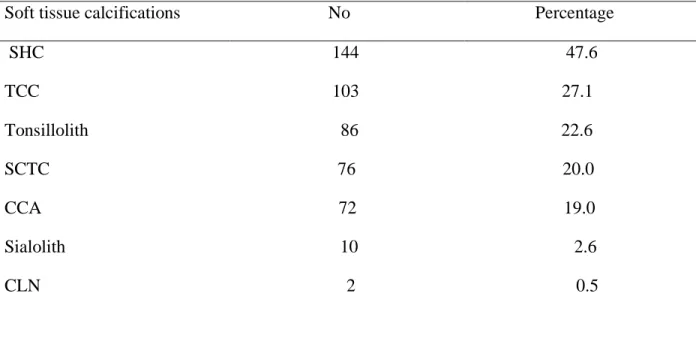
Results
A total of 380 patients who met the criteria were included in the study. The age range was from 30- 87. For age dependent data analysis, patients were divided into three groups. Group one (30-54years), Group two (55-64 years) and Group three (65-87). In our study, there were 139 males and 241 females. Of the 380 study scans reviewed, the most prevalent finding was ossified stylohyoid ligament (OSHC) (N= 144, 47.6%), followed by triticeal calcification (n=103,
27.1%), tonsillolith (n=86, 22.6%), Superior cornu of thyroid (n= 76, 20 %) CAC (n=72, 19.0%), Sialolith (n=10, 2.6 %) calcified lymph node (n=2, 0.5%) (Table1).
19
This is statistically significant, p < 0.001 (Table 2) which demonstrates that the CAC will tend increase with age and, therefore, these results favor rejection of null hypothesis. There was higher frequency of CAC (bilaterally) that was noted in males compared with females, however this difference was not statistically significant p < 0.101 (Table 3) and therefore, these results favor null hypothesis that presence CAC is unrelated to gender.
Furthermore, logistic regression test with a 95 % confidence interval was used to investigate if there is a correlation between the CCA and other soft tissue calcifications. No statistical significance was noted with following variables: Right CAC with right tonsillolith was P=0.408, OR =1.7, 95 % CI 0.5—6.1), left CAC with left tonsillolith was p0=0.707, OR =0.7, 95 % CI 0.1—5.3) (Table. 6).
Likewise No statistical significance were noted with other variables such as OSHC: Right CAC with right OSHC was P=0.809, and left CAC with left OSHC Right was p=0.7, OR =0.7, 95 % CI 0.1—5.3 (Table 7). Right CAC with right CTC was P=0.959, OR =0.9, 95 % CI 0.1— 7.5 (Table 8), left CAC with left CTC was p=0.266. Similar findings were noted with respect to the other calcifications such as Sialolith, calcified lymph nodes and CSCT were no statistical significance is noted (Table 9, 10, 11).
Discussion
20
The results in this study found no correlation between CAC and other calcifications in the neck region. To the best of our knowledge, our study is the first to report the prevalence of CAC on CBCT and the presence of correlation with other calcifications in the neck.
In our study, we found most common type of calcification to be OSHC 47.6%, followed by triticeous calcification 27.1%, tonsillolith 22.6%, superior cornu of thyroid 20 %, CAC 19.0%, sialolith 2.6 % and calcified lymph node 0.5%.
The prevalence of CAC detected on panoramic radiographs has ranges of 0.43% to 5% depending on age, gender, and risk factors [24, 28, 32]. Friedlander et al found the prevalence of carotid atheroma calcification on panoramic radiography to be 4.5[78]. Our study demonstrated higher prevalence, which is over 19 %. A probable explanation for this is usage of a CBCT modality which permits clear visualization of the carotid region in all 3Dimensions and wide range of age of sample size in our study. In contrast with previous studies [19, 34]where males were likely to be more affected than females, our study is in accordance with these studies, especially in bilateral presentation, however it is not statistically significant difference.
In our study the tonsilloliths were found to be 22.6 %. A slightly higher prevalence was noted on the right side (9.2 %) compared with the left (7.1%). Males and older patients of 65 years old exhibited more tonsilloliths which could be because of recurrent infections. However, these findings are not statistically significant. These findings are in agreement with previous study that was done by Fauroux and et al. [79]
The reported prevalence of the ossification of stylohyoid complex varies from 2% to 30% in the literature [66]. Rizzatti-Barbosa et al. found an ossification of stylohyoid ligament
21
al reported in their study higher prevalence which is about 21.1%. [61] In our study, we found the OSHC was found to be 37.6 %. The bilateral occurrence was noted in our study found to be 36.8%. No discrimination with respect to age is noted our study. Based on previous literature the normal length of ossified stylohyoid ligament was found to be <30 mm [66] hence, we also considered 30 and > 30 mm to be a benchmark for OSHC. We further evaluated to find the length of OSHC, however no distinction was made to between three types when measuring the length OSHC, and in our study we found that 23.4 % having less than 40 mm in length and 40-85mm to be 14.2% these findings are in accordance with other studies. However, based on previous literature review [67] the presence of radiographic findings are not diagnostic to stylohyoid ligament disorder (Eagle’s syndrome). Further research is needed to evaluate the length of OSHC and its correlation with Eagles’ syndrome.
Cervical lymph node calcification is uncommon, involving just 1% of patients. [56] Our study showed only 0.5 % of subjects with a calcified lymph node, which is similar to the study by Devang.
Our study showed more calcification of TC in female patients, this is in accordance with a study by Wells et. al.[34] Comparison of SCTC with gender distribution is noted in the (Table 5) the Pearson chi square test showed there was female predominance that was noted and demonstrated a significant difference (p <0.019) in our study. These findings are in contrast to study by Mupparapu et.al where the frequency was in males is 36% and females is 19%.[22, 75] The possible reasons for more TCC and SCTC in our study reciprocal calcification that is seen in post-menopausal women [30] and possible smaller neck volume in females that may have
22 Study Limitations:
A significant strength of the present study was the thorough re-evaluation of each scan for the presence of calcifications by reviewing the scans in all three orthogonal planes (axial, sagittal and coronal), followed by evaluation of MIP images and volume rendering to confirm radiological diagnosis. Our study has several limitations such as study design which is a retrospective study in nature. No statistical power analysis was performed to determine the sample size, however the sample size in our study was substantial in comparison with similar studies. Intra observer reliability was not done in our study design. All the scans were analyzed by the author, a third year resident in the oral and maxillofacial radiology program. No second review was done by another independent reviewer, however, in doubtful cases the two
experienced board certified oral and maxillofacial radiologists were consulted. Lastly, the study was conducted in a single center, and the regional patient population may have different risk factors than that of other regions. Thus, the results may not be generalized.
Conclusions:
We recommend that all scans taken with large FOV that encompasses C3-C4 vertebra region in the region should be evaluated for not only for pathology of the jaws, but also should be evaluated for the soft tissue calcifications.
23
TABLE 1: MOST COMMONLY NOTED SOFT TISSUE CALCIFICATIONS IN THE NECK WITH MORPHOLOGICAL FEATURES ON CBCT
Differential Diagnosis Morphological Characteristics (CBCT-Axial slice)
Carotid atheroma calcification Single or multiple “rice grains” (Antero-lateral to traverse process of anterior tubercle C3-C4) Calcified lymph nodes Irregular or cauliflower appearance.
Ossified stylohyoid complex Round hyperdense (oropharynx) (C2-C4)
Tonsillolith Small round hyperdense (oropharynx)
Sialolith of submandibular gland Round, smooth or concentric laminations (lingual to mandible)
Calcified triticeous cartilage Single discrete oval or round calcifications (medial to greater cornu of hyoid)
Calcified superior cornu of thyroid
24
TABLE 2. DESCRIPTIVE ANALYSIS AND FREQUENCY OF SOFT TISSUE CALCIFICATIONS FROM 380 CBCT SCANS
25
TABLE 3. PRESENCE OF CALCIFIED CAROTID ARTERY ATHEROMAS AND OTHER CALCIFICATIONS DETECTED ON CONE BEAM COMPUTED
TOMOGRAPHY SCANS Count Percent
Total 380 100.0
Calcified carotid atheroma
Absent 308 81.1
Right, unilateral 21 5.5
Left, unilateral 20 5.3
Bilateral 31 8.2
Tonsillolith
Absent 294 77.4
Right, unilateral 35 9.2
Left, unilateral 27 7.1
Bilateral 24 6.3
Stylohyoid complex
Absent 237 62.4
Right, unilateral 1 0.3
Left, unilateral 2 0.5
Bilateral 140 36.8
Triticeal cartilage
Absent 277 72.9
Right, unilateral 19 5.0
Left, unilateral 21 5.5
Bilateral 63 16.6
Sialolith
Absent 370 97.4
Right, unilateral 7 1.8
Left, unilateral 3 0.8
Bilateral 0 0.0
Calcified lymph node
Absent 378 99.5
Right, unilateral 2 0.5
Left, unilateral 0 0.0
Bilateral 0 0.0
CSCT
Absent 304 80.0
Right, unilateral 7 1.8
Left, unilateral 4 1.1
26
TABLE 4. RELATIONSHIP OF AGE AND SOFT TISSUE CALCIFICATIONS DETECTED ON CONE BEAM COMPUTED TOMOGRAPHY RADIOGRAPHY SCANS
(N, ROW PERCENT)
Absent Unilateral Bilateral P-value (a)
CCA
<55 years 131 96.3 2 1.5 3 2.2 <0.001
55–64 years 101 78.9 6 4.7 21 16.4
≥65 years 76 65.5 13 11.2 27 23.3
Total 308 81.1 21 5.5 51 13.4
Tonsillolith
<55 years 117 86.0 6 4.4 13 9.6 0.042
55–64 years 93 72.7 16 12.5 19 14.8
≥65 years 84 72.4 13 11.2 19 16.4
Total 294 77.4 35 9.2 51 13.4
SHC
<55 years 87 64.0 0 0.0 49 36.0 0.545
55–64 years 82 64.1 1 0.8 45 35.2
≥65 years 68 58.6 0 0.0 48 41.4
Total 237 62.4 1 0.3 142 37.4
Triticeal cartilage
<55 years 101 74.3 4 2.9 31 22.8 0.478
55–64 years 88 68.8 9 7.0 31 24.2
≥65 years 88 75.9 6 5.2 22 19.0
Total 277 72.9 19 5.0 84 22.1
Sialolith
<55 years 133 97.8 1 0.7 2 1.5 0.366
55–64 years 125 97.7 2 1.6 1 0.8
≥65 years 112 96.6 4 3.5 0 0.0
Total 370 97.4 7 1.8 3 0.8
Calcified lymph node
<55 years 136 100 0 0.0 0 0.0 0.101
55–64 years 128 100 0 0.0 0 0.0
≥65 years 114 98.3 2 1.7 0 0.0
Total 378 99.5 2 0.5 0 0.0
CSCT
<55 years 101 74.3 2 1.5 33 24.3 0.151
55–64 years 104 81.3 2 1.6 22 17.2
≥65 years 99 85.3 3 2.6 14 12.1
Total 304 80.0 7 1.8 69 18.2
27
TABLE 5. THE RELATIONSHIP OF GENDER AND THE PRESENCE OF SOFT TISSUE CALCIFICATIONS DETECTED ON CONE BEAM COMPUTED
TOMOGRAPHY RADIOGRAPHY (N, ROW PERCENT)
Absent Unilateral Bilateral P-value (a) CCA
Male 105 75.5 9 6.5 25 18.0 0.101
Female 203 84.2 12 5.0 26 10.8
Total 308 81.1 21 5.5 51 13.4
Tonsillolith
Male 107 77.0 11 7.9 21 15.1
Female 187 77.6 24 10.0 30 12.5 0.648
Total 294 77.4 35 9.2 51 13.4
Stylohyoid ligament
Male 80 57.6 0 0.0 59 42.5
Female 157 65.2 1 0.4 83 34.4 0.234
Total 237 62.4 1 0.3 142 37.4
Triticeal cartilage
Male 116 83.5 7 5.0 16 11.5
Female 161 66.8 12 5.0 68 28.2 0.001
Total 277 72.9 19 5.0 84 22.1
Sialolith
Male 134 96.4 4 2.9 1 0.7 0.519
Female 236 97.9 3 1.2 2 0.8
Total 370 97.4 7 1.8 3 0.8
Calcified lymph node
Male 137 98.6 2 1.4 0 0.0 0.062
Female 241 100 0 0.0 0 0.0
Total 378 99.5 2 0.5 0 0.0
CSCT
Male 121 87.1 3 2.2 15 10.8 0.018
Female 183 75.9 4 1.7 54 22.4
Total 304 80.0 7 1.8 69 18.2
28
TABLE 6. ASSOCIATION OF CAROTID ATHEROMA CALCIFICATION AND TONSILLOLITH, STRATIFIED BY RIGHT- AND LEFT- SIDES
CCA Absent
(%)
Present
(%) P-value
OR (95% CI) Right Tonsillolith Absent (%) 327 (94.8) 18 (5.2)
0.408 1.7
(0.5-6.1) Present (%) 32 (91.4) 3 (8.6) Left Tonsillolith Absent (%) 334 (94.6) 19 (5.38)
0.707 0.7
(0.1-5.3) Present (%) 32 (91.4) 3 (8.6)
29
TABLE 7. ASSOCIATION OF CAROTID ATHEROMA CALCIFICATION AND OSHC, STRATIFIED BY RIGHT- AND LEFT- SIDES
CCA Absent
(%)
Present
(%) P-value
OR (95% CI) Right OSHC Absent (%) 358 (94.5) 21 (5.5)
0.809 ----
Present (%) 1 (100.0) 3 (0.0) Left OSHC Absent (%) 358 (94.6) 20 (5.3)
0.7 0.7
(0.1-5.3) Present (%) 2 (100.0) 0 (0.0)
30
TABLE 8. ASSOCIATION OF CAROTID ATHEROMA CALCIFICATION AND TC, STRATIFIED BY RIGHT- AND LEFT- SIDES
CCA Absent
(%)
Present
(%) P-value
OR (95% CI) Right TCC Absent (%) 341 (94.5) 20 (5.5)
0.959 0.9
(0.1-7.5) Present (%) 32 (91.4) 3 (8.6) Left TCC Absent (%) 339 (94.6) 20 (5.6)
0.266 ----
Present (%) 21 (100.0) 0 (0.0)
31
TABLE 9. ASSOCIATION OF CAROTID ATHEROMA CALCIFICATION AND SIALOLITH, STRATIFIED BY RIGHT- AND LEFT- SIDES
CCA Absent
(%)
Present
(%) P-value
OR (95% CI) Right Sialolith Absent (%) 352 (94.4) 21 (5.6)
0.518 ----
Present (%) 7 (100.0) 0.0 (0.0) Left Sialolith Absent (%) 357 (94.6) 20 (5.3)
0.682 ----
Present (%) 3 (100.0) 0 (0.0)
32
TABLE 10. ASSOCIATION OF CAROTID ATHEROMA CALCIFICATION AND CLN, STRATIFIED BY RIGHT AND LEFT SIDES
CCA Absent
(%)
Present
(%) P-value
OR (95% CI) Right Calcified lymph node Absent (%) 357 (94.4) 21 (5.6)
0.732 ----
Present (%) 2 (100.0) 0.0 (0.0) Left Calcified lymph node Absent (%) 360 (94.6) 20 (5.3)
0.682 ----
Present (%) 0 (0.0) 0 (0.0)
33
TABLE 11. ASSOCIATION OF CAROTID ATHEROMA CALCIFICATION AND CSCT, STRATIFIED BY RIGHT- AND LEFT-SIDES
CCA Absent
(%)
Present
(%) P-value
OR (95% CI) Right CSCT Absent (%) 353 (94.4) 20 (5.4)
0.306 ----
Present (%) 2 (100.0) 0.0 (0.0) Left CSCT Absent (%) 376 (100.0) 0 (0.0)
<0.001 ---- Present (%) 0 (0.0) 4 (100.0)
34
TABLE12: LENGTH OF THE STYLOHYOID COMPLEX LENGTH MEASURED ON CBCT
35
FIGURE 1. THE HISTOGRAM DEPICTING PERCENTAGE OF SUBJECTS BY AGE IN YEARS, WITH NORMAL CURVE SUPERIMPOSED
0 5 10 15 20
Pe
rce
n
t
o
f
su
b
je
ct
s
30 40 50 60 70 80 90
36
FIGURE.2: PANORAMIC RADIOGRAPH DEMONSTRATES BILATERAL CAROTID ATHEROMA CALCIFICATION
37
38
39
40
41
42
43
44
45
FIGURE.11: PANORAMIC RADIOGRAPH DEMONSTRATING CALCIFIED LYMPH NODE
46
FIGURE.12: MIP IN THE SAGITTAL VIEW DEMONSTRATING MULTIPLE CALCIFICATIONS: SIALOLITH, TONSILLOLITH,
47
48
49
50
51
FIGURE.17: PANORAMIC RADIOGRAPH DEMONSTRATING TRITICEOUS CARTILAGE (BLACK ARROW) AND CALCIFIED SUPERIOR CORNU OF
52
53
54
55
56
APPENDIX A: EVALUATION OF SOFT TISSUE CALCIFICATIONS USING THE
MODIFIED RADIOGRAPHIC CRITERIA (WHITE, STUART C., MICHAEL PHARAOH.
ORAL RADIOLOGY: PRINCIPLES AND INTERPRETATION, 7TH EDITION. MOSBY, 2014. VITAL BOOK FILE.)
Soft tissue calcifications Radiographic criteria
Calcified lymph node Lobulated/ cauliflower shaped- May appear in a chain fashion (sagittal view)
Sialolith May appear as a Smooth, cylindrical entity lingual to mandible (axial view)
Tonsillolith Multiple small ill-defined, punctate, may appear in clusters (coronal view)
OSHC Long, linear thin extension from base of skull to hyoid- (sagittal view)
CAC Multiple, irregular entities in the carotid space – C3-C4 region lateral to greater cornu of hyoid (axial view)
Triticeous cartilage Round, well defined, smooth- medial to the greater cornu of hyoid (axial view)
Superior cornu of thyroid calcification
57 REFRENCES
1. Thunty, K.H. Early Pioneers of Oral and Maxillofacial Radiology. 2013 Dec 9 2013 [cited 2013 3-17-2014]; Available from:
http://c.ymcdn.com/sites/www.aaomr.org/resource/resmgr/aaomr_history/early_pioneers _of_oral_and_m.pdf.
2. Pedroso, L.A., et al., Impact of cone-beam computed tomography on implant planning and on prediction of implant size. Braz Oral Res, 2013. 28(1): p. 46-53.
3. Miracle, A.C. and S.K. Mukherji, Conebeam CT of the head and neck, part 2: clinical applications. AJNR Am J Neuroradiol, 2009. 30(7): p. 1285-92.
4. Centurion, B.S., et al., How to assess tonsilloliths and styloid chain ossifications on cone beam computed tomography images. Oral Dis, 2013. 19(5): p. 473-8.
5. Bornstein, M.M., et al., Cone beam computed tomography in implant dentistry: a
systematic review focusing on guidelines, indications, and radiation dose risks. Int J Oral Maxillofac Implants, 2014. 29 Suppl: p. 55-77.
6. Gupta, J. and S. Ali, Cone beam computed tomography in oral implants. Vol. 4. 2013. 2-6.
7. Tyndall, D.A., et al., Position statement of the American Academy of Oral and Maxillofacial Radiology on selection criteria for the use of radiology in dental
implantology with emphasis on cone beam computed tomography. Oral Surg Oral Med Oral Pathol Oral Radiol, 2012. 113(6): p. 817-26.
8. Horner, K., Cone-beam computed tomography: time for an evidence-based approach.
Prim Dent J, 2013. 2(1): p. 22-31.
9. Demirbuga, S., et al., Use of cone-beam computed tomography to evaluate root and canal morphology of mandibular first and second molars in Turkish individuals. Med Oral Patol Oral Cir Bucal, 2013. 18(4): p. e737-44.
10. Bayer, S., et al., Prevalence of findings compatible with carotid artery calcifications on dental panoramic radiographs. Clin Oral Investig, 2011. 15(4): p. 563-9.
11. Tyndall, D.A. and S. Rathore, Cone-beam CT diagnostic applications: caries,
periodontal bone assessment, and endodontic applications. Dent Clin North Am, 2008. 52(4): p. 825-41, vii.
58
13. Agrawal, J.M., et al., CBCT in orthodontics: the wave of future. J Contemp Dent Pract, 2013. 14(1): p. 153-7.
14. Kaeppler, G., et al., Diagnostic efficacy of cone-beam computed tomography for
mandibular fractures. Oral Surg Oral Med Oral Pathol Oral Radiol, 2013. 116(1): p. 98-104.
15. AlHadidi, A., et al., Comparison of two methods for quantitative assessment of mandibular asymmetry using cone beam computed tomography image volumes.
Dentomaxillofac Radiol, 2011. 40(6): p. 351-7.
16. Parashar, V., et al., Cone Beam Computed Tomography in Dental Education: A Survey of U.S., U.K., and Australian Dental Schools. Journal of Dental Education, 2012. 76(11): p. 1443-1447.
17. Scarfe, W.C., et al., Maxillofacial cone beam computed tomography: essence, elements and steps to interpretation. Aust Dent J, 2012. 57 Suppl 1: p. 46-60.
18. V.E, R., H. K, and W. H.V, Screening panoramic radiology of adults in general dental pratice: radiological findings. Br Dent J, 2001(190(9)): p. 495-501.
19. Fisher, M., et al., Carotid plaque pathology: thrombosis, ulceration, and stroke pathogenesis. Stroke, 2005. 36(2): p. 253-7.
20. Kamikawa, R.S., et al., Study of the localization of radiopacities similar to calcified carotid atheroma by means of panoramic radiography. Oral Surg Oral Med Oral Pathol Oral Radiol Endod, 2006. 101(3): p. 374-8.
21. Lell, M.M., et al., New Techniques in CT Angiography. RadioGraphics, 2006. 26(suppl_1): p. S45-S62.
22. Mupparapu, M., Ossification of Laryngeal Cartilages on Lateral Cephalometric Radiographs. Angle Orthodontist, 2005. 75(2).
23. de Moura, M.D., et al., Tonsillolith: a report of three clinical cases. Med Oral Patol Oral Cir Bucal, 2007. 12(2): p. E130-3.
24. MacDonald, D., et al., Diagnosis and management of calcified carotid artery atheroma: dental perspectives. Oral Surg Oral Med Oral Pathol Oral Radiol, 2012. 114(4): p. 533-47.
59
26. Baumann-Bhalla, S., et al., Recognizing calcifications of the carotid artery on panoramic radiographs to prevent strokes. Schweiz Monatsschr Zahnmed, 2012. 122(11): p. 1016-29.
27. Friedlander, A.H., et al., Do carotid atheromas on panoramic images prognosticate arterial calcifications on mammograms: acknowledged markers of future adverse cardiovascular events? Oral Surg Oral Med Oral Pathol Oral Radiol, 2012. 114(4): p. 526-32.
28. Cohen, S.N., et al., Carotid calcification on panoramic radiographs: An important marker for vascular risk. Oral Surgery, Oral Medicine, Oral Pathology, Oral Radiology, and Endodontology, 2002. 94(4): p. 510-514.
29. What is stroke ? 2014 [cited 2014 3-27]; Available from: http://www.stroke.org/site/PageServer?pagename=stroke.
30. Friedlander, A.H., et al., Association of calcified carotid atheromas visualized on panoramic images and aortic arch calcifications seen on chest radiographs of postmenopausal women. J Am Dent Assoc, 2014. 145(4): p. 345-51.
31. Damaskos, S., et al., Reliability of panoramic radiograph for carotid atheroma detection: a study in patients who fulfill the criteria for carotid endarterectomy. Oral Surg Oral Med Oral Pathol Oral Radiol Endod, 2008. 106(5): p. 736-42.
32. Pornprasertsuk-Damrongsri, S., et al., The prevalence of carotid artery calcifications detected on panoramic radiographs in patients with metabolic syndrome. Oral Surg Oral Med Oral Pathol Oral Radiol Endod, 2009. 108(4): p. e57-62.
33. Almog, D.M., et al., Previously unappreciated carotid artery stenosis diagnosed by cone beam computerized tomography. J Oral Maxillofac Surg, 2013. 71(4): p. 702-5.
34. Wells, A.B., Incidence of soft tissue calicifications of the Head and Neck region on Maxillofacial Cone Beam Computed Tomography. 2011.
35. Friedlander, A.H., et al., Radiation-associated carotid artery atherosclerosis: case report and review of contemporaneous literature. Spec Care Dentist, 2009. 29(2): p. 75-9. 36. Ram, S., et al., Pseudo bilateral tonsilloliths: a case report and review of the literature.
Oral Surgery, Oral Medicine, Oral Pathology, Oral Radiology, and Endodontology, 2004. 98(1): p. 110-114.
37. Caldas, M.P., et al., Tonsillolith--report of an unusual case. Br Dent J, 2007. 202(5): p. 265-7.
60
39. Oda, M., et al., Prevalence and imaging characteristics of detectable tonsilloliths on 482 pairs of consecutive CT and panoramic radiographs. BMC Oral Health, 2013. 13: p. 54. 40. Sezer, B., Z. Tugsel, and C. Bilgen, An unusual tonsillolith. Oral Surg Oral Med Oral
Pathol Oral Radiol Endod, 2003. 95(4): p. 471-3.
41. Giudice, M., et al., An unusual tonsillolithiasis in a patient with chronic obstructive sialoadenitis. Dentomaxillofac Radiol, 2005. 34(4): p. 247-50.
42. Misirlioglu, M., et al., Bilateral and pseudobilateral tonsilloliths: Three dimensional imaging with cone-beam computed tomography. Imaging Sci Dent, 2013. 43(3): p. 163-9. 43. de Oliveira Cde, N., et al., Bilateral tonsilloliths and calcified carotid atheromas: case
report and literature review. J Craniomaxillofac Surg, 2013. 41(2): p. 179-82.
44. Boffano, P. and C. Gallesio, Surgical treatment of a giant sialolith of the Wharton duct. J Craniofac Surg, 2010. 21(1): p. 134-5.
45. Huang, T.C., et al., Multiple, large sialoliths of the submandibular gland duct: a case report. Aust Dent J, 2009. 54(1): p. 61-5.
46. Bodner, L., Giant salivary gland calculi: Diagnostic imaging and surgical management.
Oral Surgery, Oral Medicine, Oral Pathology, Oral Radiology, and Endodontology, 2002. 94(3): p. 320-323.
47. Burke, C.J., R.H. Thomas, and D. Howlett, Imaging the major salivary glands. Br J Oral Maxillofac Surg, 2011. 49(4): p. 261-9.
48. Ngu, R.K., et al., Salivary duct strictures: nature and incidence in benign salivary obstruction. Dentomaxillofac Radiol, 2007. 36(2): p. 63-7.
49. Abdullah, A., F.F. Rivas, and A. Srinivasan, Imaging of the salivary glands. Semin Roentgenol, 2013. 48(1): p. 65-74.
50. Pagani, D., et al., Dysphagia and Submandibular Swelling. The Journal of the American Dental Association, 2010. 141(9): p. 1089-1093.
51. Ledesma-Montes, C., et al., Giant sialolith: case report and review of the literature. J Oral Maxillofac Surg, 2007. 65(1): p. 128-30.
52. Rai, M. and R. Burman, Giant submandibular sialolith of remarkable size in the comma area of Wharton's duct: a case report. J Oral Maxillofac Surg, 2009. 67(6): p. 1329-32. 53. Baurmash, H.D., Submandibular salivary stones: current management modalities.
61
54. Soares, E.C., et al., Giant salivary calculus of the submandibular gland. Otolaryngol Head Neck Surg, 2009. 140(1): p. 128-9.
55. Mehta, A., et al., Calcified Lymph Nodes Causing Clinically Relevant Attenuation Correction Artifacts on PET/CT Imaging. J Radiol Case Rep, 2010. 4(2): p. 31-7. 56. Devang, G., Imaging of cervical Lymph nodes in Head and Neck Cancer: Basics.
Radiologic Clincs of North America, 2006. 44(1).
57. Aydin, U., Tuberculous lymph node calcification detected on routine panoramic radiography: a case report. Dentomaxillofacial Radiology, 2003. 32(4): p. 252-254. 58. Mandel, L., Tuberculous cervical node calcifications mimicking sialolithiasis: a case
report. J Oral Maxillofac Surg, 2006. 64(9): p. 1439-42.
59. Barbara, E.L. and S.M. Peter, The Spectrum of Benign and Malignant Etiologies of Cervical Node Calcification. AJNRonline.org, 1999. 172.
60. Bar, T., et al., Calcifications simulating sialolithiasis of the major salivary glands.
Dentomaxillofac Radiol, 2007. 36(1): p. 59-62.
61. Monsour, P. and W. Young, variability of styloid process and stylohyoid ligament in panoramic radiographs. 0ral Surgery, Oral Medicine, Oral Pathology, 1986. 61: p. 522-526.
62. Pereira, F.L., et al., Styloid-stylohyoid syndrome: literature review and case report. J Oral Maxillofac Surg, 2007. 65(7): p. 1346-53.
63. Piagkou, M., et al., Eagle's syndrome: a review of the literature. Clin Anat, 2009. 22(5): p. 545-58.
64. Matsumoto, F., et al., Endoscopy-assisted transoral resection of the styloid process in Eagle's syndrome. Case report. Head Face Med, 2012. 8: p. 21.
65. Prasad, K.C., et al., Elongated styloid process (Eagle's syndrome): A clinical study.
Journal of Oral and Maxillofacial Surgery, 2002. 60(2): p. 171-175.
66. Cumali Gokce, Y.S., Prevalence of styloid proces Elongation on Panoramic
Radiography in the Turkey Population from Cappadocia Region. European Journal of Dentistry, 2008. 2: p. 18-22.
67. J.G.C, L. and R. L, Mineralization of stylohyoid ligament complex in patinets with temporomandibular disorders and asymptomatic individuals: a comparative study.
62
69. Friedlander, A.H. and I.K. Friedlander, Identification of stroke prone patients by panoramic radiography. Aust Dent J, 1998. 43(1): p. 51-4.
71. Koenig, L.J., Diagnostic Imaging: Oral and Maxillofacial. 1 ed. 2012: Amirsys.
72. Carter, L.C., Discrimination between calcified triticeous cartilage and calcified carotid atheroma on panoramic radiography. Oral Surg Oral Med Oral Pathol Oral Radiol Endod, 2000. 90(1): p. 108-10.
73. Ahmad, M., R. Madden, and L. Perez, Triticeous cartilage: prevalence on panoramic radiographs and diagnostic criteria. Oral Surg Oral Med Oral Pathol Oral Radiol Endod, 2005. 99(2): p. 225-30.
74. Di Nunno, N., et al., Anomalies and alterations of the hyoid-larynx complex in forensic radiographic studies. Am J Forensic Med Pathol, 2004. 25(1): p. 14-9.
75. Kosuri, K.C., V. Nelluri, and T. Huban, Ossified Cartilago Thyreoidea and its Clinical Insight: A Cadaveric Study. International Journal of Bioassays, 2013. 2(7): p. 1044-1047. 76. White, S.C. and M.J. Pharoah, Oral Radiology Principles and Interpretation. 6 ed. 2009:
Mosby Elsevier.
77. Ludlow, J.B. and M. Ivanovic, Comparative dosimetry of dental CBCT devices and 64-slice CT for oral and maxillofacial radiology. Oral Surg Oral Med Oral Pathol Oral Radiol Endod, 2008. 106(1): p. 106-14.
78. Friedlander, A.H., et al., Ultrasonographic confirmation of carotid artery atheromas diagnosed via panoramic radiography. J Am Dent Assoc, 2005. 136(5): p. 635-40; quiz 682-3.
79. Fauroux, M.A., Prevalence of palatine tonsilloliths: a retrospective study on 150
consecutive CT examinations. Dento-maxillo-facial radiology, 2013. 42(7): p. 20120429. 80. Enciso, R., et al., Comparison of cone-beam computed tomography incidental findings