SMOKING AND VARIATION IN BREAST TUMOR BIOMARKER EXPRESSION
Eboneé Nicole Butler
A dissertation submitted to the faculty at the University of North Carolina at Chapel Hill in partial fulfillment of the requirements for the degree of Doctor of Philosophy in the
Department of Epidemiology in the Gillings School of Global Public Health.
Chapel Hill 2017
ii © 2017
iii
ABSTRACT
Eboneé Nicole Butler: Smoking and Variation in Breast Tumor Biomarker Expression (Under the direction of Melissa A. Troester)
Purpose: Smoking is a suspected risk factor for breast cancer, with hypothesized links to estrogen-mediated, genotoxic, and growth-factor dependent mechanisms. Each mechanism can be modeled by overexpression of ER, p53, and EGFR, respectively. This dissertation examines associations between smoking and biomarkers for each mechanistic pathway. Methods: Our population-based study included 1,970 women diagnosed with invasive breast cancer in central and eastern North Carolina.Single and multigene biomarker outcomes were characterized as binary (+/-) or continuous measures for protein or mRNA. Single gene measures included ER/ESR1, p53/MDM2, and EGFR. Multigene mRNA signatures included a luminal score (LS); a p53 signature used to describe wild-type (Wt) or mutant (Mut) activity; and an algorithm-based proliferation score (PS). We used logistic and linear regression models to estimate associations between smoking and biomarker outcomes.
Results: (Aim 1)When compared with never smokers,the odds of ER+, ESR1+, and LS+ tumors were nearly doubled among current smokers, those who smoked 20 or more years, and those who smoked within 5 years of diagnosis. Quantitative levels of ESR1 mRNA were highest among current smokers compared to never smokers overall and among women with ER+ breast cancer; however, we did not observe associations between smoking and
iv
v
I dedicate this dissertation to my Granny, Ms. Eddie Mae Johnson. I love you.
“You are the light of the world—like a city on a hilltop that cannot be hidden. No one lights a lamp and then puts it under a basket. Instead, a lamp is placed on a stand, where it gives light to everyone in the house. In the same way, let your good deeds shine out for all to see, so that
everyone will praise your heavenly Father.
vi
ACKNOWLEDGEMENTS
The completion of this dissertation would not be possible without the support of many people. I would first like to thank my advisor, Dr. Melissa Troester, for her teaching, support, and wisdom. I am grateful for her guidance as I’ve grown in scholarship. I would also like to thank the members of my dissertation committee for their invaluable
contributions: Dr. Jeannette Bensen has encouraged me to think about the myriad of
biological processes that may influence observed associations between risk factors and tumor biomarker expression; Dr. Mengjie Chen has exposed me to new methods in bioinformatics that have been a great complement to my studies in epidemiology; I am thankful to Dr. Kathleen Conway for the many hours she spent discussing my research interests and objectives, which helped me to better articulate my research aims; and I am grateful to Dr. Andrew Olshan who has been a great academic advisor and department chair during my time at UNC.
vii
to land. I am also grateful to Ms. Chandra Caldwell. Thank you for your pep talks and for encouraging me to stay the course.
viii
TABLE OF CONTENTS
LIST OF TABLES ... xii
LIST OF FIGURES ... xiii
LIST OF ABBREVIATIONS ... xv
CHAPTER 1: SPECIFIC AIMS ... 1
CHAPTER 2: BACKGROUND ... 4
2.1 Two Etiologic Types of Breast Cancer ... 5
2.2 Smoking and Breast Cancer Risk ... 7
2.3 Smoking and Breast Tumor Biomarkers Linked to Pathogenesis... 8
2.3.1 Intrinsic Subtype ... 9
2.3.2 Estrogen Receptor ... 10
2.3.3 Tumor protein p53 ... 11
2.3.4 Epidermal Growth Factor Receptor ... 12
2.4 Exposure-Time-Windows and Breast Cancer Risk ... 13
2.5 Summary ... 15
2.6 Literature Review Tables ... 15
CHAPTER 3: METHODS ... 30
3.1 Overview ... 30
3.2 Study Design ... 30
ix
3.3.1 Intrinsic subtype, multigene mRNA and IHC biomarkers... 33
3.3.2 Estrogen-mediated biomarkers ... 34
3.3.3 Genotoxic biomarkers ... 34
3.3.4 Growth-factor dependent biomarkers ... 35
3.4 Exposure Assessment ... 35
3.5 Covariate Assessment ... 36
3.6 Data Analysis ... 36
3.6.1 Linear and logistic regression models ... 37
3.6.2 Binary outcomes and categorical measures of smoking ... 37
3.6.3 Cumulative smoking exposure and time-windows analysis ... 37
3.6.4 Parametric latency functions of smoking and breast cancer risk ... 38
3.7 Power Analysis ... 39
3.8 Summary ... 42
3.8.1 Limitations ... 42
3.8.2 Strengths ... 42
3.9 Addendum ... 43
CHAPTER 4: SMOKING AND ESTROGEN-MEDIATED BIOMARKERS... 44
4.1 Introduction ... 44
4.2 Methods ... 46
4.2.1 Study Population ... 46
4.2.2 Study Design ... 47
4.2.3 Data Analysis ... 50
x
4.4 Discussion ... 54
4.5 Addendum ... 58
CHAPTER 5: SMOKING, P53, EGFR, AND RELATED BIOMARKERS ... 70
5.1 Background ... 70
5.2 Methods ... 71
5.2.1 Study Population ... 71
5.2.2 Outcome Assessment ... 72
5.2.3 Exposure Assessment... 75
5.2.4 Data Analysis ... 75
5.3 Results ... 76
5.3.1 Quantitative P53 Protein and MDM2 mRNA Expression ... 76
5.3.2 Smoking and P53 Protein Binary Subtypes ... 77
5.3.3 Smoking and P53 mRNA Binary Subtypes ... 77
5.3.4 Smoking, Quantitative p53 Protein and MDM2 mRNA Expression ... 77
5.3.5 Smoking and EGFR Protein Binary Subtypes ... 78
5.3.6 Smoking and Quantitative EGFR protein and EGFR mRNA ... 78
5.3.7 Smoking and Proliferation Binary Subtypes... 78
5.4 Discussion ... 79
5.5 Addendum ... 83
CHAPTER 6: DISCUSSION ... 97
6.1 Summary ... 97
6.2 Main Findings ... 97
xi
6.2.2 Smoking and biomarkers for p53, EGFR, and cell proliferation ... 98
6.3 Breast Tumor Biomarker Expression ... 100
6.3.1 Classifying continuous variables into categorical or binary groups ... 100
6.3.2 Discordance between related biomarkers ... 101
6.3.3 Etiology vs. Progression ... 102
6.3.4 Single vs. Multigene Biomarkers ... 104
6.4 Conclusions ... 105
xii
LIST OF TABLES
Table 2.1. Temporal and dose-dependent measures of smoking and breast cancer risk. ... 16
Table 2.2. Associations between smoking and risk of breast cancer intrinsic subtype. ... 21
Table 2.3. Associations between smoking and risk of ER-defined breast cancer. ... 24
Table 2.4. Associations between smoking and risk of p53-defined breast cancer. ... 28
Table 3.1. Molecular characterization of breast tumors in CBCS III ... 33
Table 3.2. Expected distributions of subtypes in CBCS III. ... 40
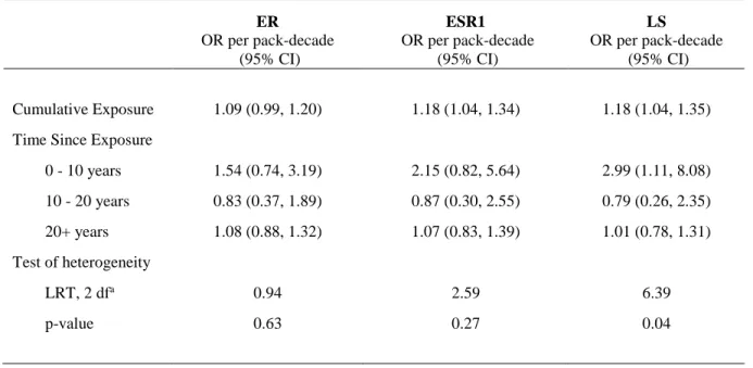
Table 4.1. Estimated odds ratios and 95% confidence intervals for cumulative smoking exposure and ER-defined breast cancer subtypes. ... 63
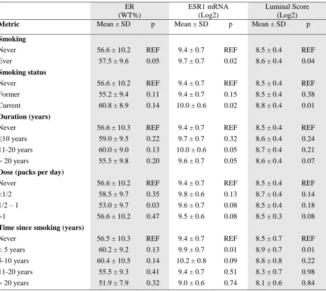
Table 4.2. Estimated biomarker expression values for the effect of categorical smoking measures. ... 65
Table 4.3. Estimated biomarker expression values for the effect of categorical smoking measures. ... 66
Table 5.1. Age, race, and smoking characteristics of CBCS III study participants. ... 84
Table 5.2. Estimated odds ratios and 95% confidence intervals for p53 protein-defined breast cancer subtypes (adjusted for age and race). ... 87
Table 5.3. Estimated odds ratios and 95% confidence intervals for p53 Mut or Wt (mRNA) breast cancer subtypes (adjusted for age and race). ... 88
Table 5.4. Estimated odds ratios and 95% confidence intervals for EGFR IHC-defined breast cancer subtypes (adjusted for age and race). ... 93
xiii
LIST OF FIGURES
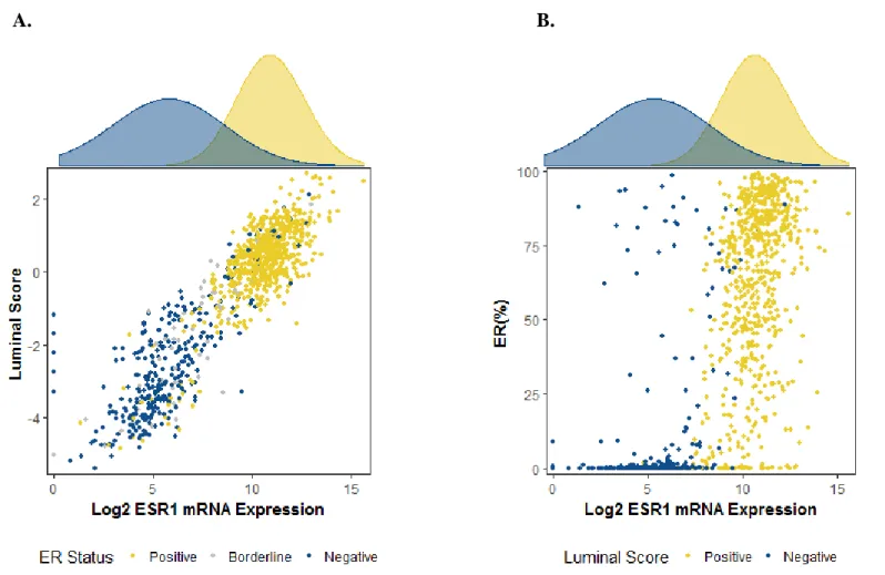
Figure 3.1. Power distributions for theoretical case-case odds ratio. ... 41 Figure 4.1. Relationships between ER IHC status, ESR1 mRNA
expression (log2), and luminal score (median-centered). ... 59 Figure 4.2. Categorical smoking metrics and association with ER, ESR1,
and LS breast cancer subtypes ... 60 Figure 4.3. Temporal associations between pack-decades of cigarettes
smoked and luminal score positive (LS+) breast cancer. ... 64 Figure 4.4. Distribution of ER protein by never, former, or current smoking status. ... 67 Figure 4.5. Distribution of ESR1 mRNA by never, former, or current smoking status. ... 68 Figure 4.6. Distribution of the luminal score by never, former, or
current smoking status. ... 69 Figure 5.1. P53 protein expression (%), by p53 mRNA signature binary classification. ... 85 Figure 5.2. Density graphs for MDM2 mRNA expression (Log2), by
p53 mRNA signature binary classification. ... 86 Figure 5.3. Boxplots displaying the distribution of weighted percent p53 protein (%)... 89 Figure 5.4. Boxplots displaying the distribution of MDM2 mRNA (log2) ... 90 Figure 5.5. Histogram of EGFR protein expression values among 1,964
breast tumors in CBCS III... 91 Figure 5.6. Histogram of EGFR mRNA expression values among 1,011
breast tumors in CBCS III... 92 Figure 5.7. Boxplots displaying the distribution of weighted percent
EGFR protein (%) ... 94 Figure 5.8. Boxplots displaying the distribution of EGFR mRNA (log2) ... 95 Figure 6.1. Distribution of estrogen-receptor (ER) protein expression
values, as measured by immunohistochemistry. ... 107 Figure 6.2. Distribution of p53 protein expression values. ... 108 Figure 6.3. Distribution of epidermal growth factor receptor (EGFR)
xiv
xv
LIST OF ABBREVIATIONS
CBCS Carolina Breast Cancer Study CI Confidence interval
CK 5/6 Cytokeratin 5/6
CPD Cigarette packs per day DNA Deoxyribonucleic acid DF Degrees of freedom
EGFR Epidermal Growth Factor Receptor ER Estrogen-receptor (protein)
ESR1 Estrogen-receptor (DNA/RNA)
HER2 Human Epidermal Growth Factor Receptor-2
HR Hazard ratio
IHC Immunohistochemistry IRR Incidence rate ratio
LS Luminal score
LRT Likelihood ratio test
MUT Mutant
OR Odds ratio
PAM50 Prediction Analysis of Microarray 50 PR Progesterone-receptor
PS Proliferation score
mRNA Messenger ribonucleic acid TMA Tissue microarray
TNBC Triple-negative breast cancer TP53 Tumor protein 53 (p53)
1
CHAPTER 1: SPECIFIC AIMS
Smoking is a suspected risk factor for breast cancer, based on weak-to-moderate measures of association, and the detection of tobacco smoke particulates in breast tissues of smokers. However, the US Surgeon General concludes that there is insufficient evidence to suggest a causal relationship between active smoking and breast cancer risk. Indeed,
epidemiologic studies have yielded mixed results. Because many breast cancers are estrogen dependent, studies that report earlier menopause and lower levels of circulating estrogens among smokers support an “anti-estrogenic” effect, and would suggest inverse risk.
However, few epidemiologic studies have supported an “anti-estrogenic” hypothesis; studies have more commonly suggested a positive association between smoking and breast cancer risk, consistent with tissue culture and animal experiments, showing that cigarette smoke causes DNA-damage, disrupts cell-cycle regulation, and is linked to malignant
transformation.
2
estrogen-mediated, genotoxic, and growth factor-mediated. Thus, a critical examination of smoking and breast cancer risk would benefit by examining intrinsic subtype and tumor biomarkers linked to pathogenesis. Moreover, such studies should carefully incorporate information on dose and timing of exposure. The examination of temporal and
dose-dependent patterns of smoking in relation to biomarker-defined breast cancer subtypes may identify time-windows of susceptibility that are associated with specific breast cancer subtypes.
The current proposal uses data from the population-based Carolina Breast Cancer Study (CBCS), which combines molecular biology and epidemiology to examine genetic and environmental risk factors for breast cancer. The CBCS has collected protein and RNA expression data on genes involved in breast tumor biology, specifically: estrogen receptor (ER) protein and RNA expression; p53 protein and p53-dependent RNA expression; and epidermal growth factor receptor (EGFR) protein and RNA expression. These hypotheses will be evaluated while simultaneously considering breast cancer intrinsic subtype.
3
of i) luminal and basal-like intrinsic subtypes and; ii) ER protein expression and ESR1 RNA levels.
Aim 2a: To examine the relationships between temporal and dose-dependent patterns smoking, breast cancer intrinsic subtype, and p53 expression. Rationale: Studies have
reported that breast cancer patients who were self-reported smokers at time of diagnosis had higher prevalence of specific TP53 mutations compared to their non-smoking counterparts. P53 expression regulates genomic stability and the DNA-damage response and may be an important mechanism underlying the relationship between smoking and breast cancer risk. Hypothesis: Temporal and dose-dependent variation in smoking is associated with variations in risk of p53 mutant cancers. Approach: To evaluate the temporal and dose-dependent association between smoking and p53+ breast cancer risk as measured by IHC and p53-dependent RNA signature.
4
CHAPTER 2: BACKGROUND
This year nearly 250,000 US women will be diagnosed with breast cancer1. Of these cases, only 1 in 4 will be attributed to high-penetrance germline mutations or familial clusters of the disease. The remaining three-fourths will have no known markers of heritable
susceptibility, leaving patients, communities, and researchers to grapple with understanding how environments and individual behaviors influence breast cancer risk. The identification of breast cancer intrinsic subtypes adds to the difficult task of understanding breast cancer etiology, as each subtype is hypothesized to have a distinct risk factor profile. It follows that a critical evaluation of any risk factor must consider the inherent heterogeneity across breast tumors, including differential expression of biomarkers linked to pathogenesis. Smoking is a suspected cause of breast cancer and has been linked to breast tumors that arise via estrogen-mediated, genotoxic, and growth-factor dependent mechanisms. In this proposal, we will examine the association between smoking exposure and breast tumor expression of
5
2.1 Two Etiologic Types of Breast Cancer
The heterogeneous nature of breast cancer has been well-established with the identification of distinct and reproducible intrinsic subtypes2. Gene expression studies have identified at least four subtypes that occur with predictable frequencies in representative populations of US women3-5, namely: Luminal A, Luminal B, HER2-enriched (HER2E), and basal-like breast tumors. Luminal A tumors are most common, occurring in approximately seventy percent of cases. In comparison to other subtypes, Luminal A tumors are the most genetically diverse and are characterized by high estrogen signaling, respond to hormone therapy, and may carry the most favorable prognoses6. Luminal B and HER2E tumors account for approximately ten and five percent of breast cancer cases, respectively. These two subtypes are characterized by overexpression or amplification of HER2 and may respond to hormone therapy and the monoclonal antibody, trastuzumab. However, Luminal B tumors have higher levels of estrogen signaling when compared with HER2E tumors. Estimates for the prevalence of basal-like tumors range between 10 and 20 percent of breast cancer cases. Basal-like tumors are characterized by low estrogen-signaling, lack targeted therapies, and are associated with clinical markers for aggressive disease. Though Luminal A, Luminal B, and HER2E tumors are defined by distinct gene expression profiles, each expresses proteins that are found predominantly in the luminal epithelial layer of the mammary gland and are thought to share luminal epithelial origins. By contrast, tumors that are classified as basal-like express proteins that are most abundant in the basal/myoepithelial layer of the mammary gland.
6
to suggest the existence of two main etiologic types – luminal and basal-like – based on estrogen-receptor expression and average age at onset7,8. Basal-like breast cancers are
estrogen-receptor negative (ER-) and have a younger average age at onset relative to luminal breast cancers, which are estrogen-receptor positive. This observation supports arguments that breast cancers of luminal epithelial and basal/myoepithelial origins represent two distinct etiologic classes of disease. In addition, a growing number of genomic platforms have
identified two distinct breast cancer clusters when representative samples of breast tumors are compared with tissues from other cancer types9. These clusters are characterized by tumors that are ER+ and ER-, which map to the luminal and basal-like phenotypes, respectively.
Several epidemiologic studies have examined associations between traditional breast cancer risk factors and breast tumors stratified by ER status (i.e., ER+ vs. ER-) or luminal and basal-like designations. Early age at menarche, African American race, and young age are associated with increased risks of Basal-like or triple-negative breast cancers10-13; these non-modifiable risk factors may reflect unmeasured risk factor profiles that increase
susceptibility to the basal-like phenotype. Lower body mass index (BMI) and breastfeeding among women with high parity are two modifiable risk factors associated with inverse risk of the basal-like breast cancer type10,12-16. Alcohol intake has been consistently linked to
7
2.2 Smoking and Breast Cancer Risk
Although the prevalence of cigarette smoking has steadily decreased since the 1950s, approximately 50% of women in the United States report a history of ever smoking and 14% are self-reported current smokers21. The Surgeon General’s 2014 report on the “health consequences of smoking” suggests there is sufficient evidence to identify mechanisms by which cigarette smoke could cause breast cancer, based on data from animal studies; the report concludes, however, that current population-based evidence is insufficient to infer causation. This conclusion has been attributed to inconsistent results from epidemiologic investigations, including the lack of an observed dose-response relationship. Indeed, epidemiologic studies of smoking and breast cancer risk have yielded a mix of positive and null findings, suggesting little or no increased risk of disease. And as in most studies of smoking and cancer incidence, duration – but not dose – has been more consistently associated with risk.
Several contemporary studies of smoking and breast cancer risk have reported positive associations in both age- and ethnically-diverse populations of women (Table 2.1). Investigators from the Multiethnic Cohort Study, Cancer Prevention Study II, Black
8
investigations by addressing biases inherent to observational study designs, including minimizing the potential for recall bias and control for confounding or effect measure modification due to established breast cancer risk factors. However, few studies have considered the heterogeneous nature of breast cancer as a source of bias in etiologic
investigations and its potential to mask associations between smoking and distinct molecular subtypes of breast cancer.
2.3 Smoking and Breast Tumor Biomarkers Linked to Pathogenesis
Tobacco smoke includes more than 70 carcinogens that have been evaluated by the International Agency for Research on Cancer (IARC), and which comprise eight chemical classes 20. Two of the largest classes – the polycyclic aromatic hydrocarbons (PAHs) and the N-nitrosamines – are thought to be responsible for cancer initiation in lung tumors.
Moreover, PAHs, N-nitrosamines – and their predecessor, nicotine – have been examined in human breast tissue and tissue culture for their ability to transform normal breast epithelium to cancer22-24. Metabolized forms of PAHs and the tobacco-specific N-nitrosamines can form covalent bonds at susceptible nucleotide binding sites to form DNA-adducts21. If the affected cell evades an arsenal of DNA repair mechanisms (e.g., nucleotide excision repair), the resultant adduct can yield single-base point mutations that may render a gene’s protein product non-functional. Further, smoking exposure may also result in chromosomal breaks and loss of heterozygosity, leading to DNA copy number aberration. Thus, protein and RNA expression levels of key genes may provide clues to understanding the etiology of smoking and breast cancer risk.
9
overexpression of the estrogen-receptor, TP53, and epidermal growth factor receptor (ER, p53, and EGFR), respectively. The overexpression of each marker reflects aberrant changes in cell-cycle regulation and homeostatic disruption of the tumor microenvironment, which allow a single cancer cell to gain selective advance and multiply through clonal expansion. By examining smoking exposure in relation to biomarkers linked to pathogenesis, we may identify etiologically-relevant subtypes that have been masked in prior epidemiologic studies of smoking and breast cancer risk.
2.3.1 Intrinsic Subtype
Triple subtypes – defined as the joint expression of the estrogen receptor (ER), progesterone receptor (PR), and human epidermal growth factor receptor 2 (HER2) – have been used as surrogates for intrinsic subtype in studies of breast cancer etiology. Each marker is measured by IHC or mRNA assay and is designated as positive (+) or negative (-), based on clinicopathologic cut points for overexpression. Luminal types are typically defined as tumors that are ER+ and or PR+, irrespective of HER2 expression. And triple negative tumors, surrogates for Basal-like tumors, are negative for all three markers. Although triple subtypes strongly correlate with gene expression profiles for intrinsic subtype, varying levels of discordance exist25. For example, approximately 75% of triple negative tumors are
10
Cancer Study, triple subtypes can be further refined by IHC assessment of cytokeratin 5/6 (CK 5/6) and epidermal growth factor receptor (EGFR), which are characteristic of the Basal-like phenotype. Future studies of smoking and breast cancer risk can also benefit from gene expression profiling of tumors, as described by the PAM50 gene signature.
2.3.2 Estrogen Receptor
With a large majority of breast cancers driven by exposure to estrogen, it has been suggested that the anti-estrogenic properties of cigarette smoke counteract its carcinogenic effects, leading to null or inconsistent associations of smoking and breast cancer risk among smokers26. This hypothesis is supported by the observation that smokers report earlier age at menopause and have lower levels of circulating estrogens. Nevertheless, epidemiologic investigations have commonly demonstrated a positive association between smoking and breast cancer risk, particularly for ER+ tumors (Table 2.3). In a recent population-based case-control study of women in the Seattle Puget-Sound metropolitan area, Kawai et al. reported a 40% increased risk of ER+ breast cancer among ever smokers (OR 1.4, 95% CI = 1.0 to 1.9), but found no association between smoking and ER- tumors. Similarly, in the Cancer
11
older studies of smoking and breast cancer risk in Swedish and Swiss populations have demonstrated positive associations between smoking and ER- breast cancer. Thus, a careful investigation should consider era and characteristics of population of interest.
2.3.3 Tumor protein p53
12 2.3.4 Epidermal Growth Factor Receptor
Until recently, nicotine - the most abundant and pharmacologically active component of cigarette smoke – had not been implicated in the development of breast cancer. Mouse xenograft models have shown that normal mammary cells transform to neoplastic cells upon exposure to nicotine and its derivatives27. And similar to findings from lung cancer studies28, breast epithelial cells treated with nicotine show increased rates of cell proliferation through activation of EGFR29, providing a measurable biological endpoint for a hypothesized
association between nicotine and EGFR+ breast cancer risk. EGFR overexpression is present in nearly three-fourths of basal like breast tumors, predicting increased risk of recurrence and poorer overall survival. Although less common, EGFR-positivity is also observed among Luminal A, Luminal B, and human epidermal growth factor receptor-2 (HER2) enriched intrinsic subtypes to varying degrees; however, its prognostic value for these breast tumor subtypes has not been defined. A recent study of basal-like marker expression (i.e., EGFR and/or CK 5/6) in luminal tumors showed that the distribution of traditional breast cancer risk factors (i.e., family history of breast cancer, age at menarche, parity, age at first full-term pregnancy, number of live births, breast feeding, and BMI) did not differ according to EGFR expression30. Although there were no apparent etiologic differences, Luminal tumors that expressed EGFR had more favorable clinical features when compared with tumors that did not express EGFR.
13
PR and were positively correlated with chromosome 7 polysomy, HER2 gene amplification, and EGFR protein expression31-35. EGFR+ tumors have also been shown to have inverse associations with disease-free survival, are more likely to have positive associations with lymph node status, and may be associated with specialized histological breast cancer types. In addition, EGFR+ tumors may be associated with higher proliferative fractions, increased aneuploidy, and increased tumor size. Although there is little data regarding the
epidemiology of EGFR+ disease, investigators for study of early stage breast cancer patients reported positive associations between high EGFR tumor expression, African American race, and young age.
The proposed study will be the first to investigate the association between smoking, concomitant nicotine exposure, and risk of breast cancer characterized by overexpression of EGFR. It is important to emphasize that prior investigations of smoking and breast cancer intrinsic subtype found no link between smoking and basal-like breast cancer, of which nearly three-fourths overexpress EGFR36. Recall, however, that EGFR may be overexpressed for all intrinsic subtypes to varying degrees25,36,37. Thus, dichotomizing breast cancers by EGFR expression (i.e., EGFR+ and EGFR-) will allow us to specifically assess whether smoking is associated with EGFR+ breast cancer across subtypes.
2.4 Exposure-Time-Windows and Breast Cancer Risk
14
of breast cancer diagnosis (Table 2.1). Both cohort and case-control studies have
incorporated measures of smoking initiation with respect to categories of age and smoking cessation relative to time-windows preceding disease diagnosis. These exposure-time-windows are defined by the investigator and observed associations may be sensitive to selected cut points. Improper specification of induction and latency periods leads to non-differential exposure misclassification and biases effect estimates toward the null38. Thus, methods that allow researchers to examine temporal patterns of smoking without selecting time periods a priori may be beneficial in studies of smoking and breast cancer risk.
The current proposal seeks to explore the temporal relationship between smoking and breast cancer risk. Logistic regression models that include parametric latency functions can be used to evaluate variation in disease risk by time since exposure. The inclusion of a parametric latency function in our logistic regression model will allow us to calculate time weighted exposure estimates using maximum likelihood estimation, where the highest weights are assigned during the period where smoking is associated with the greatest risk of EGFR positive breast cancer. In addition, non-parametric functions (e.g., B-splines) – which do not impose a specified probability distribution – can be used to visualize trends that describe differential breast cancer risk along the course of a woman’s smoking history. Further, as we explore temporal patterns of smoking in relation to biomarker-defined breast cancer types, we may observe associations that allow us to hypothesize temporal associations for the activation of a given mechanistic pathway with respect to smoking exposure.
15
smoking is associated with an early carcinogenic event. By using latency models for protracted exposures, we will identify exposure time frames where smoking is most etiologically relevant to biomarker-defined breast tumors.
2.5 Summary
Identifying the component causes of breast cancer remains one of the greatest public health challenges of the 21st century. Researchers have proposed smoking as a probable risk factor the disease; however, a definitive relationship between smoking and breast cancer risk has not been established. In this proposal, we will consider the heterogeneous nature of breast cancer and its potential to mask associations between smoking and distinct molecular
subtypes of breast cancer. We will also examine temporal patterns of smoking exposure using data-driven approaches that incorporate maximum likelihood estimation to identify critical exposure-time-windows. Results from our study may help to elucidate associations masked in prior studies.
2.6 Literature Review Tables
16
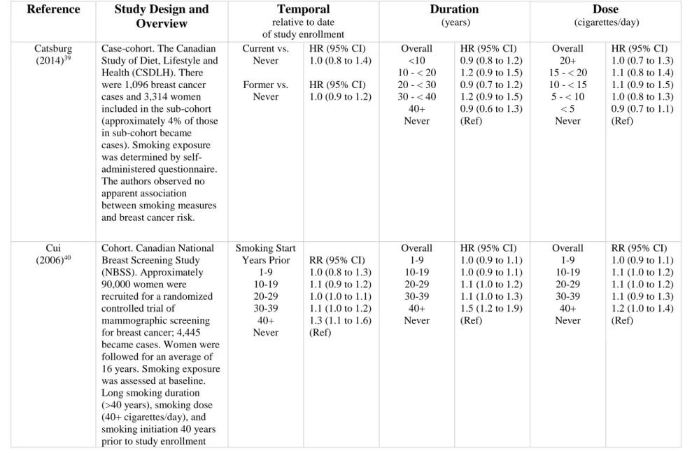
Table 2.1. Temporal and dose-dependent measures of smoking and breast cancer risk.
Reference Study Design and Overview
Temporal
relative to date of study enrollment
Duration (years) Dose (cigarettes/day) Catsburg (2014)39
Case-cohort. The Canadian Study of Diet, Lifestyle and Health (CSDLH). There were 1,096 breast cancer cases and 3,314 women included in the sub-cohort (approximately 4% of those in sub-cohort became cases). Smoking exposure was determined by self-administered questionnaire. The authors observed no apparent association between smoking measures and breast cancer risk.
Current vs. Never
Former vs. Never
HR (95% CI) 1.0 (0.8 to 1.4)
HR (95% CI) 1.0 (0.9 to 1.2)
Overall <10 10 - < 20 20 - < 30 30 - < 40
40+ Never
HR (95% CI) 0.9 (0.8 to 1.2) 1.2 (0.9 to 1.5) 0.9 (0.7 to 1.2) 1.2 (0.9 to 1.5) 0.9 (0.6 to 1.3) (Ref)
Overall 20+ 15 - < 20 10 - < 15 5 - < 10
< 5 Never
HR (95% CI) 1.0 (0.7 to 1.3) 1.1 (0.8 to 1.4) 1.1 (0.9 to 1.5) 1.0 (0.8 to 1.3) 0.9 (0.7 to 1.1) (Ref)
Cui
(2006)40
Cohort. Canadian National Breast Screening Study (NBSS). Approximately 90,000 women were recruited for a randomized controlled trial of
mammographic screening for breast cancer; 4,445 became cases. Women were followed for an average of 16 years. Smoking exposure was assessed at baseline. Long smoking duration (>40 years), smoking dose (40+ cigarettes/day), and smoking initiation 40 years prior to study enrollment
Smoking Start Years Prior 1-9 10-19 20-29 30-39 40+ Never
RR (95% CI) 1.0 (0.8 to 1.3) 1.1 (0.9 to 1.2) 1.0 (1.0 to 1.1) 1.1 (1.0 to 1.2) 1.3 (1.1 to 1.6) (Ref) Overall 1-9 10-19 20-29 30-39 40+ Never
HR (95% CI) 1.0 (0.9 to 1.1) 1.0 (0.9 to 1.1) 1.1 (1.0 to 1.2) 1.1 (1.0 to 1.3) 1.5 (1.2 to 1.9) (Ref) Overall 1-9 10-19 20-29 30-39 40+ Never
17
Reference Study Design and Overview
Temporal
relative to date of study enrollment
Duration
(years)
Dose
(cigarettes/day)
(continued) was associated with
increased breast cancer risk.
Dossus
(2014)41
Cohort. European Prospective Investigation into Cancer. Of the 322,988 women enrolled in the study, 9,822 developed breast cancer over an average follow-up period of 11 years. Smoking was assessed by baseline questionnaire. The authors observed a slight association between current or former smoking and increased breast cancer risk. There was a trend between increasing smoking duration and increased risk among current smokers. This trend was not evident among former smokers although the highest categories of smoking duration were associated with increased risk for this group. The highest categories of smoking dose were also associated with increased breast cancer risk for current and former smokers.
Current vs. Never
Former vs. Never
HR (95% CI) 1.1 (1.0 to 1.1)
HR (95% CI) 1.1 (1.0 to 1.1)
Current Smokers 0-10 10-20 20-30 >30 Never Former Smokers 0-10 10-20 20-30 >30 Never
HR (95% CI) 0.9 (0.7 to 1.3) 1.0 (0.8 to 1.1) 1.0 (1.0 to 1.1) 1.1 (1.0 to 1.2) (Ref)
HR (95% CI) 1.1 (1.0 to 1.2) 1.0 (0.9 to 1.1) 1.1 (1.0 to 1.2) 1.0 (0.9 to 1.1) (Ref) Current Smokers <6 6-10 10-15 ≥15 Never Former Smokers <6 6-10 10-15 ≥15 Never
HR (95% CI) 1.0 (0.9 to 1.1) 1.0 (0.9 to 1.2) 1.1 (1.0 to 1.2) 1.1 (1.0 to 1.2) (Ref)
18
Reference Study Design and Overview
Temporal
relative to date of study enrollment
Duration (years) Dose (cigarettes/day) Gaudet (2013)42
Cohort. The Cancer Prevention Study II (CPS-II) Nutrition Cohort. 97,786 women were enrolled in 1992 to examine cancer incidence and mortality. Median follow-up was 14 years 3,721 invasive breast cancers occurred. Both current and former smoking were associated with slight increased risks of breast cancer. Among former smokers, long duration (31 to 70 years) was associated with breast cancer risk. However, duration was not associated with risk among current smokers. There was no apparent association between smoking dose and breast cancer risk.
Current vs. Never
Former vs. Never
HR (95% CI) 1.3 (1.1 to 1.4)
HR (95% CI) 1.1 (1.1 to 1.2)
Current Smokers 1-40 40-49 50-73 Never Former Smokers <1-10 11-20 21-30 31-70 Never
HR (95% CI) 1.2 (0.9 to 1.5) 1.0 (0.9 to 1.4) 1.0 (0.9 to 1.6) (Ref)
HR (95% CI) 1.2 (1.1 to 1.3) 1.2 (1.1 to 1.3) 1.1 (1.1 to 1.3) 1.0 (1.2 to 1.4) (Ref) Current Smokers 1-9 10-19 20-29 30-39 40-90 Never
HR (95% CI) 1.2 (0.9 to 1.7) 1.0 (0.8 to 1.3) 1.2 (1.0 to 1.5) 1.1 (0.7 to 1.8) 1.4 (0.8 to 2.4) (Ref)
Gram
(2015)43
Cohort. The Multiethnic Cohort (MEC) Study. 83,300 women were enrolled and followed between 1993 and 2010. Of these, 4,484 developed invasive breast cancer. Smoking was assessed at baseline via questionnaire. Both current and former smoking were associated with slight increased risk of
Current vs. Never
Former vs. Never
HR (95% CI) 1.1 (1.0 to 1.2)
HR (95% CI) 1.0 (1.0 to 1.1)
Overall ≤20 21-30
>30 Never
HR (95% CI) 1.0 (0.9 to 1.1) 1.2 (1.1 to 1.3) 1.1 (1.0 to 1.2) (Ref) Overall ≤10 11-20 >20 Never
19
Reference Study Design and Overview
Temporal
relative to date of study enrollment
Duration
(years)
Dose
(cigarettes/day)
(continued) breast cancer. Long
smoking duration and high dose were also associated with increased risk of disease.
Nyante
(2014)44
Cohort. AARP (formerly American Association of Retired Persons). 186,150 female study participants were enrolled in 1995-96 and followed for an average of 10 years. Smoking exposure was assessed via baseline questionnaire. However, smoking duration was not assessed at baseline. Current and former smoking were associated with slight increased risk of breast cancer. There was no apparent trend between smoking dose and breast cancer risk.
Current vs. Never
Former vs. Never
HR (95% CI) 1.2 (1.1 to 1.3)
HR (95% CI) 1.1 (1.0 to 1.1)
Not reported. Current
Smokers 1-10 11-20 21-30 31-40 ≥41 Never Former Smokers 1-10 11-20 21-30 31-40 ≥41 Never
HR (95% CI) 1.2 (1.0 to 1.3) 1.2 (1.1 to 1.4) 1.1 (0.9 to 1.2) 1.1 (0.9 to 1.4) 1.4 (0.6 to 1.5) (Ref)
20
Reference Study Design and Overview
Temporal
relative to date of study enrollment
Duration (years) Dose (cigarettes/day) Rosenberg (2013)45
Cohort. The Black Women's Health Study. 52,425 women were followed for 14 years between 1997 and 2009. 1,377 breast cancer cases occurred. Smoking was assessed at baseline via questionnaire. When compared with never active or passive smokers, current and former smoking were not associated with
increased breast cancer risk. Women who smoked 20 pack-years had slight increased risk of breast cancer. Current vs. Never Active or Passive Former vs. Never Active or Passive
IRR (95% CI) 1.1(0.8 to 1.3)
IRR (95% CI) 1.1 (0.9 to 1.4)
Pack-years <10 10-19
20
IRR (95% CI) 1.1 (0.9 to 1.3) 1.0 (0.8 to 1.3) 1.2 (1.0 to 1.5)
Reported composite measure of pack-years.
Xue
(2011)46
Cohort. The Nurse's Health Study. 111,140 women were enrolled at baseline (1976) and were followed through 2006. 8,772 incident cases of breast cancer occurred. Active smoking exposure was assessed via baseline questionnaire and updated biennially. Current and former smoking were associated with increased risk of breast cancer.
Current vs. Never
Former vs. Never
HR (95% CI) 1.1 (1.0 to 1.2)
HR (95% CI) 1.1 (1.0 to 1.1)
Overall <20 20-39
≥40 Never
HR (95% CI) 1.0 (1.0 to 1.1) 1.1 (1.0 to 1.1) 1.2 (1.0 to 1.3) (Ref) Current Smokers 1-14 15-24 ≥25 Never Former Smokers 1-14 15-24 ≥25 Never
HR (95% CI) 1.0 (0.9 to 1.2) 1.1 (1.0 to 1.2) 1.1 (1.0 to 1.3) (Ref)
21
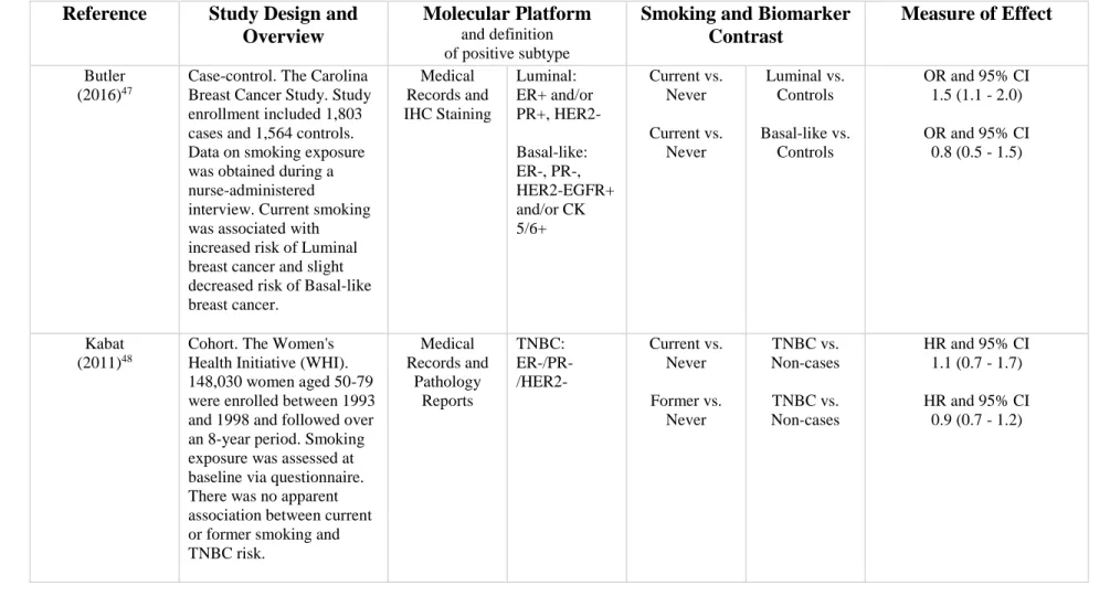
Table 2.2. Associations between smoking and risk of breast cancer intrinsic subtype.
Reference Study Design and
Overview
Molecular Platform
and definition of positive subtype
Smoking and Biomarker Contrast
Measure of Effect
Butler
(2016)47
Case-control. The Carolina Breast Cancer Study. Study enrollment included 1,803 cases and 1,564 controls. Data on smoking exposure was obtained during a nurse-administered interview. Current smoking was associated with increased risk of Luminal breast cancer and slight decreased risk of Basal-like breast cancer. Medical Records and IHC Staining Luminal: ER+ and/or PR+, HER2- Basal-like: ER-, PR-, HER2-EGFR+ and/or CK 5/6+ Current vs. Never Current vs. Never Luminal vs. Controls Basal-like vs. Controls
OR and 95% CI 1.5 (1.1 - 2.0)
OR and 95% CI 0.8 (0.5 - 1.5)
Kabat
(2011)48
Cohort. The Women's Health Initiative (WHI). 148,030 women aged 50-79 were enrolled between 1993 and 1998 and followed over an 8-year period. Smoking exposure was assessed at baseline via questionnaire. There was no apparent association between current or former smoking and TNBC risk. Medical Records and Pathology Reports TNBC: ER-/PR-/HER2- Current vs. Never Former vs. Never TNBC vs. Non-cases TNBC vs. Non-cases
HR and 95% CI 1.1 (0.7 - 1.7)
22
Reference Study Design and
Overview
Molecular Platform
and definition of positive subtype
Smoking and Biomarker Contrast
Measure of Effect
Kawai
(2014)49
Case-control. Seattle-Puget Sound metropolitan area. There were 960 cases and 938 controls. Smoking exposure was obtained via questionnaire and restricted to those that occurred prior to reference date. Current and former smoking were not associated with TNBC breast cancer risk.
ER+ or PR+: staining in ≥ 1% of tumor
cells. HER2+: FISH 3+ TNBC: ER-/PR-/HER2- Current vs. Never Former vs. Never TNBC vs. Controls TNBC vs. Controls
OR and 95% CI 1.2 (0.7 - 2.1)
OR and 95% CI 0.9 (0.6 - 1.5)
Millikan
(2008)13
Case-control. The Carolina Breast Cancer Study. Study enrollment included 1,803 cases and 1,564 controls. Data on smoking exposure was obtained during a nurse-administered
interview. Smoking duration was not differentially associated with Luminal A or Basal-like breast cancer.
Medical Records and IHC Staining Luminal A: ER+ and/or PR+, HER2- Basal-like: ER-, PR-, HER2-EGFR+ and/or CK 5/6+ Years <10 11-19 20+ Never Basal-like vs. Luminal A
OR (95% CI)
0.9 (0.6 to 1.5) 1.1 (0.7 to 1.7) 0.7 (0.5 to 1.1)
(Ref)
Tariq
(2014)50
Prospective cohort. Tumor registry at the University of Florida at Jacksonville (2000-2005). Smoking status (ever = current or past) was recorded in the tumor registry and was not associated with TNBC tumors.
Tumor registry TNBC: ER-/PR-/HER2-
Ever vs. Never TNBC vs.
Non-TNBC
Proportion 20% vs. 28%
23
Reference Study Design and
Overview
Molecular Platform
and definition of positive subtype
Smoking and Biomarker Contrast
Measure of Effect
Turkoz
(2013)51
Cross-sectional. Department of Medical Oncology at Hacettepe University, Institute of Oncology. The study identified 1,884 invasive cases that were eligible for analysis. Smoking exposure was obtained during physician-led interview.
ER+ or PR+: staining in ≥ 5% of tumor
cells.
HER2+: FISH 3+
Luminal: ER+ or PR+
TNBC: ER-/PR-/HER2-
Ever vs. Never
Ever vs. Never
Luminal vs. Non-Luminal
TNBC vs. Non-TNBC
OR and 95% CI 1.0 (0.7 - 1.4)
24
Table 2.3. Associations between smoking and risk of ER-defined breast cancer.
Reference Study Design and
Overview
Molecular Platform
and definition of positive subtype
Smoking and Biomarker Contrast
Measure of Effect
Cooper
(1989)52
Case-control. Adelaide, South Australia. The study included 451 cases
identified through the South Australian Central Cancer Registry between 1982 and 1984. There were 451 age-matched controls. Smoking exposure data was obtained in-person interview. Former smoking exposure was associated with increased risk of ER- breast cancer.
Saturation analysis assay
ER+: ≥ 10 fmol/mg Current vs. Never Former vs. Never Current vs. Never Former vs. Never ER+ vs. Controls ER+ vs. Controls ER- vs. Controls ER- vs. Controls
OR and 95% CI 1.3 (0.8 - 2.0)
OR and 95% CI 0.9 (0.6 - 1.4)
OR and 95% CI 1.3 (0.7 - 2.5)
OR and 95% CI 1.9 (1.0 - 3.6)
Gaudet
(2013)53
Cohort. The Cancer Prevention Study II (CPS-II) Nutrition Cohort. 97,786 women were enrolled in 1992 to examine cancer incidence and mortality. Median follow-up was 14 years 3,721 invasive breast cancers occurred. Both current and former smoking were associated with slight increased risks of breast cancer. Current and former smoking were associated with increased risk of ER+ breast cancer. However, neither were associated with risk of ER- breast cancer.
Medical Records and
Pathology Reports
SEER (NOS) Current vs.
Never Former vs. Never Current vs. Never Former vs. Never ER+ vs. Controls ER+ vs. Controls ER- vs. Controls ER- vs. Controls
OR and 95% CI 1.2 (1.0 - 1.5)
OR and 95% CI 1.1 (1.0 - 1.3)
OR and 95% CI 0.9 (0.5 - 1.4)
25
Reference Study Design and
Overview
Molecular Platform
and definition of positive subtype
Smoking and Biomarker Contrast
Measure of Effect
Kabat
(2011)48
Cohort. The Women's Health Initiative (WHI). 148,030 women aged 50-79 were enrolled between 1993 and 1998 and followed over an 8-year period. Smoking exposure was assessed at baseline via questionnaire. Former smokers had a slight increased risk of ER+ breast cancer.
Medical Records and
Pathology Reports
ER+: NOS Current vs.
Never Former vs. Never ER+ vs. Non-cases ER+ vs. Non-cases
HR and 95% CI 1.1 (0.9 - 1.3)
HR and 95% CI 1.1 (1.1 - 1.2)
Kawai
(2014)49
Case-control. Seattle-Puget Sound metropolitan area. There were 960 cases and 938 controls. Smoking exposure was obtained via questionnaire and restricted to those that occurred prior to reference date. Current and former smoking were associated with increased risk of ER+ breast cancer.
Medical Records and
Pathology Reports
ER+: nuclear staining in ≥ 1% of tumor cells. Current vs. Never Former vs. Never ER+ vs. Controls ER+ vs. Controls
OR and 95% CI 1.4 (1.0 - 1.9)
OR and 95% CI 1.3 (1.0 - 1.7)
Manjer
(2001)54
Cohort. Malmo, Sweden. 10,902 women born between 1926 and 1949 with average age at enrollment of 50 years. Women were enrolled between 1974 and 1992 for average follow-up of 12 years; 268 incident cases with available tumor tissue
IHC ER+:
Immuno-reactivity Current vs. Never Former vs. Never Current vs. Never ER+ vs. Controls ER+ vs. Controls ER- vs. Controls
RR and 95% CI 0.9 (0.6 - 1.2)
RR and 95% CI 1.0 (0.7- 1.5)
26
Reference Study Design and
Overview
Molecular Platform
and definition of positive subtype
Smoking and Biomarker Contrast
Measure of Effect
(continued) occurred. A
self-administered questionnaire was used to obtain data on smoking exposure. Current and former smoking exposure was associated with increased risk of ER-, but not ER+, breast cancer.
Former vs. Never
ER- vs. Controls
RR and 95% CI 2.7 (1.4 - 5.0)
Morabia
(1998)55
Case-control. Geneva, Switzerland. 372 cases diagnosed between 1992 and 1993; 1,059 controls were included. Data on smoking exposure was obtained during in-person interview. Ever smokers who smoked 20 or more cigarettes per day were at increased risk of developing ER+ and ER- breast cancer.
IHC ER+: nuclear
staining in ≥ 20% of tumor cells.
Ever 20+ cpd vs. Never
Ever 20+ cpd vs. Never
ER+ vs. Controls
ER- vs. Controls
OR and 95% CI 2.4 (1.4 - 4.5)
OR and 95% CI 4.3 (1.4 - 13.0)
Nishino
(2014)56
Case-control. Miyagi Cancer Center Hospital. 1,309 breast cancer cases and 3,878 controls. Smoking exposure was assessed via questionnaire. Current or former smoking was not associated with either of the 4 ER/PR subtypes.
Medical Records and
Pathology Reports
IHC and EIA
ER+/PR+: NOS Current vs. Never ER+/PR+ vs. Controls ER+/PR-vs. Controls ER+/PR-vs. Controls ER-/PR- vs. Controls
Null Associations for current and former smoking for all combinations of ER+/- and
27
Reference Study Design and
Overview
Molecular Platform
and definition of positive subtype
Smoking and Biomarker Contrast
Measure of Effect
Nyante
(2014)44
Cohort. AARP (formerly American Association of Retired Persons). 186,150 female study participants were enrolled in 1995-96 and followed for an average of 10 years. Smoking exposure was assessed via baseline questionnaire. SEER Cancer Registry ER+/PR+: NOS Current vs. Never Current vs. Never Current vs. Never ER+/PR+ vs. Controls ER-/PR+ vs. Controls ER-/PR- vs. Controls
OR and 95% CI 1.0 (0.9 - 1.2)
OR and 95% CI 1.4 (1.0 - 1.8)
28
Table 2.4. Associations between smoking and risk of p53-defined breast cancer.
Reference Study Design and
Overview
Molecular Platform
and definition of positive subtype
Smoking and Biomarker Contrast
Measure of Effect
Conway
(2002)57
The Carolina Breast Cancer Study. 456 invasive breast cancer cases were evaluated for specific TP53 mutations. Of these, 108 breast cancers (or 24%) harbored specific TP53 mutations. 71% of mutations were missense; the remaining were deletions or insertions. Relative to non-smokers, current smokers were more likely to harbor p53 mutations. Gene Sequencing p53+: presence of somatic mutation in exons 4-8 Current vs. Never Former vs. Never
p53+ vs. p53-
p53+ vs. p53-
OR and 95% CI 2.1 (1.2 - 3.8)
OR and 95% CI 0.6 (0.4 - 1.2)
Furberg
(2002)58
The Carolina Breast Cancer Study. The authors examined smoking exposure in relation to overexpression of p53 protein among 683 cases. There was no apparent association between smoking status and p53 tumor expression. The authors also investigated the associations between smoking dose, duration, and p53 expression and observed no apparent associations.
IHC p53+: dark
nuclear protein staining in ≥ 10% of tumor cells.
Current vs. Never
Former vs. Never
p53+ vs. p53-
p53+ vs. p53-
OR and 95% CI 0.8 (0.6 - 1.3)
29
Reference Study Design and
Overview
Molecular Platform
and definition of positive subtype
Smoking and Biomarker Contrast
Measure of Effect
Gammon
(1999)59
The Long Island Breast Cancer Study. Investigators examined the prevalence of p53
overexpression in breast tissues of young women (age < 45 years). Current smoking - but not former smoking - was associated with increased risk of p53 overexpression in breast tumors.
IHC p53+:
moderate to strong nuclear protein staining in ≥ 10% of tumor cells.
Current vs. Never
Former vs. Never
p53+ vs. p53-
p53+ vs. p53-
OR and 95% CI 2.0 (1.1 - 3.5)
OR and 95% CI 1.4 (0.8 - 2.4)
Mordukhovich
(2010)60
The Long Island Breast Cancer Study Project. 859 invasive breast tumors were evaluated for TP53 mutations (exons 5 - 8). Of these, 151 harbored a p53 mutation. Current and former smokers were slightly less likely to have a p53 mutation, when compared with never smokers. Gene Sequencing p53+: presence of somatic mutation in exons 5-8 Current vs. Never Former vs. Never
p53+ vs. p53-
p53+ vs. p53-
OR and 95% CI 0.7 (0.4 - 1.2)
30
CHAPTER 3: METHODS
3.1 Overview
Breast cancer is not a single disease, but exists as a collection of genetically distinct subtypes. These subtypes have prognostic and predictive value in clinical settings and may also provide clues to breast cancer etiology. Homogenous classifications of breast cancer may have masked associations in prior studies of smoking and breast cancer risk; thus, a critical examination would benefit by evaluating smoking in relation to etiologically-relevant subtypes. In addition, it is important to consider dose and timing of exposure. By examining temporal and dose-dependent patterns of smoking, we may identify etiologically-relevant time periods that are associated with risk of specific breast cancer subtypes. Using data from phase III of the Carolina Breast Cancer Study (CBCS III), we seek to examine temporal and dose-dependent relationships between smoking exposure, breast cancer intrinsic subtype, and breast tumor expression of biomarkers linked to pathogenesis. Phase III is a case only study of approximately 3,000 women diagnosed with invasive breast cancer between 2008 and 2013 in central and eastern North Carolina. The CBCS includes self-reported data on
smoking exposure and biomarker data obtained from tumor specimens, including protein and RNA expression data on genes with suspected links between smoking and breast
carcinogenesis.
3.2 Study Design
31
genetic risk factors for breast cancer. Breast cancer cases were identified by a rapid case ascertainment system, implemented through collaboration between Lineberger
Comprehensive Cancer Center (LCCC) and the North Carolina Central Cancer Registry (NCCCR). To be eligible for inclusion, patients must have been female and received a first and primary diagnosis of breast cancer between May 1, 2008 and October 31, 2013. The patient also must have resided in the 44-county study region and been between the ages of 20 and 74 at the time of diagnosis.
To examine potential risk factor differences by age and race, the CBCS employed a randomized recruitment strategy that was designed to oversample young and African American women. The patient’s primary physician was contacted to obtain permission to invite the patient into the study and the overall expected response rate is 70%. Patients who declined participation were not substantially different from those who chose to participate in the study. In total, 2,998 women were enrolled in CBCS III. Study participants were asked to consent to a nurse-administered in-person interview that took place in the study participant’s home or another pre-arranged location. During the in-person interview the nurse
administered a questionnaire that included items on family and personal medical history, reproductive history, smoking, alcohol, diet, medication use and occupational history. Upon consent, the nurse also collected a blood sample and objective anthropometric measurements of height (m), weight (kg, waist (m), and hip (m) circumference. The average time between study enrollment and interview was 6 months.
32
pathology reports and medical records from the treating facilities. Clinical and pathological data abstracted from medical records and pathology reports included tumor size, stage, and node status. For all cases, a single pathologist (Dr. Joseph Geradts) determined tumor grade.
3.3 Outcome Assessment
The CBCS includes protein and RNA expression data on genes used to define intrinsic subtype and genes involved in mechanistic pathways that may link smoking to breast cancer risk (Table 3.1). We will examine binary and continuous outcomes of biomarker expression for intrinsic, estrogen-mediated, genotoxic, and growth-factor dependent mechanistic pathways. Specifically, we will use gene expression signatures and protein-specific cut points to characterize tumors as positive (+) or negative (-) for the given biomarker or biomarker pathways. We will use continuous measures of protein and counts of RNA transcripts to examine whether smoking exposure modulates biomarker expression. Tissue microarrays (TMAs) were constructed for immunohistochemical (IHC) staining of estrogen receptor (ER), progesterone receptor (PR), human epidermal growth factor receptor-2 (HERreceptor-2), cytokeratin 5/6 (CK 5/6), epidermal growth factor receptor (EGFR), and p53. Automated quantification of staining was performed using Genie classifier and protein-specific algorithms61. RNA was extracted from the same tumor specimens used to construct the TMAs, using the Qiagen RNeasy FFPE kit and protocol. RNA expression data were obtained via Nanostring assay. TMA construction and IHC analyses were conducted at Tissue Pathology Laboratory (TPL) and the Immunohistochemical Core Laboratory (ICL) at UNC Chapel Hill. Nanostring assays were performed in the laboratory of Dr. Melissa
33
Table 3.1. Molecular characterization of breast tumors in CBCS III
Sp
ec
if
ic
Aim
s Specific
Aims Aim 1 Aim 2
Pathway a. Intrinsic b.
Estrogen-Mediated a. Genotoxic
b. Growth-Factor Dependent Biomarker
comparison Luminal vs. Basal ER+ vs. ER- p53+ vs. p53-
EGFR+ vs. EGFR- M o lecula r Cha ra ct er iza tio
n Single gene
IHC Nielsen37 (2004) Hammond62 (2010) Williams63
(2017) 10%
Single gene
mRNA N/A Continuous NA Continuous
Multigene IHC signature
Nielsen37
(2004) NA NA NA
Multigene mRNA signature
Parker64
(2009) NA
Troester65
(2006) NA
Note. NA-Not Applicable
3.3.1 Intrinsic subtype, multigene mRNA and IHC biomarkers
Breast cancer intrinsic subtype was measured using the RNA-based “PAM50 signature”64. Here, differential expression of the 50-gene signature is used to categorize breast cancers into 4 intrinsic subtypes: Luminal A, Luminal B, HER2E, and Basal-like. Each case was classified based upon highest Pearson correlation with a centroid defined for each subtype. For the analyses outlined in this proposal, we combined Luminal A, Luminal B, and HER2E tumors, since each have suspected luminal epithelial origins and may represent a single etiologic subtype8. Together, these tumors represent the Luminal breast cancer intrinsic subtype and will be compared to Basal-like breast tumors.
34
overexpression. Automated quantification of ER and PR protein expression was determined by Genie classifier and the Aperio nuclear v9 algorithm; HER2 and EGFR quantification was determined using the Aperio membrane v9 algorithm (Aperio Technologies, Vista, CA). Definiens Tissue Studio® was used to quantify CK5/6 staining. Intrinsic subtypes were designated as follows: Luminal breast cancers were defined as (ER+ and/or PR+, regardless of HER2 status); and Basal-like tumors were defined as (ER-, PR-, HER2-, EGFR+ and/or CK5/6+). We will compare Luminal and Basal-like breast tumors.
3.3.2 Estrogen-mediated biomarkers
We will use a cut point of ≥ 1% of tumor cells with nuclear protein staining for ER to define borderline/positive ER status, as recommended by the American Society of Clinical Oncology (ASCO) and College of American Pathologists (CAP)62. Specifically, we will define tumors with 1% to < 10% expression as ‘borderline positive’ and tumors with ≥ 10% expression as ‘positive’. Automated quantification of ER protein expression was determined by a Genie classifier and the Aperio nuclear v9 algorithm (Aperio Technologies, Vista, CA). In addition, we will examine quantiles and continuous measures of ER protein staining in relation to smoking exposure. Specifically, we will examine whether smoking exposure modulates protein expression of ER in breast tumors. We will also examine whether smoking exposure modulates ER mRNA expression.
3.3.3 Genotoxic biomarkers
35
The mutant or wildtype designations characterize downstream biologic activity following p53 loss or activation, respectively.
We will use a cut point of ≥ 10% of tumor cells with nuclear protein staining for p53 as described by Williams et al.63. Automated quantification of p53 protein expression was determined by a Genie classifier and the Aperio nuclear v9 algorithm (Aperio Technologies, Vista, CA). As described above for ER protein staining, we will also examine continuous measures of p53 protein staining in relation to smoking exposure.
3.3.4 Growth-factor dependent biomarkers
We will explore the relationships between categorical and continuous measures of EGFR protein and RNA and measures of smoking exposure. Automated quantification of EGFR protein expression was determined by a Genie classifier and the Aperio membrane v9 algorithm (Aperio Technologies, Vista, CA).
3.4 Exposure Assessment
36
cessation, where applicable (recency). Together, these data will be used to derive categorical, ordinal, and continuous measures of smoking (e.g. pack-years).
3.5 Covariate Assessment
We selected potential confounders of the relationship between smoking and breast cancer risk based on a review of the literature. These include: 1. First-degree family history of breast cancer defined as breast cancer diagnosis for mother or full female siblings16; 2. Alcohol consumption defined as number of drinks consumed per week15,17,67; 3. Breast feeding characterized by age at first breast feeding13; 4. Body Mass Index (BMI kg/m2)13; 5. Income to family ratio calculated as the household income divided by the number present in the household68; 6. Parity defined as number of full-term births13,15; 7. Years of oral
contraceptive use10; 8. Years of hormone replacement therapy use10; 9. Physical activity; 10. Age; and 11. Race.
To identify a minimal adjustment set among the eleven potential confounders, we conducted directed acyclic graph analysis using DAGitty software (version 2.3). The smallest minimal adjustment set is as follows: alcohol, breast feeding, family history, income, parity, physical activity, age, and race. For continuous variables, we will explore various coding modalities to identify the coding scheme that results in the best fit and most parsimonious model. Covariates will be included in regression models as confounders based on whether there is a 10% or greater change in estimate when added to the model.
3.6 Data Analysis
37
expression of biomarkers linked to estrogen-mediated (Aim 1b), genotoxic (Aim 2a), and growth-factor mediated (Aim 2b) pathways. The examination of temporal and dose-dependent patterns of smoking in relation to biomarker-defined subtypes may identify exposure-time-windows that are associated with specific mechanistic events.
3.6.1 Linear and logistic regression models
We will use linear regression to model the relationship between smoking exposure and continuous measures of protein or RNA expression. We will test assumptions of linearity by examining higher order polynomials of smoking exposure. Smoking exposure will be modeled as a continuous measure of pack-years, defined as the product of the number of cigarettes smoked per day (dose) and the total number of years smoked (duration). We will examine the association between pack-years and biomarker expression defined as weighted percent cells of positive and the b) H-score. An H-score is a weighted measure of the number of weakly (1), moderate (2), and strongly stained (3) cells in the tissue sample and the ranges between 0 and 300. In addition to measures of percent positivity we will also examine pack-years in relation to quantiles of gene expression using generalized logit models with ordinal outcomes.
3.6.2 Binary outcomes and categorical measures of smoking
38
3.6.3 Cumulative smoking exposure and time-windows analysis
For each binary breast cancer classification, we will use generalized logit models to estimate ORs and 95% CIs for risk of breast cancer subtype defined as a trend per pack-year for a) cumulative pack-years smoked and b) within exposure-time-windows defined by time since exposure. c) We will also use latency functions and associated graphs to visualize the temporal relation between smoking exposure and subtype-specific breast cancer risk. Evidence of an association between smoking and breast cancer risk proximal to time of diagnosis may allow us to infer that smoking is associated with a cancer promotion event for a given biomarker pathway. By contrast, if smoking exposure is associated with breast cancer risk at a distal point from date of diagnosis, we may infer that smoking is associated with cancer initiation or promotion events. Previous case-control studies of smoking and breast cancer risk have described an increased risk of disease among women who quit smoking 5 to 10 years prior to date of case/control selection42. For our analysis, we selected three time-windows that would accommodate available sample size: <10 years; 10-20 years; > 20 years. We will conduct likelihood ratio tests to determine whether our time windows analyses improve model fit, when compared cumulative exposure models.
3.6.4 Parametric latency functions of smoking and breast cancer risk
39
maximum likelihood estimation (MLE); the highest weights are assigned during the time period where smoking is associated with the greatest risk of subtype-specific breast cancer. That is, our modeling strategy will allow us to identify peak risk according to recency of exposure for each biomarker-defined breast cancer type. We will examine hierarchical models with bilinear and log-normal latency functions. If our B-spline from analysis 2c demonstrates a multimodal relationship between smoking exposure and breast cancer risk, we will explore different probability distributions for our parametric latency function.
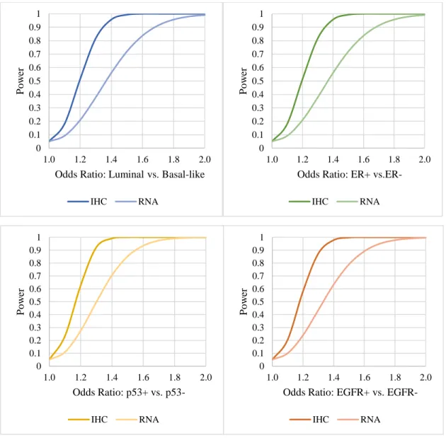
3.7 Power Analysis
40
Table 3.2. Expected distributions of subtypes in CBCS III.
Specific Aim Contrast Ratio*
Aim 1a. Luminal vs. Basal 4:1
Aim 1b. ER+ vs. ER- 4:1
Aim 2a. p53+ vs. p53- 1:2
Aim 2b. EGFR+ vs. EGFR- 1:3
41
Figure 3.1. Power distributions for theoretical case-case odds ratio. 0 0.1 0.2 0.3 0.4 0.5 0.6 0.7 0.8 0.9 1
1.0 1.2 1.4 1.6 1.8 2.0
P
o
w
er
Odds Ratio: Luminal vs. Basal-like
IHC RNA 0 0.1 0.2 0.3 0.4 0.5 0.6 0.7 0.8 0.9 1
1.0 1.2 1.4 1.6 1.8 2.0
P
o
w
er
Odds Ratio: ER+
vs.ER-IHC RNA 0 0.1 0.2 0.3 0.4 0.5 0.6 0.7 0.8 0.9 1
1.0 1.2 1.4 1.6 1.8 2.0
P
o
w
er
Odds Ratio: p53+ vs.
p53-IHC RNA 0 0.1 0.2 0.3 0.4 0.5 0.6 0.7 0.8 0.9 1
1.0 1.2 1.4 1.6 1.8 2.0
P
o
w
er
Odds Ratio: EGFR+ vs.
42
3.8 Summary
3.8.1 Limitations
Although our study has several benefits, it is important to acknowledge limitations. The CBCS III study design did not include the recruitment of a control group that could be used to assess the baseline exposure distribution. Thus, our case-case odds ratios use a subset of cases as the referent group and the distribution of smoking exposure in this group may not reflect that of the population from which the case groups arise. To address this potential pitfall, we will perform a sensitivity analysis using controls from CBCS phases I and II. Controls from these study time periods were enrolled between 1993 and 2001. Cases in phase III were enrolled between 2008 and 2013. We will use generalized logistic regression
functions with polytomous outcomes to calculate both case-control and case-case odds ratios. If the case-case odds ratios in our sensitivity analysis are similar to those which are observed in our phase III case-only analysis, we may infer that controls from the early phases are exchangeable for controls that would have been collected for phase III and are suitable to be used in phase III to evaluate associations between risk factors and subtype-specific breast cancer risk. If the case-case odds ratios from the primary and sensitivity analyses are not comparable, we will report the case-case odds ratios and include a detailed description of this limitation.
3.8.2 Strengths
43
development. Further, examining temporal patterns of smoking in relation to biomarker-defined breast cancers may allow us to infer whether the expression of a given biomarker is associated with smoking exposures that are proximal or distal to time of diagnosis. Together, the strengths of this study may help to elucidate associations masked in prior investigations.
3.9 Addendum
44
CHAPTER 4: SMOKING AND ESTROGEN-MEDIATED BIOMARKERS
4.1 Introduction
Epidemiologic studies have demonstrated distinct risk factor profiles for breast cancer subtypes classified according to estrogen-receptor (ER) status11,13. Defined by an
immunohistochemical (IHC) threshold of 1 to 10 percent staining of examined tumor cells, ER-positive (+) breast tumors account for more than 70% of all breast cancer cases
diagnosed in the United States (US), making this disease group an important public health focus4,62. Pre-diagnostic smoking exposure has been linked to the ER+ subtype in some epidemiologic studies, with increased risks ranging between 10% to 50%44,49,69. Further, prospective studies of breast cancer survivors have suggested that smoking exposure prior to diagnosis may influence survival outcomes, particularly among women with ER+ disease, and presumably through reduced efficacy of anti-estrogenic therapies. For example, in a prospective study of Swedish breast cancer patients, ER+ women who smoked before diagnosis and who were treated with an aromatase inhibitor (AI) had a 3-fold increased risk of experiencing distant metastases or death, when compared to ER+/AI-treated non-smokers [Hazard Ratio (HR) and 95% CI (3.0 and 1.4 to 6.1)]70. Together, these epidemiologic and clinical findings could be used to suggest that smoking exposure may be linked to estrogen metabolism in breast tumors and subsequent regulation of ER.