The Journal of Clinical Investigation
R e s e a R c h a R t i c l eIntroduction
Type I NKT cells (NKTs) are an evolutionarily conserved subset of innate lymphocytes that express invariant T cell receptor (TCR) α-chain Vα24-Jα18 and react to self- or microbial-derived glyco-lipids presented by the monomorphic HLA class I–like molecule CD1d (1–4). The potential importance of NKTs for tumor immu-nity and immunotherapy has been demonstrated in multiple models of cancer in mice and in early-stage clinical trials in can-cer patients (5–10). In contrast to T cells, NKTs effectively traffic to the tumor site and can mediate antitumor responses via either direct killing of CD1d+ tumor cells, inhibition of tumor-supportive macrophages, or transactivation of NK cells (11). Several studies
have revealed strong positive associations between the numbers of tumor-infiltrating or circulating NKTs and improved disease outcome in patients with diverse tumor types (6, 12–15). Con-versely, tumor progression is often accompanied by a decrease in NKT number or functional activity (16), or the downregulation of CD1d expression on malignant cells (17). To counteract these tumor escape mechanisms, we developed methods to expand pri-mary human NKTs to clinical scale ex vivo and to redirect their cytotoxicity against tumor cells via transgenic expression of chi-meric antigen receptors (CARs) (18). Similar to the observations reported in CAR-T cell clinical trials (19, 20), there is a strong cor-relation between the antitumor efficacy and in vivo persistence of CAR-NKT products in a xenogenic tumor model (18). However, the mechanisms that govern ex vivo expansion and subsequent in vivo persistence of human NKTs remain largely unknown, imped-ing rational design of NKT-based cancer immunotherapy.
Recent global transcriptional profiling studies demonstrated that NKTs, though they share properties with T and NK cells, are a distinct population of lymphocytes (21). In the mouse, the developmental program and functional differentiation of NKTs have been characterized quite extensively during the last decade, as summarized in recent reviews (4, 22). Several key features of murine NKTs have also been confirmed in their human counter-parts. Both in mice and in humans, NKTs diverge from T cells at the stage of CD4+CD8+ (double-positive, DP) thymocytes. Unlike T cells, which are positively selected by thymic epithelial cells, NKTs are selected by CD1d-expressing DP thymocytes (23). The expression of promyelocytic leukemia zinc finger transcription
Vα24-invariant natural killer T cells (NKTs) localize to tumors and have inherent antitumor properties, making them attractive chimeric antigen receptor (CAR) carriers for redirected cancer immunotherapy. However, clinical application of CAR-NKTs has been impeded, as mechanisms responsible for NKT expansion and the in vivo persistence of these cells are unknown. Here, we demonstrated that antigen-induced expansion of primary NKTs in vitro associates with the accumulation of a CD62L+ subset and exhaustion of CD62L– cells. Only CD62L+ NKTs survived and proliferated in response to secondary stimulation. When transferred to immune-deficient NSG mice, CD62L+ NKTs persisted 5 times longer than CD62L– NKTs. Moreover, CD62L+ cells transduced with a CD19-specific CAR achieved sustained tumor regression in a B cell lymphoma model. Proliferating CD62L+ cells downregulated or maintained CD62L expression when activated via T cell receptor alone or in combination with costimulatory receptors. We generated HLAnull K562 cell clones that were engineered to express CD1d and costimulatory ligands. Clone B-8-2 (HLAnullCD1dmedCD86high4-1BBLmedOX40Lhigh) induced the highest rates of NKT expansion and CD62L expression. B-8-2–expanded CAR-NKTs exhibited prolonged in vivo persistence and superior therapeutic activities in models of lymphoma and neuroblastoma. Therefore, we have identified CD62L as a marker of a distinct NKT subset endowed with high proliferative potential and have developed artificial antigen-presenting cells that generate CD62L-enriched NKTs for effective cancer immunotherapy.
CD62L
+
NKT cells have prolonged persistence and
antitumor activity in vivo
Gengwen Tian,1 Amy N. Courtney,1 Bipulendu Jena,2 Andras Heczey,1 Daofeng Liu,1 Ekaterina Marinova,1 Linjie Guo,1 Xin Xu,1
Hiroki Torikai,2 Qianxing Mo,3 Gianpietro Dotti,4 Laurence J. Cooper,2 and Leonid S. Metelitsa1,4
1Department of Pediatrics and Department of Pathology and Immunology, Baylor College of Medicine, Houston, Texas, USA. 2Division of Pediatrics, MD Anderson Cancer Center, Houston, Texas, USA.
3Division of Biostatistics, Dan L. Duncan Cancer Center, Department of Medicine, and 4Center for Cell and Gene Therapy, Department of Medicine, Baylor College of Medicine, Houston, Texas, USA.
Note regarding evaluation of this manuscript: Manuscripts authored by scientists associated with Duke University, The University of North Carolina at Chapel Hill, Duke-NUS, and the Sanford-Burnham Medical Research Institute are handled not by members of the editorial board but rather by the science editors, who consult with selected external editors and reviewers.
Conflict of interest: G. Tian, A.N. Courtney, A. Heczey, D. Liu, G. Dotti, and L.S. Metelitsa are co-inventors on pending patent applications that relate to the use of NKT cells in cancer immunotherapy. Some of the technology used in the generation of the artificial antigen-presenting cells for NKT cells was advanced through research conducted at the MD Anderson Cancer Center under the direction of L.J. Cooper. In January 2015, the tech-nology was licensed for commercial application to Ziopharm Oncology Inc. and Intrexon Corp. in exchange for equity interests in each of these companies for which L.J. Cooper and B. Jena are entitled to receive a portion. On May 7, 2015, L.J. Cooper was appointed as the Chief Executive Officer at Ziopharm. L.J. Cooper is now a Visiting Scientist at MD Anderson. B. Jena is a co-inventor of anti-Id CD19-CAR mAb (patent pending).
Submitted: June 29, 2015; Accepted: March 22, 2016.
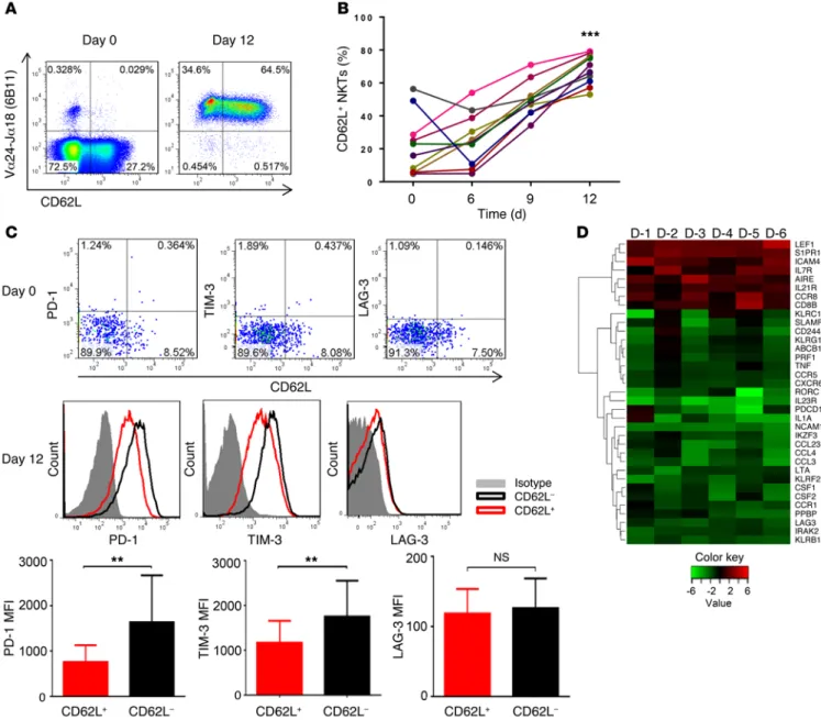
superior developmental potential and could support ex vivo expan-sion of NKTs for therapeutic applications. To test this hypothesis, we performed immunophenotyping of primary NKTs immediately after isolation from peripheral blood and at different time inter-vals in culture after stimulation with αGalCer, which consistently produced higher frequency and absolute numbers of NKTs com-pared with the use of a T cell expansion protocol based on CD3/ CD28 stimulation (Supplemental Figure 2). Despite a notable interindividual variability of CD62L expression in freshly isolated NKTs, we observed a striking accumulation of CD62L+ fraction of NKTs from 33.63% ± 27.62% in freshly isolated NKTs to 69.92% ± 10.57% on day 12 of the culture (P < 0.001, Figure 1, A and B). Although CD62L was more frequently expressed on CD4+ NKTs both before and after culture, the accumulation of CD62L+ cells could not be explained by a preferential expansion of CD4+ NKTs. Indeed, at the end of a 12-day culture, the frequency of CD62L+, CD4+, and CD62L+CD4+ NKTs increased 3.9 ± 1.8–fold, 1.9 ± 0.7– fold, and 3.8 ± 1.9–fold, respectively. Consistent with these results, we also observed an enrichment of the CD62L+CD4– subset at day 12 compared with day 0 (Supplemental Figure 1B). Further multi-parameter characterization of NKTs showed that CD62L was often coexpressed with CCR7 before culture, but CCR7 expression was progressively lost during culture (Supplemental Figure 1C). We also found that nearly all NKTs expressed both CD27 and CD28 before culture. While CD27 was downregulated in about half of NKTs by day 12 of culture irrespective of CD62L status, CD28 expression remained intact (Supplemental Figure 1C).
CD62L– cells expressed higher levels of CD161 and CD56 (NK-like differentiation), but a lower level of IL-7Rα (Supple-mental Figure 1D). While freshly isolated NKTs rarely expressed exhaustion markers (PD-1, LAG-3, or TIM-3) in either the CD62L+ or the CD62L– subset, the CD62L– subset preferentially expressed PD-1 and TIM-3 at day 12 of NKT culture (P = 0.0043, 0.0184, Figure 1C). Moreover, immune-related gene expression analysis (nCounter platform) of CD62L+ and CD62L– NKTs sorted on day 12 revealed mRNA upregulation of genes associated with T cell survival/memory (e.g., LEF1, S1PR1, IL7RA, IL21R) in CD62L+ NKTs and with exhaustion/terminal differentiation in CD62L– NKTs (e.g., PD1, LAG3, TIM3, CD244, CD161, CD56; Figure 1D and Supplemental Table 1). The transcription factor lymphoid enhancer factor 1 (LEF1) was the top immune-related gene over-expressed in CD62L+ compared with CD62L– NKTs. Intracellular flow cytometry analysis demonstrated that CD62L+ NKTs uni-formly expressed LEF1, whereas a major fraction of CD62L– cells was LEF1-negative. Since LEF1 was recently shown to mediate expansion of murine NKT precursors in part via transcriptional activation of GATA3 gene expression (39), we analyzed the level of GATA3 protein in human NKTs in relation to LEF1 and CD62L levels. We found that GATA3 expression strongly correlated with the expression of LEF1 and CD62L in human NKTs (Supplemen-tal Figure 1E). Interestingly, CD62L+ and CD62L– NKTs expressed the same level of PLZF, a transcriptional master regulator of NKT functional differentiation (21). Thus, the CD62L+ subset predomi-nantly accumulates in culture upon antigenic stimulation of pri-mary NKTs, and loss of CD62L expression is associated with NK-like terminal differentiation, exhaustion, and downregulation of pro-proliferative transcriptional regulators.
factor (PLZF) immediately after positive selection enables intra-thymic expansion and effector-memory–like differentiation of NKTs (24). Peripheral NKTs are long-lived lymphocytes, and their post-thymic maintenance largely depends on slow IL-15–mediat-ed homeostatic proliferation (25, 26). In human peripheral blood, NKTs are divided into 2 major functional subsets based on CD4 expression: CD4+ and CD4– (mostly CD8/CD4–double-negative, DN) (27). The CD4+ subset is highly enriched in neonate NKTs and undergoes fewer homeostatic divisions compared with the CD4– subset in adults (26), suggesting that CD4+ NKTs could contribute to the long-term persistence of adoptively transferred therapeu-tic NKTs under certain conditions. However, ex vivo expansion of human NKTs in response to antigenic stimulation, e.g., with α-galactosylceramide (αGalCer), produces similar numbers of CD4+ and DN NKTs (28). NKTs also exhibit an NK-like linear dif-ferentiation with acquisition of CD161 and then CD56 expression. Like in T cells, the expression of CD56 is associated with terminal differentiation and the loss of proliferative potential (29).
In contrast to peripheral T cells, which have a well-estab-lished developmental hierarchy from naive to central-memory to effector-memory to terminal effector cells (30), NKTs are broadly described as cells with “activated memory” phenotype without the naive state (31, 32). In cord blood, the majority of NKTs are CD4+ and coexpress CD45RO with CD62L and CCR7 without immedi-ate effector function (26, 31, 33), thus resembling central-memo-ry CD4 T cells. In adult peripheral blood, NKTs are equally split between CD4+ and CD4– subsets (albeit with significant interindi-vidual variability). Adult NKTs lack a clear demarcation between “memory” and “effector” states, as they variably express memory markers and have immediate effector functions such as cytokine production and cytotoxicity (26, 33). The majority of adult NKTs even in the elderly express CD28 (34), making them distinct from terminally differentiated T effector cells (35).
Recent reports have demonstrated that CD62L+ central mem-ory T cells have stem cell properties and superior therapeutic activ-ity in cell therapy products (36–38). The functional significance of CD62L expression in NKTs remains unknown. In this study, we found that the CD62L+ subset is required for NKT ex vivo expan-sion and in vivo persistence. Importantly, when engineered to express CD19-specific CAR (CAR.CD19), CD62L+ but not CD62L– CAR.CD19 NKTs produced sustained tumor regression in a B cell lymphoma model in NOD/SCID/IL-2Rγ(null) (NSG) mice. We also found that CD62L+ NKTs could be maintained during ex vivo expansion when provided with certain costimulatory ligands. With this knowledge, we engineered costimulatory artificial antigen-pre-senting cells (aAPCs) that can be used to generate NKTs and CAR-NKTs with superior therapeutic activity in patients with cancer.
Results
CD62L+ cells accumulate in culture upon antigenic stimulation of
The Journal of Clinical Investigation
R e s e a R c h a R t i c l eTh0-like polarization (a balanced production of IFN-γ and IL-4, typical of the whole population of peripheral blood NKTs), whereas the polarization profile of CD62L– cells could not be determined unambiguously because of the low absolute amounts of each cyto-kine. Despite strong upregulation of IL23R mRNA expression in the CD62L– subset as determined by the nCounter analysis (Figure 1D; a potential Th17 polarization), we detected neither IL-23R pro-tein expression on the cell surface of NKTs by FACS nor production of IL-17 upon TCR-stimulation by ELISA (data not shown).
CD62L+ NKTs have superior survival and proliferative capacity
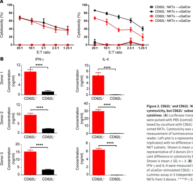
upon secondary stimulation. To examine whether the accumula-CD62L+ and CD62L– NKTs have comparable cytotoxicity, but
the CD62L+ subset is the main source of cytokines. We magnetically
sorted NKTs from primary culture into CD62L+ and CD62L– sub-sets and examined their functional properties. Figure 2A dem-onstrates that either both subsets were equally cytotoxic (3 of 6 donors) or CD62L– NKTs were more cytotoxic than CD62L+ NKTs (3 of 6 donors) against CD1d+ DAOY medulloblastoma cells when the target cells were pulsed with αGalCer. The analysis of cytokine production in αGalCer-stimulated NKTs revealed much higher lev-els of both IFN-γ and IL-4 production by CD62L+ compared with CD62L– subsets (P < 0.001, Figure 2B). CD62L+ cells exhibited a
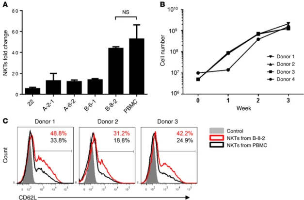
Figure 1. Accumulation of CD62L+ subset in culture after in vitro antigenic stimulation of primary NKTs. (A) CD62L expression was examined by FACS in primary NKTs from freshly isolated PBMCs (day 0) and 12 days after stimulation with αGalCer and in vitro expansion in culture (n = 10). (B) Kinetics of CD62L expression in NKTs at the indicated intervals after primary stimulation (as in A) from individual donors (n = 10). (C) Expression of PD-1, TIM-3, and LAG-3 on NKT surface was measured by FACS on days 0 and 12 (top panel). Representative plots from 1 of 4 donors (middle panel) or mean ± SD of mean fluorescence intensity (MFI) for all donors (n = 4, bottom panel). (D) On day 12 after primary stimulation, NKTs were magnetically sorted into CD62L+
and CD62L– subsets followed by RNA isolation and gene expression analysis using Human Immunology Panel version 2 and nCounter Analysis System
(NanoString). The heat map shows the log2 fold changes (CD62+/CD62L–) of genes with P values less than 0.02 and average fold change greater than 2.
bottom panel). Therefore, CD62L+ NKTs survive and proliferate in response to antigenic stimulation and are responsible for NKT numeric expansion in culture.
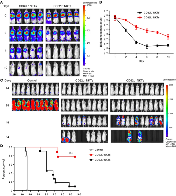
CD62L+ subset is responsible for in vivo persistence and
thera-peutic activity of NKTs. To determine the role of the CD62L+ sub-set in the in vivo persistence of adoptively transferred NKTs, we transduced NKTs with firefly luciferase and magnetically sorted them into CD62L+ and CD62L– subsets. We then adoptively trans-ferred the sorted cells to NSG mice. Longitudinal bioluminescent imaging demonstrated that the signal from CD62L– cells could be detected until day 2, whereas CD62L+ cells remained detectable up to day 10 (P < 0.001, Figure 4, A and B). Next, we compared the in vivo therapeutic potential of CD62L+ and CD62L– NKT sub-sets in a model of CAR-redirected immunotherapy for lymphoma. NKTs were transduced with a CD19-specific CAR containing 4-1BB costimulatory endodomain (CAR.CD19, Supplemental Fig-ure 3) followed by sorting into CD62L+ and CD62L– subsets. NSG mice were i.v. injected with luciferase-transduced CD19+ Daudi lymphoma cells and 4 days later were divided into 2 groups to receive CD62L+ or CD62L– CAR.CD19 NKTs. Both CD62L+ and CD62L– CAR.CD19 NKTs prolonged the survival of treated ani-mals compared with untreated control (P < 0.001). Importantly, tion of CD62L+ NKTs during in vitro expansion is due to their
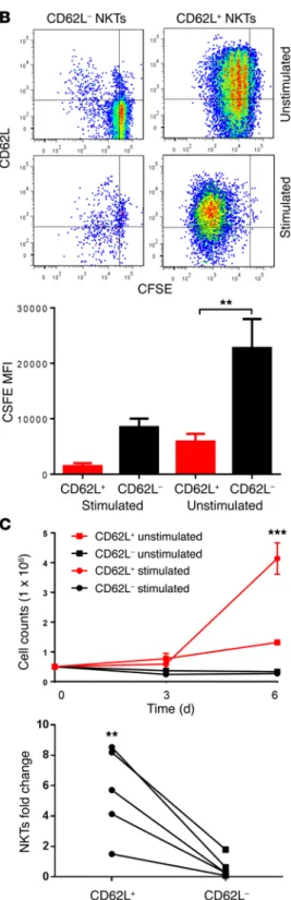
preferential survival or proliferation in response to antigenic stimulation, we measured their rate of cell death and prolifera-tion after stimulaprolifera-tion of the sorted cells with APCs that had been pulsed with αGalCer. On day 3 after stimulation, 31% ± 21% and 74% ± 7.5% of CD62L+ and CD62L– NKTs underwent apoptosis, respectively (Figure 3A). We also observed a much greater rate of proliferation in CD62L+ versus CD62L– subsets as measured by CFSE dilution on day 6 (Figure 3B). Moreover, the major-ity of cells that survived and proliferated in the CD62L– group expressed CD62L, suggesting that these cells were progenies of a small subset of CD62L+ cells in the original CD62L– fraction. Consistent with these results, we observed a striking difference in the numbers of NKTs generated after a 6-day culture of sort-ed CD62L+ versus CD62L– NKTs with IL-2 alone or with TCR-stimulation. Indeed, Figure 3C (top panel) demonstrates that CD62L+ cells underwent 2.5- and 8-fold numeric expansion with IL-2 alone or with TCR-stimulation, respectively. In contrast, CD62L– NKTs failed to expand in either condition. Although the degree of NKT proliferation in response to antigenic stimulation varied from donor to donor, the CD62L– subset contributed little or nothing to NKT expansion in all 5 tested donors (Figure 3C,
Figure 2. CD62L+ and CD62L– NKTs have comparable cytotoxicity, but CD62L+ subset is the main source of cytokines. (A) Luciferase-transduced CD1d+ DAOY cells
were pulsed with PBS (control) or αGalCer overnight fol-lowed by coculture with CD62L+ or CD62L– magnetically
sorted NKTs. Cytotoxicity was analyzed after 4 hours by measurement of luminescence intensity with a plate reader. Left plot is a representative of 3 donors (in triplicates) with no difference in cytotoxicity between NKT subsets. Shown is mean ± SD, n = 3. Right plot is a representative of 3 donors (in triplicates) with a signifi-cant difference in cytotoxicity between NKT subsets. Shown is mean ± SD, n = 3. (B) The concentrations of IFN-γ and IL-4 were measured in 24-hour supernatants of αGalCer-stimulated CD62L+ or CD62L– NKTs by the
The Journal of Clinical Investigation
R e s e a R c h a R t i c l eWhile resting NKTs express CD28 (Supplemental Figure 1C), they express little or no late costimulatory receptors such as 4-1BB and OX40 (Supplemental Figure 4A). However, stimulation of NKTs with αGalCer-pulsed autologous peripheral blood mononuclear cells (PBMCs) resulted in rapid induction of 4-1BB in all NKTs and OX40 in CD4+ NKTs (Supplemental Figure 4A). Because CD62L is transiently downregulated within the first 24–48 hours after TCR-stimulation (data not shown), we analyzed the kinetics of 4-1BB and OX40 expression in CD62L+ and CD62L– NKTs that only CD62L+ CAR-NKTs induced sustained tumor regression,
with 7 of 9 treated animals alive, 5 of which were tumor-free, for at least 3 months. In contrast, all 10 mice treated with CD62L– CAR-NKTs succumbed to tumor progression (P < 0.001, Figure 4, C and D). Thus, CD62L+ NKTs have extended in vivo persistence and superior therapeutic potential compared with CD62L– NKTs.
Costimulation maintains CD62L+ NKTs and prevents
exhaus-tion. There is growing evidence that costimulation plays a criti-cal role in the activation, survival, and expansion of NKTs (40). Figure 3. CD62L+ NKTs have superior survival and proliferative capacity upon secondary stimulation. (A) CFSE-labeled NKTs were magnetically sorted into CD62L+
and CD62L– subsets as confirmed by post-sorting FACS (top plot) and stimulated with
irradiated αGalCer-pulsed APCs. On day 3 after stimulation, staining for annexin V and 7-AAD was analyzed in NKTs by FACS after gating on CFSE+ events. Results are
from a representative of 5 donors tested (middle panel). The corresponding bar graph (bottom panel) shows mean ± SD of percent annexin V+ NKTs on day 3 (n = 5). (B) Cell
Figure 4. CD62L+ NKTs have superior in vivo persistence and antitumor activity. (A) Luciferase-transduced NKTs were sorted into CD62L+ and CD62L–
subsets and injected to NSG mice. NKT in vivo persistence was monitored with bioluminescence imaging. Data are from 1 of 2 independent experiments, 5 mice per group. (B) Mean ± SD of bioluminescence photon count on indicated days after injection of CD62L+ or CD62L– NKTs. Data are from the same
experiment shown in A (P = 0.008, paired t test). (C) Each mouse received i.v. injection of 2 × 10⁵ luciferase-transduced Daudi lymphoma cells (day 0)
fol-lowed by (day 4) i.v. injection of 107 CAR.CD19-transduced NKTs with IL-2 (1,000 U/mouse) or PBS as a control. Tumor growth was monitored using
The Journal of Clinical Investigation
R e s e a R c h a R t i c l eFigure 5. Costimulation maintains CD62L+ NKTs and prevents exhaustion. (A) NKTs were sorted into CD62L+ and CD62L– subsets and stimulated and
examined for expression of 4-1BB and OX40 by FACS before and 3 days after stimulation with αGalCer. Shown are plots from a representative of 4 donors. (B) CD62L+ NKTs were stimulated on plates coated with the indicated agonistic mAbs. Shown is mean ± SD (n = 4) of fold change in absolute NKT number
on day 7 after stimulation compared with day 0. (C) CD62L+ NKTs were stimulated as in B and analyzed for CD62L expression (black) versus isotype control
(gray) on day 7. Shown are representative overlay histograms (left panel) and mean ± SD of percent CD62L+ cells, n = 4. (D) CD62L+ NKTs were stimulated as
or both. OX40 was not tested in these settings because we could not obtain an anti-OX40 mAb with an agonistic activity. First, we found that costimulation with either anti-CD28, anti–4-1BB mAb, or both increased the number of NKTs generated in culture within 7 days compared with stimulation with anti-CD3 alone at 20 ng/ml (P < 0.001, Figure 5B). In the absence of costimulation, increasing the concentration of OKT3 from 20 ng/ml to 1 μg/ml did not affect the number of NKTs generated, while costimulation was effective in increasing NKT number only when it was com-bined with the lower concentration of OKT3 (Supplemental Fig-ure 5). Importantly, on day 7 after stimulation with OKT3 alone less than half the NKTs were positive for CD62L. The addition of anti-CD28, anti–4-1BB, or anti-CD28 with anti–4-1BB mAbs resulted in the retention of CD62L expression on 58% ± 7.1% (P = 0.026), 73% ± 9.2% (P = 0.0036), and 73% ± 6.1% (P = 0.0002) of NKTs, respectively (Figure 5C). Coordinately with the retention of CD62L expression, NKTs provided with costimulatory signals expressed significantly less PD-1 (P < 0.05, Figure 5D). Therefore, engagement of costimulatory receptors during antigenic stimula-tion supports CD62L expression in proliferating NKTs and pre-vents their exhaustion.
Generation of HLAnull CD1d-expressing aAPCs with
costimula-tory ligands. We hypothesized that, given the observed protective effect of costimulation on TCR-activated NKTs, the optimal com-bination of costimulatory ligands could enable the generation of an effective aAPC for clinical-scale expansion of NKTs with high were sorted before stimulation. We found that 71.13% ± 18.66%
and 51.98% ± 18.83% of CD62L+ and CD62L– NKTs, respectively, upregulated OX40 within 72 hours after stimulation (P = 0.0072, Figure 5A). Similarly, stimulated CD62L+ NKTs expressed a higher level of 4-1BB compared with CD62L– NKTs (P = 0.011, Figure 5A). OX40 was preferentially upregulated in the CD4+ subset of either CD62L+ or CD62L– NKTs, whereas 4-1BB was upregulated in all NKTs (Supplemental Figure 4B).
Next, we asked whether costimulation could counteract exhaustion of in vitro expanded NKTs. CD62L+ sorted NKTs were stimulated on plates coated with an anti-CD3 agonistic mAb, OKT3, alone or in combination with mAbs for CD28, 4-1BB, Table 1. Phenotype of aAPC clonesA
Clone name Phenotype
22 CD1d+
A-2-1 CD1d+CD86+
A-6-2 CD1d+CD86+4-1BBL+
B-6-1 CD1d+CD86+OX40L+
B-8-2 CD1d+CD86+4-1BBL+OX40L+
AHLAnull K562 cells were transduced with cDNA of human CD1d alone or
with the indicated costimulatory ligands followed by single-cell sorting by FACS, clonal expansion, and immunophenotyping by flow cytometry.
Figure 6. Generation of functional characterization of costimulatory aAPCs for NKT expansion. (A) CD1d+HLAnull K562 clones were transduced with the
The Journal of Clinical Investigation
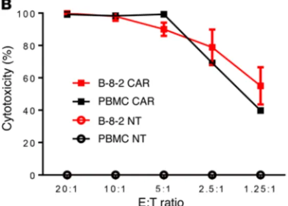
R e s e a R c h a R t i c l eFunctional properties of NKTs expanded with aAPCs. Numer-ous studies have demonstrated that the therapeutic potential of NKTs largely relies on their potent effector functions including cytotoxicity and cytokine production (11). To examine the effect of aAPCs on the functionality of NKTs, we expanded NKTs using PBMCs or B-8-2 and compared resultant NKTs using in vitro cytotoxicity and cytokine quantification assays. First, we tested direct NKT cytotoxicity against CD1d+ DAOY medulloblastoma cells (41). Figure 7A demonstrates that NKTs expanded with B-8-2 and PBMCs mediated equally potent cytotoxicity against DAOY cells, killing 90% of tumor cells at a 5:1 effector-to-target ratio. We then transduced NKTs that were expanded with B-8-2 or PBMCs with CAR.CD19 (Supplemental Figure 7) and found that both CAR-NKT preparations were equally cytotoxic against CD19+ Raji lymphoma cells, which were not killed by the paren-tal NKTs (Figure 7B). Next, we stimulated NKTs with αGalCer presented by the corresponding APCs used for their expansion and measured production of Th1/Th2 cytokines using a multi-plex Luminex assay. Figure 7C demonstrates that, compared with NKTs expanded and restimulated with PBMCs, those expanded and restimulated with B-8-2 produced significantly higher amounts of IFN-γ and GM-CSF, equal amounts of IL-4, and less IL-10 (P < 0.001). We also performed NKT stimulation with an agonistic 6B11 mAb, which binds to the CDR3 region of their TCR (42), and found that while IL-10 in PBMC-stimulated NKTs was largely derived from the PBMCs, B-8-2–expanded NKTs still produced larger amounts of IFN-γ and GM-CSF than those expanded with PBMCs (data not shown). Therefore, B-8-2– therapeutic potential. To test this hypothesis, we rendered K562
cells HLAnull by eliminating the HLA-Cw3 gene (the only HLA allele) using an HLA-C–specific zinc finger nuclease (ZFN) fol-lowed by transduction with CD1d, CD86, 4-1BBL, and OX40L cDNA. HLAnullCD1d+ clones expressing 1, 2, or all 3 costimula-tory ligands were generated after single-cell FACS sorting, pro-ducing a total of 161 clones (Supplemental Figure 6A, and Meth-ods section). Because the level of cell surface expression of the transduced ligands varied, we first compared the aAPC activity of clones within each subgroup. We then compared the best per-formers from each subgroup to one another (Table 1). Figure 6A demonstrates that clone B-8-2 with the phenotype HLAnullCD1dmed CD86high4-1BBLmedOX40Lhigh (Supplemental Figure 6B) consis-tently induced the highest rate of NKT expansion compared with other clones (P < 0.001), producing numbers of NKTs similar to the numbers produced by autologous PBMCs (P > 0.05, Fig-ure 6A). Next, we tested whether the B-8-2 clone was capable of propagating NKTs to clinically sufficient numbers. Figure 6B demonstrates that 1 round of restimulation with B-8-2 produced 272.32 ± 111–fold expansion of NKTs from 4 donors (range from 156 to 424) within 3 weeks, similar to the NKT numbers gener-ated after stimulation with autologous PBMCs (18). However, NKTs expanded with B-8-2 contained significantly higher fre-quencies of CD62L+ cells compared with PBMC-expanded NKTs (P = 0.0089, Figure 6C). Therefore, we selected B-8-2, an aAPC clone that coexpressed 3 costimulatory ligands and produced the highest rate of NKT in vitro expansion with the maximal preser-vation of CD62L expression.
Figure 7. Functional characterization of NKTs expanded with B-8-2. (A) Luciferase-transduced CD1d+ DAOY cells were pulsed
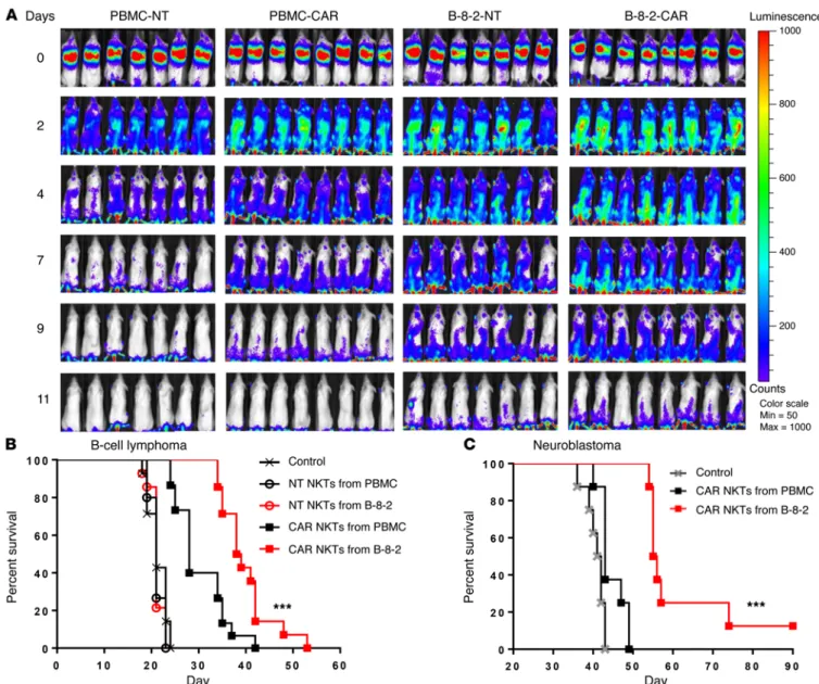
CD19 followed by adoptive transfer to NSG mice that were pre-implanted with Raji lymphoma cells. Figure 8A demonstrates that CAR expression had little effect on NKT persistence, but NKTs propagated with B-8-2 persisted significantly longer than those propagated with PBMCs regardless of CAR expression (P < 0.001). Importantly, B-8-2–expanded CAR.CD19 NKTs significantly pro-longed animal survival compared with PBMC-expanded CAR. CD19 NKTs in 2 B cell lymphoma models (P < 0.001, Figure 8B and Supplemental Figure 8). To test whether the observed thera-peutic superiority of NKTs that we had expanded with our engi-neered aAPCs also applies to their use against other types of cancer and target antigens, we used a metastatic model of neuro-propagated NKTs possess high levels of CD1d-dependent and
CAR-redirected cytotoxicity and a Th1-biased cytokine profile. aAPC-expanded NKTs have superior in vivo persistence and ther-apeutic activity. Accumulating evidence indicates that the clinical efficacy of CAR-T cell therapy against B cell malignancies criti-cally depends on the in vivo persistence of transferred cells (43– 46), which is influenced by the type of costimulation (44, 47) and phenotypic composition of CAR-T cells (48). Since we observed that the expansion of NKTs with B-8-2 affects their phenotypic composition by increasing the content of CD62L+ cells, we com-pared the in vivo persistence of NKTs expanded with PBMCs and B-8-2. NKTs were transduced with luciferase alone or with CAR.
Figure 8. aAPC-expanded NKTs have superior in vivo persistence and antitumor activity. (A) NKTs were transduced with a CD19-specific CAR followed by expansion of transduced (CAR) or nontransduced (NT) NKTs with PBMC or B-8-2 restimulation. Four groups of mice then received i.v. injection of 2 × 10⁵
Raji cells (day 0) followed by (day 3) i.v. injection of the respective NKT preparations (107 cells per mouse) with IL-2 (1,000 U/mouse). NKT persistence was
monitored by bioluminescent imaging on the indicated days. Bioluminescent images of 7 mice per group are shown from 1 of 2 independent experiments. (B) Mice received i.v. injection of 2 × 10⁵ luciferase-transduced Raji cells (day 0) followed by (day 2) i.v. injection of 107 NT or CAR.CD19 NKTs with IL-2
(1,000 U/mouse). (C) Mice received i.v. injection of 106 luciferase-transduced CHLA-255 cells (day 0) followed by (day 4) i.v. injection of 107 NT or CAR.GD2
The Journal of Clinical Investigation
R e s e a R c h a R t i c l eCD62L may represent a common marker of cells responsible for the long-term maintenance of peripheral NKT and T cells. Graef et al. demonstrated that CD62L+ central memory T cells pos-sess stem cell properties; they could propagate themselves while giving rise to effector-memory and effector T cells (36). In the most recently published study, Sommermeyer et al. demonstrated that human CAR.CD19-expressing CD8 or CD4 T cells generated from naive and central-memory subsets were more effective against Raji lymphoma xenografts compared with those generated from effec-tor-memory subsets (38). The authors also found that combining the most potent CD4+ and CD8+ CAR-expressing subsets produced synergistic antitumor activity in vivo. Activation of NKTs has been shown to lead to transactivation of NK and CD8 T cells in murine models and in human clinical trials (6, 54), so that the combination of CAR-NKTs with other defined subsets of CAR-expressing lym-phocytes may be a useful therapeutic strategy.
LEF1 and IL7RA were among the immune-related genes that were most overexpressed in CD62L+ compared with CD62L– NKTs. In a recent report, Carr et al. demonstrated a unique function of LEF1 in the expansion of murine Vα14-invariant (iNKT) cells dur-ing stage 2 of their thymic development via direct transcriptional activation of CD127 and c-MYC gene expression (39). Consistent with our observation of the coordinated expression of LEF1 and GATA3 in human NKTs, these investigators also found that LEF1 upregulates transcription factor GATA3, which is required for IL-4 production in iNKT2 cells as well as for dual production of IL-4 and IFN-γ in iNKT1 cells. The latter cells closely resemble human peripheral blood NKTs, in which we found that CD62L+ cells pref-erentially express GATA3 and produce high levels of both cyto-kines. Taking the Carr et al. study with murine NKTs and our own results with human NKTs, we suggest that LEF1 plays a critical role at early stages of NKT development and controls their number and function. The high level of LEF1 expression in CD62L+ and its loss in CD62L– subsets of human NKTs is consistent with a model of linear progression from less differentiated CD62L+ NKTs with a preserved proliferative potential and Th0-like cytokine profile toward terminally differentiated CD62L– NKTs with diminished ability to proliferate and produce cytokines.
There is growing evidence that costimulation plays a critical role in the development, activation, and functional responses of NKTs in murine models (40, 55). However, little is known about the expression of costimulatory receptors in human NKTs. In this study, we focused on a set of costimulatory receptors that have pronounced prosurvival properties in human T cells: CD28, 4-1BB, and OX40 (56–58). First, we confirmed that freshly isolated human NKTs express CD28 (34) and showed that they coexpress CD27, thereby resembling the corresponding stage of memory T cell differentiation with preserved functional potential (35). To our knowledge, the current study is the first to characterize the base-line and post-stimulation kinetics of 4-1BB and OX40 in human NKTs. We found that both receptors are undetectable in freshly isolated NKTs but are induced following TCR-stimulation. Like T cells (59), human NKTs preferentially upregulated OX40 in the CD4+ subset. The majority of NKTs also upregulated 4-1BB, which is preferentially upregulated in the CD8+ subset of T cells (60). Importantly, we found that costimulation of individual costimula-tory receptors could inhibit the loss of CD62L expression and res-blastoma with a multi-drug-resistant CHLA-255 human
neuro-blastoma cell line (49) and NKTs expressing a GD2-specific CAR. Figure 8C demonstrates that CAR.GD2 NKTs that were expanded with aAPCs had potent antitumor activity compared with untreat-ed control (P < 0.001), whereas those expanduntreat-ed with PBMCs were ineffective. Therefore, expansion of NKTs using B-8-2 aAPCs confers a qualitative advantage to NKTs, enabling superior in vivo persistence and antitumor activity.
Discussion
A critical gap in knowledge of human NKT biology has slowed down the development of effective NKT-based cancer immuno-therapy. We found that NKT numeric ex vivo expansion and sub-sequent in vivo persistence — essential requirements for effective NKT-based cell and gene therapy applications — depend on the CD62L+ subset of peripheral blood NKTs. Only CD62L+ NKTs survive and proliferate in response to repeated TCR-stimulation, while CD62L– cells undergo early exhaustion and cell death. Although continuous stimulation of NKTs is associated with the loss of CD62L expression, activation of costimulatory receptors during TCR-stimulation can counteract this process. Specifically, we experimentally determined a unique combination of CD86, 4-1BBL, and OX40L molecules and the levels of their coexpres-sion on the surface of an aAPC that enables highly efficient clin-ical-scale NKT expansion with maximal preservation of CD62L expression. CAR-NKTs generated using the aAPCs developed in the course of this study demonstrated extended in vivo persis-tence and superior therapeutic activity against in vivo models of lymphoma and neuroblastoma.
We show that the CD62L+ subset is responsible for the numeric expansion of NKTs upon antigenic stimulation ex vivo. The fol-lowing findings support the above conclusion: (a) the fraction of CD62L+ cells is dramatically increased after primary NKT stimu-lation; (b) the majority of sorted CD62L– cells undergo apoptosis, whereas sorted CD62L+ cells proliferate in response to identical stimulation; and (c) CD62L– NKTs exhibit signs of terminal differ-entiation (CD161 and CD56 expression) in freshly isolated PBMCs and rapidly acquire an exhaustion phenotype upon in vitro stimu-lation as evidenced by marked upregustimu-lation of PD-1 and TIM-3 expression and diminished ability to produce cytokines. When sim-ilar characteristics are observed in T cell therapeutic products, they are associated with a subsequent lack of persistence or of objective responses after adoptive transfer to cancer patients (50, 51).
could be maximized. Further enhancement of CAR-NKT per-sistence and antitumor efficacy could be achieved by providing optimal costimulatory signals within CAR constructs such as CD28 or 4-1BB costimulatory endodomains that are critically important for the clinical efficacy of CAR.CD19 T cells (19, 20). The optimal costimulatory CAR design for NKTs is yet to be found. It will be interesting to determine the role of the CD62L+ subset in CAR-mediated NKT proliferation and persistence. Ulti-mately, the development of an effective NKT-based CAR-redi-rected cancer immunotherapy will likely require the coordinated use of costimulation provided by aAPCs during ex vivo expan-sion and CAR after adoptive transfer to patients.
Methods
Cell lines and culture conditions. The parental K562 cells were provided by C. June (University of Pennsylvania, Philadelphia, PA). Daudi, Raji, DAOY, and 293T cells were purchased from American Type Culture Col-lection (ATCC). The CHLA-255 cell line was previously described (49). K562, Daudi, Raji, and CHLA-255 cells were cultured in RPMI, whereas DAOY and 293T cells were maintained in Iscove’s Modified Dulbecco’s Medium (IMDM; Invitrogen). Both types of medium were supplemented with 10% FBS (HyClone), 2 mM GlutaMAX-1 (Gibco-BRL).
NKT isolation, transduction, expansion, and sorting. To analyze the cord blood NKTs we used discarded cord blood units obtained from the MD Anderson Cancer Center Cord Blood Bank according to the protocols approved by the IRBs at MD Anderson Cancer Center and Baylor College of Medicine (BCM). PBMCs of healthy donors (at least 18 years old) were isolated by gradient centrifugation from buffy coats purchased from Gulf Coast Regional Blood Center. NKTs were puri-fied by anti-iNKT microbeads (Miltenyi Biotec). The negative PBMC fraction was irradiated (40 Gy) and aliquoted. NKTs were stimulated with an aliquot of autologous PBMCs pulsed with 100 ng/ml αGalCer (Kyowa Hakko Kirin). Recombinant IL-2 (200 U/ml; National Can-cer Institute Frederick) was added every other day in complete RPMI (HyClone RPMI 1640, 10% heat-inactivated FBS, and 2 mM Gluta-MAX). NKTs were expanded for 10 days and then restimulated with autologous PBMCs (irradiated with 40 Gy) or aAPCs (irradiated with 100 Gy) when indicated. On day 3 after restimulation, 24-well, non– tissue culture plates were coated with retronectin (Takara Bio) and, after washing, inoculated with 1 ml of retroviral supernatant contain-ing CAR.CD19 and spun for 60 minutes at 4,600 g. The viral superna-tant was then removed, and stimulated NKTs were added to the wells in complete media and 200 U/ml recombinant human IL-2. Cells were removed from the plate after 48 hours, washed, resuspended at the concentration 106 cells/ml in complete RPMI with 200 U/ml IL-2, and
plated for continued expansion. NKT number was determined by try-pan blue (Life Technologies) counting. When indicated, NKTs or CAR-NKTs were labeled with CD62L-PE mAb (GREG-56; BD Biosciences) and anti-PE microbeads (Miltenyi) followed by magnetic sorting into CD62L+ and CD62L– subsets according to the manufacturer’s
instruc-tions. The phenotype of the sorted cells was determined by FACS. Retroviral constructs and retrovirus production. CAR.CD19 and CAR. GD2 constructs were made as previously described (18, 73) and con-tained a single-chain variable fragment from the CD19-specific antibody FMC-63 or the GD2-specific antibody 14G2a connected via a short spac-er dspac-erived from the IgG1 hinge region to the transmembrane domain derived from CD8α, followed by signaling endodomain sequences of
cue NKTs from exhaustion. We also observed an additive effect on the maintenance of CD62L+ NKTs when CD28 and 4-1BB were simultaneously activated.
Several types of aAPCs have been developed from the K562 cell line for clinical-scale numeric expansion of T and NK cells for adoptive immunotherapy applications (61–63). In the design of K562-based aAPCs for NKTs, we sought to selectively acti-vate and expand NKTs while avoiding competition with T and NK cells. K562 cells express only 1 HLA allele, HLA-Cw3, which is known to activate NK cells (64). Hence, the deletion of the HLA-Cw3 gene and transgenic expression of CD1d in HLAnull K562 cells enabled antigen presentation to NKTs without activa-tion of NK or convenactiva-tional T cells. Importantly, the B-8-2 clone we experimentally selected with the optimal combination of costimulatory ligands (and their expression levels) demonstrat-ed aAPC qualities for NKTs that excedemonstrat-eddemonstrat-ed those of the autolo-gous PBMCs. We did, however, observe a differential effect on NKT expansion between plate-bound agonistic mAbs and the corresponding ligands for costimulatory receptors. While costimulation with 1 or 2 mAbs for CD28 or 4-1BB enhanced the rate of NKT expansion with retention of CD62L, expression of ligands for 3 receptors (CD28, 4-1BB, and OX40) on K562 cells was needed to achieve such an effect. This could be explained by higher affinity of mAbs to the receptors compared with the natural ligands or expression of inhibitory molecules on K562 cells that counteract NKT activation.
B-8-2–expanded NKTs and CAR-NKTs had superior in vivo persistence, one of the critical requirements for the efficacy of adoptive cancer immunotherapy. Indeed, we found that B-8-2– expanded NKTs expressing anti-CD19 and anti-GD2 CARs had superior therapeutic activity in B cell lymphoma and neuroblas-toma models, respectively. In this study, we do not compare NKTs or CAR-NKTs with T or CAR-T cells because the xenogenic model in NSG mice does not allow such a comparison. It is well docu-mented that adoptive transfer of human T or CAR-T cells to NSG mice leads to the development of xeno-graft-versus-host disease (GvHD) (18, 65, 66). The latter report revealed that unless deplet-ed from CD45RA+ naive cells before ex vivo expansion, human T cells with or without anti-CD19 CAR cause severe xenogeneic GvHD in addition to antileukemia activity. In contrast, the persis-tence of human NKTs or CAR-NKTs in mice is not supported by the artifact of xenoreactivity to murine tissues (18).
The Journal of Clinical Investigation
R e s e a R c h a R t i c l esystem as previously described (76). A clone with an intermediate level of CD1d expression induced the highest rate of NKT proliferation and was further modified with lentiviral vectors encoding CD86, 4-1BBL, or OX40L followed by single cell sorting and clonal expansion. The generated library of clones was divided into groups based on expres-sion of 1, 2, or 3 costimulatory molecules.
Gene expression analysis. Total RNA was collected using TRIzol reagent (Qiagen). Gene expression analysis was performed using Immunology Panel version 2 (NanoString) at BCM Genomic and RNA Profiling Core using nCounter Analysis System. Data were analyzed using nSolver 2.0 software (NanoString).
In vivo experiments. The colony of NSG mice was originally obtained from The Jackson Laboratory and maintained at BCM Ani-mal Care facility. Tumor growth was initiated by i.v. injection of 2 × 10⁵ luciferase-transduced Raji lymphoma cells. On day 3, mice were
treated with 4 × 10⁶ to 8 × 10⁶ CAR-NKTs followed by i.p. injection of
IL-2 (1,000 U/mouse) every 3 days. Tumor growth was assessed twice per week by bioluminescent imaging (Small Animal Imaging core facility, Texas Children’s Hospital). For the in vivo persistence experi-ments, NKTs were cotransduced with CAR.CD19 and luciferase using retroviral constructs, i.v. injected to tumor-free or tumor-bearing mice, and monitored using bioluminescent imaging twice per week. Animal experiments were performed according to IACUC-approved protocols.
Statistics. For in vitro and in vivo experiments, we used 2-sided, paired Student’s t test to evaluate continuous variable of 2 groups, and 1-way ANOVA with post-test Bonferroni to evaluate continuous variables of more than 2 groups. Survival was analyzed by the Kaplan-Meier method and the log-rank (Mantel-Cox) test to compare pairs of groups. Statistics were computed using GraphPad Prism 5.0 (Graph-Pad Software). Differences were considered significant when the P value was less than 0.05.
Study approval. The cord blood units were obtained from MD Anderson Cancer Center Cord Blood Bank according to the protocols approved by the IRBs at MD Anderson Cancer Center (H-16320) and Baylor College of Medicine (H-20911). Written informed consent was received from all participating women prior to inclusion in the study under the protocol H-16320. Cord blood units not suitable for clinical use (usually because of low cell counts) were either discarded or used for research purposes under the protocol H-20911 at BCM.
Animal experiments were performed according to the protocol AN-5194 approved by the IACUCs of BCM.
Author contributions
NKT isolation, expansion, transduction, and in vitro functional testing were done by GT, ANC, AH, DL, and XX. In vivo experi-ments were performed by GT, ANC, EM, and LG. Design and genetic engineering of K562-based aAPCs were performed by BJ, HT, GT, LJC, and LSM. Functional testing of aAPC clones and selection of B-8-2 clone was accomplished by GT. CAR constructs were made by GD. Data analysis including statistical analysis was performed by GT, ANC, LSM, and QM. The manuscript was writ-ten by GT and LSM and edited by GD and LJC.
Acknowledgments
We thank Malcolm Brenner and Catherine Gillespie (Center for Cell and Gene Therapy, BCM, Houston, TX) for helpful discus-sions and scientific editing. This work was supported by grants
4-1BB fused with ζ chain. Retroviral supernatants were produced by transfection of 293T cells with a combination of CAR-containing plas-mid, RDF plasmid encoding the RD114 envelope, and PegPam3 plasmid encoding the MoMLV gag-pol as previously described (74).
Proliferation and apoptosis assays. NKTs were labeled with CFSE (Invitrogen) and stimulated with αGalCer-pulsed PBMCs, aAPC clones, or plates coated with 20 ng/ml anti-CD3 (OKT3) alone or in combination with 0.5 μg/ml anti-CD28 (CD28.2) and/or 1.5 μg/ml anti–4-1BB (h41BB-M127) (BD Biosciences). Cell proliferation was examined on days 3 and 6 by measurement of CFSE dilution using flow cytometry. In addition, early and late stages of apoptosis were measured on day 3 by staining with annexin V and 7-AAD (BD Biosci-ences) followed by flow cytometry.
Multiplex cytokine quantification assay. NKTs were stimulated for 24 hours with either APCs or agonistic antibody–coated plates (clone 6B11; BD Biosciences). Supernatants were collected and analyzed with a Human Cytokine/Chemokine Immunoassay kit (Millipore) using the Luminex assay according to the manufacturer’s instructions.
Flow cytometry. Immunophenotyping was performed using the following mAbs: HLA-C EMR8-5, CD1d 42.1, CD86 2331, 4-1BBL C65-485, OX40L ik-1, CD3 HIT3a, Vα24-Jα18 6B11, CD4 SK3, CD62L DREG-56, CD134 ACT35, CD137 4B4-1, PD-1 EH12.1, GATA3 L50-823, PLZF R17-809, CCR7 3D12, CD28 CD28.2, CD27 L128, IL-7R HIL-7R-M21, CD161 DX12, CD56 B159 (BD Biosciences), LAG-3 polyclonal, TIM-3 344823 (R&D Systems), and rabbit anti-LEF1 EP2030Y mAb (Abcam). Fluorochrome- and isotype-matching antibodies suggested by BD Biosciences or R&D Systems were used as negative controls. The expression of CAR.CD19 on NKTs was determined using anti-Id (clone 136.20.1) CAR.CD19-specific mAb (75) and goat anti-mouse IgG (BD Biosciences). The expression of CAR.GD2 on NKTs was detected using the anti-idiotype 1A7 mAb, which was produced in house from murine hybridoma HB-11786 (ATCC) followed by staining with a secondary rat anti-mouse IgG1-PE mAb (BD Biosciences). The analysis was per-formed on an LSR-II 5-laser flow cytometer (BD Biosciences) using BD FACSDiva software version 6.0 and FlowJo 7.2.5 (Tree Star).
In vitro cytotoxicity assay. The cytotoxicity of parental and CAR. CD19 NKTs against DAOY or Raji cells was evaluated using 4-hour luciferase assay as previously described (41).
Generation of aAPC clones. We rendered K562 cells HLAnull by
elim-inating the HLA-C gene from the K562 cell genome using an HLA-C– specific ZFN as previously described (76). Briefly, ZFNs containing 5 or 6 fingers were designed and assembled using an established archive of prevalidated 2-finger and 1-finger modules (Sangamo BioScience). The coding sequence of HLA-C was scanned for locations at which 2 such ZFNs (designated as ZFN-L and ZFN-R) could be targeted to sites that were separated by 5 bp and located on opposite DNA strands. The nucleotide targets for candidate ZFN pairs were then checked for divergence from other HLA coding sequences. Genes encoding the ZFNs were assembled using a PCR-based procedure and cloned into a plasmid. For the modification of K562 cells, expression vectors encod-ing HLA-C–targetencod-ing ZFNs (Supplemental Figure 4A) were introduced by Nucleofection (Lonza) according to the manufacturer’s protocol. After electroporation, cells were immediately placed in prewarmed complete medium. HLA-C–negative clones were obtained using limit-ing dilution, and HLA deletion was confirmed by Surveyor nuclease assay (76). A clone of HLAnull K562 cells was then electroporated with
C.1760.06, Houston, Texas 77030, USA. Phone: 832.824.4395; E-mail: [email protected].
L.J. Cooper’s present address is: Ziopharm Oncology Inc., Boston, Massachusetts, USA.
G. Dotti’s present address is: Department of Microbiology and Immunology, University of North Carolina, Chapel Hill, North Carolina, USA.
from the National Institutes of Health (RO1 CA116548 to L.S. Metelitsa; R01 CA142636 to G. Dotti; P50 CA126752 to L.S. Metelitsa and G. Dotti), Cancer Prevention and Research Institute of Texas (RP130588 to L.S. Metelitsa), Alex’s Lemonade Stand Foundation for Childhood Cancer (to L.S. Metelitsa), and Cookies for Kids’ Cancer Foundation (to L.S. Metelitsa).
Address correspondence to: Leonid S. Metelitsa, Department of Pediatrics, Baylor College of Medicine, 1102 Bates Avenue,
1. Porcelli S, Yockey CE, Brenner MB, Balk SP. Anal-ysis of T cell antigen receptor (TCR) expression by human peripheral blood CD4-8- α/β T cells demonstrates preferential use of several V beta genes and an invariant TCR α chain. J Exp Med. 1993;178(1):1–16.
2. Lantz O, Bendelac A. An invariant T cell receptor
α chain is used by a unique subset of major histo-compatibility complex class I-specific CD4+ and
CD4–8- T cells in mice and humans. J Exp Med.
1994;180(3):1097–1106.
3. Bendelac A, Lantz O, Quimby ME, Yewdell JW, Bennink JR, Brutkiewicz RR. CD1 recogni-tion by mouse NK1+ T lymphocytes. Science.
1995;268(5212):863–865.
4. Kim EY, Lynch L, Brennan PJ, Cohen NR, Brenner MB. The transcriptional programs of iNKT cells. Semin Immunol. 2015;27(1):26–32. 5. McEwen-Smith RM, Salio M, Cerundolo V. The regulatory role of invariant NKT cells in tumor immunity. Cancer Immunol Res. 2015;3(5):425–435.
6. Dhodapkar MV. Harnessing human CD1d restricted T cells for tumor immunity: progress and challenges. Front Biosci (Landmark Ed). 2009;14:796–807.
7. Exley MA, Nakayama T. NKT-cell-based immu-notherapies in clinical trials. Clin Immunol. 2011;140(2):117–118.
8. Motohashi S, Okamoto Y, Yoshino I, Nakayama T. Anti-tumor immune responses induced by iNKT cell-based immunotherapy for lung can-cer and head and neck cancan-cer. Clin Immunol. 2011;140(2):167–176.
9. Yamasaki K, et al. Induction of NKT cell-specific immune responses in cancer tissues after NKT cell-targeted adoptive immunotherapy. Clin. 2011;138(3):255–265.
10. Taniguchi M, Harada M, Dashtsoodol N, Kojo S. Discovery of NKT cells and development of NKT cell-targeted anti-tumor immunotherapy. Proc Jpn Acad Ser B Phys Biol Sci. 2015;91(7):292–304. 11. Metelitsa LS. Anti-tumor potential of type-I
NKT cells against positive and CD1d-negative tumors in humans. Clin Immunol. 2011;140(2):119–129.
12. Metelitsa LS, et al. Natural killer T cells infiltrate neuroblastomas expressing the chemokine CCL2. J Exp Med. 2004;199(9):1213–1221. 13. Tachibana T, et al. Increased intratumor Vα
24-positive natural killer T cells: a prognostic factor for primary colorectal carcinomas. Clin Cancer Res. 2005;11(20):7322–7327.
14. Molling JW, et al. Low levels of circulating invari-ant natural killer T cells predict poor clinical
out-come in patients with head and neck squamous cell carcinoma. J Clin Oncol. 2007;25(7):862–868. 15. Cariani E, et al. Immunological and molecular
correlates of disease recurrence after liver resec-tion for hepatocellular carcinoma. PLoS One. 2012;7(3):e32493.
16. Tahir SM, et al. Loss of IFN-γ production by invariant NK T cells in advanced cancer. J Immu-nol. 2001;167(7):4046–4050.
17. Dhodapkar MV, et al. A reversible defect in natu-ral killer T cell function characterizes the pro-gression of premalignant to malignant multiple myeloma. J Exp Med. 2003;197(12):1667–1676. 18. Heczey A, et al. Invariant NKT cells with chimeric
antigen receptor provide a novel platform for safe and effective cancer immunotherapy. Blood. 2014;124(18):2824–2833.
19. Kalos M, June CH. Adoptive T cell transfer for cancer immunotherapy in the era of synthetic biology. Immunity. 2013;39(1):49–60. 20. Dotti G, Gottschalk S, Savoldo B, Brenner MK.
Design and development of therapies using chi-meric antigen receptor-expressing T cells. Immu-nol Rev. 2014;257(1):107–126.
21. Cohen NR, et al. Shared and distinct transcrip-tional programs underlie the hybrid nature of iNKT cells. Nat Immunol. 2013;14(1):90–99. 22. Constantinides MG, Bendelac A. Transcriptional
regulation of the NKT cell lineage. Curr Opin Immunol. 2013;25(2):161–167.
23. Gapin L, Matsuda JL, Surh CD, Kronenberg M. NKT cells derive from double-positive thymo-cytes that are positively selected by CD1d. Nat Immunol. 2001;2(10):971–978.
24. Savage AK, et al. The transcription factor PLZF directs the effector program of the NKT cell lin-eage. Immunity. 2008;29(3):391–403. 25. Matsuda JL, et al. Homeostasis of Vα 14i NKT
cells. Nat Immunol. 2002;3(10):966–974. 26. Baev DV, et al. Distinct homeostatic
require-ments of CD4+ and CD4– subsets of Vα
24-invariant natural killer T cells in humans. Blood. 2004;104(13):4150–4156.
27. Lee PT, Benlagha K, Teyton L, Bendelac A. Dis-tinct functional lineages of human Vα24 natural killer T cells. J Exp Med. 2002;195(5):637–641. 28. Metelitsa LS, et al. Human NKT cells mediate antitumor cytotoxicity directly by recognizing target cell CD1d with bound ligand or indirectly by producing IL-2 to activate NK cells. J Immunol. 2001;167(6):3114–3122.
29. Loza MJ, Metelitsa LS, Perussia B. NKT and T cells: coordinate regulation of NK-like pheno-type and cytokine production. Eur J Immunol. 2002;32(12):3453–3462.
30. Sallusto F, Geginat J, Lanzavecchia A. Central memory and effector memory T cell subsets: function, generation, and maintenance. Annu Rev Immunol. 2004;22:745–763.
31. D’Andrea A, et al. Neonatal invariant Vα24+ NKT
lymphocytes are activated memory cells. Eur J Immunol. 2000;30(6):1544–1550.
32. Kronenberg M, Gapin L. The unconven-tional lifestyle of NKT cells. Nat Rev Immunol. 2002;2(8):557–568.
33. Eger KA, Sundrud MS, Motsinger AA, Tseng M, Van KL, Unutmaz D. Human natural killer T cells are heterogeneous in their capacity to reprogram their effector functions. PLoS One. 2006;1:e50. 34. DelaRosa O, et al. Vα24+ NKT cells are
decreased in elderly humans. Exp Gerontol. 2002;37(2–3):213–217.
35. Okada R, Kondo T, Matsuki F, Takata H, Takigu-chi M. Phenotypic classification of human CD4+
T cell subsets and their differentiation. Int Immu-nol. 2008;20(9):1189–1199.
36. Graef P, et al. Serial transfer of single-cell-derived immunocompetence reveals stemness of CD8(+) central memory T cells. Immunity. 2014;41(1):116–126.
37. Wang X, et al. Phenotypic and functional attributes of lentivirus-modified CD19-specific human CD8+ central memory T cells
manufactured at clinical scale. J Immunother. 2012;35(9):689–701.
38. Sommermeyer D, et al. Chimeric antigen recep-tor-modified T cells derived from defined CD8 CD4 subsets confer superior antitumor reactivity in vivo. Leukemia. 2016;30(2):492–500. 39. Carr T, Krishnamoorthy V, Yu S, Xue HH, Kee
BL, Verykokakis M. The transcription factor lym-phoid enhancer factor 1 controls invariant natural killer T cell expansion and Th2-type effector dif-ferentiation. J Exp Med. 2015;212(5):793–807. 40. van den Heuvel MJ, Garg N, Van KL, Haeryfar
SM. NKT cell costimulation: experimental prog-ress and therapeutic promise. Trends Mol Med. 2011;17(2):65–77.
41. Liu D, et al. Medulloblastoma expresses CD1d and can be targeted for immunotherapy with NKT cells. Clin Immunol. 2013;149(1):55–64. 42. Exley MA, et al. Selective activation,
expan-sion, and monitoring of human iNKT cells with a monoclonal antibody specific for the TCRα-chain CDR3 loop. Eur J Immunol. 2008;38(6):1756–1766.
The Journal of Clinical Investigation
R e s e a R c h a R t i c l e44. Grupp SA, et al. Chimeric antigen receptor-modi-fied T cells for acute lymphoid leukemia. N Engl J Med. 2013;368(16):1509–1518.
45. Brentjens RJ, et al. CD19-targeted T cells rapidly induce molecular remissions in adults with chemotherapy-refractory acute lymphoblastic leukemia. Sci Transl Med. 2013;5(177):177ra138. 46. Kochenderfer JN, et al. B-cell depletion and
remissions of malignancy along with cytokine-associated toxicity in a clinical trial of anti-CD19 chimeric-antigen-receptor-transduced T cells. Blood. 2012;119(12):2709–2720.
47. Savoldo B, et al. CD28 costimulation improves expansion and persistence of chimeric antigen receptor-modified T cells in lymphoma patients. J Clin Invest. 2011;121(5):1822–1826.
48. Xu Y, et al. Closely related T-memory stem cells correlate with in vivo expansion of CAR.CD19-T cells and are preserved by IL-7 and IL-15. Blood. 2014;123(24):3750–3759.
49. Ara T, et al. Critical role of STAT3 in IL-6-medi-ated drug resistance in human neuroblastoma. Cancer Res. 2013;73(13):3852–3864. 50. Gattinoni L, et al. Acquisition of full effector
function in vitro paradoxically impairs the in vivo antitumor efficacy of adoptively transferred CD8+
T cells. J Clin Invest. 2005;115(6):1616–1626. 51. Klebanoff CA, et al. Central memory self/
tumor-reactive CD8+ T cells confer superior
antitumor immunity compared with effec-tor memory T cells. Proc Natl Acad Sci U S A. 2005;102(27):9571–9576.
52. Der Vliet HJ, et al. Human natural killer T cells acquire a memory-activated phenotype before birth. Blood. 2000;95(7):2440–2442. 53. Constantinides MG, Picard D, Savage AK,
Bend-elac A. A naive-like population of human CD1d-restricted T cells expressing intermediate levels of promyelocytic leukemia zinc finger. J Immunol. 2011;187(1):309–315.
54. Vivier E, Ugolini S, Blaise D, Chabannon C, Brossay L. Targeting natural killer cells and natural killer T cells in cancer. Nat Rev Immunol. 2012;12(4):239–252.
55. Uldrich AP, et al. NKT cell stimulation with gly-colipid antigen in vivo: costimulation-dependent expansion, Bim-dependent contraction, and
hyporesponsiveness to further antigenic chal-lenge. J Immunol. 2005;175(5):3092–3101. 56. Acuto O, Michel F. CD28-mediated
co-stimula-tion: a quantitative support for TCR signalling. Nat Rev Immunol. 2003;3(12):939–951. 57. Kroczek RA, Mages HW, Hutloff A. Emerging
paradigms of T-cell co-stimulation. Curr Opin Immunol. 2004;16(3):321–327.
58. Redmond WL, Ruby CE, Weinberg AD. The role of OX40-mediated co-stimulation in T-cell activation and survival. Crit Rev Immunol. 2009;29(3):187–201.
59. Croft M. Control of immunity by the TNFR-relat-ed molecule OX40 (CD134). Annu Rev Immunol. 2010;28:57–78.
60. Lynch DH. The promise of 4-1BB (CD137)-medi-ated immunomodulation and the immunothera-py of cancer. Immunol Rev. 2008;222:277–286. 61. Suhoski MM, et al. Engineering artificial
antigen-presenting cells to express a diverse array of co-stimulatory molecules. Mol Ther. 2007;15(5):981–988.
62. Thomas AK, Maus MV, Shalaby WS, June CH, Riley JL. A cell-based artificial antigen-present-ing cell coated with CD3 and CD28 anti-bodies enables rapid expansion and long-term growth of CD4 T lymphocytes. Clin Immunol. 2002;105(3):259–272.
63. Maus MV, et al. Ex vivo expansion of polyclonal and antigen-specific cytotoxic T lymphocytes by artificial APCs expressing ligands for the T-cell receptor, CD28 and 4-1BB. Nat Biotechnol. 2002;20(2):143–148.
64. Barakonyi A, et al. Cutting edge: engagement of CD160 by its HLA-C physiological ligand trig-gers a unique cytokine profile secretion in the cytotoxic peripheral blood NK cell subset. J Immunol. 2004;173(9):5349–5354. 65. King MA, et al. Human peripheral blood
leu-cocyte non-obese diabetic-severe combined immunodeficiency interleukin-2 receptor gamma chain gene mouse model of xenogeneic graft-versus-host-like disease and the role of host major histocompatibility complex. Clin Exp Immunol. 2009;157(1):104–118.
66. Chan WK, et al. Chimeric antigen receptor-redirected CD45RA-negative T cells have potent
antileukemia and pathogen memory response without graft-versus-host activity. Leukemia. 2015;29(2):387–395.
67. Ramos CA, Heslop HE, Brenner MK. CAR-T cell therapy for lymphoma. Annu Rev Med. 2016;67:165–183.
68. Pegram HJ, Smith EL, Rafiq S, Brentjens RJ. CAR therapy for hematological cancers: can success seen in the treatment of B-cell acute lymphoblas-tic leukemia be applied to other hematological malignancies? Immunotherapy. 2015;7(5):545–561. 69. Pillai AB, George TI, Dutt S, Teo P, Strober S.
Host NKT cells can prevent graft-versus-host disease and permit graft antitumor activity after bone marrow transplantation. J Immunol. 2007;178(10):6242–6251.
70. Morris ES, et al. NKT cell-dependent leukemia eradication following stem cell mobiliza-tion with potent G-CSF analogs. J Clin Invest. 2005;115(11):3093–3103.
71. de Lalla C, et al. Invariant NKT cell reconstitu-tion in pediatric leukemia patients given HLA-haploidentical stem cell transplantation defines distinct CD4+ and CD4– subset dynamics and
correlates with remission state. J Immunol. 2011;186(7):4490–4499.
72. Casorati G, de Lalla C, Dellabona P. Invariant natural killer T cells reconstitution and the control of leukemia relapse in pediatric haploi-dentical hematopoietic stem cell transplantation. Oncoimmunology. 2012;1(3):355–357.
73. Pulè MA, Straathof KC, Dotti G, Heslop HE, Rooney CM, Brenner MK. A chimeric T cell anti-gen receptor that augments cytokine release and supports clonal expansion of primary human T cells. Mol Ther. 2005;12(5):933–941. 74. Vera J, et al. T lymphocytes redirected against
the kappa light chain of human immunoglobulin efficiently kill mature B lymphocyte-derived malignant cells. Blood. 2006;108(12):3890–3897. 75. Jena B, et al. Chimeric antigen receptor
(CAR)-specific monoclonal antibody to detect CD19-specific T cells in clinical trials. PLoS One. 2013;8(3):e57838.