A PAIR OF TRIM PROTEINS REGULATE AXON GUIDANCE IN CONCERT
Nicholas Patrick Boyer
A dissertation submitted to the faculty at the University of North Carolina at Chapel Hill in partial fulfillment of the requirements for the degree of Doctor of Philosophy in the Curriculum of
Neuroscience in the School of Medicine.
Chapel Hill 2018
ii
© 2018iii
ABSTRACTNicholas Patrick Boyer: A Pair of TRIM Proteins Regulate Axon Guidance in Concert (Under the direction of Stephanie L. Gupton)
Axon guidance shapes formation of stereotyped central nervous system fiber tracts. Axons are directed by growth cones at their distal ends, which detect extracellular guidance cues through transmembrane receptors localized to the tips of bundled actin-rich filopodia. The cue netrin-1 binds to its receptor deleted in colorectal carcinoma (DCC) to induce changes in the dynamics of filopodia decorating growth cones. Alteration in filopodia is crucial to the attractive axon guidance functions of netrin-1, and requires function of the actin polymerase vasodilator stimulated phosphoprotein (VASP). Phosphorylation of VASP increases acutely in response to netrin-1, however this phosphorylation is not detected on the time scale over which filopodia respond to netrin, suggesting other regulatory mechanisms.
We have identified two E3 ubiquitin ligases of the tripartite motif protein (TRIM) family, TRIM9 and TRIM67, which together regulate the nondegradative ubiquitination of VASP and are required for appropriate axonal and filopodial response to netrin-1, as well as development of netrin-1 dependent axon tracts in vivo. Both TRIMs localize to growth cone filopodia tips during development and interact directly with VASP. VASP is ubiquitinated in the presence of TRIM9, which alters the mobility and localization of VASP at filopodia tips. Deubiquitination of VASP is necessary for increase in filopodia number following netrin-1 treatment. TRIM67, conversely, reduces the ubiquitination of VASP, potentially by competing for TRIM9’s binding site for VASP. Deletion of Trim9 or Trim67 causes
axons to be insensitive to guidance by netrin-1 in vitro.
iv
v
vi
ACKNOWLEDGMENTS
I would like to acknowledge all the members of the Gupton lab who were incredibly helpful in helping to develop and conduct this work over the years. This includes all of our lab technicians; Charles Park, Christopher Carey Hanlin, Caroline Monkiewicz and Vong Thoong: all of our
vii
PREFACEviii
TABLE OF CONTENTS
LIST OF TABLES ... xiv
LIST OF FIGURES ... xv
LIST OF ABBREVIATIONS AND SYMBOLS ... xviii
CHAPTER 1: REVISITING NETRIN-1: ONE WHO GUIDES (AXONS) ... 1
1 Axon response to the environment ... 1
2 Netrin-1, the classical guidance cue ... 2
2.1 Attraction and repulsion ... 2
2.1.1 Attraction:Repulsion switching ... 3
2.1.2 Switch 1: Membrane receptor levels ... 4
2.1.3 Switch 2: Intracellular secondary messengers ... 5
2.1.4 Switch 3: Netrin-1 concentration ... 6
2.1.5 Switch 4: Extracellular environment ... 6
2.2 Chemotaxis and haptotaxis? ... 7
3 Netrin-1 receptors and their mechanisms ... 9
3.1 Deleted in Colorectal Cancer (DCC) ... 9
3.1.1 DCC interactions with the cytoskeleton ... 10
3.1.2 DCC signaling pathways: receptor-linked kinases and GEFs ... 11
ix
3.2 Neogenin ... 15
3.3 The UNC5 family ... 16
3.4 Down Syndrome Cell Adhesion Molecule (DSCAM) ... 18
4 New netrin-1 modifiers ... 18
CHAPTER 1 FIGURES ... 20
CHAPTER 2: THE E3 UBIQUITIN LIGASE TRIM9 IS A FILOPODIA OFF SWITCH REQUIRED FOR NETRIN DEPENDENT AXON GUIDANCE ... 30
1 Introduction ... 30
2 Results ... 32
2.1 Identification of TRIM9 as a novel Ena/VASP interaction partner ... 32
2.2 TRIM9 binds and colocalizes with Ena/VASP proteins in cortical neurons ... 32
2.3 TRIM9 ubiquitinates VASP but not Mena or Evl ... 33
2.4 Deletion of Trim9 disrupts growth cone filopodia, netrin response and VASP localization ... 34
2.5 TRIM9 regulates filopodia density through VASP ... 35
2.6 Filopodia response to netrin requires VASP ubiquitination and DUB activity ... 37
2.7 TRIM9 decreases filopodia stability ... 37
2.8 TRIM9 and ubiquitination alter VASP dissociation from filopodia tips ... 38
2.9 TRIM9 is required for axon turning toward netrin ... 39
3 Discussion ... 40
x
3.2 TRIM9 coordinates cytoskeletal dynamics and membrane delivery
during axonal development……… ... ……….41
3.3 Regulation of filopodia and axon guidance by post-translational modification... 42
3.4 Conservation and divergence of the netrin pathway in axon guidance ... 43
CHAPTER 2 FIGURES ... 44
CHAPTER 2 SUPPLEMENTAL FIGURES ... 57
CHAPTER 3: MAMMALIAN TRIM67 FUNCTIONS IN BRAIN DEVELOPMENT AND BEHAVIOR ... 67
1 Introduction ... 67
2 Results ... 68
2.1 Phylogenetic analysis of class I TRIMs reveals evolutionary conservation of TRIM67 ... 68
2.2 Generation of Trim67-/-mice ... 69
2.3 TRIM67 interacts with TRIM9 and DCC ... 70
2.4 Cortical commissural fiber tracts ... 71
2.5 Altered subcortical anatomy in Trim67-/- brains ... 71
2.6 Behavioral phenotyping of Trim67-/- mice ... 72
2.7 Impaired spatial memory and cognitive flexibility ... 72
2.8 Deficits in social novelty preference, but not sociability ... 73
2.9 Impairments in sensorimotor gating ... 74
2.10 Trim67-/- mice have impaired muscle function ... 74
xi
3 Discussion ... 76
3.1 A role for TRIM67 in brain development and maintenance ... 77
CHAPTER 3 FIGURES ... 79
CHAPTER 3 TABLES ... 99
CHAPTER 4: TRIM67 INHIBITS VASP UBIQUITINATION ... 101
1 Introduction ... 101
2 Results ... 103
2.1 TRIM67 is involved in netrin-dependent axon guidance ... 103
2.2 Netrin increases TRIM67 localization to filopodia tips ... 104
2.3 Axonal netrin-1 responses require TRIM67... 105
2.4 Functional analysis of TRIM67 protein domains ... 106
2.5 Filopodia growth dynamics are regulated by TRIM67 ... 107
2.6 TRIM67 interacts and localizes with the filopodial actin polymerase VASP ... 108
2.7 TRIM67 antagonizes VASP ubiquitination ... 109
2.8 VASP ubiquitination slows VASP dynamics at filopodia tips ... 110
2.9 TRIM67 interacts with the coiled-coil domain of TRIM9 ... 110
3 Discussion ... 112
3.1 TRIM67 regulates brain development ... 112
3.2 TRIM67 and TRIM9, but possibly neither alone, are evolutionarily conserved ... 113
xii
3.4 The puzzle of VASP ubiquitination ... 114
CHAPTER 4 FIGURES ... 115
CHAPTER 4 SUPPLEMENTAL FIGURES ... 125
CHAPTER 4 TABLES ... 133
EXPERIMENTAL PROCEDURES ... 138
1 Animals ... 138
2 Yeast Two Hybrid ... 138
3 Cortical Neuron Culture ... 139
4 Plasmids, Antibodies and Reagents ... 139
5 Selection of Knockout HEK293 Cells Generated by CRISPR/Cas9 Technology ... 140
6 Transfection Procedures ... 141
7 Immunoblotting, Co-immunoprecipitation, Binding Assays, Ubiquitination Assays ... 142
8 Microscope descriptions ... 143
9 Elevated Plus Maze ... 143
10 Marble-bury Test ... 144
11 Olfactory Test ... 144
12 Hotplate test ... 144
13 Open Field Assay ... 145
14 Acoustic Startle and Prepulse Inhibition ... 145
15 Accelerating Rotarod ... 146
xiii
17 Rolling Wire-hang ... 146
18 Three-Chamber Sociability Assay ... 146
19 Morris Water Maze ... 147
20 Colocalization and Growth Cone Analysis ... 148
21 Filopodia Dynamics Measurements ... 149
22 FRAP Fluorescence Recovery Calculation ... 149
23 Neuroanatomical Imaging ... 150
24 Histological Measurements ... 150
25 Protein Sequence Comparison ... 152
26 Explant Analysis ... 152
27 Axon Turning Assay ... 153
28 Statistics ... 153
xiv
LIST OF TABLES
Table 1.1 – Timecourse of behavioral assays conducted on Trim67+/+ and Trim67-/- littermates ... 99
Table 1.2 – Swim speeds of mice during Morris water maze trials ... 100
xv
LIST OF FIGURES
Figure 1.1 – Netrin-1 as an attractive or repulsive, adhered or soluble guidance cue ... 20
Figure 1.2 – Known netrin-1 receptor dimers ... 21
Figure 1.3 – Attraction-repulsion switches for netrin-1 ... 22
Figure 1.4 – Concentration dependence of netrin-1 attraction and repulsion ... 23
Figure 1.5 – Signaling pathways downstream of DCC dimerization in netrin-1 signaling ... 24
Figure 1.6 – Theoretical mechanical activation of FAK ... 26
Figure 1.7 – Signaling pathways downstream of UNC5-DCC dimerization ... 27
Figure 1.8 – Netrin-1 receptor interactions with the cytoskeleton ... 28
Figure 1.9 – Draxin-netrin-1 mediated axon fasciculation ... 29
Figure 2.1 – TRIM9 is a brain-enriched Ena/VASP interaction partner... 44
Figure 2.2 – VASP ubiquitination occurs in the presence of Trim9 and is lost following netrin treatment ... 46
Figure 2.3 – Deletion of Trim9 increases growth cone size and filopodia density and alters VASP localization to filopodia tips ... 48
Figure 2.4 – TRIM9 constrains filopodia density through VASP ... 50
Figure 2.5 – VASP deubiquitination is required for netrin-dependent increases in filopodia density ... 52
Figure 2.6 – Ubiquitination of VASP reduces filopodia lifetime and the rate of VASP dissociation from filopodia tips ... 53
Figure 2.7 – Deletion of Trim9 disrupts axon guidance ... 55
xvi
Figure 2.S2 – Mena and Evl are not ubiquitinated by TRIM9 and VASP ubiquitination is not lost in the absence of DCC and is not reduced in single, double
or triple VASP lysine mutants ... 58
Figure 2.S3 – TRIM9 regulation of growth cone filopodia occurs downstream of DCC ... 60
Figure 2.S4 – TRIM9-dependent filopodia density regulation is cytoskeletal and specific to netrin-1 ... 62
Figure 2.S5 – Filopodia density is not increased by VASP overexpression in the absence of Trim9 ... 64
Figure 2.S6 – Filopodial protrusion dynamics are not regulated by TRIM9 ... 65
Figure 2.S7 – The fimbria and density of dendritic spines are not altered by deletion of Trim9 ... 66
Figure 3.1 – TRIM67 is evolutionarily conserved ... 79
Figure 3.2 – Generation of Trim67-/- mouse and TRIM67 brain localization ... 81
Figure 3.3 – TRIM67 is present in multiple murine brain regions ... 83
Figure 3.4 – TRIM67 is expressed in developing cortex and interacts with TRIM9 and DCC ... 84
Figure 3.5 – Trim67 deletion reduces the thickness of certain fiber tracts ... 86
Figure 3.6 – Reduction in total brain weight and hypotrophy of multiple brain areas occurs with loss of Trim67 ... 87
Figure 3.7 – Mouse growth, sensory ability and general locomotion are not affected by Trim67 deletion... 89
Figure 3.8 – Loss of Trim67 leads to impairments in spatial learning and memory ... 90
Figure 3.9 – Trim67-/- mice display normal sociability, but impaired social novelty preference ... 92
Figure 3.10 – Prepulse inhibition of acoustic startle is impaired by loss of Trim67 ... 93
xvii
Figure 3.12 – Malformation of caudate putamen in Trim67-/- mice ... 95
Figure 3.13 – Deletion of Trim67 is associated with a reduction in thalamus size ... 97
Figure 4.1 – Trim67 is required for axonal development and guidance in vivo and in vitro ... 115
Figure 4.2 – TRIM67 localizes to filopodia tips, and this localization is enhanced by netrin-1 ... 117
Figure 4.3 – TRIM67 is required for axon and growth cone responses to netrin-1 ... 118
Figure 4.4 – TRIM67 regulates filopodial growth and dynamics ... 120
Figure 4.5 – TRIM67 inhibits the ubiquitination of the actin polymerase VASP ... 121
Figure 4.6 – TRIM67 competitively inhibits the TRIM9 interaction with VASP ... 123
Figure 4.7 – TRIM67 functions upstream of TRIM9 in the regulation of VASP and filopodia ... 124
Figure 4.S1 – TRIM67 growth cone response to morphogens ... 125
Figure 4.S2 – All TRIM67 domains are required to fully rescue growth cone response to netrin-1 ... 127
Figure 4.S3 – All TRIM67 domains are required to rescue axon branching in response to netrin-1 ... 129
Figure 4.S4 – TRIM67 interacts with all members of the Ena/VASP family, and does not regulate VASP or TRIM9 protein levels ... 130
xviii
LIST OF ABBREVIATIONS AND SYMBOLS AKAP79 PKA anchoring protein 79
AMPK AMP-activated protein kinase
ANOVA analysis of variance
AU Airy unit
BBCCC b-box, coiled-coil and COS domains
BoNTA botulinum neurotoxin A
Ca2+ calcium ion
CaMKII Ca2+-calmodulin-dependent protein kinase II
cAMP cyclic adenosine monophosphate
CaN calcineurin
CC coiled-coil
cGMP cyclic guanosine monophosphate
CMV cytomegalovirus
CPu caudate putamen
CRMP collapsin response mediator protein
CytoD cytochalasin D
DAG diacylglycerol
DCC deleted in colorectal cancer/carcinoma
DOCK1 DOCK180
xix
ECM extracellular matrixERK1 extracellular signal-related kinase 1
ERK2 extracellular signal-related kinase 2
ERM ezrin-radixin-moesin
EVH1 Ena/VASP homology domain 1
Evl Ena/VASP-like protein
FAK focal adhesion kinase
FGF-2 fibroblast growth factor 2
FN3 fibronectin type-III
FP4 amino acid sequence (D/E)FPPPPX(D/E)(D/E)
FRAP fluorescence recovery after photobleaching
GAG glycosaminoglycan
GFP green fluorescent protein
GST glutathione-S-transferase
HSPG heparin sulfate proteoglycan
JNK1 Jun N-terminal kinase
KCl potassium chloride
L1CAM L1 cell adhesion molecule
MAPK mitogen-activated protein kinase
MARCKS myristoylated alanine-rich C kinase substrate
xx
MEK1 MAP kinase kinase 1Mena mammalian homologue of enabled
MICAL molecule interacting with CasL
MLCK myosin light-chain kinase
MyoII myosin II
MyoX unconventional myosin X
NCK1 NCK adaptor protein 1
netrin netrin-1
N-WASP neuronal Wiskott-Aldrich syndrome protein
PAK1 p21-activated kinase 1
PI(4,5)P2 phosphatidyl inositol 4,5-bisphosphate
PI3Ks phosphatidylinositol-3-kinases
PICK1 protein interacting with C-kinase 1
PKA protein kinase A
PKB/Akt protein kinase B
PKCα protein kinase Cα
PLCɣ phospholipase Cɣ
PLEKHH1 human homolog of required for motor axon guidance 1
PLSD Fisher’s protected least-significant difference test
PP1 protein phosphatase 1
xxi
RGM repulsive guidance moleculeSFK Src family kinases
SHP2 tyrosine-protein phosphatase non-receptor type 11
SIM structured illumination microscopy
SNAP25 synaptosomal associated protein 25
sps substitutions per site
TIRF total internal reflection fluorescence (microscopy)
TRIM tripartite motif (protein/domain)
TRIM67 tripartite motif protein 67
TRIM9 tripartite motif protein 9
Trio triple domain functional protein
UNC5 uncoordinated locomotion 5
VAMP2 vesicle associated membrane protein 2
VAMP7 vesicle associated membrane protein 7
VASP vasodilator stimulated phosphoprotein
1
CHAPTER 1: REVISITING NETRIN-1: ONE WHO GUIDES (AXONS)*
1 Axon response to the environment
Development of an animal nervous system, from that of the nematode Caenorhabditis elegans to larger mammals such as humans, requires that each neuron connect to proper target cells. This is accomplished by the extension of a specialized projection, the axon, from the neuronal cell body. The growing axon traverses relatively long distances, up to several thousand times the diameter of the cell body, and reaches the correct region to produce the stereotyped circuits found across members of a species. A complex, cytoskeletal rich structure at the end of a developing axon, the growth cone, is responsible for not only extending the axon, but also detecting and responding to the extracellular signals that direct pathfinding. These signals, frequently in the form of glycoproteins secreted into or presented attached to the extracellular matrix, are ligands for receptors on the surface of the growth cone and trigger a variety of intracellular responses, including membrane remodeling through exocytosis and endocytosis, cytoskeletal reorganization, and modification of protein expression and degradation, both locally in the axon and throughout the neuron. For
thorough, recent reviews on growth cone regulation, see “Regulation of plasma membrane expansion during axon formation” on membrane remodeling and addition1, “Actin based growth cone motility and
guidance” on actin responses in the growth cone2, “Mechanochemical regulation of growth cone
motility” on mechanosensation and mechanotransduction by growth cones3, and “Axon Guidance
Pathways and the Control of Gene Expression” on regulation of gene expression4. This review will
specifically focus on the mechanisms by which the guidance molecule netrin-1 produces axon guidance responses.
* This chapter previously appeared as a review in Frontiers in Cellular Neuroscience. The original citation is as
2
2 Netrin-1, the classical guidance cueOne of the defining discoveries in the field of axon guidance demonstrated that axonal outgrowth promoted by a previously unknown and presumably diffusible extracellular cue, was biased in the direction of the cue source5. Purification of the major factors from chick brain that promoted
axon outgrowth in embryonic rat spinal cord explants yielded two proteins homologous to the C. elegansunc-6 gene product required for axon guidance6, which were named netrin-1 and netrin-2
after the Sanskrit “netr” meaning “one who guides”5. Further work would show that these were indeed
axonal guidance cues7–13, and netrin-1 has since been one of the most well-studied members of this class of proteins with roles in not only axon guidance, but also axon branching14, synaptogenesis15,
cell migration16, cell survival17, and axon regeneration18. This review, however, focuses on the
function of netrin-1 as an axon guidance cue. Axon guidance by netrin-1 has been implicated in multiple developing brain regions and developing neuronal types, making it one of the most
characterized, diversely functioning guidance cues. Fascinatingly, whereas many axon guidance cues have been found to act predominantly as either attractive or repulsive, and as either
diffusible/chemotactic or adhesive/haptotactic molecules, evidence of the function of netrin-1 has never placed it squarely into one category (Figure 1). This diversity in function renders netrin-1 an ideal candidate for studies on mechanotransduction in axon guidance, as recent studies have emphasized the importance of substrate adhesion in netrin-1 function in vivo19–21.
2.1 Attraction and repulsion
Even from the earliest description of the C. elegans genes unc-5 (UNC5 in mammals), unc-6
(NTN1 in mammals), and unc-40 (DCC and NEO in mammals, frazzled in Drosophila), data
suggested that a ventral source of unc-6 both attracts and repulses axons6. Deletion of unc-6 affects
guidance of axons that extend dorsally (repulsion) or ventrally (attraction). Dorsal guidance is specifically impaired by deletion of unc-5 and ventral guidance is impaired by deletion of unc-40,
3
netrin-15, with the highest concentrations promoting less robust outgrowth. A
concentration-dependent bimodal response is also observed in the turning of embryonic cortical murine axons in a stable gradient of netrin-1 in vitro22. Elegant experiments by a number of labs over the years have
established that this bifunctionality of netrin-1 signaling is dependent upon the receptors presented by the axonal growth cone. Netrin binding to the receptor deleted in colorectal cancer (DCC) results in attractive responses, via homodimerization of DCC (covered in detail in later sections)23–26, whereas heterodimerization between DCC and receptor uncoordinated locomotion 5 (UNC5) converts this attractive response into repulsion27–29. Intriguingly, UNC5 can also mediate shorter-range repulsive responses to netrin-1 in the absence of DCC30. This repulsive response requires association between
UNC5 and the co-receptor, down syndrome cell adhesion molecule (DSCAM)31. The structures and
outcomes of known netrin-1 receptor dimers are summarized in Figure 2.
An important feature of many netrin-1 signaling pathways, both attractive and repulsive, appears to be interaction between the cytoplasmic domains of dimerized receptors (Figure 2). Netrin-1 induces homodimerization of DCC, bringing their cytoplasmic tails into close proximity29. The close
apposition of these domains is thought to create an assembly platform for the association of further signaling effectors. In the case of netrin-induced repulsion the association of the intracellular P1 domain of DCC and a DCC-binding domain of UNC5 is also required (Figure 2)32. Whether netrin-1
repulsion by an UNC5/DSCAM complex analogously involves association between intracellular domains of these two receptors remains to be determined. Though the repertoire of receptors that govern the attractive or repulsive responses to netrin-1 have been identified, the differences in intracellular signaling and mechanotransduction responses between these two modes are comparatively less well understood.
2.1.1 Attraction:Repulsion switching
These experiments leave us with a glaring question: what determines whether exposure to netrin-1 results in attraction or repulsion of an axon? There are several possibilities for this “switch”;
4
signal of intracellular status (such as Ca2+, cGMP or cAMP) that may activate or inhibit signaling
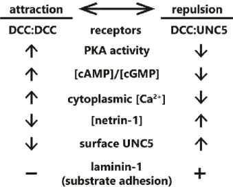
pathway components, favoring either attraction or repulsion; 3) the relative affinities of individual receptor types for netrin-1, along with the extracellular concentration of netrin-1; or 4) the presence of other molecules in the extracellular environment. Current evidence suggests that all four of these mechanisms are capable of switching netrin-1 responses between attraction and repulsion. Known mechanisms that convert such netrin-1 responses are summarized in Figure 3.
2.1.2 Switch 1: Membrane receptor levels
Modulation of receptor expression levels or presentation on the surface of cells is a common mechanism for tuning responses to extracellular ligands33–37. Altered expression levels and/or surface levels of netrin receptors have also been implicated in modulating response to netrin-1. For example, the DNA repair gene Rad51 upregulates expression of UNC5B and UNC5C in mouse primary cortical neurons, and negatively regulates netrin-dependent axon branching38, typically viewed as an
attractive response to netrin. Opposing this, endocytic internalization and membrane depletion of UNC5, and not DCC, converts repulsive netrin-1 responses to attraction. This UNC5 internalization is triggered by protein interacting with C-kinase 1 (PICK1)-dependent recruitment of active protein kinase Cα (PKCα) to the plasma membrane, which phosphorylates UNC5 residues S408 and
S58739,40. Multiple studies suggest that surface levels of DCC are altered by exposure to netrin-1,
however the specific response varies between studies. Multiple studies in embryonic rat spinal commissural and cortical neurons, demonstrate that exposure to netrin-1 increases DCC localization to the plasma membrane41 potentiated by, but not requiring, protein kinase A (PKA) activation42,43. A
single study in dissociated rat embryonic cortical neurons suggests that DCC is ubiquitinated, internalized, and then degraded after netrin-1 exposure44. However, whether this affects subsequent
netrin-1 responses was not shown. Intriguingly KCl-induced depolarization of cortical neurons, but not spinal commissural neurons, leads to increased plasma membrane levels of DCC and increases axon outgrowth in response to netrin-1. These responses require activity of PKA, PKC, and
5
studies are required to define how expression levels, surface localization, and protein stability of DCC are modulated by netrin in diverse cell types, and in scenarios in which netrin is perceived by the growth cone as attractive or repulsive.
2.1.3 Switch 2: Intracellular secondary messengers
Intracellular levels of the secondary messenger, cAMP, which promotes the activity of PKA, may trigger an attractive-repulsive switch in response to netrin. Inhibition of PKA in Xenopus spinal neurons with small molecules KT5720 or Rp-cAMPS causes a typically attractive netrin-1 gradient to repulse axons45. The authors conducted a dose-response experiment using Rp-cAMPS, a potent and
specific competitive inhibitor of cAMP-dependent activation of PKA, to investigate whether the change from netrin-1 attraction to repulsion was “switch” or a “dial”. This revealed that the turning response
transitions abruptly between 1 and 5 µM Rp-cAMPS, rapidly plateauing at higher concentrations: such a sigmoidal response suggests a switch-like mechanism45. Later experiments found that PKA
inhibition with a considerably higher dose of KT5720 reduces attractive responses to netrin-1 in rat spinal commissural neurons in a spinal explant, but does not switch the response to repulsion46. The
differences in experimental conditions make interpretation of these data difficult, however they imply that either higher concentration of PKA inhibitor, some non-neuronal component of the spinal explant, or differences between rat and frog spinal neurons (such as differing expression of netrin-1 receptor isoforms), alters the role of PKA in the netrin-1 attraction/repulsion switch.
Netrin-1 response is modulated by other secondary messengers in addition to cAMP/PKA. Axon guidance experiments in isolated Xenopus spinal commissural neurons revealed that attractive netrin-1 response relied on both Ca2+ release from the endoplasmic reticulum and Ca2+ influx through
the plasma membrane, and that blockade of Ca2+ influx into the cytoplasm converted the attractive
response to repulsion47. Ca2+-dependent repulsion also requires cGMP. Using cyclic nucleotide
6
these classic experiments on netrin-1 attraction and repulsion with new tools to optogenetically manipulate the spatial and temporal distribution of secondary messenger levels is warranted.
2.1.4 Switch 3: Netrin-1 concentration
Many axon guidance or outgrowth studies reveal bimodal responses of axons occur with increasing netrin concentrations5,12,45,46,49, suggesting that the concentration of netrin-1 may
determine which receptors, and thus intracellular pathways, are recruited. Indeed in a microfluidically isolated gradient of netrin-1, embryonic murine cortical axons closer to the source of netrin-1 (higher concentration) are repelled, whereas those at the lower end of the concentration gradient are attracted22, supporting the notion of a concentration dependent response. Although further work is
needed to establish this mechanism, biophysical experiments have demonstrated that netrin-1 binds with higher affinity to DCC than to UNC529. This could lead to increased UNC5/DCC
heterodimerization at higher concentrations of netrin-1, inducing repulsive axon guidance
responses27–29. The attractive and repulsive forces leading to directional axon outgrowth under this paradigm are summarized in Figure 4. Structural studies on the interaction between netrin-1, DCC and UNC5 suggest that although DCC can bind two sites on netrin-1, only one of these can interact with UNC529, which would preclude the formation of UNC5 homodimers. Therefore UNC5-dependent,
DCC-independent short-range axon repulsion in response to netrin-1 may require dimerization with additional receptors such as DSCAM, discussed later in this review. Alternatively, high concentrations of netrin-1 may saturate DCC, and prevent DCC homodimerization and attractive responsiveness.
2.1.5 Switch 4: Extracellular environment
Surprisingly little is known regarding modification of netrin-1 attraction and repulsion by the composition of the extracellular environment. One other axon guidance molecule, draxin, has been shown to antagonize netrin-1 and is proposed to render netrin-1 a fasciculation cue50,51, however any
7
glycosaminoglycans (GAGs), a major component of the extracellular matrix, yet once again the role of these glycoproteins in axon guidance is unknown. Draxin and GAGs will be discussed in more detail later in this review. The integrin ligand laminin-1 is one of the few known extracellular components that switches netrin-1 dependent attraction to repulsion. Xenopus retinal growth cones, normally attracted to netrin-1, will be repulsed if laminin-1 is present in the extracellular matrix (ECM) or substrate52. This response is also likely sensitive to secondary messengers, as in the presence of
substrate-adhered laminin-1, blockade of Ca2+ release, which is dependent on Ca2+
-calmodulin-dependent protein kinase II (CaMKII), calcineurin (CaN) and protein phosphatase 1 (PP1), switches a repulsive netrin-1 response to attraction in Xenopus spinal neurons53. This suggests an intriguing
modulation of internal signaling by extracellular matrix components; indeed in chick ciliary ganglion neurons, treatment with laminin induces an influx of Ca2+54. Whereas a Ca2+ influx blockade can
switch netrin-1 attraction to repulsion47, a further increase of Ca2+ beyond the attractive netrin-1
response regime may switch the response once more. The integration of signaling events from a complex extracellular environment represents a rich area for future studies into axon guidance. The requirement for substrate adhesion of laminin-1 may also indicate that the netrin-1 attractive-repulsive switch is in part reliant on mechanotransduction pathways, however this remains to be investigated. This is an especially intriguing area for study of mechanical regulation of axon guidance, as many mechanotransduction proteins are known to be ion channels, which could modify the intracellular environment55–57.
2.2 Chemotaxis and haptotaxis?
A new axis recently emerged for netrin-1 during haptotactic axon guidance. The dogma of the field has long posited that netrin-1 is a diffusible cue, supported by assays in which axons extend from explants toward netrin-1 secreted by distant patches of cells5,10,21,58,59 or diffusing from enriched
agarose blocks58, and by the presence of a gradient of netrin-1 protein in embryonic chick spinal
cord60. Supporting chemotaxis, fragments of netrin-1 lacking the major ECM-binding domains are
8
suggesting that there may be adhesion-independent effects of 1 on the axon. However, netrin-1 also binds the extracellular matrix10 or cell membranes60 and guides axons locally12,62, suggesting a
potential role for netrin as a haptotactic cue that promotes mechanotransduction. Supporting this possibility, when beads covalently linked to netrin-1 are presented to an extending spinal
commissural axon in vitro, the growth cone exerts force on the bead63. If the bead is immobilized,
growth cones reorient toward the bead. Adhesion of netrin-1 to the substrate is suggested to be necessary for attractive axon guidance in spinal commissural neurons, as inhibiting netrin adhesion with heparin blocks this attractive response, and deletion of the highly positively charged C-terminal extracellular matrix-binding C domain of netrin-1 reduces axon outgrowth61. This attraction to
adhesive netrin involves non-muscle myosin II (MyoII)-dependent mechanotransduction, as
blebbistatin treatment blocked the generation of forces on netrin-1 beads by the growth cone. Indeed, netrin-1 signaling through DCC activates MyoII via indirect activation of myosin light-chain kinase (MLCK)64. MyoII also promotes mechanical activation of focal adhesion kinase (FAK), an important
downstream effector of netrin-1 signaling through DCC61 (covered in more detail in a later section).
The original studies on the function of netrin-1 in vivo relied on a hypomorphic gene trap allele of Ntn1 that maintained low levels of netrin-1 protein65, however recent development of mice
carrying a floxed Ntn1 allele has allowed for tissue-specific and complete loss of netrin-166,67. Three
recent papers have galvanized the potential role of netrin-1 as a haptotactic cue in vivo by selectively deleting netrin-1 from the floor plate and/or ventricular zone of the spine and hindbrain using floxed
Ntn1 alleles, and assessing the midline crossing of commissural axons19–21 (for an additional mini-review on Dominici et al. and Varadarajan et al., see Morales, 2018). All three groups found that netrin-1 expression in the floor plate, originally thought to be the source of the attractive gradient of netrin-1 responsible for commissural crossing60,69, is not necessary for commissure formation. Rather,
netrin-1 deposited on the pial surface by ventricular zone neural progenitors forms a path for axons to reach the site of the commissure19,20. This deposition of netrin-1 on a surface supports a haptotactic
guidance role, though it does not invalidate earlier experiments showing a chemotactic function of netrin-1. Indeed netrin-1 is deposited on the extending axons in a DCC-dependent manner,
9
whether netrin-1 functions as a chemotactic cue, and potentially suggest that netrin-1 may signal differently through chemotactic and haptotactic mechanisms, or perhaps that other modulators of netrin-1 guidance are critical in situ. Clearly further experiments are required to determine the role of chemotactic and haptotactic netrin-1 responses in vitro and in vivo, however the data accumulated over the past 30 years suggest that both mechanisms are important during nervous system development.
3 Netrin-1 receptors and their mechanisms
Netrin-1 acts through a repertoire of membrane-spanning receptors with extracellular
domains that bind netrin and cytoplasmic domains that interact with effector proteins. These receptors include DCC (Frazzled in Drosophila, Unc-40 in C. elegans), its paralog neogenin in vertebrates, the UNC5 family (Unc5/Unc-5 in both Drosophila and C. elegans), and DSCAM (DSCAM in Drosophila
and C. elegans).
3.1 Deleted in Colorectal Cancer (DCC)
DCC is a transmembrane receptor of the immunoglobulin superfamily highly expressed in spinal commissural neurons24, the retina70,71, and many projection neurons of the fore- and midbrain
during embryonic development72. DCC functions as a receptor for netrin-1 in both growth cone
attraction and repulsion23–29. The extracellular portion of DCC consists of four Ig-like N-terminal
domains followed by six fibronectin type-III (FN3) repeats24. Structural studies reveal that netrin-1
binds in the area of the fifth and sixth FN3 domains, and that attractive axon guidance requires binding to two of these sites29,73,74. The binding of a single molecule of netrin-1 to two receptors
induces DCC homodimerization29. As there are at least three binding sites on DCC for netrin-1, and
three binding sites on netrin-1 for DCC29,75,76, netrin-1 may link dimers to produce the larger-order
10
the integrin/laminin receptor/ligand system79,80, and could represent a mechanism for the regulation of
mechanical forces on the netrin-1/DCC complex.
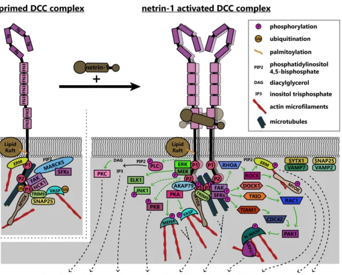
The increase in receptor valence due to netrin-1:DCC multimerization likely brings the intracellular domains of the receptors into close apposition29. The intracellular region of DCC contains
three domains termed P1, P2, and P3, which are conserved among DCC family proteins25. When
multiple DCC molecules coalesce due to netrin-1-dependent clustering, the P3 domains interact81,
and these binding-site-rich intracellular domains form a scaffold for the recruitment of downstream effectors and regulatory proteins82. As the intracellular domains of DCC are required for repulsive
netrin-1 dependent axon responses as well as attraction, parsing discreet downstream signaling pathways is complicated. This section specifically addresses mechanisms associated with either attractive axon guidance or increased axon outgrowth in response to netrin-1 downstream of DCC. Asymmetrical changes in the shape and rate of extension of the growth cone reorient outgrowth during turning; this involves dramatic and regulated remodeling of the plasma membrane1 and
underlying cytoskeleton83, which is orchestrated by both chemical and mechanical transduction
downstream of netrin/DCC, summarized in Figure 5.
3.1.1 DCC interactions with the cytoskeleton
The P3 domain of DCC is a hotspot for interaction with many binding partners of DCC that are poised to promote cytoskeletal and membrane remodeling, including the unconventional myosin X (MyoX), the nonreceptor tyrosine kinase FAK, the E3 ubiquitin ligase TRIM9, F-actin binding ezrin-radixin-moesin (ERM) proteins, and p120RasGAP, which are situated to modulate chemical signaling, mechanotransduction, or both. The MyTH4-FERM domain of MyoX binds to the P3 domain of DCC and to microtubules, whereas the head/motor domain of MyoX translocates along filamentous actin; as such, MyoX translocates DCC to the periphery of cells and tips of filopodia84,85. MyoX is also
required for netrin-1 dependent axon outgrowth and guidance of spinal commissural neurons84. Now
that conditional alleles for MyoX exist86, exploring the roles of MyoX in netrin dependent
11
reciprocally regulates MyoX, increasing association with actin filaments and promoting filopodial formation87. The modulation of MyoX localization, and potentially function, by DCC represents an
intriguing direction for future studies into the effect of extracellular ligands on intracellular force generation, however future studies need to confirm the netrin dependency of MyoX enhanced actin binding.
Netrin-dependent remodeling of the actin and microtubule cytoskeletons are critical points in axon guidance that may also be regulated by mechanotransduction. The formation of filopodia in netrin-1 dependent axon guidance relies on the Ena/VASP family of actin polymerases88,89 which,
along with DCC, localize to the tips of growth cone filopodia90–94. Increases in the length and number
of filopodia involves PKA phosphorylation of Mena at S236, corresponding to S157 in VASP89,95.
VASP, but not Mena or the third Ena/VASP family member EVL, is also regulated in netrin-1 dependent axon guidance by non-degradative TRIM9-dependent ubiquitination91. Ubiquitination of
VASP is associated with reduced filopodia lifetime and reduced VASP dynamics and localization to filopodia tips. The PKA-dependent phosphorylation of Ena/VASP proteins downstream of DCC also requires the function of PKA anchoring protein 79 (AKAP79) and ERM actin-binding proteins95,96. In
addition to regulation of actin-binding proteins, involvement of the microtubule cytoskeleton is also implicated in netrin-1/DCC responses: a recent study found that DCC interacted with neuron-specific β-III-tubulin in a netrin-1 dependent manner97. By mutating sites on β-III-tubulin necessary for this
binding, the group found that DCC:β-III-tubulin interactions are required for axon branching of cortical
neurons and guidance of spinal commissural neurons.
3.1.2 DCC signaling pathways: receptor-linked kinases and GEFs
Netrin-dependent dimerization of the DCC P3 domains results in clustering of FAK and adaptor protein NCK1, which are constitutively bound to DCC58,82,98,99. This leads to activation of FAK
12
membrane101. Intriguingly, FAK and SFK are maintained in a ready pool when not phosphorylated by
the membrane-associated scaffolding protein myristoylated alanine-rich C kinase substrate
(MARCKS)102, which interacts with PIP2 and β-actin and promotes lamellipodium formation103. This
provides another link between the actin cytoskeleton and the membrane to transduce internal mechanical forces to the cell surface for remodeling, through the interaction between FAK and actin.
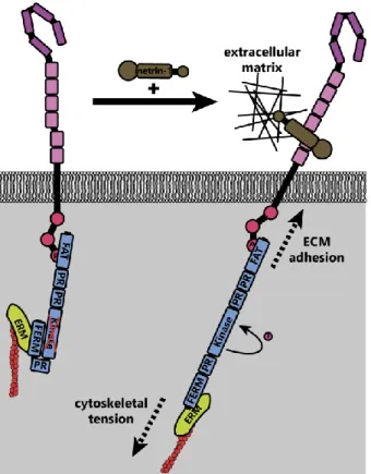
Activation of FAK downstream of netrin-1 may be mechanically induced61. The C-terminal
FAT domain of FAK binds to the P3 domain of DCC, whereas the extracellular region of DCC is linked to the extracellular matrix through netrin-1 (Figure 6); FAK also indirectly interacts with the actin cytoskeleton through associations between its N-terminal FERM domain and the F-actin binding ERM proteins and Arp2/3 complex104,105. These interactions together form a mechanotransductive
complex centered on FAK. Forces generated by the treadmilling of connected actin filaments or contractile forces generated by myosins, expose the FAK kinase domain and allow
autophosphorylation of FAK106. Further experiments are needed to identify the source of the
mechanical forces that activate FAK upon netrin-1 binding to DCC. Additionally, atomic force
microscopy could be conducted on netrin-1/DCC/FAK/actin complexes or FAK under tension in vitro, or models made in silico to determine the structure of FAK under tension and in a relaxed state.
Similar studies are needed to determine the extent to which mechanical forces are required for the activation of other proteins linking DCC to cytoskeletal components. For example netrin-dependent phosphorylation of DCC pY1418 recruits members of the ERM family96,107, linking DCC to
the actin cytoskeleton. ERM proteins bridge filamentous actin to the plasma membrane through interaction with the transmembrane receptors CD44108 and L1 cell adhesion molecule (L1CAM)109,
each of which have roles in nervous system development110,111. The ERM family member moesin also
interacts with microtubules112, providing an additional link between the netrin-1/DCC complex, the
13
Activation of ERM113–116 and MYOII117,118 enacts reorganization and traction forces, respectively, on
the actin cytoskeleton.
Once activated by FAK, the SFKs Src and Fyn recruit to the DCC signaling complex the Rho GEFs DOCK180 (DOCK1)119 and triple domain functional protein (Trio)120–125 to promote the
activation of the Rho GTPase Rac1. Similarly the GEF Tiam-1 is recruited94,126–128 to activate Cdc42.
In contrast, RhoA activity is decreased throughout the cell after application of netrin-1 in rat spinal commissural neurons129. Interestingly, another study shows that netrin-1 treatment can also activate
RhoA downstream of DCC in rat cortical neurons, leading to activation of Rho kinase (ROCK) and subsequent phosphorylation of and activation of ERM proteins107 and myosin II (MYOII)64. Together
these GTPases regulate the actin cytoskeleton to promote netrin-dependent spreading of lamellipodia and formation of filopodia129. Activated Rac1 and Cdc42 form a complex with and activate
p21-activated kinase 1 (PAK1), leading to the activation of the nucleation promotion factor, neuronal Wiskott-Aldrich syndrome protein (N-WASP)127. N-WASP then activates the filamentous actin
nucleation and branching activity of the Arp2/3 complex, a major driver of lamellipodial
protrusions130,131. This pathway appears to be evolutionarily conserved, as the Rac-like GTPases
CED-10 and MIG-2, and the GEF UNC-73 (ortholog of mammalian Trio) in C. elegans are also required for unc-6 dependent axon guidance88,120. However, mammalian Trio is required for attractive
axon guidance, whereas the C. elegans ortholog is involved in repellant guidance88,121. While
GEF/GTPase activation is one of the most thoroughly studied components of netrin-1:DCC signaling, there remain unanswered questions to be investigated. For example, how do we reconcile the decrease in RhoA activity seen globally129 with the observation that RhoA is activated downstream of
DCC107? Two potential explanations are that the activation and inhibition of specific GTPases are
regulated differently depending upon timing after netrin-1 treatment, or that other signals dictate the direction of regulation. More work must be done to understand the intricacies of regulatory pathways and timelines in the context of DCC, as well as axon guidance receptors in general.
14
(MEK1), extracellular signal-related kinases 1 and 2 (ERK1/2), and Jun N-terminal kinase (JNK1) with DCC133–135. This is specific, as activation of another MAPK family member, p38 is not triggered by netrin-1133. Palmitoylation of the transmembrane domain of DCC, and its association with lipid rafts is
required for the activation of ERK136. Activation of ERK is necessary for attractive axon guidance and
leads to activation of transcription factors such as ELK1, suggesting a possible role of netrin-1/DCC signaling in transcriptional regulation133. DCC pY1418 is also a binding site for p120RasGAP, which is
required to tightly control Ras and ERK activities in neurons during attraction of cortical axons toward netrin-1137.
3.1.3 DCC signaling pathways: plasma membrane remodeling
Recent work has elaborated on an important role of membrane reorganization and addition during netrin dependent axonal morphogenesis, along with regulation of membrane composition. Several groups described addition of DCC to the plasma membrane through exocytosis in response to netrin-141–43. The delivery of membrane is critical for axon guidance and outgrowth, which involve rapid plasma membrane expansion138–140. Exocytosis frequency increases in response to netrin-1141,
and may occur downstream of DCC, FAK77,90, SFKs, and ERK1/2142. Early reports suggested that
syntaxin-1 and vesicle associated membrane protein 7 (VAMP7, TI-VAMP) were required for this DCC-dependent increase in exocytosis in spinal commissural neurons, whereas synaptosomal associated protein 25 (SNAP25) and vesicle associated membrane protein 2 (VAMP2) were not involved143,144. However, later reports show that SNAP25 is required for SNARE complex formation
and exocytosis in netrin-1 dependent axon branching in embryonic cortical neurons, and that
SNAP25 is regulated in this context by TRIM9138,141. Studies also show an increase in the exocytosis
frequency of VAMP2 containing vesicles in response to netrin-1 which, intriguingly, were regulated by TRIM9 specifically in neurites77,138. Tension on the cell membrane has been shown to increase the
efficiency of exocytosis145; this membrane tension can be generated by increased cytoskeletal
15
increase in exocytosis in response to netrin-1 provides another avenue to investigate the role of intracellular force generation on axon guidance.
The plasma membrane lipid composition is also modified by netrin-1 signaling. Downstream of DCC, phospholipase Cɣ (PLCɣ) is phosphorylated and activated; this activation is required for axon
outgrowth in response to netrin-1147,148. Activation of PLCɣ leads to hydrolysis of PI(4,5)P2 into
diacylglycerol (DAG) and inositol 1,4,5-trisphosphate, which among many other functions, lead to the activation of PKC and induce endoplasmic reticulum Ca2+ release, respectively149,150. As mentioned,
PKC activation induces endocytosis of UNC5, which may promote increased DCC-only
homodimerization and thus attractive outgrowth39,40. Intracellular Ca2+ release is also required for
netrin-1 dependent attraction47. Interestingly, since PI(4,5)P2 is also required for autophosphorylation
and activation of FAK by DCC101, this may represent an intrinsic homeostatic negative feedback loop
that decays the DCC signal, giving it a finite lifetime. In addition to phosphatidylinositol hydrolysis, netrin-1 treatment also induces activation of PI3Ks in a DCC-dependent manner, and subsequent activation of protein kinase B (PKB/Akt)147,148, which is necessary for nervous system development151.
3.2 Neogenin
Neogenin is well characterized as a receptor for the repulsive guidance molecule (RGM)152.
In contrast, although neogenin also binds netrin-1, most information regarding the function of neogenin in netrin-1 dependent axon guidance derives from comparisons made to DCC, as they are similar proteins with identical domain structures. In the spinal cord of the chicken, an organism in which there is no DCC, neogenin may fully replace DCC function153. However, whether this
comparison is valid in species with both paralogs remains to be seen. Some differences in protein functions have been noted. For example, whereas DCC promotes MyoX movement along basal actin filaments and induces basal filopodia formation, neogenin promotes MyoX movement toward apical filaments, and induces apical filopodia formation84,85. Further studies into the cause of this distinct
16
neogenin interacts with DOCK1 as DCC does119. Unlike DCC however, neogenin does not promote
PI(4,5)P2 hydrolysis in response to netrin-1147, and therefore does not recruit an identical repertoire of
proteins. Neogenin appears to facilitate spinal commissure formation alongside DCC in mice75, as
well as ventral forebrain axon tracts in Xenopus154. Expression studies of neogenin in the developing
mouse show a broad expression among many types of maturing neurons, suggesting the receptor may act in a variety of processes in addition to netrin-1 axon guidance155. Future studies are needed
to address the functional outcome of netrin-1 binding to neogenin, as this likely forms an isolated dimer as opposed to the clustering observed with DCC75. As neogenin can functionally replace DCC
in the chicken spinal cord, any differences between the signaling pathways and force transduction capacities of these two proteins could provide invaluable insight into which mechanisms are necessary for the formation of a spinal commissure.
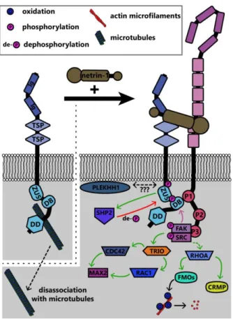
3.3 The UNC5 family
The UNC5 family of repulsive netrin receptors consists of one isoform in invertebrates (UNC5 in Drosophila and Unc-5 in C. elegans) and up to four in vertebrates (UNC5A, UNC5B, UNC5C, UNC5D). UNC5 proteins function in repulsive axon guidance in response to netrin-1, through heterodimerization with DCC for long-range repulsion27–29, and potentially heterodimerization with
DSCAM for short-range repulsion30,31. The signaling pathways discussed in this section are
summarized in Figure 7. The intracellular domain of UNC5 contains a binding site for the P1 domain of DCC; association between UNC5 and DCC mediates long-range netrin-1 dependent axon
repulsion29,32. DCC independent repulsion effects also require the ZU-5 motif of the UNC5
intracellular domain156 and the adaptor protein required for motor axon guidance 1 (Max-1, human
homolog PLEKHH1)157, both of which may scaffold downstream proteins into a signaling complex.
17
guidance in vivo156,159. Phosphorylation of UNC5C recruits tyrosine-protein phosphatase non-receptor
type 11 (PTPN11 / SHP2), which then dephosphorylates UNC5C158. Thus phosphorylation may lead
to activation of proteins downstream of UNC5, but additionally activates negative feedback through phosphatases like SHP2 as possible homeostatic mechanisms.
Rho family GTPases are also regulated downstream of Unc5 through distinct repertoires of GEFs. In neurite outgrowth in mouse neuroblastoma cells, RhoA is activated by netrin-1 binding to UNC5A, and to a lesser extent, Rac1 and Cdc42 are activated as well160. In C. elegans, netrin-1
binding to Unc-5 similarly leads to activation of the Trio ortholog Unc-73, which in repulsive netrin-1 responses can act as a GEF for the GTPases Ced-10 and Mig-2 (as opposed to Tiam-1, which occurs in attractive netrin-1 responses126,128 to inhibit growth cone protrusion120. Downstream of these
GTPases, the PAK Max-2 is required for netrin-1 dependent repulsion161. Recent work demonstrates
that additional effectors of these GTPases in netrin-1 repulsion include Unc-33 (ortholog of collapsin response mediator proteins (CRMPs)), which regulates remodeling of both the actin and microtubule cytoskeletons162, and Unc-44 (ortholog of Ankyrin), an adaptor protein connecting integral membrane
proteins to the spectrin cytoskeleton120,163. Genetic analysis suggests the flavin monooxygenases
Fmo-1, Fmo-4, Fmo-5 and Ebhp-1 are required for netrin-dependent repulsion in C. elegans164. The C. elegans Rac orthologs activate the flavin monooxygenases, which oxidize a variety of
substrates165. A similar family of monooxygenases, the molecule interacting with CasL (MICAL)
protein family, has been shown in Drosophila and in vertebrates to regulate repulsive axon guidance to semaphorins by oxidative dismantling of F-actin166–168. However, whether the C. elegans
monooxygenases similarly oxidize F-actin or other relevant substrates in netrin-1 dependent axon guidance has yet to be investigated. In addition to regulation of the actin cytoskeleton, UNC5C also interacts with β-III-tubulin, an interaction that may modulate netrin-1 dependent axon repulsion169.
18
3.4 Down Syndrome Cell Adhesion Molecule (DSCAM)DSCAM is a transmembrane cell adhesion molecule which also acts as a receptor for netrin-1170–172. DSCAM acts with UNC5 as a co-receptor for netrin-1, to mediate short-range repulsive
responses31. DSCAM is required for midline crossing of spinal commissural neurons in Xenopus, and
neurons exogenously expressing DSCAM respond to attractive netrin-1 independent of DCC173.
However, DSCAM is not required for all netrin-1 mediated guidance, such as in mouse spinal commissural neurons174. In Drosophila, DSCAM functions as a midline receptor for netrin and
possibly for an unidentified guidance cue operating in parallel to netrin and the DCC ortholog Frazzled175. DSCAM interacts with UNC5C in response to netrin-1, and this pair mediates axonal
growth cone collapse in mouse cerebellar granule cells by promoting the phosphorylation of FAK, Fyn, and PAK131,176. Evidence also suggests that DSCAM may coordinate with DCC to regulate
interactions with microtubules177 and the activation of JNK1 in response to netrin-1135. Possibly
independently of either UNC5 or DCC, DSCAM interacts with AMP-activated protein kinase (AMPK), which is phosphorylated in response to netrin-1 treatment178. DSCAM promotes axonal fasciculation
and dendritic tiling through neurite-neurite adhesion179–181. Further studies are required to
mechanistically define how DSCAM mediates diverse functions during netrin-1 dependent morphogenesis.
4 New netrin-1 modifiers
Recently identified netrin-1 modifiers provide additional complexity to netrin-dependent axonal morphogenesis. The protein draxin, secreted from the floor plate, binds soluble netrin-1 and acts as an antagonist in the developing spinal cord by binding to netrin50. Structural studies
demonstrate that a draxin/netrin-1 complex binds two DCC molecules from neighboring axons (in trans) simultaneously, promoting fasciculation as opposed to attractive axon guidance or outgrowth51,
as illustrated in Figure 9. In light of recent studies that revealed floor-plate-derived netrin-1 is
19
study. Studies on draxin in the central nervous system suggest a similar role there as well. Deletion of draxin in mice leads to partially penetrant agenesis of the corpus callosum, a major midline brain commissure182. Concomitant heterozygous deletion of Dcc and draxin (Dcc+/-draxin+/-) results in a
much stronger agenesis phenotype than either single heterozygous knockout.
Structural studies suggest that netrin-1 may require heparin sulfates as co-ligands, as the C-terminal extracellular matrix domain of netrin-1 interacts with GAGs73,183, and X-ray crystallography of
netrin-1 in complex with DCC reveals a bound sulfate29. A recent study also shows that the heparin
sulfate proteoglycan (HSPG) glypican binds to DCC and modulates netrin-1 dependent axon
guidance in vivo184. Curiously, this interaction with netrin-1/DCC relies on the N-terminal peptide-only
20
CHAPTER 1 FIGURES
21
22
Figure 3. Summary of known “switches” between attractive and repulsive function of netrin-1.
Switches span from receptor concentrations at the plasma membrane, cytoplasmic second
23
Figure 4. The concentration of netrin-1 may switch attraction to repulsion responses. Attractive and repulsive forces generated by DCC and UNC5 dimers are represented as green and red arrows, respectively, leading to an axon outgrowth vector (black arrows). As the concentration of netrin-1 increases, more DCC homodimers are recruited. At higher netrin-1 concentrations, the nonspecific receptor binding site begins to recruit UNC5 to dimers, as this binding site has a lower affinity for UNC5 than for DCC. At the highest concentrations of netrin-1, saturation of receptors with ligand may result in receptors being maintained as monomers as opposed to dimerization, preventing
downstream signaling that requires a dimer. By virtue of the concentration dependence of attraction and repulsion responses, axons are guided into a concentration in which forces are balanced.
24
Figure 5. Model of known signaling pathways and interactions downstream of DCC in netrin-1 dependent axon guidance. DCC interacts with several enzymes and adaptor proteins and the actin and microtubule components of the cytoskeleton in the absence of netrin-1, forming a “primed”
25
kinase anchoring protein 79; ARP2/3, actin-related protein 2/3 complex; CDC42, cell division control protein 42; DCC, deleted in colorectal cancer; DOCK1, dedicator of cytokinesis 1; ELK1, ETS transcription factor; ERK, mitogen activated protein kinase; ERM, ezrin-radixin-moesin; FAK, focal adhesion kinase; FN3, fibronectin type 3 domain; Ig, immunoglobulin domain; JNK1, c-Jun N-terminal kinase 1; MARCKS, myristoylated alanine-rich C kinase substrate; MEK, mitogen activated protein kinase kinase; MENA, mammalian enabled; MYOII, myosin II; MYOX, unconventional myosin X; NCK1, NCK adaptor protein 1; N-WASP, neuronal Wiskott-Aldrich syndrome protein; PAK1, protein associated kinase 1; PKA, protein kinase A; PKB, protein kinase B; PKC, protein kinase C; PLCɣ,
phospholipase C gamma; RAC1, Ras-related C3 botulinum toxin substrate 1; RHOA, Ras homolog gene family member A; ROCK, Rho associated protein kinase; SFKs, Src family kinases; SNAP25, synaptosomal associated protein 25; SYTX1, syntaxin-1; TIAM1, T-lymphoma invasion and
26
Figure 6. Theoretical model of FAK activation by mechanical forces (adapted from Moore et al. 2012). In the inactive state, the N-terminal FERM domain of FAK interacts with the kinase domain, preventing activation. Upon netrin-1 binding, tension on the actin cytoskeleton (through actin
27
Figure 7. Summary of known signaling pathways downstream of UNC5 in repulsive netrin-1 signaling. Interaction between the intracellular DB domain of UNC5 and the P1 domain of DCC produces a scaffold similar to that in DCC homodimers. FAK and Src are phosphorylated and activated as in attractive netrin-1 signaling, however the functional outcomes are different.
Abbreviations: CDC42, cell division control protein 42; CRMP, collapsin response mediating protein; DB, DCC-binding domain; DD, death domain; FAK, focal adhesion kinase; FMOs, Flavin
28
29
30
CHAPTER 2: THE E3 UBIQUITIN LIGASE TRIM9 IS A FILOPODIA OFF SWITCH REQUIRED FOR NETRIN DEPENDENT AXON GUIDANCE†
1 Introduction
During embryonic development, growth cones at the tips of extending axons respond to extracellular cues to direct axon growth185. In the mammalian cortex, the secreted guidance cue
netrin-1 (netrin) and its receptor DCC (deleted in colorectal cancer) promote attractive axon
guidance81,186 and deficiency of the murine gene encoding netrin-1 orDCC induces cortical projection
defects26,65. DCC localizes to the tips of filopodia94, bundled filamentous actin (F-actin) rich
protrusions that decorate the growth cone periphery and contribute to axon guidance. Furthermore, DCC is required for netrin-dependent increases in filopodia density89 . In addition to guidance
receptors, the filopodia tip complex contains cytoskeletal regulatory proteins that modulate filopodial growth and stability90. Cytoskeletal dynamics contribute to the extension and turning of growth cones,
but how the function of the tip complex is regulated by netrin is not known.
Ena/VASP actin regulatory proteins localize within the tip complex and are essential in netrin response, filopodial formation, neuritogenesis and axon fiber tract formation in the murine
cortex89,187,188. Mammals have three Ena/VASP orthologs: Mena, VASP, and EVL92,188, which promote
formation of unbranched F-actin through binding and protecting the barbed end from capping and promoting polymerization189–192. This family is characterized by an N-terminal Ena/VASP Homology 1 (EVH1) domain that binds proteins with the sequence (D/E)FPPPPX(D/E)(D/E) (abbreviated FP4), a proline-rich (Pro) domain, and an EVH2 domain that binds monomeric and F-actin and mediates tetramerization193. Ena/VASP function is required for netrin-dependent increases in growth cone
† This chapter previously appeared as an article in Developmental Cell. The original citation is as follows:
Menon S#, Boyer NP#, Winkle CC, McClain LM, Hanlin CC, Pandey D, Rothenfußer S, Taylor AM, Gupton SL.
31
filopodia89. Ena/VASP proteins are acutely phosphorylated in response to netrin, however this
phosphorylation is not detected when filopodia density increases, suggesting unidentified mechanisms regulate Ena/VASP function.
We identified murine TRIM9 as a direct binding partner of DCC that regulates cortical axon branching in response to netrin141. Like Ena/VASP, TRIM9 localizes to filopodia tips in cortical
neurons. The interaction between TRIM9 and DCC is conserved in invertebrates, where the TRIM9
ortholog is required for netrin responses194,195. Their similar localization and requirement in netrin
responses suggests that TRIM9 and Ena/VASP may cooperate within filopodia in response to netrin. TRIM9 is a member of the tripartite motif (TRIM) family of E3 ubiquitin ligases, which mediate covalent linkage of ubiquitin to substrates. Ubiquitin addition can trigger proteasomal degradation or alternatively modify substrate localization, trafficking or function196–198. Additionally, ubiquitination can
be reversed by deubiquitinases (DUBs199). Although TRIM9 exhibits ligase activity200, its substrates
and the consequences of its ligase activity are unknown.
32
2 Results2.1 Identification of TRIM9 as a novel Ena/VASP interaction partner
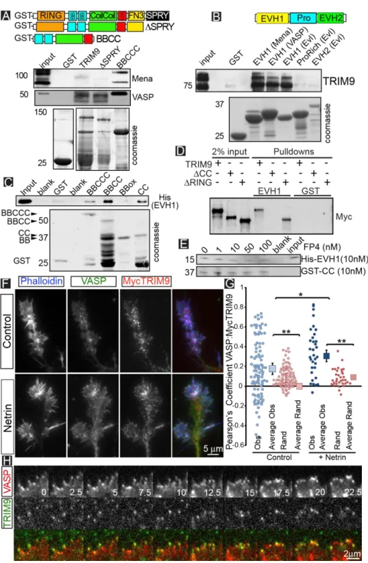
A yeast two-hybrid screen using an embryonic mouse brain cDNA library and EVL as bait identified four independent clones containing sequences corresponding to amino acids 45-532 of TRIM9. TRIM proteins share a conserved N-terminal TRIM motif with an E3 ubiquitin ligase RING domain, 1-2 BBox domains, and a coiled-coil (CC) domain that mediates homo- and
hetero-multimerization (Fig1A). In TRIM9, the TRIM motif is followed by a COS box, fibronectin type III (FN3) and SPRY domains201. Since the SPRY domain of TRIM9 directly interacts with DCC141, the
interaction between Ena/VASP and TRIM9 may link netrin to filopodia.
2.2 TRIM9 binds and colocalizes with Ena/VASP proteins in cortical neurons
To confirm the interaction between TRIM9 and Ena/VASP predicted by yeast two hybrid, we incubated GST-tagged TRIM9 variants in embryonic mouse brain lysate (Fig1A). A GST-tagged protein containing the BBox-CC-COS domains of TRIM9 (BBCCC) bound endogenous Mena and VASP, whereas GST did not. GST-TRIM9 and a variant lacking the SPRY domain (ΔSPRY) failed to
precipitate Mena or VASP. Even though TRIM9 lacks an FP4 motif, GST-EVH1 domains of all three Ena/VASP members precipitated endogenous TRIM9 from embryonic brain lysate, but GST-Pro or GST-EVH2 did not (Fig1B). This pattern of binding may suggest conformational changes or post-translational modifications in TRIM9 or Ena/VASP proteins are required to permit binding, or that terminal domains of the proteins modulate binding. However, direct binding assays demonstrated that BBCCC was able to precipitate His-EVH1 (Fig1C). This interaction was maintained with GST-BBCC and GST-CC, but not GST-BBox, indicating that the CC domain is the minimal binding region of TRIM9. We confirmed this observation with precipitation of Myc-TRIM9 variants expressed in HEK293 cells (Fig1D). GST-EVH1 precipitated Myc-TRIM9 and Myc-TRIM9 lacking the RING domain (TRIM9ΔRING) but not Myc-TRIM9 lacking the CC motif (TRIM9ΔCC). Tes and Abi are the only