DISSECTING THE HUMAN ANTIBODY RESPONSE TO DENGUE VIRUS
Adamberage Ruklanthi de Alwis
A dissertation submitted to the faculty of the University of North Carolina at Chapel Hill in partial fulfillment of the requirements for the degree of Doctor of Philosophy in the Department of Microbiology and Immunology
Chapel Hill 2013
Approved by:
Aravinda M. de Silva, MPH, PhD Stephen H. Clarke, PhD
ABSTRACT
ADAMBERAGE RUKLANTHI DE ALWIS: Dissecting the Human Antibody Response to Dengue Virus
(Under the direction of Aravinda M. de Silva)
(EDI-II) of the E protein. Due to the role of the EDI-DII hinge region in DENV fusion and entry, this epitope offers a functional advantage. As hypothesized, we observed that all neutralizing EDI-DII hinge-binding monoclonal antibodies that were isolated blocked DENV infection at a step post-attachment, while the mechanism of neutralization by human polyclonal sera was more variable. In parallel, we found that the weakly neutralizing, cross-reactive group of antibodies was responsible for antibody-mediated enhancement of infection by heterotypic DENV serotypes. Further investigation mapped these enhancing cross-reactive antibodies to the DENV surface glycoproteins prM and E protein. These studies shed some light on the protective and enhancing DENV epitopes targeted by the human immune response, and set the stage for a safer and efficacious human vaccine against DENV.
ACKNOWLEDGEMENTS
“We are like dwarfs sitting on the shoulders of giants. We see more, and things that are more distant, than they did, not because our sight is superior or because we are taller than they, but because they raise us up, and by their great stature add to ours”
Bernard of Chartres, 12th Century
Messer for being one of the greatest scientific minds around me. Thank you Bill for all the advice, support and encouragement you have given me. I thank my thesis committee (Ron Swanstrom, Stanley Lemon, Ed Collins, Stephen Clark) for all the scientific advice and support they have given. Most of all, I thank my committee for supporting me in twisting Arvinda’s arm to allow me to be a part of the human monoclonal project (lol). I thank Laura White for always, always supporting me in all my scientific endeavors, and also for introducing me to a special someone. I thank Dixie Flannery and the administration staff (especially Theresa Duffy and Lisa Best); without their reminders I would not have turned in any official paperwork and would have missed all the deadlines important for completing this dissertation. I thank the Royster Society of Fellows for financially supporting me in the last year of my thesis work.
To all my collaborators (which there were many), I have thoroughly enjoyed working with them to unravel the mysteries of the human antibody response to dengue virus: 1) Scott Smith and James Crowe, for the insane number of human monoclonal antibodies we generated and characterized together, 2) Katherin Williams and Eva Harris, for always pushing me and encouraging me in the ADE mouse work, 3) Bill Messer, Jeremy Huynh and Ralph Baric for escape mutant and infectious clone work, 4) Kristen Kahle and Benjamine Doranz for the antibody mapping work.
TABLE OF CONTENTS
LIST OF TABLES ... xiv
LIST OF FIGURES ... xv
LIST OF ABBREVIATIONS ... xviii
CHAPTER ONE Introduction... 1
1.1 Dengue: Epidemiology and clinical disease... 1
1.2 DENV genome and life cycle... 3
1.3 DENV surface glycoproteins... 4
1.4 Virus Structure ... 7
Mature virion... 8
Immature virion... 9
Major viral rearrangements: maturation & fusion... 10
1.5 Human immune response to natural DENV infections... 12
Primary DENV infections... 12
Secondary DENV infections... 13
1.7 Mechanisms of DENV neutralization ... 18
Antibody affinity... 19
Epitope accessibility due to complex virion structure... 19
DENV structural dynamics... 20
Epitope accessibility & DENV maturation... 20
Complement protein, C1q, & decrease in threshold number... 21
Pre-attachment and post-attachments blocking Abs... 22
1.8 Antibody dependent enhancement of DENV: the other edge of the double-edged sword... 23
1.9 Prospective DENV vaccines... 26
1.10 Objectives of this dissertation... 28
CHAPTER TWO In-depth analysis of the antibody response of individuals exposed to primary dengue virus infection... 59
2.1 Overview... 59
2.2 Introduction ... 60
2.3 Materials and Methods ... 61
Viruses, recombinant proteins and immune sera... 61
Epitope mapping of EDIII binding hMAbs... 63
2.4 Results... 64
Identification of DENV-reactive memory B cells following primary infection... 65
Isolation and characterization of DENV-specific hMAbs from donor 033... 65
Characterization of Human MAbs generated from donor 013... 67
Epitope mapping donor 013 hMAbs binding to EDIII... 68
2.5 Discussion ... 69
CHAPTER THREE Identification of human neutralizing antibodies that bind to complex epitopes on dengue virions ... 87
3.1 Overview... 87
3.2 Introduction ... 88
3.3 Methods ... 90
Serum Samples... 90
Virus and rE Proteins... 90
Depletion of DENV-Specific Abs from Human Immune Sera... 90
Depletion of DENV rE-Specific Abs from Human and Monkey Immune Sera... 91
Detection of DENV or rE-Binding Abs by ELISA... 91
Depletion of Heterologous DENV-Specific Abs from Immune Sera... 92
Depletion of DENV Recombinant E Protein-Binding Abs from Immune Sera... 93
Characterization of hMAbs That Strongly Neutralize DENV... 95
Generation of DENV Mutants That Escape Neutralization by hMAbs... 96
3.5 Discussion ... 97
3.6 Supporting Information ... 100
CHAPTER FOUR Further characterization of the neutralizing human antibody response to dengue virus ... 126
4.1 Overview... 126
4.2 Introduction ... 126
4.3 Methods ... 129
Virus and cell lines... 129
Human DENV-immune sera and MAbs... 129
Generation of neutralization escape mutants... 130
Epitope mapping using mutant prM-E proteins... 130
Mapping antibodies using an infectious clone... 131
ELISA binding assays... 132
Further characterization of the 5J7 epitope... 134
The EDI-DII hinge region is a major target of type-specific protection in humans... 135
Potently cross-protective human MAbs target domain II of the E protein... 137
Insights into the mechanism of protection of strongly neutralizing human antibodies... 139
Antibodies that do not target the structural DENV proteins are also protective in vivo... 141
4.5 Discussion ... 142
CHAPTER FIVE Mapping enhancing antibodies produced by the human immune response after primary dengue virus infections... 173
5.1 Overview ... 173
5.2 Introduction... 173
5.3 Methods... 175
Virus and cell lines... 175
Human sera and Fabs... 176
Depletion of virus-specific antibodies from human sera... 176
ADE assay in AG129 mice... 178
5.4 Results ... 178
ADE of DENV in K562 cells and the AG129 mouse model... 178
Depletion of cross-reactive antibodies ablated ADE of heterotypic DENV virus... 179
Depletion of rE-specific antibodies decreased ADE of heterotypic virus... 180
Competition ADE assays with prM-binding Fab indicates a role for prM-binding Abs in ADE... 181
5.5 Discussion ... 182
CHAPTER SIX Discussion ... 198
6.1 A snapshot of the human antibody response to DENV... 198
6.2 Lessons learnt from protective antibodies following natural DENV infections in humans. ... 199
Role of EDI-DII hinge-specific Abs in type-specific protection... 199
DENV EDI-DII hinge-specific Abs and mechanism of neutralization... 201
Role of DII-binding Abs in cross-reactive protection... 203
LIST OF TABLES
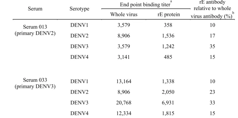
Table 2.1 Relative levels of virus and rE protein binding antibody in
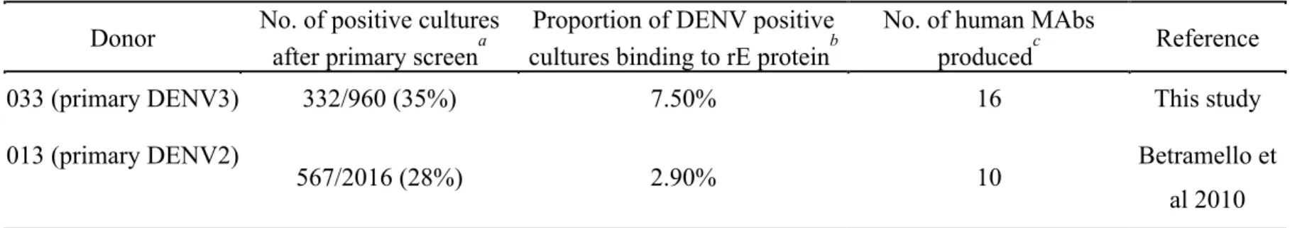
immune sera ... 77 Table 2.2 Screen for isolating DENV-specific human MAbs... 78 Table 2.3 Epitope mapping DENV2 EDIII reactive MAbs
from donor 013... 79 Table 2.S1 Dengue immune human sera used in the present study ... 80 Table 2.S2 Properties of MAbs from donor 033 (Primary DENV3 infection) ... 81 Table 3.1 Homologous DENV serotype neutralization titers of
immune sera depleted of cross-reactive antibodies from subjects
following primary infection... 111 Table 3.2 Homologous DENV serotype neutralization titers of primary
immune sera depleted of rE-binding antibodies ... 112 Table 3.3 Binding and neutralization properties of isolated strongly
neutralizing human MAbs ... 113 Table 3.S1 A panel of late convalescent DENV-immune sera from
individuals with past primary DENV2 or DENV3 infections... 114 Table 3.S2 Binding properties of DENV-immune sera with and without
rE-binding antibodies to the homologous virus serotype... 115 Table 4.1 Effects of swapping DENV3 EDI-DII hinge with DENV4 residues on
neutralization by DENV-immune human sera... 159 Table 4.2 Binding and neutralization characteristics of two broadly
neutralizing human MAbs that were isolated... 160 Table 4.3 Successful generation of several DENV2 neutralization escape
mutants against 1C19 and 1N5... 161 Table 4.4 Neutralization properties of DENV by whole MAb versus fAb
LIST OF FIGURES
Figure 1.1 DENV genome and life cycle... 32
Figure 1.2 DENV E glycoprotein structure and its arrangement on the mature virion ... 34
Figure 1.3 DENV E protein undergoes large conformational changes during virus maturation and viral fusion ... 35
Figure 1.4 The antibody response in humans following natural primary DENV infections... 36
Figure 1.5 Epitope mapping of DENV-specific mouse MAbs... 37
Figure 1.6 Structural dynamics, virion maturation and presence of complement affect the antibody threshold of DENV neutralization... 38
Figure 1.7 Antibody dependent enhancement of DENV ... 39
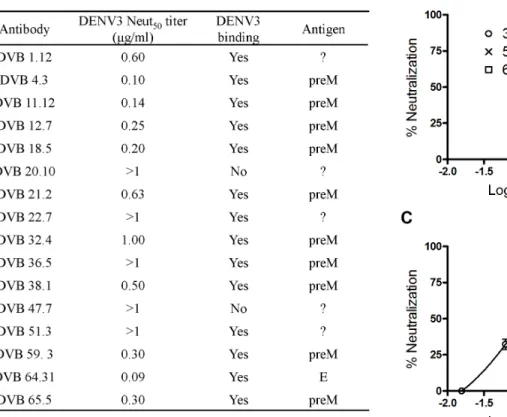
Figure 2.1 Antigens recognized by hMAbs produced from donor 033... 73
Figure 2.2 DENV3 neutralization by donor 033 human MAbs... 74
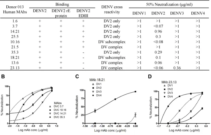
Figure 2.3 Binding and neutralization properties of donor 013 hMAbs ... 75
Figure 2.4 Epitope mapping of anti-DENV2 hMAbs binding to EDIII... 76
Figure 3.1 Binding and neutralization properties of primary immune sera depleted of total or cross-reactive DENV-binding Abs ... 105
Figure 3.2 Binding and neutralization properties of primary immune sera depleted of rE-binding Abs ... 107
Figure 3.3 Epitope mapping of escape mutants generated from type-specific neutralizing hMAbs ... 109
Figure 3.S4 Binding properties of rE-depleted sera to the homotypic DENV ... 117 Figure 4.1 Additional mapping data for 5J7 ... 147 Figure 4.2 Twenty-six amino acid residues on the DI-DII hinge of
DENV3 E protein was successfully mutated to the corresponding
residues in DENV4... 148 Figure 4.3 Mapping 1N5 and 1C19 through neutralization escape
mutants ... 150 Figure 4.4 Epitope mapping of 1C19 using a DENV3 prM-E
mutant library ... 152 Figure 4.5 Neutralization differences of 5J7 fAb in Vero versus
U937+DC-SIGN is not due to Fcγ receptors ... 153
Figure 4.6 Pre-attachment and post-attachment neutralization analysis of strongly neutralizing human monoclonal antibodies ... 155 Figure 4.7 Neutralization analysis of virus-depleted human serum
in AG129 mice... 157 Figure 5.1 Primary DENV immune sera enhance heterotypic serotypes
at high serum concentrations... 185 Figure 5.2 In the AG129 mouse model, heterotypic sera enhance
DENV2 infection and causes ADE-induced lethality, while homotypic sera protects from DENV challenge... 186 Figure 5.3 Removal of cross-reactive antibodies from primary
DENV-immune human sera, removes enhancement of
heterotypic DENV infection in vitro... 187 Figure 5.4 Removal of cross-reactive antibodies from primary
DENV3-immune sera significantly protected AG129 mice from ADE of DENV2 ... 188 Figure 5.5 Recombinant E-binding cross-reactive antibodies make
up a significant portion of the heterotypic virus-enhancing
Figure 5.7 Between 25-50% of heterotypic virus enhancement by primary DENV-immune sera can be attributed to prM-binding
antibodies... 192
LIST OF ABBREVIATIONS AA Ab ADE cryo-EM CD CNBr CS DC-SIGN DENV DF DHF DSS E E85 EBV EDI EDI-II or EDII EDIII amino acid antibody
antibody dependent enhancement cryo-electron microscopy
cluster of differentiation cyanogen bromide conserved stem region
dendritic cell-specific intercellular adhesion molecule-3-grabbing non-integrin
dengue virus dengue fever
dengue hemorrhagic fever dengue shock syndrome envelope protein
85% of E protein Epstein barr virus
FRNT GC hMAb IFN IgG IL LDC MAb mMAb Neut50 NS PBMC prM/M rE PCR RT-PCR TBEV TGN UTR VEEV VRP-rE
focus reduction neutralization titer genome copies
human monoclonal antibody interferon
Immunoglobulin G interleukin
Langerhans dendritic cells monoclonal antibody
mouse monoclonal antibody 50% Neutralization titer nonstructural protein
peripheral blood mononuclear cells pre-membrane/ membrane protein recombinant envelope protein polymerase chain reaction
reverse transcription polymerase chain reaction tick-borne encephalitis virus
trans-Golgi network
untranslated terminal region
Venezuelan equine encelphalitis virus
CHAPTER ONE
Introduction
1.1 Dengue: Epidemiology and clinical disease
Dengue is a re-emerging neglected disease that affects individuals in over 100
countries and is the pre-eminent arthropod-borne viral disease of humans (47, 89). Dengue
disease is caused by dengue virus (DENV), which exists as four closely related serotypes
(named DENV1 through DENV4). DENV spreads efficiently between humans primarily
through the mosquito vectors Aedes aegypti and Aedes albopictus. DENV is estimated to infect over 390 million individuals globally each year (8, 11, 89, 131). The mosquito vectors
reside in subtropical and tropical regions; thereby placing 2.5 billion individuals at risk from
acquiring a DENV infection. Unfortunately, there are currently no approved therapeutics or
vaccines against DENV.
Indian subcontinent and Southeast Asia (51). Since the 1970s, severe forms of dengue have
spread from five to over 100 countries (reviewed in (44)). It is estimated that at least 500,000
cases of DHF and DSS cases (with 5% mortality) occur globally each year (131). This
continual rise in morbidity, mortality and geographical spread is due to increasing
urbanization, globalization, lack of basic infrastructure, and the spread of the mosquito
vectors to new territories.
Successful transmission of DENV by the mosquito vector requires circulating titers of
around 107-109 mosquito infectious doses in an infected person (34). When an Aedes. species mosquito injects DENV into the skin of a new susceptible human, the virus undergoes the
first round of replication in the resident skin dendritic cells, called Langerhans dendritic cells
(LDCs) (92). The infected LDCs then migrate to the draining lymph node where the virus
infects monocytes and macrophages (162). Within the lymphatic and circulatory system, the
virus is then disseminated to other organs, such as the liver, spleen and bone marrow (64).
Dengue manifests as a broad spectrum of clinical symptoms in humans. Between
50-95% of DENV infections are asymptomatic or present sub-clinical symptoms. Symptomatic
DENV infections are predominantly cases of DF, with classical symptoms of fever,
headache, eye pain, myalgia, athralgia, rash, nausea and abdominal pain, following the 3-7
day viral incubation period. Most patients recover after fever subsides, while a small
proportion goes on to develop a systemic vascular leak syndrome (i.e. DHF and or DSS),
with petechiae, thrombocytopenia and shock (108). Prior classification of DENV-induced
Therefore, the WHO recently revised the guidelines, and now patients are diagnosed as either
having dengue or severe dengue (SD) (109). Hence in the present document, we will use SD
and DHF/DSS interchangeably.
1.2 DENV genome and life cycle
DENV belongs to the arthropod-borne, single-stranded positive-sense RNA
(+ssRNA) virus family called flaviviruses. Other significant human pathogenic flaviviruses
include West Nile virus (WNV), yellow fever virus (YFV), Japanese encephalitis virus (JEV)
and tick-borne encephalitis virus (TBEV) (45). Important work on these related flaviviruses
have contributed significantly to our understanding of the life cycle and structure of DENV
(reviewed in (120)). Similarly in the presently described work, we have on numerous
occasions delved into published material on related flaviviruses to gain further insight while
studying the human antibody response to DENV.
Flaviviruses attach to the surface of the host plasma membrane and enter the host cell
through receptor-mediated endocytosis (149). The internalization receptor for DENV is as
yet unknown. However, several important attachment factors that aid DENV infection in vitro have been identified; these include C-type lectin (DC-SIGN, dendritic cell-specific ICAM-3 grabbing non-integrin) (142), glycosaminoglycans (Heparin Sulphate, manose
receptor in macrophages) (19, 28, 96, 114) and phosphatidylserine receptors (93). As the
virion travels through the endosomal pathway, the low pH environment within the endosome
triggers large conformational changes on the virion surface, leading to viral fusion with the
into a single polypeptide. As shown in Figure 1.1A, the viral polypeptide (with protein
sequence C-prM-E-NS1-NS2A-NS2B-NS3-NS4A-NS4B-NS5) is subsequently processed by
viral and host proteases into three structural (C, capsid; prM, membrane precursor; E,
envelope) and seven nonstructural proteins (NS) (reviewed in (120)). During protein
synthesis on the rough endoplasmic reticulum (ER), several viral membrane proteins are
anchored into the ER membrane and the process of viral assembly begins within the ER
lumen (Figure 1.1B). Viral translation initiates formation of convoluted membrane structures,
where the viral NS proteins form replication complexes and actively replicate the RNA
genome (88, 146). The newly transcribed viral genomic RNA strands are encapsulated within
a nucleocapsid, which in turn is packaged within an ER-membrane derived envelope
containing prM and E proteins in a trimeric conformation (Figure 1.1B and C) (reviewed in
(120)). These assembled viral particles are immature and non-infectious (Figure 1.1C). While
the immature virions are transported through the trans-Golgi network (TGN), they undergo
glycosylation modifications and processing by the low pH-dependent host protease, furin
(Figure 1.1B and C) (133, 164). Mature, infectious virions are subsequently exocytosed from
the host cell.
1.3 DENV surface glycoproteins
When a DENV virion is initially assembled, each virion surface is embedded with
heterodimers of the two glycoproteins, prM and E. The prM protein primarily functions to
prevent premature fusion of the E glycoprotein with host cell membranes, and chaperones the
the flavivirus family, there is high prM protein sequence conservation (~70%) among the
four DENV serotypes (31). The prM protein contains approximately 166 amino acids
arranged in three domains; 1) the pr peptide (91 amino acids) which folds into a 7 stranded β
-sheet, 2) the M portion, which is predicted to consist of an amphipathic helix, and 3) two
C-terminal transmembrane segments (M-T1 and M-T2) (Figure 1.2C), where M-T2 contains
the signal sequence for the secretion of E protein (78, 91). X-ray crystallography and
cryo-electron microscopy (cryo-EM) structures indicate that in the immature virus, the pr peptide
resides on the distal end of the E protein and covers the fusion peptide to prevent viral fusion
within the TGN membranes (78, 170). During DENV maturation (Figure 1.1C), the prM
protein is cleaved by host furin into the soluble pr peptide and the membrane-anchored M
protein (~75 amino acids) (2, 164). Structural studies indicate that the M protein is partially
hidden below the surface-exposed E proteins in the mature virion (as shown in the cartoon
representation in Figure 1.2C), and thereby presumably inaccessible to antibodies and
predicted to play an insignificant antigenic role (168).
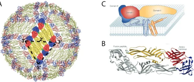
The flavivirus E glycoprotein (E) (~495 amino acids) is the most surface-exposed
protein on the mature, infectious virion particle (Figure 1.2A) (75). E protein plays critical
roles in determining cell tropism, attachment, fusion and entry into host cells. The E protein
is also the most antigenically significant protein across members of the flaviviral group and
the major target of neutralizing antibodies (122). The initial identification of four DENV
serotypes was based on the antigenic differences in the E protein. The existence of four
1) the ectodomain, 2) the two amphipathic helices (E-H1 and E-H2) and conserved stem
region (CS), and 3) the two C-terminal transmembrane helices (E-T1 and E-T2) (103, 168).
Since the helices and CS region are both hidden by the E ectodomain and membrane
associated, very little structural information is known about these regions (168). Several
molecular and biochemical studies have shown that these regions are involved in viral
assembly (i.e. through prM-E heterodimer stability, and membrane association), low pH
rearrangement events and fusion (12, 70, 83, 98, 115, 171). It is also assumed that because
these helices are not surface exposed they are inaccessible to antibodies and therefore,
irrelevant for studying neutralizing antibody responses.
When the ectodomain region of the E protein (80% of E protein) is expressed
without the helices, the resultant recombinant E protein is soluble and secreted (27). The
x-ray crystallography structures of several flaviviral E proteins were solved using the soluble E
ectodomain, also termed recombinant E protein (rE) (Figure 1.2B) (97, 99, 117). The E
ectodomain is folded into three distinct β-barrel domains (named domain I, II and III); the
N-terminal domain I (EDI) is an eight-stranded β-barrel, connected on one side with four
peptide chains to the dimerization domain (EDII, domain II) and on the other side with one
peptide linker to the immunoglobulin (Ig)-like domain (EDIII, domain III). Domain II
contains the highly conserved fusion peptide, which is inserted into the host membrane
during viral fusion and entry. Domain III of the E protein has the receptor binding sites (25,
by x-ray crystallography as stabilizing the E protein dimers by stretching across the dimer
interface to cover a region of the fusion peptide (97, 100, 113, 117).
The E protein ectodomain under neutral pH has been solved by x-ray crystallography
for both DENV2 and DENV3 (97, 99). Despite a two amino acid deletion in EDI of the
DENV3 E protein, the E protein structures of both serotypes were found to share very high
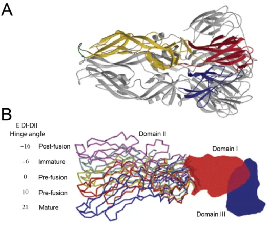
structural similarity. One of the few structural differences was observed when the E
structures of the two serotypes were super-imposed on each other; it was noted that a 10°
difference in rotation at the EDI-II hinge region of the E protein translated into a 13Å and
18Å difference in position of the fusion loop and EDIII, respectively (99). Therefore, it is
important to keep in mind that the EDI-DII hinge region is highly flexible and undergoes
large conformational changes during viral maturation and the low pH-induced fusion process
(Figure 1.3B) (168).
1.4 Virus Structure
Similar to other flaviviruses, the assembled, infectious DENV virion is 500Å in
diameter. Each virion consists of the viral genomic RNA-containing nucleocapsid enveloped
in a lipid membrane containing the two membrane-embedded glycoproteins, (prM/M and E
protein) (75). The DENV genome encodes a single open reading frame of 11 kb in length,
with untranslated terminal regions (UTRs) that are critical for regulation of genome
replication and translation. Highly asymmetrically charged capsid (C) proteins package this
DENV RNA for virion assembly (87). A host ER membrane-derived lipid envelope
E proteins (75, 102, 168, 170). This is further evident by the routine observation of subviral
particle formation in virus-infected or prM-E transfected cells in culture, where 30 prM-E
heterodimers are sufficient to arrange into particles with an icosahedral symmetry (T=1), induce fusion and stimulate protective immunity (2, 23, 35, 72, 103).
Mature virion
Recent x-ray crystallography and cryo-EM images have revolutionized our
understanding of the DENV virion structure (75, 97-99, 170). The DENV virus is made of
180 heterodimers of prM/M and E proteins, ordered into an icosahedral structure. The
surface exposure and arrangement of the glycoproteins on the particle is dependent on the
maturity of the virus (Figure 1.1C) and the pH of the surrounding environment (75, 103, 117,
170). The earliest cryo-EM studies of DENV were of the mature, infectious virion (75). In
the mature virion, E protein monomers are arranged into head-to-tail dimers, with the fusion
loop of one monomer hidden below EDI and EDIII of the adjacent monomer (see Figure
1.2B). Three sets of these dimers are arranged into herringbone-structured rafts, and 30 of
these repeated raft structures assemble into the infectious virion (Figure 1.2A). As the E
monomers are assembled into the virion particle, the hinge angle between EDI and EDII
reduces by about 10-20° (Figure 1.3B), and the EDIII is pushed outwards. It was recently
found that the M protein, which is hidden below the surface-exposed E protein, also has a
vital role to play in keeping the virion intact (169). The N-terminal 20 amino acid region of
M protein contains three pH-sensing histidine residues that at neutral pH latch onto the
Cryo-EM studies of TBEV subviral particles lead to the hypothesis that flaviviral
particles are assembled in a T=3 symmetry (35). However, cryo-EM studies of the infectious
virion show that the orientation of the 30 rafts in the virion form 5-3-2 axes of symmetry; the
EDIII of five E proteins come together to form a 5-fold axis, the EDI of three E proteins form
the 3-fold axis and two E proteins come together to form the 2-fold axis (Figure 1.3A) (75).
Therefore in the assembled virion, not all the E protein monomers are in the same chemical
environment, and this could have significant consequences on attachment, fusion and
neutralizing epitopes available for antibody binding.
Immature virion
Surface structure of the immature DENV virus particle is vastly different from that of
the mature DENV virus (Figure 1.1C). In the immature prM-E heterodimers, the uncleaved
prM extends along the length of the E protein, and the pr peptide β-barrel structure sits on the
fusion loop at the distal tip of EDII preventing premature fusion events (78). In the immature
virion, these prM-E heterodimers arrange into 60 repeated trimer structures. Unlike the
smooth outer surface of the mature virion, these trimers give the immature virus an irregular,
spiky surface and an external diameter of 600 Å (i.e. 100 Å greater than the mature virion)
(170). The highly flexible hinge region between DI and DII of E protein in the immature
virus differs by 30°, when compared to the mature virion, and about 5-15° as compared to the
crystal structures of the soluble E protein (75, 97-99, 170). Differences in the EDI-DII hinge
angle, E protein epitope exposure and the presence of uncleaved prM could have significant
culture-derived DENV virions were found to be only partially mature, with almost 90% of
the virions containing uncleaved prM (6, 66, 67, 121). The minimum degree of viral maturity
required for infectivity is unknown. Recent unpublished data from our lab indicates that the
ratio of mature and immature virus may have vast consequences on attachment and
infectivity of different cell lines. Furthermore, It is unknown whether natural in vivo
infections in humans or mosquitoes also produce DENV virions with a range of maturity.
Major viral rearrangements: maturation & fusion
During the DENV lifecycle, the virion particle undergoes two major conformational
changes: 1) during viral maturation in the TGN (reviewed in Perera, R et al, 2008 (111)), and 2) during viral fusion with the host membrane in the late endosomes. Elegant
biochemical studies coupled with studies of virus structure have given us some insight into
the conformational changes the virus experiences during these events. 1) Viral maturation
occurs in two important steps. Spiky immature virus buds from the ER and enters the TGN,
where a gradient decrease in the surrounding pH causes pH-sensing histidines in the
C-terminal ectodomain region of prM to induce latching on to the membrane-facing side of the
E proteins, causing a 30° rotation at the EDI-DII hinge region and forcing the E proteins to
lie flat on the membrane surface (Figure 1.3B) (111, 164, 169). This results in the
rearrangement of 60 prM-E trimers into 90 E protein dimers (Figure 1.1C). This low
pH-induced virion conformation exposes the furin cleavage site, enabling efficient processing of
prM into pr peptide and M protein (133, 164). The pr peptide prevents premature fusion by
The second major structural rearrangements of the surface glycoproteins occur during
viral fusion with the host membrane in the late endosomes (Reviewed in Harrison, SC, 2008 (56)). In the recent years, DENV membrane fusion and the resulting conformational changes in the E, have been studied extensively. Flaviviral fusion is a class II fusion process (117,
134, 135). The structural rearrangements associated with membrane fusion begin when the
pH drops in the endosomal compartment, leading to protonation of several histidine residues
in both E and M glycoproteins (2, 38). This leads to disruption of the E protein raft structures
and dissociation of E protein dimers into monomers. As a result, EDII of E protein is pushed
upwards, exposing the fusion loop at the tip of EDII. The E protein monomers rearrange to
form the low pH-induced unstable trimer structure. As the virus passes through the
endosomal pathway, the viral fusion loop is inserted into the late endosomal membranes
enriched with anionic lipids (166). After the insertion of the fusion loop into the outer leaflet
of the host endosomal membrane, EDIII folds towards EDII and brings the viral membrane
and host membrane together to form the unstable hemifusion intermediate. This is followed
by the formation of the fusion pore, and the release of the RNA-containing viral nucleocapsid
into the cytosol. X-ray crystallography has been able to capture only the pre-fusion and
post-fusion trimer conformations (Figure 1.3)(98, 117). Therefore, while biochemical studies have
tried to fill in the gaps of our understanding of this fusion process (79, 130), there is still
much work to be done in this area. Furthermore, during the membrane fusion process, the
EDI-DII hinge undergoes a large angular change of ~35° (Figure 1.3) (171). Therefore, it is
1.5 Human immune response to natural DENV infections
DENV causes an acute self-limited infection, which a majority of individuals clear
asymptomatically. Studies have shown that the human adaptive immune response is
instrumental in the resolution of DENV infection and prevention of re-infection with the
same serotype. A human cohort study showed that during secondary infections,
cross-reactive CD8+ T cells contributed to severe disease through a process known as “original
antigenic sin” (101). However, several recent studies indicate that CD8+ T cells might play a
more protective role during secondary infections (163). Therefore, the role of T cells in
primary and secondary infections is unclear and requires further examination. Protection
against DENV has been directly correlated with the neutralizing antibody response.
Furthermore, natural infections with DENV lead to the life-long production of protective
antibodies, preventing re-infection with the serotype of infection. Therefore, it is widely
accepted that in humans neutralizing antibodies are the primary mediators of protection
against DENV.
Primary DENV infections
The human antibody response following a DENV infection is very similar to other
acute viral infections. During a primary infection, DENV initially stimulates an IgM response
within 4-5 days of fever. DENV-specific IgM reaches a peak titer at about 2 weeks and
decays to undetectable levels around 2-3 months after infection (Figure 1.4)(63). Isotype
switching occurs and a rapidly rising DENV-specific IgG response is detected within two
the four DENV serotypes, this observed cross-reactivity in the antibody response is hardly
surprisingly. Unfortunately, these cross-binding antibodies prevent the use of simple binding
assays (such as conventional ELISAs or dipstick assays) to identify the infecting DENV
serotype, and require the use of more expensive and technically challenging assays, such as
reverse transcription polymerase chain reaction (RT-PCR) and virus neutralization. These
cross-reactive antibodies provide a transient cross-protection against the DENV serotypes not
seen by the immune system (i.e. heterotypic serotypes) (Figure 1.4). Human
DENV-challenge studies in the 1950s, along with epidemiological and more recent computational
investigations have calculated this transient individual and herd population cross-immunity
as lasting for approximately 6-12 months and 2-3 yrs, respectively (1, 26, 126). Unlike the
cross-protective IgG response, the type-specific neutralizing IgG response can be detected
even 60 years after infection, and is generally assumed to provide life-long immunity against
the serotype of infection (48, 62, 63, 107). Nevertheless, the main basis of current vaccine
platforms is to stimulate balanced, life long, neutralizing responses to each of the four DENV
serotypes.
Secondary DENV infections
Due to the lack of long-lasting cross-serotype protection after primary DENV
infections, individuals are susceptible to secondary infections from heterotypic serotypes (14,
128). Pre-existing sub-neutralizing, cross-serotype binding antibodies to heterotypic
serotypes lead to a higher probability of severe disease during secondary infections, which is
infections. Soon after a secondary infection, both naïve B cells and DENV-specific memory
B cells are stimulated, and a rapid elevation in neutralizing antibody titers towards the
primary infection serotype is observed; a phenomenon explained by “original antigenic sin”
(36, 54, 95). This has strong implications for vaccine strategies, since boosting with
heterologous serotypes will preferentially amplify the antibody repertoire towards the
serotypes of initial vaccination. A common feature of secondary infections is the eventual
development of a broad cross-immunity to include serotypes yet unseen by the immune
system. Furthermore, severe dengue in tertiary and quaternary infections are rare, suggesting
the presence of long-lasting antibodies targeting cross-protective epitopes after secondary
infections (3, 155).
DENV Antigens targeted by the human antibody response
Natural DENV infections stimulate the development of specific antibodies towards
several structural and non-structural proteins. By western blot binding analysis, primary
DENV infections were found to raise antibodies primarily towards E, NS1, NS3 and NS5
proteins, while secondary infections broaden the response to include NS1, prM and C
proteins as well (21, 147). It is unclear whether secondary infections truly induce a broader
antibody response, or whether the overall higher level of antibodies following a secondary
infection enables the detection of anti-NS1, prM and C antibodies. It is unlikely that
antibodies directed against the intracellular proteins, NS3 and NS5 proteins, contribute to
protection or enhancement of the disease (141). On the other hand, NS1 protein is a soluble
challenge (60, 129). Furthermore, several vaccine designs expressing DENV NS1 protein
(either alone or in combination with E protein) also induced protective responses against
DENV (22, 24, 161). Similar protective roles of NS1 protein have been shown in several
other flaviviruses (32). However, anti-NS1 antibodies have also been implicated in severe
disease by promoting vascular leakage. DENV NS1 was shown to stimulate autoimmune
antibodies that not only bind NS1, but also cross-react with platelets and endothelial cells,
leading to endothelial cell damage and inflammation (reviewed in (81)).
The DENV E protein is the most surface-exposed protein on the infectious virion, and
the principal target of neutralizing antibodies (21, 147). Therefore, E protein has been the
subject of intense research for several decades. A significant portion of these circulating
anti-E protein antibodies are directed towards the highly conserved fusion loop of anti-E protein, and
are cross-reactive (76, 82, 107). Since primary infections only provide long-term protection
against the serotype of infection, it is highly unlikely that these cross-reactive, fusion
loop-binding antibodies are cross-neutralizing. Results using mouse model systems led to many
studies investigating the possibilities of EDIII as being the primary target of neutralizing
antibodies made in humans. Unexpectedly, these studies with DENV-immune human sera
showed that natural infections produced only a few anti-EDIII antibodies, and this small
proportion of anti-EDIII antibodies contributed to a minor proportion (10-15%) of the
neutralizing capacity of the polyclonal sera (153, 154). Similarly, mutating a critical lateral
ridge epitope on EDIII in WNV did not significantly affect neutralization titers in infected
Memory B-cells and human monoclonal antibodies
For many decades, the DENV antibody field has been restricted to using
DENV-immune human polyclonal sera or mouse monoclonal antibodies (MAbs) to map epitopes
targeted by the antibody response. Due to the inherent complexity of polyclonal sera, only a
limited number of studies have tried to map the DENV epitopes recognized by
DENV-immune human sera, and even fewer studies have tried to map the neutralizing epitopes. It is
only in the past decade, that the technology to efficiently generate human MAbs from human
peripheral memory B-cells has been worked out. Generation of human MAbs from memory
B-cells in peripheral blood mononuclear cells (PBMCs) must follow several critical steps; 1)
isolation of antigen-specific memory B cells, 2) Epstein-Barr virus (EBV) transformation of
the isolated memory B cell, 3) fusion of EBV-transformed B-cell with a fusion partner to
create a hybridoma cell line, and 4) purification of the secreted antibody. Memory B-cells
specific to an antigen of interest are usually particularly rare, making the isolation of these
cells particularly difficult. Until recently, EBV transformation of memory B-cells was
inefficient, only transformed CD21+ B-cell subsets, and led to transformed B-cells with poor
growth, low antibody production and chromosomal instability. Recent studies found that the
addition of CpG dramatically increases the efficiency of EBV transformation (7, 57, 144).
Furthermore, recent improvements in cell fusion technologies (such as the generation of
heterohybridomas) have dramatically improved the number of suitable fusion partners
available (165). The above-mentioned improvements in human MAb generation have led to
1.6 Lessons from DENV-specific mouse MAbs
Until recently, a majority of studies investigating the epitopes of antibody
neutralization or enhancement in DENV were conducted by characterizing DENV-specific
mouse MAbs raised via viral inoculation or vaccination with recombinant E (rE) protein
(reviewed in (33, 155)). Mouse MAbs have been mapped primarily to the most
surface-exposed E protein. Few prM and NS1 mouse MAbs have been isolated, but these were found
to be only weakly or moderately neutralizing in vitro. Isolated mouse MAbs have been mapped to all domains of E protein; 1) a few were cross-reactive, weakly neutralizing and
bound the fusion loop in EDII, 2) a few strongly neutralizing MAbs bound the lateral ridge of
EDI, 3) several strongly neutralizing MAbs bound the dimer interface, and 4) most
importantly, a large proportion of the strongly neutralizing mouse MAbs targets the EDIII
(Figure 1.5) (25, 80, 85, 123, 138, 139).
There are two overlapping epitopes within the EDIII that are recognized by
neutralizing mouse MAbs: 1) the 4-looped lateral ridge region of EDIII, and, 2) the A-strand
of EDIII (Figure 1.5B). The lateral regions of EDIII have been identified as containing high
variability between serotypes, with high conservation within serotypes. Therefore it is
understandable that the strongly neutralizing mouse MAbs that have been mapped to the
lateral ridge of EDIII are type-specific (42, 138, 139). Many of the mouse MAbs that mapped
to the A-strand epitope within EDIII are sub-complex binding (i.e. MAbs binding to two or
three serotypes, but not to all four serotypes), and only moderately neutralizing (43, 85, 139).
noted above, recent studies indicated that natural infections in humans were found to only
rarely elicit EDIII-binding antibodies, and that human neutralizing antibodies targeted an
epitope that is yet unmapped.
1.7 Mechanisms of DENV neutralization
Despite differences in the neutralizing epitopes targeted by humans and mice, the use
of mouse MAbs in mechanistic studies has vastly expanded our knowledge of the
mechanisms of DENV neutralization (reviewed in (33)). The antibody-mediated
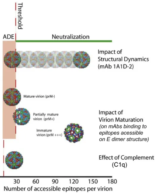
neutralization of flaviviruses (including DENV) is a “multiple-hit” phenomenon, where
neutralization entails a virion to be bound with an antibody stoichiometry that exceeds a
required threshold number of bound antibodies for neutralization (Figure 1.6). Work by
Pierson and colleagues using WNV and a panel of anti-EDIII mouse MAbs, have calculated
the antibody threshold number for flaviviruses as being ~30. This means that for successful
antibody neutralization of a flavivirus particle that contains 180 E protein epitopes, there
needs to be > 30 antibody molecules bound to that virion (94, 112). It is hypothesized that
when antibodies are bound to virions at a stoichiometry less than the threshold number, then
there is the risk of ADE (33). It is unclear whether this threshold requirement of at least 30
antibody molecules to neutralize a virion is true for all epitopes. Furthermore, the effect on
the threshold number when a polyclonal mix of antibodies is binding to several different
epitopes on the same virus (as is the case when DENV enters a DENV-immune human body)
is also not known. Nonetheless, exceeding the antibody threshold requirement of
Antibody affinity
Antibody affinity defines the percentage of epitopes on the virion that are occupied
by that antibody at a given concentration. Therefore, at any given concentration, antibodies
with higher affinity will bind to the virion in greater numbers than low affinity antibodies.
Similarly, E protein mutations that reduce antibody affinity, will lower the number of viral
epitopes occupied at any given concentration, and reduce the neutralization potential of that
antibody. Several studies have observed that decreases in antibody affinity due to
intra-serotype, strain variation (i.e. 4-10% within E protein) lead to reduced neutralization of those
strains (13, 138, 152). It is unclear whether these intra-serotype variations also affect in vivo
protection against re-infection from any strain within a particular serotype.
Epitope accessibility due to complex virion structure
Due to the complex structural properties of flaviviruses (and more particularly
DENV), epitope accessibility is multifaceted. As detailed above, the 180 E proteins are
arranged in a dense 5-3-2-fold axes of symmetry, dividing the E proteins on the mature virion
into three different chemical environments. Therefore, depending on the epitope recognized
by a particular antibody, steric constraints of that epitope would decrease the epitope’s
accessibility for binding (20, 68, 69, 85, 106). For example, cryo-EM structures of the
neutralizing mouse MAb, E16, in complex with WNV showed that although E16 binds the
highly exposed lateral ridge region on EDIII, it can only occupy 120 of the available 180
EDIII epitopes on the mature virion. It was observed that E16 could not occupy the EDIII
hand, in the mature virion the highly conserved fusion peptide is buried beneath EDI and
EDIII of the opposing E protein, and inaccessible to binding by anti-fusion loop MAbs, such
as E53. Therefore, MAbs similar to E53 need to bind almost all accessible epitopes to exceed
the threshold of neutralization, making these MAbs only weakly neutralizing (20, 137).
DENV structural dynamics
Despite our earlier detailed description of the mature flavivirus virion, we know that
proteins assembled into viral particles are not static structures. In fact, viral surface proteins
are thought to be capable of constant movement or “breathing” (65, 160). This dynamic
movement was shown to also be true of DENV, and enable the exposure of epitopes that
would otherwise have been inaccessible. The mouse MAb, 1A1D-2, is potently neutralizing,
DENV-subcomplex binding and more specifically, recognizes an epitope on the A-strand of
EDIII (85, 123). Detailed mapping of 1A1D-2 showed that this MAb binds an epitope that is
hidden in the mature virion. Experiments at 4°C showed that 1A1D did not bind the virus at
low temperatures, while 1A1D binds and efficiently neutralized DENV at 37°C. Cryo-EM
studies showed that “breathing” of the DENV virion at 37°C allowed binding of 1A1D-2,
which caused E proteins to rotate in a manner that further exposed its epitope (85). Studies
with 1A1D-2 and the more recently described DENV1-E11 MAb are evidence of the effects
of DENV structural dynamics on the accessibility of epitopes to antibody binding (Figure
1.6) (4, 85).
virus population is higher than previously assumed, with up to 90% of the virions containing
uncleaved prM (66). Although the minimum requirement of cleaved prM for infectivity is
unknown, a majority of partially mature virions are observed to be infectious (66). The
arrangement of E proteins during the mature E dimer conformation is vastly different from
the immature prM-E trimer conformation (Figure 1.1C). This vast difference in conformation
has drastic consequences on epitope accessibility (Figure 1.6), with many instances where a
subset of the virus population is resistant to neutralization even at saturating antibody
concentrations (105, 112). For example, it was observed that the fusion loop was more
exposed in the immature E trimer conformation. Hence, several anti-fusion loop MAbs are
better at neutralizing more immature virus, leaving completely mature virions resistant to
neutralization from these MAbs (105). Although no detailed cryo-EM mapping of an
anti-prM antibody has been conducted yet, it is also easily conceivable that antibodies targeting
prM are especially sensitive to DENV maturation.
Complement protein, C1q, & decrease in threshold number
It has been known for years that serum complement components can boost the
neutralization of flaviviruses by antibodies (reviewed in (33)). However, it was only within
the past few years that the complement protein responsible for this observed increase in
neutralization was identified as being C1q. As part of the classical complement pathway, the
~460 kDa C1q multimeric protein activates the pathway by binding the Fc-portion of
antibodies. In humans, C1q preferentially binds to IgG3 and IgG1 with higher affinity, than
and therefore, decreasing the antibody stoichiometry or threshold number required for
neutralization (Figure 1.6) (94). The effects of C1q on antibody neutralization should be
considered when testing DENV-specific MAbs and DENV-immune polyclonal sera in in vitro neutralization assays.
Pre-attachment and post-attachments blocking Abs
DENV-specific MAbs have been documented to neutralize DENV using two main
mechanisms. Many MAbs prevent a productive DENV infection by binding regions on the
virion surface that are involved in receptor attachment, and are named pre-attachment
blocking antibodies. Another group of MAbs (named post-attachment antibodies) allow the
attachment of DENV to host cells, but block an essential step in the entry process and
prevent viral fusion (25, 40, 58, 106, 123, 136, 143, 151). Post-attachment antibodies bind
regions on the virus that are critical for the low pH-induced large conformational change
required for fusion. MAbs that neutralize post-attachment bind several different epitopes on
the E protein, including EDIII and the fusion loop (40, 123, 136, 143, 151). Both
pre-attachment and post-pre-attachment blocking MAbs can be strongly neutralizing (25, 40, 58, 106,
123, 136, 143, 151). Only one study has investigated the dominant mechanism of
neutralization in DENV-immune polyclonal human sera (58). This study found that human
serum from acutely DENV-infected patients primarily blocked attachment of DENV (58).
However, this study used serum samples that were not conducive to investigate long-term
protection. Therefore, it is still unclear which mechanism of neutralization is dominant in
1.8 Antibody dependent enhancement of DENV: the other edge of the double-edged sword.
One of the most compelling explanations for the higher proportions of severe disease
during secondary heterotypic DENV infections (when compared to primary infections) is the
phenomenon of ADE (52, 53, 150). As shown in Figure 1.7, ADE of DENV infection is
expected to occur when pre-existing sub-neutralizing antibodies in the body (such as from a
primary infection) bind to a heterotypic virus during a subsequent infection, and facilitate the
entry of the virus through Fc receptor (FcR)-mediated endocytosis into FcR-bearing myeloid
cells (such as monocytes and macrophages). Through mechanisms that are largely unclear
and possibly furin cleavage-dependent, the antibody-bound virus escapes the phagolysosome
and establishes a productive infection within the host cell (Figure 1.7) (reviewed in (52)). It
is important to remember that primary infections can also sometimes lead to DHF/ DSS,
which means that severe DENV-induced disease is multi-factorial and ADE is only one
important mediator of severe disease.
Apart from inducing severe secondary infections, there are several other
epidemiological observations that tie the theory of ADE to DENV infections. 1) In DENV
endemic regions, infants between the ages of 6 and 12 months are a high-risk group for
severe forms of DENV such as DHF or DSS (17, 18, 71). Since the infant immune system is
facing DENV for the first time, this occurrence is independent of a cellular adaptive immune
response. One of the explanations for a time-dependent severe disease phenotype in infants is
DENV-mediated DENV infection for several months (17, 18, 71). 2) Severe disease in both
secondary infections and infant cases are observed to reach high serum viremia titers (150).
This is not always due to higher virulence of the infecting virus strain (49). Several studies
have clearly observed that a DENV infection through the ADE pathway (as opposed to the
conventional receptor-mediated entry pathway) leads to significantly higher replication and
secretion of infectious virus (41, 55).
ADE of DENV infection is easily reproducible in cell culture using FcR-bearing
cells and DENV-specific MAbs or DENV-immune polyclonal sera. Many cell lines, both
primary (PBMCs, primary dendritic cells, monocytes and macrophages) and immortalized
cells (K562, U937, THP-1), are presently being used to recapitulate the ADE phenomenon in
vitro (6, 10, 16, 41, 74, 124, 145, 159). A majority of these immortalized cell lines are only
permissive to DENV infection in the presence of DENV-specific antibodies, hence removing
the complexity of traditional mechanisms of entry while studying ADE. It was initially
postulated that the higher viremia when infected through ADE is not due to more efficient
FcR-mediated uptake of virus and higher numbers of infected cells (aka extrinsic ADE) (reviewed in (155)). However, recent in vitro studies show that entry of DENV through ADE
is not very efficient, and does not infect more cells than the traditional method of entry (145).
Instead the FcR-mediated mechanism of uptake of virus into the host cell suppresses the
anti-viral responses within that cell, which allows greater anti-viral replication and output (a process
124). Whether FcR-mediated uptake is more immune suppressive than the traditional method
of receptor-mediated entry, is yet unclear. Further studies need to be conducted to define the
exact molecular mechanism of how the virus escapes the phagolysosomes, and the
proceeding steps that lead to a suppression of the cellular anti-viral response.
The concept of ADE (as opposed to the competing ‘cytokine storm’ explanation) causing severe disease in DENV infected patients has been a topic of much debate for several
decades (118). Because DENV is an obligate pathogen of humans (including non-human
primates) and the mosquito vector, there is no natural DENV small-animal model, which has
hampered in vivo investigations of ADE. Although DENV infection is generally asymptomatic in non-human primates, DENV replicates to low titers in several non-human
primate models. DENV infection following passive transfer of DENV-immune sera or
DENV-specific MAbs into monkeys showed signs of ADE through a 10-100-fold increase in
viremia, when compared to virus only treated monkeys (41, 50). However, since non-human
primates do not show severe DENV-like symptoms, they are not considered a very good
animal model for dengue disease/DHF. One of the most successful animal models for
studying ADE of DENV is the type I and II IFN receptor knockout mouse model, AG129
(148). When AG129 mice are passively injected with DENV-specific MAbs or polyclonal
sera and then challenged with a mouse adapted virus strain, within 5 to 6 days the mice
develop a lethal vascular leakage disease that is similar to DHF in humans. During ADE, The
infected cells within the AG129 are FcR-bearing cells (such as monocytes and macrophages
Therefore, although the absence of type I and II IFN-receptors makes the AG129 mouse
model imperfect for studying immune responses to DENV, it represents the human DHF
disease closely and serves well as a passive antibody transfer model for mapping out the
DENV epitopes targeted by enhancing antibodies. Almost all DENV-specific MAbs or
polyclonal sera, whether neutralizing or not, when diluted out far enough, will enhance
DENV infection. Therefore, it is critical to identify the heterotypic DENV epitopes targeted
by antibodies in human sera at physiological concentrations. Determining the targets of
enhancing antibodies in human sera will hopefully enable the design of safer,
non-DENV-enhancing vaccines.
1.9 Prospective DENV vaccines
The presence of four related serotypes, each capable of inducing cross-reactive
antibodies that at sub-neutralizing concentrations can enhance infection of heterotypic virus,
makes DENV vaccine development a very challenging task. Vaccine developers have several
factors to keep in mind when trying to create a DENV vaccine (reviewed in (104)). First,
since most DENV endemic countries have all four DENV serotypes co-circulating, it is
imperative that the vaccine elicits a protective response against each of the four serotypes.
Second, this protective response needs to be life-long. If antibody levels fall to
sub-neutralizing levels, this places the vaccinee at risk of enhanced DENV infection and
therefore, more severe disease. Third, as with all vaccines, a DENV vaccine must be safe and
produce minimum symptoms in the recipients. Fourth, the immunization protocol must
poor health care, any approved DENV vaccine must be economical and affordable for
individuals residing in these countries.
Scientists are using many strategies to develop a DENV vaccine. Nonliving vaccine
platforms being tested against DENV are inactivated subunit, subviral particles and whole
virus vaccine. Although, nonliving vaccines have the advantage of being safe, these vaccines
are not very efficient at stimulating strong adaptive responses, and hence require adjuvants
and serial boosts. Furthermore, subunit vaccines currently being developed using E
ectodomain protein do not present the immune system with the same repeated E protein raft
structure that is displayed on the whole virion. Several studies have pursued the development
of subviral particle through the expression of prM and E proteins, where vaccine recipients
are either immunized with purified subviral particles (84) or with viral vectors (such as
adenovirus, vaccinia, or alphavirus) that express DENV prM and E proteins (37, 116, 157,
158). Although subviral particles have a T=1 symmetry, they also lack some of the complex
epitopes formed when E assembles into the virion particle. Whether this repeated E protein
raft structure is essential for raising life-long DENV neutralizing antibodies is unclear.
The three DENV vaccines most advanced in clinical trials are live attenuated
vaccines. One is a live, attenuated DENV vaccine (LAV) developed by the National Institute
of Health. This vaccine contains a DENV4 genetic background with attenuation mutational
deletions within the 3’UTR region. The prM and E proteins from the three other serotypes
were inserted into this attenuated DENV4 background. This vaccine has just passed phase I
DENV2 strain, PDK-53, into which the prM and E proteins from the other three serotypes
are inserted. This vaccine is also in clinical trials at the moment. The third LAV,
ChimeriVax, is a chimeric virus between DENV and the highly successful (and presently
licensed) yellow fever vaccine strain, YFV 17D (46). The prM and E proteins are expressed
in the YFV 17D background, thereby producing 4 chimeric, monovalent viruses. This
ChimeriVax vaccine, designated CYD-TDV (licensed by Sanofi Pasteur) has reached phase
IIB clinical trials. Unfortunately, these clinical trial data, published late last year, showed that
this vaccine had an overall protective efficacy of only ~30% against natural DENV
infections, with no protection against DENV2 infections (125). Several hypotheses about
genotypic differences between immunized and circulating viruses are being put forward
(unpublished data from Sanofi Pasteur). Since we know that the most successful DENV
immunizations in humans at present are natural infections, we need to ask another vital
question; are the vaccines being developed eliciting similar T cell and B cell responses as
natural wild type DENV infections? Unfortunately, with very little known about the DENV
epitopes targeted by protective T cells and neutralizing antibodies, the answer to this question
is still pending.
1.10 Objectives of this dissertation
At a time when DENV is endemic in over 100 countries with over 390 million new
infections globally, we known very little about the neutralizing and enhancing human
antibody response after a natural DENV infection. The lack of a suitable animal model led to
123, 138, 139). However, as described above, more recent studies have clearly shown that
EDIII is not the target of human neutralizing antibodies following natural flaviviral infections
(107, 127, 153, 154). Therefore, there is a significant knowledge gap in our understanding of
the epitopes targeted by the human antibody response after a natural DENV infection. One of
the goals of this dissertation is to expand our knowledge of the human antibody response
after primary DENV infections. To achieve this goal, we have devised a simple technique to
fractionate DENV-specific antibodies in human immune sera into groups, and then
characterize their binding and functional properties. Additionally, in collaboration with other
groups, we have also generated, isolated and characterized the binding and functional
properties of DENV-specific human MAbs from individuals with past natural DENV
infections.
A recent study has mapped a potently neutralizing anti-WNV human MAb to a
complex viral surface epitope centered on the DI-DII hinge region of the E protein. This
epitope is not conserved on the soluble E protein and hence, this potent anti-WNV MAb does
not bind soluble E protein (69, 151). Primary DENV-immune human sera are very reactive to
soluble DENV E protein from all serotypes. Despite the large cross-reactivity across
serotypes, late convalescent sera after primary DENV infections only neutralize the serotype
of infection. This indicates that the neutralizing antibodies in human sera are very rare and
possibly very difficult to isolate. Fortunately, advances in human B-cell technologies have
vastly improved the transformation and fusion of human memory B-cells, so that even rare
antibodies. Successful characterization of the neutralizing epitopes will allow us to probe and
understand the mechanisms of DENV neutralization by human MAbs, and how they differ
from those already described using DENV-specific mouse MAbs.
Almost any DENV-specific MAb (binding E or prM protein) can enhance DENV
infection in vitro. Therefore, the importance of characterizing the enhancement properties of MAbs is unclear. What is critical is to define the epitopes of antibodies in DENV-immune
human sera that enhance infection of heterotypic serotypes. Hence, the third goal of this
dissertation is to characterize the epitopes targeted by human polyclonal antibodies that
enhance heterotypic infection. These experiments will be conducted using in vitro ADE assays and the in vivo AG129 mouse model. In summary, the primary goal of this dissertation is to expand our understanding of the neutralizing and enhancing human
antibody response to DENV, for purposes of understanding the biology of antibody-mediated
protection in DENV infection and for the rational development of successful vaccines.
Hypothesis: We hypothesize that humans exposed to primary dengue infections develop two
unique antibody sub-populations that 1) neutralize homotypic and 2) enhance heterotypic serotypes of virus. Moreover, we propose that human antibodies neutralize DENV at a post-attachment step by binding to novel conformational epitopes that include the DI-DII hinge of the dengue envelope (E) protein.
Aim 2. To map and characterize the neutralizing epitope(s) targeted by the human antibody response after a natural DENV infection, by mapping both DENV-immune human polyclonal
sera and strongly neutralizing human monoclonal antibodies. Furthermore, mechanistic
studies will be conducted to gain insight into the mechanism of DENV neutralization by
human antibodies.
Aim 3. To determine the epitopes of enhancing antibodies circulating in humans after a primary DENV infection. In vitro observations will be extended to in vivo experiments, using the AG129 ADE mouse model.