R E V I E W A R T I C L E
Aesthetic bite for the management of temporomandibular
disorders: A case series review
Giacomo Derchi1, Vincenzo Marchio1, Letizia Bolognesi2, Elisabetta Carli1, Maria Rita Giuca1, Mutlu Özcan3, Mario Bosco4
1Department of Surgical, Unit of Stomatology and Oral Surgery, Medical, Molecular and Critical Needs Pathologies, University of Pisa, Pisa, Tuscany, Italy, 2Department of Clinical, Surgical, Unit of Orthodontics and Paediatric Dentistry, Section of Dentistry, Diagnostic and Paediatric Sciences, University of Pavia,
Pavia, Lombardy, Italy, 3Dental Materials Unit, Center for Dental and Oral Medicine, Clinic for Fixed and Removable Prosthodontics and Dental Materials Science,
University of Zurich, Zurich, Switzerland, 4Department of Dentistry, University of Pavia, Pavia, Lombardy, Italy
Abstract
Background: Oral appliances (OA) are the most widely used treatment approach for temporomandibular disorders (TMDs): They are designed to relieve or prevent degenerative forces on the joint, the articular disk, and dentition. Classical OAs, however, are cumbersome and esthetically unpleasing: A more esthetic and functional
appliance might improve treatment efficacy and shorten treatment time. Aim: The aim of this case series is to introduce an esthetical device for the management of TMD.
Methodology: Three adults between 43 and 60 years old with a diagnosis of intra-articular TMD were included and were instructed to apply the device throughout the day and night. The patients were recruited at the same private dental clinic and each one signed an informed consent for both treatment and inclusion in the study. We evaluated and compared four clinical parameters (mandibular excursion, mandibular opening pattern, muscular and articular pain, and TMJ noises) between time 0 (T0) and 1, 2, 3, 6, and 12 months of using the device following the protocol of diagnostic criteria for temporomandibular joint disorders (DC/TMD). A magnetic resonance imaging exam was conducted to investigate tissue changes between T0 and 3 and 12 months of therapy.
Conclusions: For all four parameters, we observed considerable improvements in all
patients, who noticed the positive effects of the therapy themselves. The results suggest that this esthetic device was an effective treatment to manage TMD in the described
cases, also increasing patients’ quality of life. However, studies on a larger scale are
required to prove the effectiveness of this device. Clinical Significance: This case series aims to highlight the potentiality of an esthetic device used for the management of TMDs. This is because a more esthetically pleasant and comfortable device increases the
time of usage, thus improving the positive effects of the device on the TMD.
Keywords: Dental esthetics, dental occlusion, dentistry, orthodontic appliances, temporomandibular joint disorders, temporomandibular joint
Correspondence:
Dr. Vincenzo Marchio, Unit of Stomatology and Oral Surgery, Department of Surgical, Medical, Molecular and Critical Needs Pathologies, University of Pisa, Pisa, Tuscany, Italy. Phone: +39-3338152172. E-mail: [email protected]
Received 03 April 2020; Accepted 08 May 2020
doi: 10.15713/ins.ijcdmr.145
How to cite the article:
Derchi G, Marchio V, Bolognesi L, Carli E, Giuca MR, Özcan M, Bosco M. Aesthetic bite for the management of temporomandibular disorders: A case series review. Int J Contemp Dent Med Rev, vol.2020, Article ID: 010520, 2020. doi: 10.15713/ins.ijcdmr.145
Introduction
Temporomandibular disorders (TMDs) are a heterogeneous
group of diagnoses affecting the temporomandibular joint
(TMJ) and the surrounding tissues.[1]
TMDs are a Significant Public Health Issue
Epidemiological studies show that approximately 33% of the population has at least one characteristic symptom and 56% have a clinical sign.[2,3] Overall 3.6–7% of the population has TMD
with sufficient severity to cause them to seek treatment.[4] TMD
is a prevalent disorder most commonly observed in individuals between the ages of 20 years and 40 years and the number of
affected people also is increasing, perhaps due to the influence of
mental tension in today’s society.[5]
The diagnosis and evaluation of TMDs must follow a standardized protocol. The latest guidelines on this topic were published in 2014: The diagnostic criteria for TMDs (DC/ TMD)[6] derive from the previous research diagnostic criteria for TMDs (RDC/TMD),[7] which allowed the standardization and reproducibility of research in the most common forms of TMD.
This classification system is based on a biaxial model
a classification of the different forms of TMD through an
anamnestic questionnaire and an accurate clinical examination allowing the formulation of a somatic diagnosis. Axis 2 makes possible to evaluate the psychosocial impairment of the patient due to the pain experience.[8]
Since the first classification in 1986, TMD etiology and
management were often discussed, including various treatments and strategies to control TMD pain.[9-11]
In general, conservative and reversible treatments are preferable to more aggressive and irreversible approaches (e.g. occlusal interventions and surgery)[12] since current epidemiological information indicates that TMD is frequently self-limiting.[13,14]
Oral appliances (OA) are the most widely used treatment approach:[15] They are designed to relieve or prevent degenerative forces on the joint, the articular disk, and dentition.[16]
Classical splints, however, are designed to solve functional problems and thus they are cumbersome and incompatible with chewing and speech; consequently, the time of use of the device will be reduced at night and at some hours of day.
On the contrary, a bite whose purpose is to make up for esthetic problems and which is designed to be used during the day will have the key features of discretion and comfort. These properties confer the potential to increase exponentially the time of use and to potentially reduce the duration of therapy.
In this case series, patients with TMDs and dento-skeletal changes in which a predisposing occlusal vector is recognized were selected. For these cases, which require rehabilitation as
indicated in the DC/TMD protocol, a first phase of reversible
therapy is chosen to control the symptomatology.
Along with counseling and masticatory muscle exercises, a bite therapy is added.
At present, the most widely used plaque is the Michigan Splint,[17,18] but in this case series, the use of the Snap-on Smile devices is proposed because of the esthetic characteristics and the possibility of being used for a longer time during the day.
In this case report, the esthetic device used was the Snap-On Smile® (produced by DenMat Italia S.r.l. – F. Pinto 16, Salerno, Italy).
Snap-On Smile is a multi-purpose restorative appliance that requires no preparation or alterations of tooth structure, no injections, and no adhesives. It is non-invasive, making it completely reversible.[19]
The Snap-On Smile® device is made of crystallized acetyl
resin. This material is very durable and slightly flexible and increases the device’s retention by “flexing” over the heights and
contours of the existing teeth, snapping onto the gingival third of the tooth.
Using a CAD/CAM technique, the device is designed and produced following the operator instructions [Figure 1a-c].
The device has high esthetic properties, which give it the
potential to significantly increase the amount of time of use
of the device. Furthermore, if designed to create occlusal relations which allow us to obtain a stable condition of the TMJ, it may help reducing the duration of therapy and ensuring an
improvement in patients’ health by stopping the progression of the disease.
The protocol used for the realization of the Snap-On Smile® is the same for all subjects and it includes:
• Complete photographic documentation;
• Maxillary and mandibular impressions, taken with 3M™ Imprint™ 4 Penta™ Impression material;
• Wax bite, using Beauty Pink X-Hard INTEGRA MILTEX, manually guided in centric occlusion. Registration was carried out when the operator felt that the patient was relaxed and hinging freely,[20] using Roth power centric bite registration technique.[21-23]
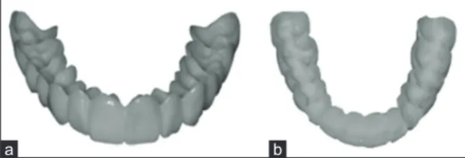
Snap-On Smile® can be designed to be complete, if it covers all the dental surfaces, or open in the frontal area; furthermore, it can be designed for the upper or lower arch [Figure 2a and b].
In this case series, all patients were clinically evaluated 6 times during the therapy following the DC/TMD. Regarding the analysis of muscle palpation reference is made to the RDC/ TMDprotocol,[7] as the authors want to emphasize the course of the algic symptomatology between the various follow-ups.
At present, magnetic resonance imaging (MRI) is the gold standard for the study of pathological processes involving the soft tissues of the TMJ;[24-29] it is of great diagnostic value in
cases of disk dislocation, disk degeneration, osteoarthritis (T2
sequences), and in cases of inflammatory reaction of synovial
tissue (T1 sequences).[25,30]
For this reason, in addition to the clinical exam, diagnosis and therapy checks have been conducted through the evaluation of MRI. All subjects underwent MRI 3 times: Once before treatment (T0), after 3 months (T1), and after 12 months of therapy (T2).
Quantitative and qualitative analyses were carried out following and integrating the traditional methods proposed by Bumann, Vargas-Pereira with more recent ones.[31-34]
Case 1
A 42-year-old woman using an upper splint for approximately 18 years, presented on July of 2016 complaining about pain
Figure 1: Device design process. (a) Digital scan of patient’s occlusion model, (b) esthetic device digital design, (c) final result
c b
a
Figure 2: The esthetic device. (a) Front view, (b) inside view
and constant neck muscles contraction, auricular fullness, and crepitus sound during phonation; the symptomatology is more intense on the left side. She was included in the study after signing an informed consent for treatment and study inclusion.
The anamnestic examination showed the following relevant data:
• A first episode of mandibular block in 1997 and a second
episode 1 month before the visit,
• Clenching evaluated objectively using Smith and Knight tooth wear index (1–2 score overall),[35-37]
• Muscle and jaw pain both at rest and during function, • Left TMJ noise during function,
• Previous orthodontic treatment.
Examination by palpation showed pain in the following structures:
• Left TMJ pole,
• Superior area of right masseter,
• Superior, middle, and inferior areas of the left masseter, • Lateral pterygoid and sternocleidomastoid on both sides.
The mouth opening and closing trajectory showed: • Uncorrected pattern,
• A pain free opening (PFO) of 37 mm,
• A maximum unassisted opening (MUO) of 43 mm, • A maximum assisted opening (MAO) of 44 mm.
On examination, crepitus of the left TMJ was present during opening and closing and clicking during laterals excursions was observed.
The objective examination and the magnetic resonance led to the diagnosis of bilateral disk displacement without reduction, bilateral osteoarthritis, myalgia, and arthralgia.
The patient reported that the previous bite never brought substantial improvements and that the symptoms have worsened since the year before.
After having explained to the patient the diagnosis and the therapeutic possibilities, a DC/TMD protocol was started with cognitive behavioral therapy (CBT), and masticatory muscles exercises and massages.
For the splint treatment the authors chose to use a maxillary Snap-On Smile, open on the vestibular side and built to confer a centric occlusion.
The position was recorded by wax while being manually guided in centric occlusion.
The registration was carried out when the operator felt that the patient was relaxed and hinging freely.[20]
The patient was instructed to use the device during both day and night, and to remove the Snap-On Smile exclusively during meals and while brushing. If the device did not cause discomfort while eating, however, the device did not have to be removed.
Follow-up appointments were scheduled for 1 month, 2 months, 3 months, 6 months, and 12 months.
At the 1-month follow-up, the patient reported a substantial reduction of muscle contraction in the neck, the disappearance of the auricular fullness, and the left crepitus during the phonation.
During the first three follow-ups (1 month, 2 months, and
3 months), clinical improvements included a gradual disappearance of muscle pain symptoms and an increase in the opening range (PFO 43.6 mm, MUO 48.5 mm, and MAO 48.8 mm).
In addition, Joint pain disappeared as early as the first
follow-up (1 month).
Regarding joint noises, the left crepitus persisted but decreased in intensity.
The patient was re-evaluated at 6 and 12 months after starting therapy.
At the 6 months follow-up, the patient reported that she felt a considerable improvement regarding all symptoms, and she no longer felt spontaneous or provoked pain.
In addition, during palpation, pain, and noises were no longer present and the opening range increased further: PFO was
44 mm, MUO was 48.6 mm, and MAO was 49 mm. These values
remained stable between this and the 12 months follow-up. After 12 months of therapy the opening pattern (OP),
although slightly deflected to the left, appeared more linear and
closer to the midline.
In addition, the patient was very satisfied and did not report
muscle pain and discomfort during function. When asked to compare the previous bite with the Snap-On Smile device, the patient’s opinion was in favor of the second because of better esthetic properties, which made it easier to wear the device
throughout the day increasing its effectiveness. The patient also
reported the ability to maintain the correct position during the night, a condition that was sometimes lost with the use of the previous bite.
Case 2
A 53-year-old woman presented on November 2016 complaining about muscular pain and a very strong headache (with varying intensity during function) which prompted the patient to
use anti-inflammatory drugs (diclofenac, and nimesulide),
pain relievers and muscle relaxants in high doses and at short intervals. This condition had worsened in the last month, forcing the patient to often be absent from work because of the pain. He was included in the study after signing an informed consent for treatment and study inclusion.
During the first visit, the patient also showed:
• Clenching, verified through objective evaluation (tooth wear
index: two score among the mouth)[35-37]
• Bruxism, previously diagnosed at another specialized clinic through polysomnography
• Previous usage of an upper occlusal splint.
Examination by palpation showed pain in the following structures:
• Right TMJ pole,
• Superior, middle, and inferior area of the right and left masseter,
• Posterior and middle area of the right and left temporalis, • Lateral pterygoid and sternomastoid on both sides.
• Uncorrected pattern, • A PFO of 29 mm,
• A MUO of 36 mm, • A MAO of 37 mm.
On examination, crepitus of both TMJs was present during opening and closing movements.
The objective examination and the magnetic resonance (MRI), according to DC/TMD, led to the diagnosis of Bilateral disk displacement with reduction, bilateral osteoarthritis, myalgia, and headache attributed to TMD.
After having explained to the patient the diagnosis and the therapeutic possibilities, a DC/TMD protocol was started with CBT, masticatory muscles exercises and massages.
For the patient’s esthetic needs, the treatment proposed by the authors was the use of a lower and complete coverage Snap-On Smile, designed to give a centric occlusion.
The patient was instructed to use the device during both day and night, and to remove the Snap-On Smile exclusively during meals and while brushing. If the device did not cause discomfort while eating, however, the device did not have to be removed.
Follow-up appointments were scheduled for 1 month, 2 months, 3 months, 6 months, and 12 months.
At the 1-month follow-up, the patient reported a reduction of headaches episodes and a reduction of muscle pain: The most
striking change was that the use of anti-inflammatory drugs by
the patient had drastically reduced and she never had to leave her workplace because of the pain.
The clinical examination of the first follow-up (1 month) showed a significant increase in the opening range (PFO 41 mm,
MUO 43 mm, and MAO 44 mm).
In successive follow-ups (2 and 3-months), the patient reported a further reduction of muscular pain, although a slight pain on palpation of the middle portion of the masseter and the right lateral pterygoid was reported.
The patient returned for the fourth follow-up (6 months) in May 2017, reporting that the headache had occurred only sporadically, and that muscle and joint pains were no longer present.
Clinical examination confirmed what the patient reported,
although the joint crepitus persisted.
At the 12 months follow-up, the situation remained stable from a symptomatologic point of view: Headaches did not occur during the previous 2 months.
At palpation, no muscle pain was reported and even the joint crepitus was no longer observed.
The opening range had stabilized, although it was slightly reduced compared to the 1 month follow-up. The values however were in the physiological range: PFO was 40 mm, MUO was 44 mm, and MAO was 45 mm. The OP, although
slightly deflected to the left, appeared more linear and closer to
the midline.
The patient was extremely satisfied, especially from the point
of view of quality of life given the absence of need for medication
and time off work.
Case 3
A 60-year-old woman presented on January 2017 complaining about muscular pain, TMJ noises, episodes of tinnitus, and dizziness all of which had become acute in recent weeks. He was included in the study after signing an informed consent for treatment and study inclusion.
During the first visit, the patient also showed
• Clenching, evaluated through objective evaluation (tooth wear index: 2–3 score among the mouth),[35-37]
• Noises at function.
In addition, the patient reported never using an occlusal splint
before, and her partner confirmed the presence of clenching
during the night.
Examination by palpation showed pain in the following structures
• Right TMJ pole,
• Middle area of right masseter (myofascial pain with trigger point),
• Temporal tendon on both sides.
The mouth opening and closing trajectory showed • A linear opening,
• A PFO of 33 mm, • A MUO of 34 mm, • A MAO of 35 mm.
On examination, crepitus of both TMJ was present during opening and closing movements.
The objective examination and the magnetic resonance led to the diagnosis of bilateral disk displacement without reduction, bilateral osteoarthritis, and myalgia.
After having explained to the patient the diagnosis and the therapeutic possibilities, a DC/TMD protocol was started with CBT, and masticatory muscles exercises and massages. The treatment proposed to the patient was the use of an upper ad complete coverage Snap-On Smile, designed to give a centric occlusion.
The patient was instructed to use the device during both day and night, and to remove the Snap-On Smile exclusively during meals and while brushing. If the device did not cause discomfort while eating, however, the device did not have to be removed.
Follow-up appointments were scheduled for 1 month, 2 months, 3 months, 6 months, and 12 months.
At the 1-month follow-up, patient returned showing a good improvement, mostly in terms of dizziness and tinnitus that disappeared.
The clinical examination of the first follow-up (1 month)
showed an increase in the opening range (PFO 35 mm, MUO 42 mm, and MAO 43 mm).
The patient reported that she was not able to use the device during meals.
Regarding joint noises, the left crepitus persisted although with decreased intensity.
At the 6-month follow-up, palpation showed only slight muscular pain in the left temporal sinus the crepitus was detected only at the left joint.
The PFO opening range was 44 mm, MUO was 46 mm, and MAO was 47 mm.
At the 12 months follow-up (January 2018), the vertigo and tinnitus remained unchanged as the patient no longer complained about episodes.
At palpation no muscle was reported to be painful and no articular noise was found.
The opening range was further increased with a PFO of
45 mm, a MUO of 48 mm, and a MAO of 49 mm.
The patient claimed to be extremely satisfied with the
treatment, especially regarding the possibility of using the device
even during the day. The only difficulty encountered concerned
its use during meals.
Discussion
Numerous studies suggest that the therapy of choice for TMDs is represented by a conservative treatment that uses occlusal devices (bites) to recreate joint stability.[38,39]
Various types of splints have been proposed in the literature and, for the cases presented in this case series, a device that can be included in the anterior repositioning splint category has been chosen.
In the cases presented, the Snap-On Smile has been constructed to confer an optimal occlusion in centric relation, trying to position the condyle in a more accurate relationship with the articulation structures.
Regardless of personal opinion or the device that can be chosen, it is fundamental for the authors to use evidence-based
scientific protocols: For this reason, the diagnosis and evaluation
of therapies are based on the DC/TMD published in 2014.[6]
All DC have been summarized in four main parameters 1. Mandibular excursion,[6]
2. Mandibular OP,
3. Muscular and articular pain caused by palpation techniques,[13] 4. TMJ noises during opening, closing, lateral, and protrusive
movements.[6]
An evaluation using imaging technique was performed using MRI and it was associated with clinical evaluations, as the DC/ TM and other studies indicate.[6]
Changes of the joint space and of the relationship between the status of the disk and joint pain were evaluated by comparing
the images obtained before treatment (first MRI) and those
obtained during treatment.
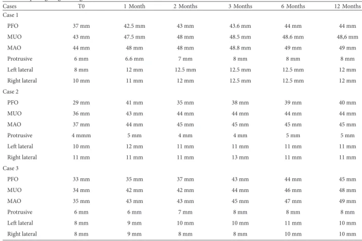
Table 1: Opening range through time for all cases
Cases T0 1 Month 2 Months 3 Months 6 Months 12 Months
Case 1
PFO 37 mm 42.5 mm 43 mm 43.6 mm 44 mm 44 mm
MUO 43 mm 47.5 mm 48 mm 48.5 mm 48.6 mm 48,6 mm
MAO 44 mm 48 mm 48 mm 48.8 mm 49 mm 49 mm
Protrusive 6 mm 6.6 mm 7 mm 8 mm 8 mm 8 mm
Left lateral 8 mm 12 mm 12.5 mm 12.5 mm 12.5 mm 12 mm
Right lateral 10 mm 11 mm 12 mm 12.5 mm 12.5 mm 12 mm
Case 2
PFO 29 mm 41 mm 35 mm 38 mm 39 mm 40 mm
MUO 36 mm 43 mm 44 mm 44 mm 44 mm 44 mm
MAO 37 mm 44 mm 45 mm 45 mm 45 mm 45 mm
Protrusive 4 mmm 5 mm 4 mm 4 mm 5 mm 5 mm
Left lateral 10 mm 12 mm 11 mm 11 mm 11 mm 11 mm
Right lateral 11 mm 11 mm 11 mm 13 mm 11 mm 11 mm
Case 3
PFO 33 mm 35 mm 37 mm 43 mm 44 mm 45 mm
MUO 34 mm 42 mm 42 mm 44 mm 46 mm 48 mm
MAO 35 mm 43 mm 43 mm 45 mm 47 mm 49 mm
Protrusive 6 mm 6 mm 7 mm 8 mm 8 mm 8 mm
Left lateral 8 mm 9 mm 10 mm 10 mm 11 mm 10 mm
For all patients, positive results have been achieved, as showed from the parameters.
In every case presented, the mandibular opening range was the most positive improvement caused by this therapy: As a
matter of fact, there was a significant increase (millimeters) just after 1 months of therapy (first follow-up) [Table 1].
Regarding the mandibular opening range, it had a pronounced improvement from a transverse point of view: The deviations, when present, became less and less evident and closer to the correct linear path [Table 1].
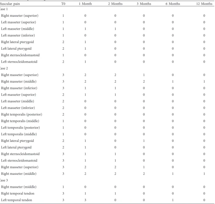
Muscle pains tended to disappear gradually, and it was the improvement best perceived by patients; joint pains, when present, followed the same trend [Tables 2 and 3].
The joint noises associated with disk dislocation were
eliminated already at the first follow-up.
However, the crepitus noises associated with degenerative pathology tended to persist as expected. This pathology has an extremely slow course and takes years to give rise to the conditions found in patients: For this reason, the reduction of noise intensity after only few months (if not after 1 month) was considered an excellent result [Table 4].
MRI evaluations reflected the results clinically obtained.
In all three cases presented, the structural improvements of the articulations were obviously scarce at the 3 months follow-up, seeing as it takes a long time for anatomical remodeling to occur.
Table 2: Muscular pain through time for all cases
Muscular pain T0 1 Month 2 Months 3 Months 6 Months 12 Months
Case 1
Right masseter (superior) 1 0 0 0 0 0
Left masseter (superior) 1 0 0 0 0 0
Left masseter (middle) 1 1 1 0 0 0
Left masseter (inferior) 1 0 0 0 0 0
Right lateral pterygoid 2 1 0 0 0 0
Left lateral pterygoid 2 1 0 0 0 0
Right sternocleidomastoid 1 0 0 0 0 0
Left sternocleidomastoid 2 1 0 0 0 0
Case 2
Right masseter (superior) 3 2 2 1 0 0
Right masseter (middle) 3 2 2 2 1 1
Right masseter (inferior) 3 1 1 0 0 0
Left masseter (superior) 2 1 1 0 0 0
Left masseter (middle) 2 0 0 0 0 0
Left masseter (inferior) 2 0 0 0 0 0
Right temporalis (posterior) 2 0 0 0 0 0
Right temporalis (middle) 1 0 0 0 0 0
Left temporalis (posterior) 1 0 0 0 0 0
Left temporalis (middle) 1 0 0 0 0 0
Right lateral pterygoid 2 1 0 1 0 0
Left lateral pterygoid 2 1 0 0 0 0
Right sternocleidomastoid 3 1 1 0 0 0
Left sternocleidomastoid 3 1 1 0 0 0
Right masseter (superior) 3 2 2 1 0 0
Right masseter (middle) 3 2 2 2 1 1
Case 3
Right masseter (middle) 1 0 0 0 0 0
Right temporal tendon 3 1 1 0 0 0
The 12-month control showed a more significant change: AA
growth and better distribution of the joint space were reported, and the thickness of the condylar cortical also increased.
Given the large number of images analyzed, only the most
significant images from the most emblematic case (Case 2) are
reported. In some cases, the comparison is between T0, T1, and T2 while in others its between T0 and T2 to show the most important changes.
For the left TMJ at T0 and T2 [Figure 3], an increase in posterior joint space and a reduction of bone marrow
hyperintensity (sign of edema and bone marrow suffering) can
be observed.
For the left TMJ at T0, T1, and T2 [Figure 4], the
disappearance of joint effusion and a better distribution of joint
space can be observed.
For the right TMJ at T0 and T2 [Figure 5], an increase in the joint space can be observed, while the disk is located in a more backward position.
For the left TMJ at T0, T1, and T2 [Figure 6], a reduction of bone marrow hyperintensity, an increase of condylar cortical bone tissue and a better disk position can be observed.
An important concept in the preliminary results of this case series is the high time of use of the device (close to 24 h/die) when compared to the traditional splint. This was made possible
Table 4: Joint noise through time for all three cases
Joint noise T0 1 Month 2 Months 3 Months 6 Months 12 Months
Case 1
Opening Crepitus L Crepitus L Crepitus L Crepitus L Crepitus L
-Closing Crepitus R - - - -
-Right lateral Click - - - -
-Left Lateral Click - - - -
-Case 2
Opening Crepitus L and R Crepitus L and R Crepitus L and R Crepitus L Crepitus L -Opening Crepitus L and R Crepitus L and R Crepitus L and R Crepitus L and R Crepitus L -Case 3
Closing Crepitus L and R Crepitus L and R Crepitus L and R Crepitus L and R Crepitus L
-Table 3: Joint pain through time for all cases
Joint pain T0 1 Month 2 Months 3 Months 6 Months 12 Months
Case 1
Left TMJ lateral pole 1 0 0 0 0 0
Dynamic left TMJ lateral pole 1 0 0 0 0 0
Case 2
Right TMJ pole 3 2 1 1 0 0
Dynamic right TMJ lateral pole 3 2 1 0 0 0
Case 3
Right TMJ lateral pole 2 0 0 0 0 0
Figure 3: Left temporomandibular joint at T0 and T2 for Case 2
Figure 4: Left temporomandibular joint at T0, T1, and T2 for Case 2
Conclusions
The positive patient feedback, associated with the clinical parameters, highlights a few important aspects: The comfort given by the Snap-On Smile causes less hindrance to the lingual movements (including contact with the palate, and fundamental while speaking) when compared with a bite with classical morphology; The retention system, which uses anatomical undercuts, and the intercuspation between the device and natural dental elements, makes it necessary to perform a conscious action (using one’s own hands) to actively displace the Snap-On from the correct position. In this way, the established occlusal condition cannot be inadvertently lost during night use; no patients had any problems regarding the indications to use the Snap-On Smile for about 24 h a day. The esthetic characteristics and the comfort of the device are reported as essential positive factors for this therapy; the use of the Snap-On Smile for 24 h a day related to the result obtained would suggest a more rapid resolution of the problem compared to a bite that is used only at night.
Based on the promising results of this case series, future
studies should evaluate the efficacy of the Snap-On Smile in a
larger subject sample and a strict protocol should be applied to avoid bias due to sample non-randomization.
References
1. Shaffer SM, Brismée JM, Sizer PS, Courtney CA. Temporomandibular disorders. Part 1: Anatomy and examination/diagnosis. J Man Manip Ther 2014;22:2-12. 2. Ingervall B, Mohlin B, Thailander B. Prevalence of symptoms
of function disturbances of the masticatory system in Swedish men. J Oral Rehabil 1980;7:185-97.
3. Nilner M, Lassing SA. Prevalence of functional disturbances
Figure 5: Right temporomandibular joint at T0 and T2 for Case 2
Figure 6: Left temporomandibular joint at T0, T1, and T2 for Case 2
and diseases of stomatognatic system in 7-14 years old. Swed Dent J 1981;5:173-87.
4. Wright EF, North SL. Management and treatment of temporomandibular disorders: A clinical perspective. J Man Manip Ther 2009;17:247-54.
5. Tvrdy P. Methods of imaging in the diagnosis of temporomandibular joint disorders. Biomed Pap Med Fac Univ Palacky Olomouc Czech Repub 2007;151:133-6.
6. Schiffman E, Ohrbach R, Truelove E, Look J, Anderson G, Goulet JP, et al. Diagnostic criteria for temporomandibular disorders (DC/TMD) for clinical and research applications: Recommendations of the international RDC/TMD consortium network and orofacial pain special interest group. J Oral Facial Pain Headache 2014;28:6-27.
7. Dworkin SF, LeResche L. Research diagnostic criteria for temporomandibular disorders: Review, criteria, examinations and specifications, critique. J Craniomandib Disord 1992;6:301-55.
8. Network Symposium to be Presented at IADR. Diagnostic Criteria for TMD (DC/TMD): A New Version of the RDC/ TMD. Spain: Barcelona, Network Symposium to be Presented at IADR; 2010.
9. Gray RJ, Quayle AA, Hall CA, Schofield MA. Physiotherapy in the treatment of temporomandibular joint disorders: A comparative study of four treatment methods. Br Dent J 1994;176:257-61.
10. Dao TT, Lavigne GJ, Charbonneau A. The efficacy of oral splints in the treatment of myofascial pain of the jaw muscles: A controlled clinical trial. Pain 1994;56:85-94.
11. Stohler CS, Zarb GA. On the management of temporomandibular disorders. A plea for a low-tech high-prudence approach. J Orofac Pain 1999;13:255-61.
12. De Leeuw R, Boering G, Stegenga B, de Bont LG. Clinical signs of temporomandibular joint osteoarthritis and internal derangement 30 years after non-surgical treatment. J Orofac Pain 1994;8:12-8.
13. Williamson EH, Rosenzweig BJ. The treatment of temporomandibular disorders through repositioning splint therapy: A follow-up study. Cranio 1998;16:222-5.
14. National Institutes of Health Technology Assessment Conference Statement. Management of temporomandibular disorders. JADA 1996;127:1595-600.
15. Conti PC, De Alencar EN, Da Mota Corrêa AS, Lauris JR, Porporatti AL, Costa YM. Behavioural changes and occlusal splints are effective in the management of masticatory myofascial pain: A short-term evaluation. J Oral Rehabil 2012;39:754-60. 16. Klasser GD, Greene CS. Oral appliances in the management of
temporomandibular disorders. Oral Surg Oral Med Oral Pathol Oral Radiol Endod 2009;107:212-23.
17. Badel T, Simonić-Kocijan S, Lajnert V, Dulčić N, Zadravec D. Michigan splint and treatment of temporomandibular joint. Med Fluminensis 2013;49:112-20.
18. Ramfjord SP, Ash M. Reflections on the Michigan occlusal splint. J Oral Rehabil 1994;21:491-500.
19. Liechtung M. The snap-on smile removable appliance a non-invasive, affordable smile enhancement. Inside Dent 2010;6:96-7. 20. Linsen SS, Stark H, Matthias A. Changes in condylar position
using different types of splints with and without a chinstrap: A case-control study. Cranio 2012;30:25-31.
In: McNeil C, editor. Science and Practice of Occlusion. Chicago, IL: Quintessence Publishing Co Inc.; 1997. p. 506-7.
22. Wood D, Elliott RW. Reproducibility of the centric relation bite registration technique. Angle Orthod 1994;64:211-20.
23. Schmitt M, Kulbersh A, Freeland T, Bever K, Pink FE. Reproducibility of the roth power centric in determining centric relation. Semin Orthod 2003;9:102-8.
24. Jank S, Zangerl A, Kloss F, Laimer K, Missmann M, Schroeder D,
et al. High resolution ultrasound investigation of the
temporomandibular joint in patients with chronic polyarthritis. Int J Oral Maxillofac Surg 2011;40:45-9.
25. Tasaki MM, Westesson PL. Temporomandibular Joint: Diagnostic accuracy with sagittal and coronal MR imaging. Radiology 1993;186:723-9.
26. Arnett FC, Edworthy SM, Bloch DA, Mcshane DJ, Fries JF, Cooper NS, et al. The American rheumatism association 1987 revised criteria for the classification of rheumatoid arthritis. Arthritis Rheum 1988;31:315-24.
27. Hatala MP, Westesson PL, Tallents RH, Katzberg RW. TMJ disc displacement in asymptomatic volunteers detected by MR imaging. J Dent Res 1991;70:278-81.
28. Bonnett RE, Carpentier P, Yung JP, Defrennes D, Pharaboz C. Clinical diagnosis compared with magnetic resonance in 242 patients with internal derangement of the TMJ. J Orofac Pain 1995;9:244-53.
29. Orsini MG, Kuboki T, Terada S, Matsuka Y, Yamashita A, Clark GT. Diagnostic value of 4 criteria to interpret temporomandibular normal joint disc position on magnetic resonance images. Oral Surg Oral Med Oral Pathol Oral Radiol Endod 1998;86:489-97.
30. Held P, Moritz M, Fellner C, Behr M, Gmeinwieser J. Magnetic
resonance of the disk of the temporomandibular joint. RM imaging protocol. Clin Imaging 1996;20:204-11.
31. Bumann A, Lotzmann U. Funktionsdiagnostik und therapieprinzipien. In: Rateitschak KH, Wolf HF, edtiors. Farbatlanten der Zahnmedizin. Vol. 12. Stuttgart: Thieme; 2000. 32. Bumann A, Vargas-Pereira MR. Metric TMJ analysis for
standardized evaluation of magnetic resonance images. J Dent Res 1997;76:237.
33. Vargas-Pereira MR. Quantitative Auswertungen Bildgebender Verfahren und Entwicklung Einer Neuen Metrischen Analyse fur Kiefergelenkstrukturen im Magnetresonanztomogramm. Germany: University Kiel; 1997.
34. Ruf S, Wüsten B, Pancherz H. Temporomandibular joint effects of activator treatment: A prospective longitudinal magnetic resonance imaging and clinical study. Angle Orthod 2002;72:527-40.
35. Wetselaar P, Lobbezoo F. The tooth wear evaluation system: A modular clinical guideline for the diagnosis and management planning of worn dentitions. J Oral Rehabil 2016;43:69-80. 36. López-Frías F, Castellanos-Cosano L, Martín-González J,
Llamas-Carreras JM, Segura-Egea JJ. Clinical measurement of tooth wear: Tooth wear indices. J Clin Exp Dent 2012;4:e48-53. 37. Smith BG, Knight JK. An index for measuring the wear of teeth.
Br Dent J 1984;156:435-8.
38. Kurita H, Ohtsuka A, Kurashina K, Kopp S. A study of factors for successful splint capture of anteriorly displaced temporomandibular joint disc with disc repositioning appliance. J Oral Rehabil 2001;28:651-7.
39. Madani AS, Mirmortazavi A. Comparison of three treatment options for painful temporomandibular joint clicking. J Oral Sci 2011;53:349-54.