R E V I E W A R T I C L E
A brief overview of cellular and molecular mechanisms of
osseointegration
Seyed Hadi Hosseini1, Mozhgan Kazemian1, Sajedeh Ghorbanzadeh2
1Department of Oral and Maxillofacial Surgery, Faculty of Dentistry, Mashhad University of Medical Sciences, Mashhad, Iran, 2Department of Endodontics, Dental School of Shahid Beheshti University of Medical Sciences, Tehran, Iran
Abstract
Osteointegration is one of the most studied issues and is considered as one of the most evaluated cases in implantology. It is important for implantologists to have an in-depth understanding of what exist in the bone-implant interface. It is a treatment plan that is compatible with standards and provides a better clinical forecast. The present study is a comprehensive review from all that have been conducted regarding various aspects of osteointegration.
Keywords: Bone-implant interface, dental implant, integrin, osseointegration Correspondence
Dr. Mozhgan Kazemian, Department of Oral and Maxillofacial Surgery, Faculty of Dentistry, Mashhad University of Medical Sciences, Mashhad, Iran. Tel: +98-51-38832300, Fax: +98-51-38829500. E-mail: [email protected]
Received 04 April 2015; Accepted 06 May 2015
doi: 10.15713/ins.ijcdmr.70
How to cite the article:
Seyed Hadi Hosseini, Mozhgan Kazemian, Sajedeh Ghorbanzadeh, “A brief overview of cellular and molecular mechanisms of osseointegration,” Int J Contemp Dent Med Rev, vol.2015, Article ID: 010415, 2015. doi: 10.15713/ins.ijcdmr.70
Introduction
Collins (1954), Southam and Selwyn (1970) refuted the claim that bone-implant connection does not contain any fi brous layer formation and within several decades of development, the layer has existed around the implant and its integration has decreased with the bone.[1,2] Professor Brånemark et al. during
1950s-1960s, while working on microcirculation of bone and lesion treatment via microscope, discovered the osteointegration process accidentally and brought new lights in implantology through better medical choices for patients that increased their performance quality and general health. What seemed signifi cant in this unexpected fi nding was bone adhesion to titanium with no
fi brous layer formation, which was inseparable without fraction.[3]
It was Professor Brånemark who used the term osteointegration for the fi rst time and since then; the term is used to explain the process of bone-titanium connection. Although in the past, introduced other terms such as “osteointegration” and “osteointegration,” it also recommends the term “osseous integration.”[4] In this article, however, we used the term
“osteointegration.”
Originally, osteointegration was defi ned by Brånemark in 1985 as a structural and functional connection between the
desired living bone and the surface of an implant that carries graft. In 1981, Albrektsson interpreted this term as a direction in the light microscopic level that establishes a connection between the living bone and the implant.[5] And in 1986, Steinemann
described it as a bone connection with resistance to shear and tensile strength.[6] In 1990, Brånemark proposed a modifi ed
defi nition - “A feasibly stable structural and functional symbiosis in a symbolic method among distinguished biological tissues with remodeling and strictly controlled synthetic compositions and it is obvious that the last special clinical function is provided before the recoiling mechanism begins.”
The Bone
Osteointegration is a developing process that indicates the formation procedure, role, and restoration adoption that, due to osteoplastic and bone osteoplastic activity, is known as a coupling agent as well.[7-10]
Astrix transcription factors.[11] Moreover, osteoblasts direct the
osteoclasts activity by the secretion of osteoprotegerin, which is a RANK trap that inhibits the reabsorption of osteoblast bone.[12,13]
Osteoclasts are bone absorbing cells and work in connection with osteoblasts, while osteocytes are new cells that are being trapped inside the new bone matrix. They make a connection with other bone cells through protruding the cell membrane in tunnels, which is called canaliculi. The role of these network cells is relatively unknown and however, they play a signifi cant part in bone absorption[14] and the sense of mechanical loading.[15]
Although epithelial bone cells mostly cover the static bone surface, yet their function is somehow unknown, and there are doubts regarding their region.[16]
Bone-implant interface events
As soon as the implant is placed in the prepared space, within a limited time as long as nanoseconds, the layer of water molecules form in its surrounding, which is considerably under the infl uence of implant surface.[17] This layer facilitates protein
and other molecule absorptions within the implant surface.[18,19]
In the second stage, within 30 s to several hours after the implant, the surface will be coated with a layer comprised of intercellular matrix proteins. Its structure, inclination, and composition depend on the surface type. These proteins initially come from blood and interstitial fl uid in wound location and then derive from cell activity in the area around the prosthesis.[20]
In the third stage, cells interaction with the implant surface occurs via a protein layer, which is initiated by cell adhesion, migration, and diff erentiation that lasts for several hours or days.[21] This phase is fi nely adjusted with extracellular matrix
(ECM) proteins, cell surface binding and cytoskeleton proteins, chemical characteristics, binding topography, and chemical ion release.[22]
ECM carries information that could be decoded by cells and cohesive structures, as well as cell shape, organizing cytoskeleton, mobility and polarity of the cell, gene expression, proliferation, and survival. The process includes collagen Type I, proteoglycans, and nano collagen proteins.[23-25]
In fact, ECM is a data transmitting method included in a number of proteins, such as collagen I, fi bronectin, thrombospondin, osteonectin, osteopontin, osteoadrin, and bone sialoprotein (BSP), as well as specifi c plasma proteins like α2HS glycoprotein,[20] which mostly acts like cell adhesion
interfaces and some as messengers with cell to cell/cell to protein interaction.[26] Moreover, protein serums like albumin are
absent which indicates selective agglomeration/sedimentation of molecules in the interface. Molecules that contain Arg-Gly-ASP or RGD sequence contribute to cell adhesion and mineral binding. This RGD sequence is present in a number of ECM proteins such as fi brin, collagen, fi bronectin, vitronectin, osteopontin, and BSP.[27]
Cellular connection is a complicated process and forms by integrin, focal adhesion, and fi lopodia. Integrins are membrane
transporters of cell surface receptors that mediate between the physical binding of cell to the outside of matrix to broadcast messages from outside in and vice versa.[28,29] They have α
and β subunits with cells, expressing various combinations of integrins.[30] Canonical cohesion of integrin is based on cell
molecule compositions participating in a messaging-based cohesion[31,32] and binding ECM to the actomyosin of cell
cytoskeleton.[33] These structures are moving and based on the
cell, could be bonded, detached, dispersed, and recovered.[33,34]
Filopodia is an actin-rich cell appendage that, along the cell, causes cohesion on rough surfaces.[35] Surface structures scan
the fi lopodia layer and stabilize the cell in line with the receiving signals from cavities with micro and sub-micrometer structures, acting as an appropriate setting during the route fi nding phase. A desirable support will shape with certain points along the
fi lopodia as well as the tip of these points. These tips that become wide and branch outward will convert the sticky structures that are known as footpads. Cell expansion interfaces with cell membrane appendages in footpads or with the bulge of a cytoplasmic disk, which means that it is similar to a lamella or lamellipodium between sticky fi lopodia.[36-38] On the other hand,
cells stick to the fl at surface through canonical cohesion. When this surface is scanned, fi lopodia receives negative signals, which go back to the cell body. The process leads to developed stress
fi bers that impose the stress along the cell body and by increasing the cells’ connection to the surrounding, they will become smoother.[35]
During the fi rst day of implant placement, it is the water molecules and platelet absorption that secretes the growth factors. Moreover, messaging and provoking osteoblasts to stick in cell level becomes possible through the aid of fi bronectin that is interfacing with canonical cohesion.[39] Pluripotent
mesenchymal cells are the fi rst to migrate along the implant level. They do not deliver osteoblasts.[40] The ability of these
cells to distinguish active osteoblasts depends on topical oxygen tension, food availability, and local regulatory growth factors; all of which depend on the angiogenesis of implant position and the physiology of transplant.[41] Migration of these cells is also
contingent to the decrease of oxygen concentration gradient toward the center of wound that is due to local ischemia and necrosis. Local ischemia and necrosis are the result of circulation stop and lack of oxygen for osteoblasts due to the breakage of capillaries. Although neutrophils are the most abundant cells to reach their peaks (maximum) within 24-48 h, yet later on macrophages become dominant. Both of these cells are involved in forming clots and tissue necrosis.
During the third day, related osteoblast transcription factors of Runx2 and Op are activated by the cells around the implant. By the 4th day, the created necrotic bone within the surgery is
reabsorbed and a certain interface zone is formed. In the 5th day,
some evidences are observable from the new bone formation and the presence of alkaline phosphatase activity, which indicates the beginning of mineralization and matrix remodeling.[42] By the
in the surface and bone cavities reach to 35.8 ± 7.2% implant connection ratio. Up to the 16th day, the implant surface becomes
fully and abundantly coated with a mixture of mineralized tissues, osteoid, and dense matrix.[43]
On the 28th day, which is the end of 4th week, the main bone
establishes a complete binding along the implant surface and also in the neck, collagen fi bers, and osteoblasts create a volume of tissue layer adjacent to the implant; while collagen fi bers incline toward themselves becoming parallel to the implant surface and cells, ECM proteins, and mineralized bone tissue appear in direct relationship with the implant and bone to reach the size of 46.3 ± 17.7% in implant ratio.[44] According
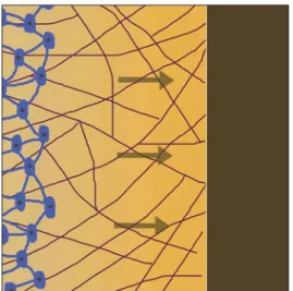
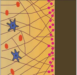
to Davies;[45] Puleo and Nanci,[20] ossifi cation occurs in two
directions, from implant surface toward the bone and from the bone toward the implant surface, which is known as bone regeneration and distance bone regeneration [Figures 1 and 2]. In the process of bone regeneration within the contact area, the bone gets shaped 30% faster. In this mode, prior to the formation of bone matrix, the implant surface clone with bone cells, and the identical mesenchyme, which was created during the remodeling
process is recognized as new bone formation. In distance bone regeneration, the new bone is formed on the surface of implant while the implant is covered by the surrounding bone. It is expected that this procedure occurs in cortical bone healing.[45]
The initial bones are formed woven, which have osteoid in their matrix. At the end of 12th week, the new bone that is formed at
the implant surface will be uniformed with a body connection of mature lamellar bone with titanium surface.[43]
Conclusion
Osteointegration is a fairly complicated process and the aspects of micro and micro molecule bone-implant interface are the matters of controversy. However, through the use of conducted tests and studies of many authors, we can claim that the treatment patterns in cortical and trabecular bones are diff erent. Cortical healing depends on the remodeling of haversian, while trabecular healing is based on osteoconduction phenomenon (bone growth on the surface) and the formation of new bone.
Bone formation in the position of bone lesion takes place due to coupling mechanism and according to frost, the mechanism of formation and reabsorption should be existing. The biomechanical milieu in position failure immediately aff ects cartilage and bone development.
Once the position of implant placement is prepared, a lesion is made and the phase of bone-implant healing is performed by a method similar to healing in the case of a broken bone, since both operations start with penetration in an intact skeletal position, an immune response, a new angiogenesis, and use skeletal progenitor cells. Nevertheless, some skeletal progenitor cells will diff erentiate into chondrocytes in cases of bone failure, while the others will change into osteoblasts that are followed by ossifi cation inside the cartilage. Around an implant, all skeletal progenitor cells that change into osteoblast are followed with intra-membranous ossifi cation. The other contradiction in implant healing is that the osteointegration process is extremely under the infl uence of implant surface, chemical composition, and implant biomechanics.[46,47]
Hence, as soon as the implant is located, platelet density takes place.[45] These platelets secrete the growth factors, such
as platelet-derived growth factor-BB, insulin-like growth factor (IGF, IGF-2), FGFs (a-FGF, b-FGF), transitional growth factor beta, BMPs, vasoactive factors of serotonin and histamine [Figure 3]. These factors are more diff erentiated, proliferated, and bound to osteoblasts with titanium level [Figure 4] and form a new matrix [Figure 5]. The development is managed by the transcription factor protein core binding-factor-alpha.[48]
There are ample evidences to prove the direct relationship between osteointegration and superfi cial topography. It is verifi ed that the rough surface made osteointegration binding better, resulting in fi lopodia, as well as a four layer increase in cbcfa1.[48] We also surmise that, an increase in the surface is
not a defi nitive factor for regulating cell growth in the bone-implant interface. Consequently, bone-implant surface topography
Figure 1: Distance osteogenesis
satellite-like rough surface cells.[35] Therefore, the bone matrix
in the bone-implant interface is formed during the bone growth on the surface [osteoconduction], simulating ossifi cation (osteoinduction), bone regeneration (osteogenesis), and bone progress (osteopromotion).[26]
Bone growth in the surface refers to the orientation of formed bone activity to the position or specifi c surface, like hydroxyapatite coating, which is retained as a framework for making cell connection and growth. Stimulation of ossifi cation includes applying mesenchymal stem cells that will be converted to osteoblasts. Implant surfaces are not stimulating. Bone regeneration is related to the stimulation of proliferation of bone progenitor cells and the stimulation of the biosynthetic activity of osteoblasts. As the fourth modifi cation, although bone progress is relatively a new term, is related to the formation of bone in topical bone positions through using techniques that are related to membrane barriers, and is only utilized in promoting clinical cells.
There are many unanswered questions; however, there is no fi eld of study in implantology that thoroughly investigates osteointegration. The current results and future research projects represent better understanding and a fi ner transparent image of what is happening in the bone-implant interface, which subsequently provides the required information for implantologists. These findings, after understanding the physiological and biological needs of bone, can supply the patients with the best possible medical treatments.
References
1. Albrektsson T, Albrektsson B. Osseointegration of bone implants. A review of an alternative mode of fi xation. Acta Orthop Scand 1987;58:567-77.
2. Brånemark R, Brånemark PI, Rydevik B, Myers RR. Osseointegration in skeletal reconstruction and rehabilitation: A review. J Rehabil Res Dev 2001;38:175-81.
3. Brånemark PI. Osseointegration and its experimental background. J Prosthet Dent 1983;50:399-410.
4. VanBlarcom CW. Th e Glossary of Prosthodontic Terms. St. Louis: Mosby; 1999.
5. Albrektsson T, Brånemark PI, Hansson HA, Lindström J. Osseointegrated titanium implants. Requirements for ensuring a long-lasting, direct bone-to-implant anchorage in man. Acta Orthop Scand 1981;52:155-70.
6. Nayab S, Shinawi L, Hobkirk J, Tate T, Olsen I, Jones F. Adhesion of bone cells to ion-implanted titanium. J Mater Sci Mater Med 2003;14:991-7.
7. Muhonen V, Heikkinen R, Danilov A, Jämsä T, Tuukkanen J. Th e eff ect of oxide thickness on osteoblast attachment and survival on NiTi alloy. J J Mater Sci Mater Med 2007;18:959-67. 8. Rodan GA, Martin TJ. Role of osteoblasts in hormonal control of
bone resorption – A hypothesis. Calcif Tissue Int 1981;33:349-51. 9. Lacey DL, Timms E, Tan HL, Kelley MJ, Dunstan CR, Burgess T,
et al. Osteoprotegerin ligand is a cytokine that regulates
osteoclast diff erentiation and activation. Cell 1998;93:165-76.
10. Cooper LF, Masuda T, Yliheikkilä PK, Felton DA.
Generalizations regarding the process and phenomenon of
Figure 3: Secretion of growth factors from platelets
Figure 4: Diff erentiation and proliferation of growth factors and attachment of osteoblasts with titanium surface
Figure 5: Formation of new bone matrix
osseointegration. Part II. In vitro studies. Int J Oral Maxillofac Implants 1998;13:163-74.
11. Komori T. Regulation of bone development and maintenance by Runx2. Front Biosci 2007;13:898-903.
12. Buckwalter J, Glimcher M, Cooper R, Recker R. Bone biology. J Bone Joint Surg Am 1995;77:1256-75.
13. Roodman GD. Advances in bone biology: Th e osteoclast. Endocr Rev 1996;17:308-32.
14. Shimizu H, Sakamoto M, Sakamoto S. Bone resorption by isolated osteoclasts in living versus devitalized bone: Diff erences in mode and extent and the eff ects of human recombinant tissue inhibitor of metalloproteinases. J Bone Miner Res 1990;5:411-8. 15. Tatsumi S, Ishii K, Amizuka N, Li M, Kobayashi T, Kohno K,
et al. Targeted ablation of osteocytes induces osteoporosis with
defective mechanotransduction. Cell Metab 2007;5:464-75. 16. Weinmann JP, Sicher H. Bone and Bones: Fundamentals of
Bone Biology. St. Louis: Mosby; 1955.
17. Singhatanadgit W. Biological responses to new advanced surgace modifi cations of endosseous medical implants. Bone Tissue Regen Insights 2009;2:1-11.
18. Shard AG, Tomlins PE. Biocompatibility and the effi cacy of medical implants. Regen Med 2006;1:789-800.
19. Th evenot P, Hu W, Tang L. Surface chemistry infl uences implant biocompatibility. Curr Top Med Chem 2008;8:270-80.
20. Puleo DA, Nanci A. Understanding and controlling the bone-implant interface. Biomaterials 1999;20:2311-21.
21. Wilson CJ, Clegg RE, Leavesley DI, Pearcy MJ. Mediation of biomaterial-cell interactions by adsorbed proteins: A review. Tissue Eng 2005;11:1-18.
22. Ratner BD, Bryant SJ. Biomaterials: Where we have been and where we are going. Annu Rev Biomed Eng 2004;6:41-75. 23. Damsky CH, Werb Z. Signal transduction by integrin receptors
for extracellular matrix: Cooperative processing of extracellular information. Curr Opin Cell Biol 1992;4:772-81.
24. Globus RK, Doty SB, Lull JC, Holmuhamedov E, Humphries MJ, Damsky CH. Fibronectin is a survival factor for diff erentiated osteoblasts. J Cell Sci 1998;111:1385-93.
25. Moursi AM, Damsky CH, Lull J, Zimmerman D, Doty SB, Aota S,
et al. Fibronectin regulates calvarial osteoblast diff erentiation.
J Cell Sci 1996;109 (Pt 6):1369-80.
26. Cooper LF. Biologic determinants of bone formation for osseointegration: Clues for future clinical improvements. J Prosthet Dent 1998;80:439-49.
27. Schwartz Z, Lohmann CH, Oefi nger J, Bonewald LF,
Dean DD, Boyan BD. Implant surface characteristics modulate diff erentiation behavior of cells in the osteoblastic lineage. Adv Dent Res 1999;13:38-48.
28. Hynes RO. Integrins: A family of cell surface receptors. Cell 1987;48:549-54.
29. Takagi J, Petre BM, Walz T, Springer TA. Global conformational rearrangements in integrin extracellular domains in outside-in and inside-out signaling. Cell 2002;110:599-11.
30. Clover J, Dodds RA, Gowen M. Integrin subunit expression by human osteoblasts and osteoclasts in situ and in culture.
J Cell Sci 1992;103 (Pt 1):267-71.
31. Abercrombie M, Heaysman JE, Pegrum SM. Th e locomotion of fi broblasts in culture. IV. Electron microscopy of the leading lamella. Exp Cell Res 1971;67:359-67.
32. Avnur Z, Geiger B. Th e removal of extracellular fi bronectin from areas of cell-substrate contact. Cell 1981;25:121-32. 33. Sastry SK, Burridge K. Focal adhesions: A nexus for
intracellular signaling and cytoskeletal dynamics. Exp Cell Res 2000;261:25-36.
34. Smilenov LB, Mikhailov A, Pelham RJ, Marcantonio EE, Gundersen GG. Focal adhesion motility revealed in stationary fi broblasts. Science 1999;286:1172-4.
35. Zhu X, Chen J, Scheideler L, Altebaeumer T, Geis-Gerstorfer J, Kern D. Cellular reactions of osteoblasts to micron- and submicron-scale porous structures of titanium surfaces. Cells Tissues Organs 2004;178:13-22.
36. Rosen JJ, Culp LA. Morphology and cellular origins of substrate-attached material from mouse fi broblasts. Exp Cell Res 1977;107:139-49.
37. Albrecht-Buehler G. Filopodia of spreading 3T3 cells. Do they have a substrate-exploring function? J Cell Biol 1976;69:275-86. 38. Adams JC. Cell-matrix contact structures. Cell Mol Life Sci
2001;58:371-92.
39. Joos U, Büchter A, Wiesmann HP, Meyer U. Strain driven fast osseointegration of implants. Head Face Med 2005;1:6.
40. Schwartz Z, Martin JY, Dean DD, Simpson J, Cochran DL, Boyan BD. Eff ect of titanium surface roughness on chondrocyte proliferation, matrix production, and diff erentiation depends on the state of cell maturation. J Biomed Mater Res 1996;30:145-55. 41. Rajpurohit R, Koch CJ, Tao Z, Teixeira CM, Shapiro IM.
Adaptation of chondrocytes to low oxygen tension: Relationship between hypoxia and cellular metabolism. J Cell Physiol 1996;168:424-32.
42. Colnot C, Romero DM, Huang S, Rahman J, Currey JA, Nanci A,
et al. Molecular analysis of healing at a bone-implant interface.
J Dent Res 2007;86:862-7.
43. Depprich R, Zipprich H, Ommerborn M, Mahn E, Lammers L, Handschel J, et al. Osseointegration of zirconia implants: An SEM observation of the bone-implant interface. Head Face Med 2008;4:25.
44. Büchter A, Joos U, Wiesmann HP, Seper L, Meyer U. Biological and biomechanical evaluation of interface reaction at conical screw-type implants. Head Face Med 2006;2:5.
45. Davies JE. Understanding peri-implant endosseous healing. J Dent Educ 2003;67:932-49.
46. Shalabi MM, Gortemaker A, Van’t Hof MA, Jansen JA, Creugers NH. Implant surface roughness and bone healing: A systematic review. J Dent Res 2006;85:496-500.
47. Pearce AI, Richards RG, Milz S, Schneider E, Pearce SG. Animal models for implant biomaterial research in bone: A review. Eur Cell Mater 2007;13:1-10.