Macedonian Journal of Chemistry and Chemical Engineering, Vol. 27, No. 2, pp. 99–106 (2008)
MJCCA9 – 520 ISSN 1857 – 5552
Received: October 1, 2008 UDC: 612.124:543.51
Accepted: November 11, 2008
Review
DISCOVERING HUMAN PROTEIN DIVERSITY
Dobrin Nedelkov
Institute for Population Proteomics and Intrinsic Bioprobes Inc. 2155 E. Conference Dr. Suite 104, Tempe, AZ 85284
[email protected] // [email protected]
Current emphasis on discovering and correlating human genetic variations lays the foundation for future stud-ies of human protein diversity. Protein posttranslational processing, along with the translation of genetic variations, results in a complex, variable human proteome. Analyzing these protein variations on a grandeur scale has become feasible with the advent of mass spectrometry. Mass spectrometry is the only detection method today that can univer-sally provide information about specific protein structural modifications, without a priori knowledge of the modifica-tion. However, high-throughput separation approaches are needed to effectively prepare the proteins for mass spec-trometric interrogation. Such are the immunoaffinity separations that target single proteins by using highly specific antibodies for their affinity retrieval from the biological fluids. The resulting combination of immunoaffinity separa-tion with MALDI-TOF mass spectrometry, termed Mass Spectrometric Immunoassay (MSIA), has been recently ap-plied in two large studies of protein diversity. The results of these studies reveal a human protein diversity that is far more complex than the variations observed at the genetic level. Assessing the human proteome variations among and within populations will be an important future undertaking with significant clinical and diagnostic implications.
Key words: protein diversity; population proteomics; protein isoforms; plasma; serum; mass spectrometry; immunoassay
ИСПИТУВАЊЕНАПРОТЕИНСКИОТДИВЕРЗИТЕТКАЈЧОВЕКОВАТАПОПУЛАЦИЈА
Откривањетоивоспоставувањетокорелацијапомеѓучовековитегенетскиваријациипретставувадобра основазаиднииспитувањанапротеинскиотдиверзитеткајчовековатапопулација. Протеинскиотдиверзитет ерезултатнапосттранслацискитепроцесикајпротеините, какоиваријациитенагенетскониво. Опсежното испитувањенаовиепротеинскиваријациистанувавозможносоприменанамасенаспектрометрија. Масената спектрометријаеединствениотметодза детекцијакојможеуниверзалнодададеинформација заодредени протеински структурни модификации, без претходно знаење за идентитетот на модификацијата. Меѓутоа, брзииефикаснисепарацискипроцесисепотребнизапротеинитесоодветнодасеподготватзамасенаанали -за. Такви сеимуноафинитетнитераздвојувања коисе применуваатза изолација напротеини одбиолошки примероци со помош на високо специфични антитела кои имаат афинитет кон анализираните протеини. Комбинацијата на имуноафинитетни раздвојувања со масената спектрометрија MALDI-TOF се нарекува масеноспектрометрискаимуноанализа (MSIA) инеодамна бешеприменета водве опсежнистудиинапро -теинскиотдиверзитет кај човековата популација. Резултатите од овие студиисугерираат големстепен на протеинскаразновидносткојаедостапокомплекснаодгенетскиотдиверзитет. Откривањетоистудирањето напротеинскитеваријациивочовековатапопулацијаќебидезначаенделодиднитепротеинскииспитувања воклиничкителабораториииводијагностиката.
Клучнизборови: протеинскидиверзитет; протеинскиизоформи; плазма; серум; масенаспектрометрија; имуноанализа
INTRODUCTION
The human genome contains little over 3 bil-lion base pairs, and an estimated 20,000–25,000 protein-coding genes. Immediately after the intial
human DNA sequence variation [3]. In recent years, the focus has moved to individual human genomes, as advances in sequencing technology and a decrease in costs have made sequencing in-dividual genomes feasible. In 2007, the complete (diploid) genome sequences of two individuals were published, those of James Watson (the co-discoverer of the DNA structure) [4] and Craig Venter, the head of the privately funded human genome project [5]. In 2008, a consortium of sci-entists sequenced the genomes of eight people from diverse ethnic backgrounds, and completed the first sequence-based map of structural varia-tions in the human genomes, such as inservaria-tions, deletions, inversions, single nucleotide polymor-phisms (SNPs), and copy number differences [6]. And in early 2008, an international research con-sortium (the “1000 Genomes Project”) was formed to create the most detailed and medically useful picture to date of human genetic variation, by se-quencing the genomes of at least a thousand peo-ple from around the world [7]. In all, the human genetic variation is such a hot topic that it was named the breakthrough of the year 2007 by the magazine Science [8]. Therefore, it is not surpris-ing that population genetics and personal genomics are being rapidly commercialized, with companies like 23andMe (www.23andme.com), deCode Ge-netics (www.decode.com), and Navigenics (www.navigenics.com) offering personalized ge-netic analysis to reveal one’s risks to specific dis-eases, traits, and other conditions, based on spe-cific regions and changes in the genetic sequence. The Genographic project, with aims to map the migratory history of the human species, is another example of how genetic data can be used to track one’s ancestry [9]. With these concerted efforts and outcomes from the investigations of the human genetic variation, we are only starting to appre-hend and appreciate the extent to which our ge-nomes differ from person to person. More impor-tantly, we are learning about the implication that these variations have in disease predisposition and development.
But the genes only control heredity and pro-vide the codes for the life’s building blocks – the proteins. If there is so much variation at the gene level, what kind of variations can be expected at the protein level? The answer to that question leads us to the much more complex world of hu-man protein diversity. The protein diversity stems from several dimensions. To start with, a single nucleotide change in the DNA sequence (i.e.,
SNP) can give rise to a different amino acid in the protein sequence, which in turn can influence pro-tein folding, processing, function, etc. Next, alter-native splicing of the pre-mRNA can produce sets of protein isoforms, each with distinct characteris-tics and functionalities. Then, following the pro-tein sequence translation from the mRNA, a large number of modifications are induced at specific sites of the protein sequence, and can include phosphorylation, glycosylation, disulfides forma-tion, side chain oxidaforma-tion, various enzymatic proc-essing, N- and C-terminal sequence truncations, etc. These posttranslational processes, as well as the overall protein expression in specific tissues, are heavily influenced by various cell processes, environmental factors, and cell and body cycles, resulting in an overall fluidic state of the human proteome. Analyzing these variations on a gran-deur scale has been very challenging – there is no PCR-equivalent technique for protein analysis. Classical biochemical approaches have been used in the past to painstakingly unveil specific protein function and structure, in a one-protein-at-a-time approach. The invention of enzyme immunoassays in the early 1970s [10] enabled researchers to read-ily determine concentration ranges for a large number of proteins in biological fluids such as human serum, plasma, and urine [11, 12]. These methods have been the cornerstone of the diagnos-tic world for over three decades, and are still con-sidered the gold standard in the clinical and refer-ence laboratories. However, there has been no easy and rapid method for detecting protein structural modifications – until the advent of mass spec-trometry.
protein, including sequence truncations, side-chain residue modifications (phosphorylation, sulfona-tion, oxidasulfona-tion, etc.), deglycosylations, chemical adducts, etc. Many of those modifications have been reported for numerous proteins, yet, to date there is virtually no data on their distribution across the general population, even for the most abundant proteins. Point-mutations at the gene level can also be detected and catalogued as those mutations oftentimes result in detectable mass shifts, even on intact proteins.
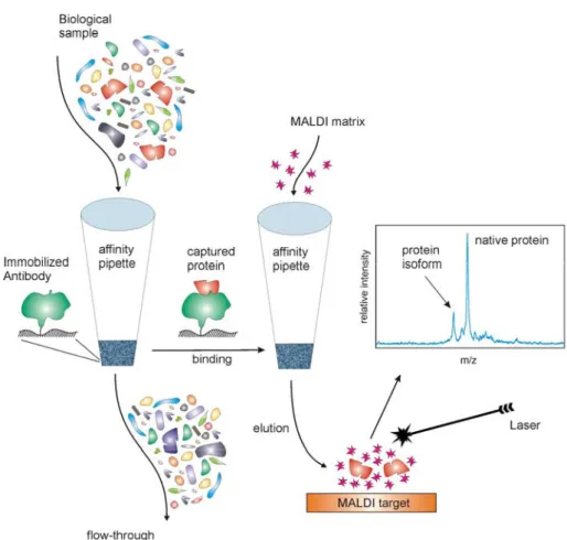
And yet, mass spectrometry is only a method of detection, and as with any other detection tech-nique, the separation processes that prepare the proteins for MS detection are critical. Standard separations such as two-dimensional gel electro-phoresis, liquid chromatography, and affinity sur-faces, have been used in combination with mass spectrometry extensively in the past 15 years. However, most of them are complex, multifaceted, and often times do not yield reproducible results, even within the same laboratory. To achieve high-throughput reproducible analyses, the fractionation approaches have to be conceptually very simple, highly-reproducible, and yield unambiguous read-ings and results. Such are the immunoaffinity separations that target single proteins by using highly specific antibodies for their affinity re-trieval from the biological fluids. Combining this immunoaffinity separation with MALDI-TOF mass spectrometric detection yields an approach termed Mass Spectrometric Immunoassay (MSIA) (Fig. 1) [15, 16]. MSIA is essentially a rational combination of micro-scale immunoaffinity cap-ture and mass spectrometry. Antibodies are sur-face-immobilized in small, porous microcolumns that are fitted at the entrance of pipettor tips. Bio-logical samples are repeatedly aspirated and dis-pensed through these affinity pipettes to expose the immobilized antibody to the protein antigen present in the sample. Once the protein is cap-tured, the affinity pipettes are rinsed to remove any loosely associated and non-specifically bound sample components, and a small volume of MALDI matrix is aspirated into the affinity pi-pettes. The pH of the matrix solution, and its com-ponents, disrupt the antibody-antigen interaction, and the antigen-containing eluate is deposited di-rectly onto a MALDI target for ensuing mass spec-trometric analysis. This combination of immunoaf-finity capture with mass spectrometry results in a dual specificity assay – the capturing antibody provides the first level of specificity, while the
mass spectrometric detection gives the assay an-other (orthogonal) measure of specificity in that each protein should register in the mass spectrum at a precise m/z value that corresponds to its mo-lecular mass. The ability to see deviations from the predicted mass of the protein in the mass spectra enables detection of protein isoforms and other posttranslational modifications that give rise to the protein diversity.
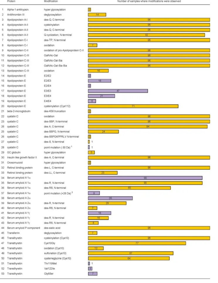
considera-tion the gender, age, and ethnicity of the individu-als who provided the samples, it was determined that the Gly6Ser mutation in transthyretin was de-tected only in individuals of Caucasian origin, which is consistent with existing knowledge about the occurrence of this common non-amyloidogenic population polymorphism in Caucasians [18]. An-other correlation was observed in regards to inter-protein variations in specific individuals: all seven individuals for which carbohydrate deficient trans-ferrin was detected were also characterized with deglycosylated antithrombin III.
Following this small scale protein diversity study, a second study of human protein diversity was recently carried out wherein the number of samples was greatly expanded in order to get an accurate view of the distribution of some of the protein modifications in the general population [19]. One thousand individuals from 4 geographi-cal regions in the United States (California, Flor-ida, Tennessee, and Texas) were selected and the protein modifications for beta-2-microglobulin (b2m), cystatin C (cysC), retinol binding protein
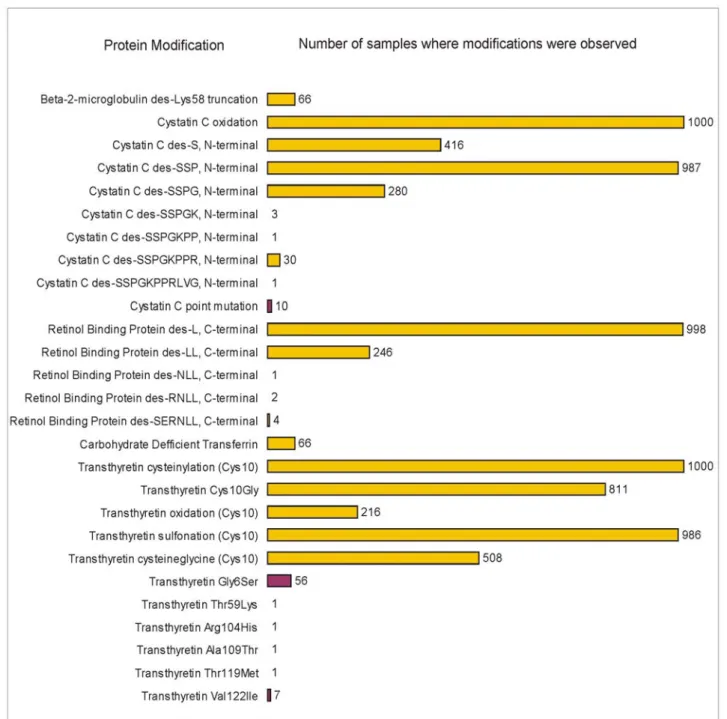
(RBP), transferrin (TRFE), and transthyretin (TTR) were delineated (in the 96-samples study, these five proteins accounted for 19 of the 53 pro-tein variants observed). The results of the study are summarized in Figure 4, which lists the protein modifications observed and the frequency of each in the 1,000 samples cohort. A total of 27 protein modifications (20 posttranslational modifications and 7 point mutations) were detected, with various frequencies in the cohort of samples. Variants re-sulting from oxidation were observed most fre-quently, along with single amino acid truncations. Least frequent were variants arising from point mutations and extensive sequence truncations. In total, six modifications were observed with high frequency (present in >80% of the samples), 5 were of medium frequency (20-50% of the sam-ples), and 16 were low frequency modifications observed in <7% of the samples. Nine of the low frequency modifications were not observed in the 96 individuals study. Thus, by increasing the size of the population it became possible to detect these low-occurrence protein modifications.
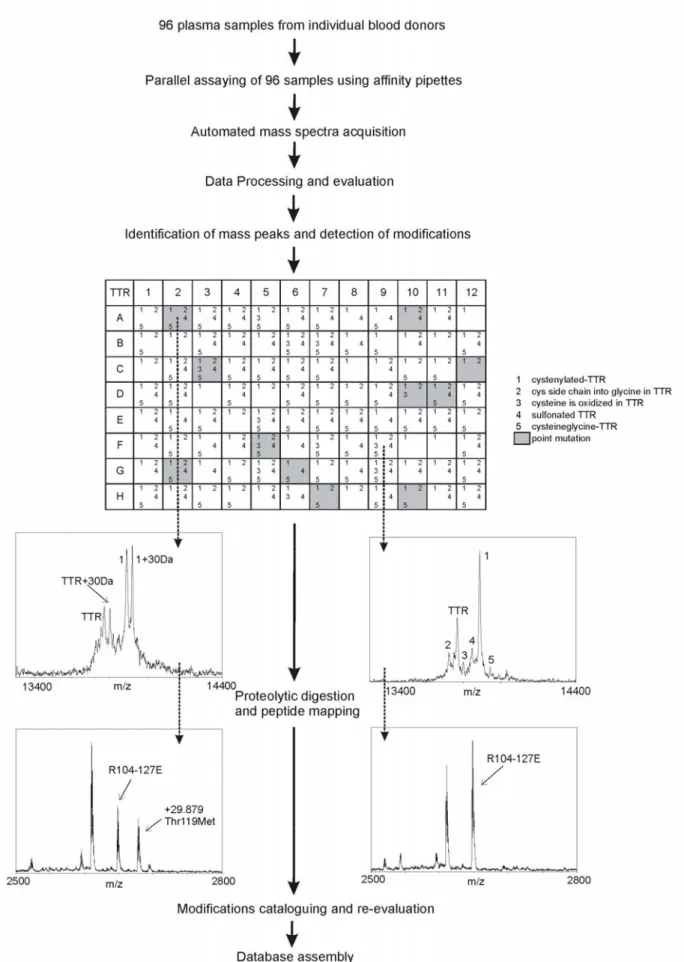
Fig. 3. Modifications observed in 18 of the 25 proteins analyzed from 96 human plasma samples (modifications were not detected for albumin, cerruloplasmin, C-reactive protein, insulin like growth factor I, lysozyme, plasminogen, and urine protein 1.
Fig. 4. Modifications observed for five proteins studied from 1,000 human plasma samples. Reprinted with permission from Nedelkov et al. (19).
When the frequencies of the modifications in the two studies were compared, an excellent corre-lation was obtained. For example, in both cohorts ~7% of the individuals were characterized with carbohydrate deficient transferrin. Upon further data analysis based on the gender, age, and geo-graphical origin of the individuals who provided the samples, it was determined that the samples obtained from California contained significantly less protein modifications than the samples ob-tained from Florida, Tennessee, and Texas, even though the samples from all four states were
re-lated to cystatin C: all 10 of the cystatin C point mutations were found in males.
Two conclusions can be made from these two systematic studies of protein modifications and variants. First, mass spectrometry is capable of detecting structural protein modifications, and, when coupled to immunoaffinity separations, it can be employed in a high-throughput systematic study of human protein diversity, in a discipline termed population proteomics [20-22]. Second, the human protein diversity is far more complex than the varia-tion observed at the genetic level. While it might be premature to declare the human proteins variation “the next big thing”, it is reasonable to predict that assessing human proteome variations among and within populations will be a paramount effort that can facilitate biomarker discovery. Such endeavor would represent a paradigm shift in proteomics with significant clinical and diagnostic implica-tions, as protein variaimplica-tions, quantitative and quali-tative, begin to be associated with specific dis-eases. The time to start these studies has arrived.
REFERENCES
[1] E. S. Lander et al., Initial sequencing and analysis of the human genome. Nature, 409, 860–921(2001).
[2] J. C. Venter et al., The sequence of the human genome. Science, 291, 1304–1351(2001).
[3] D. Altshuler et al.,A haplotype map of the human ge-nome. Nature, 437, 1299–1320 (2005).
[4] D. A. Wheeler et al., The complete genome of an indivi-dual by massively parallel DNA sequencing. Nature, 452, 872–876 (2008).
[5] S. Levy et al., The diploid genome sequence of an indi-vidual human. PLoS Biol, 5, e254 (2007).
[6] J. M. Kidd et al., Mapping and sequencing of structural variation from eight human genomes. Nature, 453, 56-64 (2008).
[7] J. Kaiser, DNA sequencing. A plan to capture human diversity in 1000 genomes. Science, 319, 395 (2008). [8] E. Pennisi, Breakthrough of the year. Human genetic
variation. Science, 318, 1842–1843 (2007).
[9] D. M. Behar, S. Rosset, J. Blue-Smith, O. Balanovsky, S. Tzur, D. Comas, R. J. Mitchell, L. Quintana-Murci, C. Tyler-Smith, R. S. Wells, The Genographic project public participation mitochondrial DNA database. PLoS Genet 3, e104 (2007).
[10] R. M. Lequin, Enzyme immunoassay (EIA)/enzyme-linked immunosorbent assay (ELISA). Clin. Chem., 51, 2415–2418 (2005).
[11] W. Y. Craig, T. B. Ledue, R. F. Ritchie, Plasma Proteins: Clinical Utility and Interpretation, Foundation for Blood Research, Scarborough, ME 2001.
[12] R. F. Ritchie (Ed.), Serum Proteins in Clinical Medicine, Foundation for Blood Research, Scarborough, ME 1999. [13] R. Aebersold, M. Mann, Mass spectrometry-based
pro-teomics. Nature, 422, 198–207 (2003).
[14] B. F. Cravatt, G. M. Simon, J. R.Yates, 3rd, The biologi-cal impact of mass-spectrometry-based proteomics. Na-ture, 450, 991–1000 (2007).
[15] D. Nedelkov, Mass spectrometry-based immunoassays for the next phase of clinical applications. Expert Rev Pro-teomics, 3, 631–640 (2006).
[16] R. W. Nelson, J. R. Krone, A. L. Bieber, P. Williams, Mass-spectrometric immunoassay. Anal. Chem. 67, 1153 –1158 (1995).
[17] D. Nedelkov, U. A. Kiernan, E. E. Niederkofler, K. A. Tubbs, R. W. Nelson, Investigating diversity in human plasma proteins. Proc. Natl. Acad. Sci. U S A, 102, 10852–10857 (2005).
[18] L. H. Connors, A. Lim, T. Prokaeva, V. A. Roskens, C. E. Costello, Tabulation of human transthyretin (TTR) variants, Amyloid, 10, 160–184 (2003).
[19] D. Nedelkov, D. A. Phillips, K. A. Tubbs, R. W. Nelson, Investigation of human protein variants and their fre-quency in the general population. Mol. Cell. Proteomics, 6, 1183–1187 (2007).
[20] D. Nedelkov, Population proteomics: addressing protein diversity in humans. Expert Rev. Proteomics, 2, 315–324 (2005).
[21] D. Nedelkov, Population proteomics: investigation of protein diversity in human populations. Proteomics, 8, 779–786 (2008).