Nerve growth factor and chemokine receptors regulate macrophage phenotype via activation of transient receptor potential and inositol triphosphate receptor channels
By
Viktoriya Zhuravleva
Senior Honors Thesis Biology Department
University of North Carolina at Chapel Hill 24 March, 2016
Approved: 11 April, 2016
Rick B. Meeker, Thesis Advisor Steve Crews, Reader
Amy Shaub Maddox, Reader
Maria Servedio, Biology Faculty Sponsor
Zhuravleva 2
Abstract
Human Immunodeficiency Virus (HIV) activates macrophages and microglia cells in the central nervous system (CNS) and triggers the secretion of neurotoxins, causing neuronal damage. Therapeutic approaches to restore cognitive function by suppressing macrophage and microglial activation have not been successful, partially due to limited knowledge of cellular mechanisms that control toxin secretion. In this investigation, we study the pathway to HIV-induced
neurotoxin release, focusing on how calcium influx and phosphorylation of the HIV co-receptor CXCR4 lead to macrophage activation, including alteration of the actin cytoskeleton resulting in the formation of ruffles and podosomes. We found that Nerve Growth Factor (NGF) increases CXCR4 phosphorylation, increases calcium signaling, and reverses the HIV-induced increase in podosome formation. To further elucidate the pathway that mediates this effect, we challenged macrophages with specific inhibitors for CXCR4 and CCR5 receptors, as well as for PI3K and Pyk2. To investigate the source of calcium leading to macrophage activation, we inhibited the P2X7 receptor, CRAC channels, TrpC channels, and IP3 receptor channels. Our data indicated
that calcium entry through the P2X7 receptor contributed to podosome formation while the IP3 receptor channel favored expression of the less toxic ruffled phenotype, Pyk2 signaling and calcium entry through the TrpC channel were necessary for both ruffling and podosomes.
Because podosome expression is correlated with toxin secretion from macrophages, determining the pathway to morphological modifications enables a better overall understanding of
Introduction
Human Immunodeficiency Virus (HIV) infection causes neurodegeneration in the central nervous system (CNS), resulting in HIV associated dementia and other cognitive deficits1. As patients survive into older age with the help of highly active antiretroviral therapies (HAART), neuronal damage is expected to progress and accelerate2. Available therapeutics are unable to cross the blood-brain barrier (BBB), allowing the CNS to become a protected reservoir of HIV2.
HIV infects cells by binding to the receptor CD4 and a chemokine co-receptor, CCR5 or CXCR4. Binding results in fusion of the cell’s membrane with that of HIV, allowing the virus entry into the cell3. Since there is little evidence of HIV crossing the blood brain barrier on its own during early infection, the process is dependent on infection of circulating monocytes. These cells cross the BBB, differentiate into macrophages and microglia, and introduce HIV into the CNS2. Once the infection is established in the CNS, the presence of HIV or HIV surface
proteins can activate macrophages and microglia through the HIV co-receptor independent of infection2. Once the cells are activated by HIV, they release unknown neurotoxins, which are the main cause of HIV-related damage to the CNS4.
Macrophages are dynamic cells that release a variety of signaling molecules and growth factors depending on external cues. HIV activates macrophages to release neurotoxins by interacting with the chemokine receptor CXCR43. Though HIV stimulates the chemokine receptor, macrophages also express the neurotrophin receptors p75NTR and TrkA, which regulate HIV-induced neurotoxin release5. In neurons, TrkA binds Nerve Growth Factor (NGF) with high affinity and p75 binds NGF with low affinity. The p75 receptor can also bind the immature proNGF6. The mature and pro-form of NGF have opposite effects on neurons; NGF typically
Zhuravleva 4
apoptosis6. The opposing effect of proNGF and NGF also applies to macrophage neurotoxin release in response to HIV. NGF stimulation results in decreased neurotoxicity while proNGF exacerbates neurotoxin release7. The changes in neurotoxin release are paralleled by changes in macrophage morphological phenotype.
Previous studies have associated macrophage cytoskeletal morphology, specifically the presence of ruffles or podosomes, with the cells’ toxicity level and acute calcium spiking
activity. Ruffled macrophages are less toxic and have more calcium spikes, while those with podosomes produce more neurotoxins and have fewer calcium spikes7. Podosomes have been linked to increased tissue invasion and secretion of matrix metalloproteases8, which unpublished studies suggest may have neurotoxic activity. Therefore, we focused on the presence of
podosomes in our studies on macrophage morphology, aiming to determine the pathway by which macrophages are activated to produce podosomes. The proposed model for podosome formation was that macrophage activation by HIV leads to CXCR4 phosphorylation, increased calcium levels and activation of Pyk2.
In this investigation we continued previous studies on the pathway by which
Methods
Primary cultures of human macrophages1
Peripheral blood mononuclear cells (PBMCs) were isolated by density gradient centrifugation on Ficoll-Paque Plus. PBMCs were washed and plated on low-adhesion tissue culture plates
(Fisher) in DMEM with 10% FBS and 20 µg/mL gentamicin. Monocytes adhered over a period of 3-5 days. Cells were then washed and cultured for 7 days in the same medium containing 15ng/mL GM-CSF (Sigma Inc.) to fully differentiate monocytes into
monocyte-derived-macrophages (MDMs). After differentiation, the monocyte-derived-macrophages were grown in standard DMEM +
1Macrophages were cultured by my mentor.
Figure 1. Proposed mechanism for the control of macrophage activation by HIV and neurotrophins. HIV and mature neurophins have opposite effects on the ability of
macrophages to exhibit a neurotoxic podosome-containing phenotype. HIV was hypothesized to stimulate and phosphorylate CXCR4, with subsequent calcium entry activation of Pyk2 and podosome formation. In a different but related pathway, NGF triggers TrkA
autophosphorylation, activation of PI3K, and increased ruffling. Extracellular calcium may enter through CRAC channels, Trp channels, or activation of the P2X7 receptor by ATP. IP3
Zhuravleva 6
10% FBS + gentamicin. Cells were subcultured at a density of 10,000-40,000 cells/cm2 as needed. After reaching the desired density, cells were fixed for staining and imaging. Western blotting2
Human monocyte-derived macrophages were stimulated with HIV, SDF, or NGF for 0, 5, 10, and 30 minutes. Cells were removed from 100mM dishes, treated with lysis buffer (1X RIPA buffer (Millipore 20-188), 1:100 PMSF, 1:1000 protease inhibitor cocktail) and sonicated. Protein concentration was measured used Bradford reagent (Bio-Rad 500-0209). Protein lysates were resolved by SDS on TGX gels (Bio-Rad 456-1034) and transferred to a nitrocellulose membrane (Bio-Rad 162-0234). The membranes were blocked in 2% milk for one hour at room temperature followed by overnight incubation of primary antibodies at 4°C. Membranes were then washed in TBS + 1% Tween and incubated in secondary antibody for one hour at room temperature. The membranes were washed again and imaged using a chemiluminescent substrate (Thermo Scientific cat# 34080) and film (Kodak 178 8207) or Image Quant LS 4000 technology. The primary antibody was pCXCR4 (ABCAM AB74012) at 1:500 dilution. Secondary Antibody was 1:4000 Goat anti-Rabbit HRP (Millipore AP132P).
Phalloidin staining of human macrophages
Cells were challenged under one of the eleven conditions listed in Table 1, and fixed with 2% paraformaldehyde. They were stained for one hour at room temperature with the filamentous actin (F-actin) stain Rhodamine Phalloidin (Cytoskeleton PHDR1), and diluted 1:100 in PBS. Cells were washed in PBS placed on slides with Fluoromount G (Southern Biotech 0100-01).
Challenge condition
Applied concentration
Challenge time
Function
Ctrl (aCSF) --- --- Mimics conditions in CNS Xestospongin-C 1 µM 2 hr IP3 receptor antagonist; prevents
release of Ca2+ from ER
A438079 4 µM 2 hr P2X7 receptor antagonist; inhibits extracellular calcium entry
LY294002 10 µM 2 hr PI 3-Kinase inhibitor
Pyr 3 1 µM 20 min TRP Channel 3 antagonist; inhibits TRPC3-mediated Ca2+ influx
ATP 1 mM 2 hr P2X7 receptor agonist; increases extracellular calcium entry AMD3100 0.02 - 0.13 µM 2 hr CXCR4 inhibitor
Maraviroc 6.4 nM 2 hr CCR5 inhibitor
Gadolinium 10 µM 2 hr CRAC Channel inhibitor YM58483 100 nM 2 hr CRAC Channel antagonist PF431396 11 nM 2 hr Pyk2 inhibitor
Macrophage Calcium Imaging
Macrophages were incubated in serum-free Dulbecco’s Modified Eagle Medium (DMEM) and loaded with the calcium indicator, Fluo-4 NW (Molecular Probes, Inc., Eugene, OR) at 1:4 dilution for 20 minutes. The macrophages were then treated with one of the drugs listed above and incubated for another 10 minutes. Time lapse digital images were captured automatically by the Metamorph System every 6 seconds for 20 minutes in the continued presence of the drug.
The first three images taken served as the baseline measurement of fluorescence at the beginning of each experiment. The increase in fluorescence intensity within each cell was then measured relative to the baseline measurements to correct for cell to cell differences in dye loading and intrinsic fluorescence. The differences in intensity between successive 6 sec measurements were determined to assess calcium spiking over time. Incremental increases greater than 2.3 standard
Zhuravleva 8
deviations from the basal calcium followed immediately by recovery were counted as spikes (>20 fluorescence units; p < 0.01).
Data Analysis and Statistics
Western blots were analyzed using average intensities of bands, as measured by Metamorph Software after imaging. Phalloidin stained macrophage images were scored visually; each macrophage in the image was classified as podosome-bearing, ruffled, or having no
specializations. Two-tailed, unpaired t-tests were calculated in Excel from a minimum of three replicates for statistical comparisons. P values < 0.05 were considered significant.
Results
Western blotting showed that NGF induces phosphorylation of CXCR4 similarly to its natural ligand SDF-1. The time course for CXCR4 phosphorylation is illustrated in Figure 2. When stimulated by SDF, CXCR4 phosphorylation peaked at about 5 minutes and remained elevated for 30 min (Figure 2A). After stimulation with NGF, CXCR4 phosphorylation peaked at about 10 minutes. There was no indication that HIV promoted CXCR4 phosphorylation (Figure 2A). CXCR4 phosphorylation was regulated by TrkA. Blocking TrkA with the specific inhibitor GW 441756 and stimulating the cells with NGF alone or NGF+HIV decreased CXCR4
M in u t e s P r o p o r ti o n o f C o n tr o l
0 1 5 1 0 3 0
0 .5 1 .0 1 .5
2 .0 N G F
S D F
HIV
C X C R 4 P h o s p h o r y la t io n
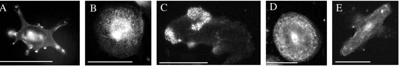
Phalloidin staining revealed that macrophages exist in several phenotypes that correlate well with toxicity. The less toxic, ruffled macrophages exhibit a less rounded shape and have highly-stained actin-rich areas on the edges (Figure 3A). More toxic macrophages have podosomes, which can be diffuse (Figure 3B), clustered (Figure 3C), or in a “belt” formation
(Figure 3D). Many macrophages also exhibit no distinctinve specializations (Figure 3E).
CTRL HIV NGF+HIV proNGF+HIV NGF+HIV+ NGF NGF+
0.0 0.5 1.0 1.5 2.0 *
p= 0.03 vs HIV
*
p= 0.004 vs NGF+HIV
p
CX
CR
4
GW 441756 GW 441756
*
*
p= 0.04 vs NGF p<0.001 vs CTRL
A
Figure 2. NGF stimulation induces phosphorylation of CXCR4 in a TrkA-dependent fashion. A) Phosphorylation of CXCR4 by SDF-1 peaked at 5 min and persisted to 30 min. NGF induced CXCR4 phosphorylation, which peaked at 10 min. HIV produced no significant changes in phosphorylation. B) The specific inhibitor for TrkA, GW 441756 decreased the phosphorylation of CXCR4 in the presence of NGF or NGF+HIV relative to NGF alone or NGF+HIV, respectively. Values are mean ± sem, n = 3-9 trials.
A
Zhuravleva 10
Blockade of CXCR4 with AMD3100 or CCR5 with Maraviroc, failed to affect podosome presence on macrophages, suggesting that other signaling pathways were crucial to the
phenotypic differentiation of these cells (Figure 4A). Blocking CCR5 decreased ruffling (Figure 4B), indicating that the receptor acts in the pathway toward ruffle formation. A greater
proportion of cells expressed podosomes in the presence of HIV. Blocking CXCR4 and challenging the macrophages with HIV increased the expression of ruffles relative to HIV (Figure 4C), with a corresponding decrease in the expression of podosomes.
B
C
D E
A B C D E
C t r l A M D 3 1 0 0 M a r a v ir o c 0 .0
0 .5 1 .0
1 .5 C h e m o k in e R e c e p t o r s
P o d o s o m e s /T o ta l C e ll s
C t r l A M D 3 1 0 0 M a r a v ir o c 0 .0
0 .5 1 .0
1 .5 C h e m o k in e R e c e p t o r s
R u ff le s /T o ta l C e ll s *
To investigate whether signaling targets downstream of the chemokine and neurotrophin receptors were necessary for podosome formation, PI3K and Pyk2 were blocked by LY294002 and PF431396, respectively. Blocking the TrkA effector, PI3K, did not affect ruffling, but it significantly decreased podosome expression (Figure 5). Inhibiting Pyk2, which is thought to mediate CXCR4 effects, greatly decreased the presence of both podosomes and ruffles on the cells (Figure 5).
CTR L HIV HIV +AM D31 00 HIV +Ma ravi roc 0.0 0.2 0.4 0.6 0.8
p=0.008 vs ctrl
*
P o d o s o m e s /T o ta l c e lls CTR L HIV HIV +AM D31 00 HIV +Ma ravi roc 0.0 0.1 0.2 0.3 0.4 0.5*
p=0.008 vs HIVRu ff le s /T o ta l Ce lls B A
Figure 4. Macrophage podosome expression is unaffected by blocking chemokine receptors. A) Blocking the CXCR4 receptor with the specific inhibitor AMD3100 and the CCR5 receptor with the specific inhibitor Maraviroc did not affect podosome formation. B) Blocking CXCR4 had no effect on macrophage ruffling, but blocking CCR5 decreased the proportion of ruffled macrophages (p=0.04). Values are normalized to control. C) HIV increased podosome expression, and blocking CXCR4 with AMD 3100 increased expression of ruffles in macrophages stimulated by HIV. Values are mean ± sem, n = 20-35 cells, 3-5 cultures.
Zhuravleva 12
C t r l P F 4 3 1 3 9 6 L Y 2 9 4 0 0 2 0 .0
0 .5 1 .0 1 .5
2 .0 S ig n a lin g P a th w a y
P o d o s o m e s /T o ta l C e ll s *** *
C t r l P F 4 3 1 4 9 6 L Y 2 9 4 0 0 2 0 .0
0 .5 1 .0 1 .5
2 .0 S ig n a lin g P a th w a y
R u ff le s /T o ta l C e ll s **
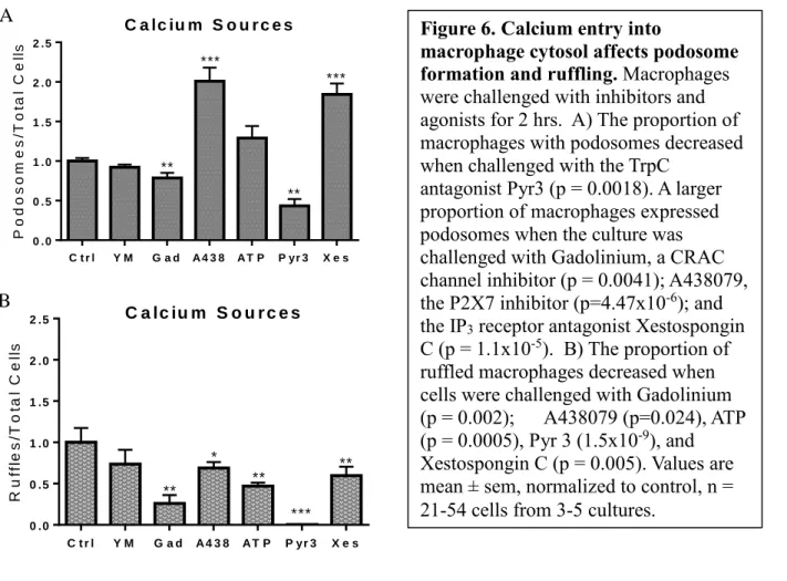
Since Pyk2 is involved in the pathway to podosome formation and requires calcium for activation, further investigations related macrophage morphology to calcium channels in order to determine the source of calcium that leads to podosomes. Phalloidin staining revealed that blocking calcium entry through the P2X7 channel with the specific inhibitor A438079 increased podosome expression and decreased ruffling (Figure 6A, 6B), though stimulating the
macrophages with the agonist ATP only decreased ruffles (Figure 6B). Inhibiting the TrpC3 channel with Pyr3 greatly decreased the presence of podosomes and ruffles (Figure 6A, 6B). Blocking the CRAC calcium channel with resulted in a small decrease in the proportion of macrophages expressing podosomes and ruffles (Figure 6A, 6B). Inhibiting calcium entry from the endoplasmic reticulum by blocking the IP3 receptor channel with Xestospongin C also
increased the proportion of macrophages with podosomes and decreased ruffling (Figure 6A, 6B).
C t r l Y M G a d A 4 3 8 A T P P yr 3 X e s 0 .0 0 .5 1 .0 1 .5 2 .0 2 .5
C a lc iu m S o u r c e s
P o d o s o m e s /T o ta l C e ll s ** *** ** ***
C t r l Y M G a d A 4 3 8 A T P P yr 3 X e s 0 .0
0 .5 1 .0 1 .5 2 .0
2 .5 C a lc iu m S o u r c e s
R u ff le s /T o ta l C e ll s ** * ** *** **
Since calcium spiking is associated with macrophage morphology, calcium activity of macrophages was measured to assess whether the channels that affect morphology also affect calcium spiking. Macrophages exhibit basal calcium spiking activity in aCSF (Figure 7A), and the activity is decreased when the TrpC channels are blocked (Figure 7B). Blocking the CRAC channel with the specific inhibitor YM 58483 had a small effect on calcium spiking (Figure 7C). Blocking the IP3 receptor channel with Xestospongin C nearly abolished calcium spiking activity
(Figure 7C), indicating that calcium spikes are dependent on ER calcium. Blocking the TrpC3 channel with Pyr3 also greatly decreased calcium spiking (Figure 7C). Placing macrophages in calcium free medium completely blocked calcium spiking activity, revealing that extracellular calcium is necessary for macrophages to produce calcium spikes (Figure 7C).
Figure 6. Calcium entry into
macrophage cytosol affects podosome formation and ruffling. Macrophages were challenged with inhibitors and agonists for 2 hrs. A) The proportion of macrophages with podosomes decreased when challenged with the TrpC
antagonist Pyr3 (p = 0.0018). A larger proportion of macrophages expressed podosomes when the culture was challenged with Gadolinium, a CRAC channel inhibitor (p = 0.0041); A438079, the P2X7 inhibitor (p=4.47x10⁻⁶); and
the IP3 receptor antagonist Xestospongin
C (p = 1.1x10⁻⁵). B) The proportion of
ruffled macrophages decreased when cells were challenged with Gadolinium
(p = 0.002); A438079 (p=0.024), ATP (p = 0.0005), Pyr 3 (1.5x10⁻⁹), and
Xestospongin C (p = 0.005). Values are mean ± sem, normalized to control, n = 21-54 cells from 3-5 cultures.
A
Zhuravleva 14
0 3 0 0 6 0 0 9 0 0 1 2 0 0
0 1 0 2 0 3 0 4 0 5 0
a C S F
T i m e ( s )
C h a n g e i n F lu o r e s c e n c e
0 3 0 0 6 0 0 9 0 0 1 2 0 0
0 1 0 2 0 3 0 4 0 5 0
P y r 3
T i m e ( s )
C h a n g e i n F lu o r e s c e n c e S p ik e s /1 0 M in
C o n t r o ls Y M X e s P yr 3 C a lc iu m f r e e 0
2 4 6 8
1 0 C a lc iu m S p ik in g A c t iv it y
Discussion
In this investigation we explored the effect of the neurotrophin NGF on phosphorylation of the HIV co-receptor CXCR4. Furthermore, we studied macrophage morphology, focusing on
A
C
Figure 7. Macrophage calcium spiking is dependent on extracellular calcium, Trp channels, and IP3 Receptor channels. Macrophages exhibit calcium spiking activity.
Incremental increases in calcium levels of macrophages show spiking activity over time, change in fluorescence ≥ 20 fluorescence units, p ≤ 0.01 compared to baseline calcium levels. A) Macrophages in aCSF have many more calcium spikes than (B) macrophages pre-treated with Pyr3, the Trp Channel inhibitor. Each colored line represents a single measured cell, 11 cells displayed per condition from matched cultures. C) Blocking the IP3 receptor channel using Xestospongin C or the Trp Channel using Pyr3 nearly abolished calcium spiking. Macrophages in calcium free medium had no spiking activity. Values are mean ± sem, n = 21-98 cells from 3-5 independent runs.
conditions that support the formation of podosomes or ruffles to indicate more toxic and less toxic macrophages, respectively. We examined the effects of various pathway inhibitors and agonists on macrophage cytoskeletal morphology to elucidate the pathway by which the cells are activated to form podosomes and release neurotoxins.
CXCR4 and CCR5
We found that NGF induced phosphorylation of CXCR4, suggesting that CXCR4 phosphorylation is protective because previous studies in the lab have shown that stimulating macrophages with NGF results in a less toxic macrophage phenotype7. This result is the opposite of previous studies, which suggested that CXCR4 activation was the first step in macrophage activation toward toxin release3. The previous studies used the HIV viral envelope, gp120, whereas we used intact virions, so the viral envelope may interact with CXCR4 differently than the intact virus. Data from previous research in the lab showed that HIV-induced podosome expression could be blocked by CXCR4 and p75 antagonists. Furthermore, phosphorylation of CXCR4 decreased when TrkA was blocked, supporting the idea that neurotrophin receptors interact with the HIV co-receptor to reduce HIV’s effect on macrophage activation.
Pathway to Podosome Formation
Pyk2 has been reported as a downstream target of HIV-chemokine receptor signaling that is necessary for podosome organization9, and calcium signaling through PI3K has been
Zhuravleva 16
deactivation could have led to a podosome decrease through a pathway independent of TrkA. Consistent with previous studies, blocking Pyk2 decreased the formation of podosomes and ruffles, revealing that Pyk2 is necessary for these changes in actin structure.
Calcium Sources
Because Pyk2 requires calcium to become active, and Del Corno, et al. (2001) indicated
that calcium influx was necessary for HIV activation of macrophages, we investigated possible sources of calcium. Although the non-specific cation CRAC channel was implicated in Del Corno’s investigations, we saw only a slight decrease in podosome formation when we blocked
these channels with gadolinium. Gadolinium is a less specific CRAC antagonist than YM58483, which produced no significant change in the expression of ruffles or podosomes. This result
indicated that calcium influx through the CRAC channel is not the main step in macrophage activation following HIV stimulation, opposing the conclusion reached by Del Corno, et al 2001. We therefore evaluated the contribution of other calcium channels thought to be active in
macrophages: P2X7, TrpC channels and the IP3 receptor channel.
When we blocked extracellular calcium entering through the P2X7 receptor by inhibiting the receptor with A438079, the presence of podosomes increased and ruffling decreased.
Stimulating the P2X7 receptor with its agonist ATP decreased ruffling and had no significant effect on podosomes. However, ATP may act at different receptors indicating that the response to ATP may be complex. Due to the conflicting results, the role of the P2X7 receptor in
macrophage activation remains unclear.
Trp Channels and IP3 Receptor Channels
membrane as well as the ER. Investigations on extracellular calcium entry targeted the non-specific cation channel TrpC. Trp channels, often activated in response to calcium release by the ER, enable individual cells to sense changes in their environment, and may therefore be
important actors in macrophage activation11. Smani, et al. found that actin modifications affect
Trp channel activity, and vice versa12. When we inhibited the TrpC channel with the antagonist
Pyr3, podosome formation and ruffling were almost abolished, indicating that macrophage activation leading to cytoskeletal ruffling or podosomes relies heavily on calcium entry through the TrpC channel.
In addition to serving as a source of extracellular calcium, Trp channels have also been implicated in coupling with ER calcium release channels12, such as IP
3. In this context, the Trp
channels may participate in refilling the ER in response to calcium depletion. When we inhibited the IP3 receptor channel with its antagonist Xestospongin C, the proportion of macrophages with
podosomes increased and the proportion with ruffles decreased. This suggests that calcium from the ER promotes ruffling and decreases podosome formation. The effect on podosomes is the reverse of the TrpC channel, which promotes their formation. These results indicate that there is an inverse relationship between IP3 and Trp channels, with the two receptors have opposing roles
in the expression of podosomes on macrophages.
Calcium Spiking Activity
Macrophage morphology has been shown to correlate with calcium spiking. Higher rates of spiking have been associated with ruffled cells and less toxicity, and HIV suppresses spiking7.
This raises the question if the spike activity represents the channels associated with
Zhuravleva 18
the expression of ruffles as well, making the decrease in calcium spiking activity consistent with the finding that ruffled cells are associated with calcium spiking7. Since Xestospongin C blocks
IP3 receptor channels and Pyr3 blocks TrpC channels, ER calcium and the refilling mechanism
through Trp channels must play an essential role in macrophage calcium spiking activity. The results indicate that calcium spikes are likely caused by ER calcium entering the cytosol through IP3 receptor channels, and if the ER is unable to refill, either due to inactivity of the Trp channels
or due to a lack of extracellular calcium, macrophage calcium spiking activity is heavily decreased. If macrophages were imaged as they were challenged with antagonists, we would expect to see initial calcium spiking with a progressive decrease when Trp channels are blocked, corresponding to release of calcium from the ER and then a rundown of calcium due to the inability of the ER calcium stores to refill. However, macrophages were treated with antagonists 10 minutes prior to imaging in these experiments to insure that we had complete receptor blockade prior to imaging. Thus, we may have missed key early changes in the calcium spike frequency.
Conclusion
The data revealed that Pyk2 and TrpC channels are necessary for the development of cytoskeletal specializations associated with neurotoxic activity. Both extracellular calcium entering through the TrpC3 channels and ER calcium release through the IP3 receptor channels
directions of this project will investigate whether HIV blocks the ability of NGF or other
agonists to phosphorylate CXCR4, determine whether there is the predicted rundown of calcium spiking activity when macrophages are treated with the Trp channel inhibitor, and further
elucidate the pathway to podosome formation and toxin secretion.
Though blocking calcium channels and kinases in the pathway to macrophage activation is useful for determining which steps lead to the formation of ruffles and podosomes, more research is needed before the channels can be targeted for therapies to combat macrophage activation toward neurotoxin release. However, the effect of Xestospongin C on macrophage activation was a promising result, as calcium entering the cell from the ER could be a target for steering macrophages from becoming activated toward the toxic phenotype. In order for this approach to be effective at decreasing HIV-induced neurotoxicity, however, the drug would still need to cross the blood brain barrier and target macrophages and microglia in the CNS. The current studies indicate that ligands targeted to the IP3 receptor may have utility in controlling how macrophages are activated toward toxic and non-toxic phenotypes.
Acknowledgements
Dr. Rick Meeker’s guidance made this research project possible, and I thank him for
creating a wonderful lab environment that fostered my love for science and enabled me to learn how to conduct research. I also owe much of my learning to Kimberly Williams, who served as my direct mentor for my first year and a half of research in the Meeker lab. I thank Dr. Amy Maddox and my peer editors in the Biology 692H course for providing valuable feedback on polishing this thesis.
Zhuravleva 20
References
1. Maung, R. et al. Genetic knockouts suggest a critical role for HIV co-receptors in models of HIV gp120-induced brain injury. J. Neuroimmune Pharmacol. 7, 306–318 (2012). 2. Ghafouri, M., Amini, S., Khalili, K. & Sawaya, B. E. HIV-1 associated dementia:
symptoms and causes. Retrovirology 3, 28 (2006).
3. Del Corno, M. et al. HIV-1 gp120 and chemokine activation of Pyk2 and mitogen-activated protein kinases in primary macrophages mediated by calcium-dependent, pertussis toxin-insensitive chemokine receptor signaling. Blood 98, 2909–2916 (2001). 4. Laskin, D. L., Sunil, V. R., Gardner, C. R. & Laskin, J. D. Macrophages and tissue injury:
agents of defense or destruction? Annu. Rev. Pharmacol. Toxicol. 51, 267–88 (2011). 5. Williams, K. S., Killebrew, D. A., Clary, G. P., Seawell, J. A. & Meeker, R. B.
Differential regulation of macrophage phenotype by mature and pro-nerve growth factor.
J. Neuroimmunol. 285, 76–93 (2015).
6. Reichardt, L. F. Neurotrophin-regulated signalling pathways. Philos. Trans. R. Soc. B Biol. Sci. 361, 1545–1564 (2006).
7. Williams, K. S., Killebrew, D. A., Clary, G. P. & Meeker, R. B. Opposing Effects of NGF and proNGF on HIV Induced Macrophage Activation. J. Neuroimmune Pharmacol.
(2015). doi:10.1007/s11481-015-9631-z
8. Linder, S. & Aepfelbacher, M. Podosomes: Adhesion hot-spots of invasive cells. Trends Cell Biol. 13, 376–385 (2003).
9. Shyu, J. F. et al. Calcitonin induces podosome disassembly and detachment of osteoclasts by modulating Pyk2 and Src activities. Bone 40, 1329–1342 (2007).
Phosphatidylinositol 3,4,5-Trisphosphate Directs Association of Src Homology 2-containing Signaling Proteins with Gelsolin. J. Biol. Chem. 276, 47434–47444 (2001). 11. Venkatachalam, K. & Montell, C. TRP Channels. Annu Rev Biochem. 387–417 (2007).
doi:10.1146/annurev.biochem.75.103004.142819.TRP