Basic investigation on performance of low-density
polymer gel dosimeter
INTRODUCTION
Low-density (LD) gel dosimeters were introduced to extend potential application range of gel dosimeters in simulating tissues with different electron density. In preliminary study, the density of Fricke gel dosimeter was reduced (1, 2). However, similar to Fricke gel dosimeters, its LD type also suffered from diffusion problem
and it could not keep a stable dose distribution
within the irradiated dosimeters (3).
With introduction of polymer gel dosimeters, LD Fricke gel was replaced by LD polymer gel
dosimeters (4). Among different polymer gel
formulas, methacrylic acid (MAc) based gels
showed higher sensitivity(5). In LD polymer gel
dosimeters, Mac based (MAGIC, MAGAT, etc.) gels are used to compensate expected loss in
F. Pak
1, A. Takavar
1*, H.A. Nedaie
1,2, H.R. Saligheh Rad
1,3,
V. Vaezzadeh
2, E. Eqlimi
1, M. Shojaee Moghadam
41Department of Medical Physics and Biomedical Engineering, Faculty of Medicine, Tehran University of Medical
Sciences, Tehran, Iran
2Department of Radiotherapy Oncology, Cancer Research Centre, Cancer Institute, Tehran University of Medical
Sciences, Tehran, Iran
3Research Center for Molecular and Cellular Imaging, Tehran University of Medical Sciences, Tehran, Iran 4Medical imaging center, Payambaran Hospital, Tehran, Iran
ABSTRACT
Background: In this study a series of basic dosimetric proper es of a low-density (LD) gel dosimeter are inves gated. The dose response is studied regarding to linearity, sensi vity, dose-rate and energy dependence as well as lung ssue equivalence. Materials and Methods: The LD gel was made by mixing the polymer gel with expanded polystyrene spheres. Energy dependence was studied at two different energies: 1.25 MeV and 6 MV photon beams which were produced by 60Co and Linac machines. Inves ga on of dose rate dependence was performed in the low, medium, and high absorbed dose regions. Also reproducibility of dose response was studied in three sets of LD gel with iden cal prepara on, irradia on and imaging procedure at three different days. Moreover the linearity and sensi vity were inves gated up to 30Gy.Results: The results showed that the dose response was reproducible. The gel response was found linear up to 22Gy with r2=0.981 and sensi vity of 0.814S-1Gy-1. In the measured ranges, the dose response of LD gel was independent of beam energy within less than ±0.02 and dose rate had no effect on the gel response. LD gel was nearly lung ssue equivalent with mass density 0.37 to 0.4g/cm3 and rela ve electron density 0.41. Conclusion: MAGAT LD gel dosimeter appears to be a promising dosimeter in all aspects of dosimetric proper es evaluated in this study. In addi on, its high linearity together with no dose rate dependence in different level of absorbed doses makes it a suitable dosimeter to measure 3D -dose distribu ons inside a non-homogeneous media.
Keywords: Lung tissue dosim etry, polym er gel dosim eter, basic radiation properties, MAGAT.
*Corresponding author:
Dr. Abbas Takavar,
Fax: +98 21 66 482654
E-mail: [email protected]
Revised: Jan. 2016
Accepted: Feb. 2016
Int. J. Radiat. Res., October 2016; 14(4): 349-353
► Original article
DOI: 10.18869/acadpub.ijrr.14.4.349
SNR as a result of the lower density (6).
MAGAT LD polymer gel dosimeters can be considered as a valuable tool for veri2ication of dose distributions in lung during radiation treatments of lung or mediastinal tumors. Before application of a certain type of polymer gel in radiation dosimetry, a systematic study of the
most important properties has to be
implemented (7, 8). Although the basic radiation
properties of MAGAT gel with unit density (UD)
had been studied before (5), recipe and structure
of LD gel have some modi2ications compared to UD gel which can affect its performance in radiation dosimetry. Existence of styrofoam beads and also further amount of anti-oxidant in recipe of LD gel dosimeter are two main source of discrepancy between MAGAT gel and its LD type.
The present work explore some of the basic properties of LD MAGAT polymer gel including the effect of radiation beam energy, dose rate, linearity and reproducibility of dose response
together with veri2ication of its tissue
equivalence which are important factors in their application radiation therapy.
MATERIALS
AND METHODS
MAGAT gel is composed of 86% deionized water, 8% gelatin (300 Bloom, Sigma Aldrich),
6% methacrylic acid (purity grade
approximately 99%, Sigma-Aldrich) and 50mM
THPC (technical grade 80% in water,
Sigma-Aldrich). The gel were fabricated
according to a procedure as described by
Haraldsson et al.(9).
Two sets of gel dosimeters with different densities were prepared. For UD polymer gel dosimeters, the prepared solution was poured into testing vials and for preparation of LD gels, the solution was injected into the vials 2illed
with polystyrene spheres (StyrofoamTM
spheres, Isopan, Regensburg, Germany). For
further density reduction compared to
Haraldsson study (9), the vials were completely
2illed with foam beads.
Samples were irradiated approximately 24 hour after manufacturing by a Varian 2100 C/D
linear accelerator (Varian Medical systems, Palo Alto, CA, USA). The vials were irradiated with doses ranged 1 to 30Gy, using 6 MV photon beams. One sample was left un-irradiated for background measurement.
24 hours before imaging the vials were kept in MRI room to prevent any error by temperature 2luctuation in the samples. MR images of the gel were obtained by a Siemens Magnetom Avanto 1.5T scanner (Siemens Medical Solutions, Erlangen, Germany). The transverse relaxation time (T2) was determined using a multi spin-echo sequence. For all measurements a time to repeat (TR) of 4000ms, 32 time echo (TE) ranging from 20 to 640ms with increment of 20 were used. To increase SNR, for each scan, data from two acquisitions
were averaged (NEX=2)(10).
Lung tissue equivalence
In therapeutic energy ranges, relative electron and mass density are the most important parameters for radiological tissue
equivalence of a dosimeter (7). In order to
investigate the lung equivalence of
LD gel dosimeter, its mass density, relative electron density to water and CT number were determined and compared with lung tissue.
Density of LD gel dosimeter was measured several times and each time a vial of the gel was weighted and its volume was determined.
The relative electron density of the LD gel
was obtained from the CT number (11) using
equation 1:
( 1)
Computed tomography of the gel samples was carried out using a CT scanner (GE HiSpeed NX/iPro, GE Medical Systems). A pulmonary protocol with a slice thickness of 3 mm was applied.
Linearity and sensitivity
Linearity of LD polymer gel was described by a linear 2it to the quasi-linear increase of R2 versus absorbed dose between 1Gy and the
maximum detected dose (10). For this purpose,
the gel vials were irradiated to doses ranging from 1 to 30 Gy.
Reproducibility
In order to investigate the dose response reproducibility, the LD gel was manufactured as described and the gel vials were irradiated to doses of 1 to 15 Gy. The experiment was repeated three times in different days, while keeping irradiation method and scanning parameters unchanged.
Dose rate dependence
For investigation of dose rate dependence of LD gel, irradiations were performed with three dose levels: low dose D=2 Gy, medium dose D=5 Gy, and high dose D=10 Gy and three different dose rates (100, 200 and 300 cGy/min).
Energy dependence
For assessing energy dependence, two sets of LD gel were prepared and irradiated to doses of 2, 5 and 10Gy. Irradiation carried out at two photon energies of 6 MV and 1.25 MeV X-ray produced by linear accelerator (dose rate=100 cGy/min) (Varian Medical systems, Palo Alto, CA,
USA) and 60Co machine (dose rate= 100 cGy/
min) ( Theraton 780, MDS/Nodion, Canada).
RESULTS
Lung tissue equivalence
LD gels had a mass density between 0.37 to
0.4g/cm3 and the CT numbers varied from
approximately −590 to −630 Houns2ield units. The relative electron density of LD gel dosimeter
was 0.41.
Linearity and sensitivity
The results of R2 measurements as a function of dose, is shown in 2igure 1. It can be seen that the LD gel dose response is linear up to 22Gy
with r2=0.981. The R2-dose sensitivity of LD gel
is 0.814S-1Gy-1.
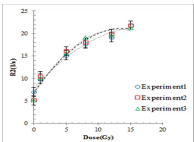
Reproducibility
Figure 2 shows the results obtained from R2 in three sets of the samples. The data shows that the dose response is reproducible over the range
of the measured dose with difference of 4% (r2:
exp1=0.967, exp2=0.927, exp3:0.937).
Energy dependence
In 2igure 3, the in2luence of the energy on dose response is visualized. It can be seen that the dose response of the LD gel is independent of the beam energy to within less than ±2% in
the measured range (r2: Co60= 0.963,
6MV=0.990).
Dose rate dependence
Figure 4 shows the R2 dose response of LD gel as a function of dose for three different dose rates and three different dose levels in LD gel. The results indicate that within the studied ranges, the dose rate did not signi2icantly affect
the response of the LD gel with SD = ±3% (r2:
DR100cGy/min=0.998, DR200cGy/min=0.997,
DR300cGy=0.998).
Figure 1.Linearity of R2 as a func on of absorbed dose up to
Figure 2. Reproducibility of low-density gel dose response in three different experiments with same prepara on, irradia on
and imaging methods.
DISCUSSION
In this study a series of basic dosimetric properties of MAGAT LD gel dosimeter, such as
linearity, sensitivity, dose rate, energy
independence and tissue equivalence were studied.
The relative electron density values obtained for LD gel dosimeter comply well with De Deene et al. results which reported an electron density
of 0.4g/ml (6). While, compared to Haraldsson et
al study (9), the gel density and CT number were
decreased signi2icantly with addition of more polystyrene spheres, still they were higher than the corresponding values for normal human lung tissue, which ranges from −770 to −875
Houns2ield units (12). The gel density could
potentially be further lowered by adding of smaller polystyrene spheres.
Dose-response of LD gel dosimeter,
increased up to 22Gy. It is in accordance with
De Deene et al results (13) but is in contrast with
Haraldsson et al. (9), who reported a linear
dose-response just for doses between 2 to 8Gy. The results of our experiment showed a sharp increase in R2 values from 0 to 2Gy, but
Haraldsson reported inhibition of
polymerization in the low-dose region (≤2Gy)
(9). Although gel preparation, irradiation and
imaging were followed according to Haraldsson study, the differences can be due to different concentration of foam beads or 2itting method
and threshold application in data processing.
Weak echo signals degrade R2 values
signi2icantly (14). In their study, two point 2it
method with no report of thresholding was used for R2 extraction, while, we used many point 2it
method (15) and those echoes with SNR less than
3 were excluded from data analysis (16).
Dose rate may vary within the volume of interest in clinical irradiation pattern and it is expected that a dosimeter be independent of dose rate in the range of the applied dose during dosimetric investigations. In this study no dose rate dependence was observed in different levels of doses for LD MAGAT gel dosimeter,
whereas, Bayreder et al. in 2006 (5) stated , dose
response of MAGAT gel depends on dose rate in medium and high dose region. Bayreder used 2 mM of THPC as antioxidant, while, in Haraldson
et al. study, 94mM of THP was recommended to
remove any probability of oxygen
contamination in LD gel. Sedaghat et al. claimed,
oxygen and antioxidant both act as radical scavengers that affect the amount of polymer formed in the gel and modifying the radiation
dose response of the dosimeter (17). In LD gel,
excess oxygen that is released by styrofoam beads, react with the extra antioxidant and somehow neutralize its inverse effect, but the amount of unreacted antioxidant left in the LD gel dosimeter can have an impact on the polymerization reaction. Although no hard evidence is available at the moment, we
Figure 4. Dose rate dependence of low-density gel for three different dose rates and three different dose levels (2,
5,10Gy). Figure 3. Energy dependence of low-density gel for two
different energies (6MV and 1.25 MeV) and three different doses (2, 5, 10Gy).
hypothesized, the extra concentration of THPC in LD gels, removed the dose rate dependence of MAGAT gel dosimeters, but further investigation is needed to explore the possibility of other sources.
No signi2icant differences were found in dose response to 1.25MeV and 6MV photon beams in this study. Unfortunately there is no available data to compare the results. Looking at previous studies on Mac polymer gel dosimeters, De
Deene et al. (18) found no signi2icant effect of
beam energy on the dose response of nMAG in 25MV.
CONCLUSION
MAGAT LD gel dosimeter appears to be a promising dosimeter in all aspects of dosimetric properties evaluated in this study. In addition, its’ high linearity together with no dose rate dependence in different level of absorbed dose make it a suitable dosimeter to measure 3D-dose distributions insidea non-homogeneous media such as lung tissue.
ACKNOWLEDGMENT
This study was supported by Grant No. 25920 from Tehran University of Medical Sciences. Con lict of interest: Declared none.
REFERENCES
1. Gum F, Scherer J, Bogner L, et al. (2002) Preliminary study on the use of an inhomogeneous anthropomorphic Fricke gel phantom and 3D magne c resonance dosimetry for verifica on of IMRT treatment plans. Phys Med Biol, 47:
N67-77.
2. Ståle Ø, Arne S, Øyvind B, Dag Rune O (2000) Dose distribu on measurements by MRI of a phantom containing lung ssue equivalent compartments made of ferrous sulphate gel. Phys Med Biol, 45: 2761-2770.
3. Olsson LE, Westrin BA, Fransson A, Nordell B (1992) Diffusion of ferric ions in agarose dosimeter gels. Phys Med Biol, 37: 2243-2252.
4. Gholami M, Shahbazi-Gahrouei D, Allahverdi Pourfallah T (2015) Dose response evalua on of a low density anoxic polymer gel dosimeter using MRI. Int. JRR, 13: 243-9. 5. Bayreder C, Georg D Fau - Moser E, Moser E Fau - Berg A,
Berg A (2006) Basic inves ga ons on the performance of a normoxic polymer gel with
tetrakis-hydroxy-methyl-phosphonium chloride as an oxygen scavenger: reproducibility, accuracy, stability, and dose rate dependence. Med Phys, 33: 2506-2518.
6. De Deene Y, Vandecasteele J, Vercauteren T (2013) Low-density polymer gel dosimeters for 3D radia on dosimetry in the thoracic region: A preliminary study. Journal of Physics: Conference Series : IOP Publishing, 012026.
7. Baldock C, De Deene Y, Doran S, et al. (2010) Polymer gel dosimetry. Phys Med Biol, 55: R1-R63.
8. Farajollahi AR, Pak F, Horsfield M, Myabi Z (2014) The basic radia on proper es of the N-isopropylacrylamide based polymer gel dosimeter. Int. JRR, 12: 347-54.
9. Haraldsson P, Karlsson A, Wieslander E, et al. (2006) Dose response evalua on of a low-density normoxic polymer gel dosimeter using MRI. Phys Med Biol, 51: 919 –928. 10. De Deene Y and Baldock C (2002) Op miza on of mul ple
spin–echo sequences for 3D polymer gel dosimetry. Phys Med Biol, 47: 3117.
11. BaLsta JJ, Rider WD, Van Dyk J (1980) Computed tomography for radiotherapy planning. Int J Radiat Oncol BiolPhys, 6: 99-107.
12. Adams H, Bernard MS, McConnochie K (1991) An appraisal of CT pulmonary density mapping in normal subjects. Clinical Radiology, 43: 238-42.
13. De Deene Y, Vergote K, Claeys C, De Wagter C (2006) Three dimensional radia on dosimetry in lung-equivalent regions by use of a radia on sensi ve gel foam: Proof of principle. Medical physics, 33: 2586-97.
14. Watanabe Y and Kubo H (2011) A variable echo-number method for es ma ng R2 in MRI-based polymer gel dosimetry. Medical physics, 38: 975-82.
15. Deene YD, Walle R, Achten E, Wagter CD (1998) Mathema cal analysis and experimental inves ga on of noise in quan ta ve magne c resonance imaging applied in polymer gel dosimetry. Signal Processing, 70: 85-101. 16. De Deene Y (2004) Fundamentals of MRI measurements
for gel dosimetry. Conference Series: IOP Publishing. Journal of Physics, 3: 87-114.
17. Sedaghat M, Bujold R, Lepage M (2011) Severe dose inaccuracies caused by an oxygen-an oxidant imbalance in normoxic polymer gel dosimeters. Phys Med Biol, 56:
601–625.
18. Deene YD, Vergote K, Claeys C, Wagter CD (2006) The fundamental radia on proper es of normoxic polymer gel dosimeters: a comparison between a methacrylic acid based gel and acrylamide based gels. Phys Med Biol, 51:
653-673.