by Alyssa Drews
Senior Honors Thesis Exercise and Sport Science
University of North Carolina at Chapel Hill
2016
Approved by:
Advisor: Brian Pietrosimone Reader: Troy Blackburn
CHAPTER 1: INTRODUCTION………..1
CHAPTER 2: LITERATURE REVIEW…...4
Epidemiology of Anterior Cruciate Ligament Injury………4
Risk Factors for an ACL Injury………...5
Effects of an ACL Injury………...6
Epidemiology of Post-traumatic Osteoarthritis……….9
Walking Speed as a Health Marker……….10
OA and Walking Speed………...11
Changes to Gait due to OA………..13
Walking Speed in Clinical Settings……….14
CHAPTER 3: METHODS………...17
Study Design………17
Participants………...17
Procedures………18
Walking Speed Analysis………..18
Statistical Analysis………...19
CHAPTER 4: RESULTS……….21
Table 1 – Demographics and covariates………..22
at 1 meter………23
Figure 3 – Correlation of ACLR individuals IKDC scores and walking speed at 2 meters..………23
CHAPTER 5: DISCUSSION………24
Main Findings………...24
Clinical Implications……….………26
Limitations………28
Further Research………..……….28
Anterior cruciate ligament (ACL) injury can cause both acute as well as long-term disability. Injury to the ACL has lifelong consequences and higher risk of the development of osteoarthritis. After undergoing an ACL reconstruction (ACLR) an individual has a 1 in 3 chance of developing osteoarthritis (OA) within the first decade after reconstruction, and a 1 in 2 chance of developing OA in the second decade.28 There are changes to gait biomechanics that contribute to abnormal knee loading due to the decreased knee flexion angles, which contribute to the development of OA in ACLR individuals.17 Along with an increased risk of OA,
individuals with ACLR have long-term disabilities that may or may not lead to the development of OA. Individuals with ACLR have decreased quadriceps strength, altered gait patterns, and structural changes to their joint.Error! Reference source not found.The decrease in quadriceps strength and power are contributing factors to long-term disabilities following ACL reconstruction and altered gait patterns decrease the overall function of the joint; along with. There is evidence to suggest that altered biomechanics and joint loading patterns may contribute to a decrease in habitual walking speeds.35
associated with functional limitations and structural breakdown, which are associated with many diseases. Slower habitual walking speeds are associated with increased joint loading during the stance phase of the gait cycle, which has been correlated with increased cartilage breakdown. 35 The cartilage breakdown has been found to be an early indicator of the onset of knee OA. Slow
walkers (≤ 1 m/s) without OA were 1.8 times more likely to develop radiographic signs of OA compared to fast walkers.45 By the time that OA progresses to a level that is considered severe, walking speed diminished to less than 1 m/s and almost 0.4 m/s slower than asymptomatic individuals.Error! Reference source not found. Data on walking speed was collected in over 1,800 adults 45+ years old and an inverse relationship was found between walking speed and prevalence of knee OA.43 Additionally, a link between slower habitual walking speeds and greater collagen breakdown after an ACL reconstruction has recently been reported.35 The breakdown of collagen in the knee is one link to the progression of OA. If this collagen breakdown is associated with slower walking speeds, slower walking speeds may be a predictor of the progression and onset of OA.
Currently no research has been conducted comparing the habitual walking speeds of individuals with ACLR and healthy individuals. Previous research has found a link between slower walking speeds in individuals with ACLR and increased collagen breakdown.35 This research, however, does not compare the walking speeds of the individuals with an ACLR to a health control group. By comparing the healthy control group with the group
ACLR’s IKDC scores and walking speed will allow for the analysis of the relationship between these two data sets.
Since walking speed is one indicator of the onset and progression of OA, it would be important to know if individuals with ACLR have a slower habitual walking speed than the healthy population. One-third of individuals with ACLR develop knee OA within the first decade after reconstruction, meaning that walking speed may have significant clinical implications in the population of individuals with ACLR. Having the ability to differentiate between walking speeds of healthy individuals and individuals with ACLR may be used clinically to distinguish individuals with greater risk of post-traumatic OA development. The purpose of this study is to determine the difference in self-selected walking speed measured in a 3D biomechanics laboratory between healthy individuals and ACLR participant 12 months following ACLR. A secondary aim is to identify the association between walking speed and self-reported function. Therefore, we hypothesize that individuals with ACLR who have slower walking speeds will report a low self-reported function. We also hypothesize that individuals with ACLR will demonstrate slower habitual walking speeds compared to a healthy control group.
CHAPTER 2: LITERATURE REVIEW
Epidemiology of Anterior Cruciate Ligament Injury
Anterior cruciate ligament (ACL) injury is one of the most commonly studied lower extremity injuries in the field of orthopedics. The ACL has been reported as the most common ligamentous injury in the knee.Error! Reference source not found. It is commonly reported that there are around 200,000 ACL injuries per year in the United States.Error! Reference source not found. It was found that in 1994 the number of ACL reconstruction procedures was 86,837 in the United States.Error! Reference source not found. The number of ACL surgeries increased in 2006 to 129,836 ACLR
procedures. This number corresponds to a rate of 43.48 people per 100,000 people undergo ACLR per year.30 The overall incident rate of ACLR per year has been reported between as low as 65,000 and as high as 175,000 per year. It was also found that in 2006, 60.0% of ambulatory ACLRs were performed on males. This number has decreased from 69.8% in 1994. The
number of inpatient ACLRs has also decreased ACLR on males from 71.1% in 1990 to 52.7% in 2007.26
played in these countries. When comparing females to males in the same sport, females have a higher incidence rate of ACL injury than males. This is true for sport exposure but in the overall population females have a lower incidence rate than males. Individuals that wish to return to sport after an ACL injury will have to undergo reconstruction to get back to a level of
competitive play. Even with ACLR there are risks and consequences that are associated with the initial injury.
Risk Factors for an ACL Injury
After an ACL injury, there are temporary and permanent consequences that the individual has to face. In a review of the Hunt Valley II meeting in 2005 several risk factors were identified with being associated with non-contact ACL injury. There are intrinsic (things from within the body) and extrinsic (things outside the body) factors that have an effect on the risk of a non-contact ACL injury. The footwear that a person wears can have both extrinsic and intrinsic factors that can contribute to ACL injury. These factors are due to the shoe-surface interaction and the compensations that the person wearing the shoe makes. Other environmental factors, such as knee braces, have yet to have enough evidence to suggest they have an effect on ACL injury. Some anatomical factors that have been associated with being a risk factor of a non-contact ACL injury are the quadriceps femoris angle, the degree of knee valgus, foot pronation, body mass index, and ACL geometry. 26 It was seen in a study, which followed 205
landing phase than the non-injured knees. It has been demonstrated that the knee valgus has a correlation with the risk of ACL strain.17
Another anatomical factor that has been evaluated post ACL injury is foot pronation. In two different studies by Woodford-Rogers et al46 and Allen and Glasoe1 a small sample of ACL injured athletes was compared to a similar sample of healthy control subjects. It was observed that the ACL injured individuals had a great navicular drop, which suggests a greater subtabular pronation and can contribute to a greater association with ACL injury. It has also been suggested by multiple studies that examined the size of the ACL and notch width that females have a smaller ACL and that there may not be correlation between ACL injury and a smaller notch size. Body mass index has also been inspected in different studies as a risk factor for ACL injury. Increased body mass index has been shown to be a predictive factor in future ACL injury in females.38 There have also been several neuromuscular factors that have been associated as risk factors with ACL injury. These factors include higher quadriceps activation during controlled laboratory studies, and muscle stiffness.14 The changes to muscular activation, biomechanics and structural support of the knee can cause temporary and permanent disability of the knee joint.
Effects of an ACL Injury
After an ACL rupture the individual may be asymptomatic but most people have pain and swelling associated with the injured knee.44 The patient has the option to leave the ACL
strength during flexion of the hamstring muscle was decreased in the operated knee one year after the procedure. It was also observed that this decrease in strength was greater at higher angles of knee flexion and that the non-operated knee did not demonstrate any significant difference in the knee flexion strength at any point post-operation. Another study investigated the quadriceps femoris strength in young athletes at return to sport post-ACLR. The subjects were broken up into high and low quadriceps strength and were compared to a healthy control group. It was observed that both the high and low quadriceps strength groups demonstrated greater limb asymmetry during landing than the healthy control group. It was observed that the high and low quadriceps groups had greater trunk flexion, decreased knee flexion excursion, and decreased knee extension moments in the ACLR limb than the healthy group. All of these findings were strongly associated with a single-leg drop-landing movement.20
In a meta-analysis of knee kinematics and joint moments during gait after an ACLR data was collected from 34 articles that met the selection criteria. It was found that there was
moderate evidence to suggest that individuals had a smaller peak knee flexion angle and smaller knee extension moments in the ACLR knee than the contralateral knee. It was discovered that most biomechanical deficits in the ACLR knee occur in the sagittal plane starting 6 months post reconstruction. The researchers also contribute the higher knee flexion angles observed during gait <6 months after reconstruction to the pain and swelling in the patellofemoral joint.
In a study by Kline et al22 the rate of torque development and knee extensor moment was
evaluated in individuals with a patellar tendon graft reconstructed ACL. It was found that the reconstructed knee had a lower rate of torque development in the quadriceps muscle than the non-reconstructed limb. It was also found that the onset and rate of the knee extensor moment in the ACL reconstructed knee were slower than the healthy knee. The delayed onset and
decreased rate of knee extensor moment has been linked to be a contributing factor to the development and early onset of post-traumatic OA in the reconstructed knee.
In a systematic review that evaluated the prevalence of OA following ACL reconstruction in 38 different studies found that an estimated 12% of the United States population that has knee OA.28 The prevalence of post-traumatic OA in the ACL injured population can be anywhere from 60% to 90%.31 In the sample that was evaluated in the systematic review OA was present
in 44% of patients that underwent an ACL reconstruction and was present in 37% of the ACL deficient patients. It was also discovered that individuals that had an ACL reconstruction were 1.73 times more likely to develop OA than the ACL deficient population. However, people who had a meniscectomy and an ACL reconstruction decreased their rate of knee OA by 7%
compared to people who had a meniscectomy and were ACL deficient. Frobell et al11 reported
Pathomechanics of Post-traumatic Osteoarthritis
Osteoarthritis is a disease that occurs in many individuals and has been associated with age and the degradation of a joint. However, there is a wide range of factors that contribute to the development of OA in each individual. These factors range from biomechanical to
biochemical factors and can be a combination of the two. One factor that has not been determined to cause OA but is a factor that can influence the disease progression is synovial inflammation, which is associated with biological markers that, when present at high levels, are indicative of synovitis. It has also been discovered that the excessive production of cytokines by the inflamed synovium play and important role in the progression of OA.34 Osteoarthritis has also been associated with ACL injury. There is a lot of research about the relationship of an ACLR and the risk of OA. There could be underlying factors during the initial injury of the ACL, such as damage to the subchondral bone or the meniscus that can influence the onset of OA.16 Other factors that cause OA are chondrocyte death, thickening and/or neovascularization of the subchondral bone, fibrosis of the joint capsule, and atrophy and/or fat infiltration of the surrounding muscles.29
after a reconstruction of the ligament. There are many studies28 that demonstrate that about
one-third of individuals with an ACLR develop OA one decade after injury and that risk increases to one-half about two decades later. After the initial tear of the ligament there is an inflammatory response, which, in most individuals, resolves on its own.44 However, the mechanics in gait are a major factor that changes in individuals after an ACL tear and after reconstruction of the ligament. The main changes in gait that are seen are the external rotation of the tibia during stance phase and increased knee flexion at heel strike.Error! Reference source not found. One contribution to this change is the location of the new graft not being in the anatomically correct position. The ACL can never be surgically placed in the same biological position or be made of the same material as the old ACL. The ACL not being in the correct biologically position can lead to external rotation of the tibia during stance phase and increased flexion with heal strike.24 It has
also been found with the use of T1RHO and MRI imaging that there are acute structural changes to
the articular cartilage after an ACL injury.Error! Reference source not found. The minor structural changes, with the biological impacts of inflammation, and the major changes to the joint kinematics all are contributing factors to the progression of OA in individuals with an ACLR. One factor that changes over the course of the disease progression is a change to the individuals walking speed.21
Walking Speed as a Health Marker
were tested for cognitive ability and motor function, taken in the form of walking speed. The participants started the study at age of 85 or younger and were tested every 2 years following their original session. It was seen that poor results on the psychomotor tests and verbal fluency test were associated with slower walking speeds.40
Another study evaluated gait speed in 9 different cohorts of the elderly to assess the association between walking speed and survival. Walking speed is a good measure to predict survival due to its use of energy, motor control, and multiple organ systems through out the body.42 Health status, motor control, mental cognition, cardiovascular health, endurance, habitual physical activity, and musculoskeletal condition are all variables that contribute to individuals self-selected walking speed.10 In a study by Hardy et al15, walking speed was
assessed along with physical health status of 439 participants with a mean age of 74. They were assessed at baseline, 3, 6, 9, and 12 months for 6 different measures. It was demonstrated that the individuals that had improvement in their walking speed over a year period of time had a 17.7% absolute reduction in risk of death over the subsequent 8 years. Furthermore, walking speed is something that can be improved and potentially indicate their overall quality of life, physical function, and decrease their medical interventions.18 As stated above, OA is one of
many impairments that can alter an individuals walking speed. OA and Walking Speed
subjects. Not only did the patients with knee OA walk slower, but they also had a 6° reduced peak knee motion, a reduced knee extension moment, and tried to reduce their knee joint loading when compared to the healthy subjects. The differences in the patients with knee OA and those without could be due to the patient changing their gait in the attempt to reduce their pain.
Assessing all of the changes to gait in people with knee OA can help to identify effective clinical practices to prevent and aid in the pain management of the disease.
A study conducted by Landry et al 25, also examined the effects of mild-to-moderate knee OA on gait compared to healthy individuals. It was found that the individuals with OA walked slower than the control group at both a self-selected speed and during a fast walk. The
biomechanical differences that were observed between the control and OA group were not increased with an increased walking speed. The changes in the knee joint kinematics may be a result of the pathomechanics of OA. Another important finding in a study done by Astephen et alError! Reference source not found., was that slower walking speeds are easily seen when comparing individuals with moderate OA to people with severe OA. The effects of mechanical loading on the articular cartilage of the joint can be found when evaluating the difference in kinetics
between the moderate OA group and the asymptomatic group. The increased mechanical load in the joint with slower walking suggests that clinical intervention in the kinetics of walking should be addressed early on in the progression of the disease.
length was also shorter as severity of OA increased, and the time in the stance phase increased with increasing severity of OA.Error! Reference source not found.The slower walking speed of those with severe OA is due to the amount of load that that the joint can take and the individual
subconsciously trying to decrease the load and pain felt at the joint.
Purser et al36, looked to determine if slower walking speeds was associated with
increased risk of OA. They were able to collect baseline data from 1,858 individuals age 45+ in the form of questionnaires and clinical examinations. These measures were then used during follow up testing. The subjects were divided into three cohorts, one without radiographic OA at baseline, one with our symptomatic OA at baseline, and without radiographic or symptomatic OA at baseline. The subjects were then timed while walking 8 feet for two trials and the the two times were averaged. It was found that there was an inverse relationship between faster walking speed and knee OA. Therefore, slower walking speed is associated with higher incidence of knee radiographic OA. It has been documented that there is increased joint loading during faster walking, which a healthy joint is able to handle but one with cartilage damage may not be able to withstand, thus resulting in slower walking speeds to decrease the load at the joint. In other studies it has been suggested that faster walking speeds can alter the overall biomechanics and neuromuscular control of gait at the knee.
Changes to Gait due to OA
the knee pain group did not walk at a significantly slower speed than the normal group. The knee pain group had lower maximum knee flexion angle during the stance phase and less quadriceps action. The lower knee flexion and less quadriceps activation show that the knee pain group was absorbing shock less effectively and putting more load on their joint during heel strike into the stance phase of the walking cycle. This micro-incoordination of the knee neuromuscular control is referred to as microklutziness and is related to the process and progression of OA. The
reduced firing of the quadriceps muscle that was found and also the hyperextension of the knee are both abnormal gait patterns for people with knee pain.
Andriacchi et al Error! Reference source not found., also investigated the gait patterns of 22 healthy subjects, 11 subjects with knee pathologies. The step length, cadence, force throughout gait cycle, and swing and stance phase characteristics were the main things that were looked at for the different gait patterns. It was observed that with an increase in walking speed there was a decrease in the time spent in the swing and stance phases. When comparing the subjects with knee pathologies to the healthy subjects the subjects with knee pathologies had shorter step-lengths and a higher cadence. Other biomechanical changes observed in other studies were decreased maximum knee flexion during gait and increased knee extension during the peak varus moment in individuals with OA.21 Patients with OA also have a larger adduction magnitude during mid-late stance phase. It was also observed that OA patients were less likely to externally rotate their tibia during early stance phase. Lastly, it was seen that these biomechanical changes were not amplified when individuals with OA walked at faster speeds.25
Walking Speed in Clinical Settings
setting until a study done by Braden et alError! Reference source not found., looked at 46 patients that entered the acute care physical therapy clinic at a hospital. The patients had to be able to walk at least 20 feet on their own and over the age of 60 to be observed for this study. The physical therapists took the walking speed data for their patients and then filled out a questionnaire about how feasible they thought measuring walking speed was and how informative they found it. The physical therapists took an initial walking speed and then a final walking speed during their last session before discharge. The data showed that there was an improvement in the patients’ walking speeds from the beginning of therapy to the end of therapy. The therapists also agreed that walking speed was not a hard test to administer to their patients and that there was valuable information that can be obtained from walking speed. Walking speed can give information on the health status of the patient and when they are ready for discharge. This was a small sample of patients but seeing that there is an improvement in walking speed during acute care can lead to looking at the implications of walking speed in inpatient rehabilitation facilities where the stay of the patient is longer.
A study preformed by Stephens-Lapsley et al41, looked at the effects of a
Walking speed as a functional test can be used in physical therapy settings and is improved after a higher intensity physical therapy program in older adults.
Walking speed is an easy measurable test that can be used in a hospital, an acute care facility, a physical therapy clinic, and other rehabilitation centers. Walking speed is indicative of present and future health status. Individuals with ACLR are at higher risk of developing post-traumatic knee OA. Evidence suggests that individuals with knee OA walk at slower speeds than those without in order to reduce the pain that they experience during gait.21 Walking speed has also been found to predict the incidence of knee OA earlier than other markers.45 Many studies have found the altered gait patterns in individuals with an ACLR and/or knee OA. There is not a study that has looked at the change in walking speed of individuals with an ACLR to evaluate their risk of developing OA post reconstruction. It has been found in a study under review that individuals with an ACLR that walked slower had higher concentrations of collagen type-II cleavage. The higher concentration of collagen type-II cleavage suggests that a slower walking speed may have a link to greater collagen breakdown post ACLR. Although this does not prove that walking speed can predict the formation of OA, it is something that can be further
investigated. It is also known that individuals with an ACLR walk with altered biomechanical gait and loading patterns. The loading patterns of the knee joint during gait have a large
CHAPTER 3: METHODS
Study design
For the primary aim of the study a case-control study design with 21 participants who have undergone an ACL reconstruction 12 months prior to data collection and 24 healthy control subjects with no history of knee injury or impairment. For the secondary aim of the study a cross-sectional observational study was used to assess the relationship between walking speed and self-reported function in the ACL reconstructed subjects.
Participants
We recruited individuals with a primary unilateral ACLR, and healthy individuals without history of knee injury or impairment between the ages of 16-35. All of the ACLR participants had undergone reconstruction a minimum of 12 months prior to data collection and had
physician approval to return to participation in physical activity. We excluded ACLR
Procedures
The current study was part of a larger ongoing project assessing three-dimensional
biomechanical outcomes following ACLR and in healthy control individuals. Demographics, including height, weight, date of birth, age, and gender were all collected form the subject prior to ACLR. All participants were asked to self-report age, and sex, while height and weight were measured in the laboratory prior to testing. ACLR individuals completed the International Knee Documentation Committee Subjective Knee Evaluation Form (IKDC) and the Tegner
Questionnaire. Written informed consent was obtained from all participants before data collection, and the university’s Institutional Review Board approved the study methods and recruitment procedures.
Walking Speed Analysis
All participants were fitted with 25 retroreflective markers (bilateral acromioclavicular joints, sternum, anterior superior iliac spines, posterior superior iliac spines, L4-L5 joint, coccyx, greater trochanters, anterior thighs, medial and lateral femoral epicondyles, anterior shanks, medial and lateral malleoli, top of 1st metacarpals, top of 5th metacarpals, and the calcaneus of both feet). Marker positions were sampled at 120 Hz using a 7-camera three-dimensional motion capture system (Vicon Nexus) and post-processed with Vicon Nexus v1.4.1 motion capture software (Vicon Motion Systems). The motion capture collection area included a six-meter walkway with three embedded force plates (40x60cm, FP406010, Bertec Corporation,
Columbus, Ohio, United States) that were placed in a staggered formation so that the both the right and left limbs could strike a single plate during one trial. Participants wore tight fitting spandex shorts and shirts supplied by the laboratory and performed all gait analysis trials
in the modeling of skeletal segments. Participants performed five practice-walking trials where they were instructed to walk at their normal self-selected speed. These trials were used to ensure that the participants could accurately strike the force plates with out altering their gait and to obtain an average speed for each participant. The walking speed of the gait trails was assessed using two sets of infrared timing gates (TF100, Trac Tronix, Lenexa, Kansas, United States). During data collection the participants were required to walk at a speed +/- 5% of the average of the five practices. The participants performed five acceptable gait trials that required 1) both the right and left foot striking and toeing off of a single force plate, and 2) maintaining a consistent gait speed within the range of the average speed found from the practice trials. Trajectories from the anterior superior iliac spine and sacral cluster markers were low pass filtered at 10 Hz (4th order recurrent Butterworth) and used to estimate the center of mass of the participant. For the purpose of more accurately calculating gait speed for data analysis, we located the point of initial ground contact for the first foot striking the force plate during each gait trial and measured the velocity of the center of mass during a one-meter distance that began 0.5 meters prior to and ended 0.5 meters after initial ground contact. This same procedure was used to measure a two-meter distance walking speed that began 1 two-meter prior to initial ground contact and ended 1 meter after contact. Both values were used to check for consistency in walking speed.
Statistical Analysis
CHAPTER 4: RESULTS
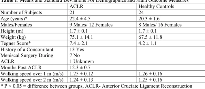
The groups did not differ in age, distribution of sex, height, or weight (Table 1). The ACLR group (22.4± 4.5 years) was older than the control group (20.3± 1.6 years; t43= 2.07,
P=0.044) and had higher Tegner scores (7.4±2.1 vs. 4.2±1.2; t38= 6.152, p<0.001). While not
statistically different, the ACLR group weighed more than the control group (75.1± 14.1 kg vs. 67.5± 11.8 kg; t43= 1.96, P=0.056). The ACLR group had 21 participants with walking speed
data but Tegner and IKDC scores were only collected for 19 of those participants. The control group had 24 participants with walking speeds but only 21 with reported Tegner and IKDC scores.
We found no difference between the groups for walking speed over a 1 (t43 = -0.347, P=
0.73) and 2-meter (t43=-0.233, P=0.816) collection distance (Table 1). After correcting for the
significant covariates (i.e. Tegner Scores and age) there was no difference found between the two groups in walking speed over 1 (F3,39= 0.356 P=0.785) and 2 meters (F3,39= 0.228 P=0.876)
(Figure 1).
There was no association between walking speed and IKDC scores in the ACLR group for both 1 (r=0.182, P=0.456) and 2 meter (r=0.186, P=0.445) distances (Figure 1 and 2). There also was no correlation found between walking speed and IKDC scores when controlling for height (r16=0.182, P= 0.47; r16=0.189, P=0.454), age (r16= 0.190, P=0.451, r16=0.2, P=0.427), and
Table 1: Means and Standard Deviations For Demographics and Main Outcome Measures
ACLR Healthy Controls
Number of Subjects 21 24
Age (years)* 22.4 ± 4.5 20.3 ± 1.6
Males/Females 9 Males/ 12 Females 8 Males/ 16 Females
Height (m) 1.7 ± 0.1 1.7 ± 0.1
Weight (kg) 75.1 ± 14.1 67.5 ± 11.8
Tegner Score* 7.4 ± 2.1 4.2 ± 1.1
History of a Concomitant Meniscal Surgery During ACLR
13 Yes 7 No 1 Unknown Months Post ACLR 12.3 ± 0.7
Walking speed over 1 m (m/s) 1.25 ± 0.12 1.26 ± 0.16 Walking speed over 2 m (m/s) 1.24 ± 0.13 1.25 ± 0.16
* P < 0.05 = difference between groups, ACLR- Anterior Cruciate Ligament Reconstruction
Figure 1. Comparison of average walking speeds between the ACLR and Control group
Figure 2. Association between 1-meter walking speed and IKDC scores in the ACLR participants.
ACLR- Anterior Cruciate Ligament Reconstruction, IKDC- International Knee Documentation Committee Subjective Knee Evaluation Form
Figure 3. Association between 2-meter walking speed and IKDC scores in the ACLR participants.
ACLR- Anterior Cruciate Ligament Reconstruction, IKDC- International Knee Documentation Committee Subjective Knee Evaluation Form 30 40 50 60 70 80 90 100
0.9 1 1.1 1.2 1.3 1.4 1.5
IKDC
2 Meter Walking Speed (m/s)
r= 0.186 P=0.445 30 40 50 60 70 80 90 100
0.9 1 1.1 1.2 1.3 1.4 1.5
IKDC
1 Meter Walking Speed (m/s)
CHAPTER 5: DISCUSSION
The primary aim of the study found that there was no significant difference between the ACLR and healthy group in walking speed at 12 months post-ACLR. The current data suggests that 12 months post-reconstruction may be too early to see changes in walking speed in an ACLR population. In a previous study,4 elderly adults in inpatient rehabilitation demonstrated the ability to increase their walking speed over the course of their acute bout of physical therapy. If a significant change in walking speed was demonstrated after an acute bout of physical
therapy, we can expect that after 6 months of physical therapy that an individual with an ACLR would be able to increase their walking speed. A previous study47 has shown that most
biomechanical deficits that are associated with an ACL injury are considered reversible with an anatomical ACLR and neuromuscular training.
for the variance related to the difference in Tegner score and age between the ACLR and healthy control group, no significant difference was demonstrated between groups for walking speed. Many previously conducted studies9, 18, 20 have evaluated the gait biomechanics of ACLR individuals but no one has evaluated the relationship between walking speed and self-reported function.
There was no significant association between IKDC scores and walking speed in this ACLR cohort. A lower IKDC score indicates more disability, meaning that ACLR individuals with a lower IKDC score are self-reporting more disability. Although there were no associations found between IKDC and walking speed in the ACLR group 12 months post-reconstruction, there could be an association between these two outcomes at a later time point following ACLR. A recent systematic review estimated that around one-third of individuals with an ACLR develop PTOA within the first decade following injury.28 It is possible that associations between gait speed and self-reported function may be apparent closer to PTOA onset or after PTOA has already developed. Following an ACLR, there have been associations found with changes in the structure, tissue metabolism, and biomechanics of the knee.7 A recent study found that there was an association between increased type II collagen metabolism and a slower habitual walking speed post ACLR.35 While the results of this study do not directly link walking speed with the development of PTOA, it is known that collagen breakdown can lead to the deterioration of the cartilage matrix.35 In the aforementioned study32, participants reported a higher IKDC score (85%) compared to our current study (82%) and no association was found between IKDC score and walking speed in either study.35
post-injury level of function at one year post-reconstruction. In a previous study23 it was found that
individuals with an ACLR returned to their pre-injury activity level 3 years after reconstruction. This study23 found that Tegner scores decreased from a score of 7 at 3-year post reconstruction follow-up exams to a score of 4 at 15-year post reconstruction follow-up exams. This shows that the participants in the current study may have reached their highest level of activity or are still trying to get to their highest level of activity 12 months after ACLR. The IKDC Subjective Knee Evaluation measurement tool provides a general measure of participant functioning and does not specifically focus on an individual’s walking ability. The main points of the IKDC have to do with the pain, swelling, and giving way that the individual experiences along with the highest level of activity that they think they can preform. There is not a question that pertains to the individual’s ability to walk about during their daily life. Without questions pertaining
specifically to habitual walking it may be more difficult to determine an association between the IKDC score and an individual’s walking speed. A previous study13 has found an association between those that report higher knee functioning on the Global Rating Scale (GRS) and
increased limb contact forces during gait on the ACLR knee. It has also been demonstrated that the self-reported function on the Knee Outcome Survey and Actives of Daily Living Scale (KOS-ADLS) of individuals with an ACLR is related to their knee flexion angles during gait.27 Further research should evaluate the association between walking speed and disability using other self-reported knee function questionnaires.
have a lower self-reported function could be reporting this due to the lack of confidence that they have in their everyday ability to preform normal tasks. This lack of confidence in physical function as it relates to the knee has been found to be troubling to 54% of people with or at high risk of knee OA.8 Having a lack of confidence in one’s ability to do normal tasks with their knee can contribute to a slower habitual walking speed.39 Walking speed has been referred to as “the 6th vital sign.” 10,32 Walking speed is able to predict present and future health status in many
different populations.10,32 Slower walking speed is associated with poor results on psychomotor tests and verbal fluency tests. 40 Walking speed has also been found to be a predictor of physical function and quality of life in older adults.18 Walking speed has been found to only decline at a rate of 1-2% per decade before the age of 62; after the age of 62 there is a sharp decline in walking speed.19
Not only do individuals that have knee OA walk slower than the healthy population, individuals with severe OA also have slower walking speeds than individuals with moderate knee OA.25 A link between severity of OA and the pain felt while doing activities of daily living (ADL) has been reported, as the more severe the OA the more pain and individual felt while doing ADLs.13 These findings show the ability of walking speed to predict the severity of OA in different individuals.25 Since walking speed is able to predict the severity of OA and presence of
OA in individuals, it is important to know the walking speed of ACLR individuals. The ACLR population is at a higher risk of developing knee OA compared to the healthy population.25 We may not be able to predict disability in ACLR individuals using their walking speed, but we may be able to predict OA development in the ACLR population before they have changes in their self-reported function. In an aforementioned study,35 it was found that slower habitual walking
speed in individuals with ACLR is associated with higher levels of C2C concentration in the knee, indicating increased type II collagen breakdown.35 Individuals with an ACLR, a slower habitual walking speed, and greater knee tissue damage should be monitored by clinicians to look for an even greater decline in walking speed over time.
walking speed at one year, there may be metabolic markers (i.e. C2C) that are associated with OA development. Further research should be conducted at different time points post-ACLR comparing habitual walking speed in ACLR to healthy controls to identify a time point when a difference in walking speed is present. The correlation between walking speed and IKDC scores should be analyzed at these later time points to see if there is also a decline in self-reported function. The identification of a time point when there is a decline in self-reported function and walking speed could help in potentially identifying the development of OA in individuals with an ACLR.
determine the effects of certain covariates (i.e. height, age, etc.) and demographics on the relationship between walking speed and self-reported function.
In conclusion, there was no difference found between the walking speed of the ACLR individuals and the healthy controls. Additionally, individuals with an ACLR who walked slower did not have more disability at one year post-reconstruction. Although there was no correlation between IKDC scores and walking speed in ACLR individuals, this population is already at greater risk of developing OA.28 Therefore, the ability to determine if there is a time point post-ACLR that an individual’s walking speed starts to decline may be helpful in
REFERENCES
1. Allen, M. K., & Glasoe, W. M. (2000). Metrecom Measurement of Navicular Drop in Subjects with Anterior Cruciate Ligament Injury. Journal of Athletic Training , 35 (4), 403-406.
2. Andriacchi, T. P., Ogle, J., & Galante, J. (1977). Walking speed as a basis for normal and abnormal gait measurements. Journal of Biomechnics , 10 (4), 261-268.
3. Astephen, J. L., Deluzio, K. J., Caldwell, G. E., Dunbar, M. J., & Hubley-Kozey, C. L. (2008). Gait and neuromuscular pattern changes are associated with differences in knee osteoarthritis severity levels. Journal of Biomechanics , 41 (4), 868-876.
4. Braden , H. J., Hilgenberg, S., Bohannon, R. W., Ko, M.-S., & Hasson, S. (2012). Gait Speed Is Limited but Improves Over the Course of Acute Care Physical Therapy. Journal of Geriatric Physical Therapy , 35 (3), 140-144.
5. Brophy, R. H., Wright, R. W., & Matava, M. J. (2009). Cost Analysis of Converting From Single-Bundle to Double-Bundle Anterior Cruciate Ligament Reconstruction. The American Journal of Sports Medicine , 37 (4), 683-687.
6. Buller, L. T., Best, M. J., Baraga, M. G., & Kaplan, L. D. (2015). Trends in Anterior Cruciate Ligament Reconstruction in the United States. Orthopaedic Journal of Sports Medicine , 3 (1), 1-8.
7. Chu, C. R., & Andriacchi, T. P. (2015). The Dance Between Biology, Mechanics, and Structure: A Systems Based Approach to Developing Osteoarthritis Prevention Strategies. Journal of Orthopaedic Research , 33 (7), 939-947.
8. Colbert C.J., Song J., Dunlop D., Chmiel J.S., Hayes K.W., Cahue S., et al. (2012), Knee confidence as it relates to physical function outcome in persons with or at high risk of knee osteoarthritis in the Osteoarthritis Initiative. Arthritis Rheum, 64,1437-46.
9. Emami Meybodi, M., Jannesari, M., Rahim Nia, A., Yaribeygi, H., Firoozabad, V., & Dorostegan, A. (2013). Knee flexion strength before and after ACL reconstruction using hamstring tendon autografts. Trauma Monthly , 18 (3), 130-133.
10. Fritz, S., & Lusardi, M. (2009). White Paper : “ Walking Speed : the Sixth Vital Sign ”. Journal of Geriatric Physical Therapy , 32 (1), 3-6.
11. Frobell, R. B., Roos, H. P., Roos, E. M., Roemer, F. W., Ranstam, J., & Lohmander, L. (2013). Treatment for acute anterior cruciate ligament tear: five year outcome of randomised trial. BMJ (Clinical research ed.) , 346, 232.
12. Fukutani, N., Iijima, H., Aoyama, T., Yamamoto, Y., Hiraoka, M., Miyanobu, K., Matsuda, S. (2016). Knee pain during activities of daily living and its relationship with physical activity in patients with early and severe knee osteoarthritis.
13. Gardinier E., Manal K., Buchanan T., Snyder-Mackler L. (2014), Clinically-relevant measures associated with altered contact forces in patients with anterior cruciate ligament deficiency. Clinical Biomechanics, 29 (5), 531-536.
15. Hardy, S. E., Perera, S., Roumani, Y. F., Chandler, J. M., & Studenski, S. A. (2007). Improvement in usual gait speed predicts better survival in older adults. Journal of the American Geriatrics Society , 55 (11), 1727-1734.
16. Hart, A. J., Buscombe, J., Malone, A., & Dowd, G. (2005). Assessment of osteoarthritis after reconstruction of the anterior cruciate ligament: a study using single-photon emission computed tomography at ten years. The Journal of Bone and Joint Surgery. British volume , 87 (11), 1483-1487.
17. Hart, H. F., Culvenor, A. G., Collins, N. J., Ackland, D. C., Cowan, S. M., Machotka, Z., et al. (2015). Knee kinematics and joint moments during gait following anterior cruciate ligament reconstruction: a systematic review and meta-analysis. British Journal of Sports Medicine .
18. Hewett, T. E., Myer, G. D., Ford, K. R., Heidt, R. S., Colosimo, A. J., McLean, S. G., et al. (2005). Biomechanical measures of neuromuscular control and valgus loading of the knee predict anterior cruciate ligament injury risk in female athletes: a prospective study. The American Journal of Sports Medicine , 33 (4), 492-501.
19. Himann, J. E. J. (1988). Medicine and science in sports and exercise: Age-related changes in speed of walking.American College of Sports Medicine. 20(2), 161-166.
20. Ithurburn, M., Paterno, M., Ford, K., Hewett , T., & Schmitt, L. (2015). Young Athletes With Quadriceps Femoris Strength Asymmetry at Return to Sport After Anterior Cruciate Ligament Reconstruction Demonstrate Asymmetric Single-Leg Drop-Landing Mechanics. The American Journal of Sports Medicine .
21. Kaufman, K., Hughes, C., Morrey, B. F., Morrey, M., & An, K. N. (2001). Gait characteristics of patients with knee osteoarthritis. Journal of biomechanics , 34 (7), 907-915.
22. Kline, P. W., Morgan, K. D., Johnson, D. L., Ireland, M. L., & Noehren, B. (2015). Impaired Quadriceps Rate of Torque Development and Knee Mechanics After Anterior Cruciate Ligament Reconstruction With Patellar Tendon Autograft. The American Journal of Sports Medicine .
23. Kostogiannis, I., Ageberg, E., Neuman, P., Dahlberg, L., Friden, T., & Roos, H. (2007, July). Activity level and subjective knee function 15 years after anterior cruciate ligament injury: a prospective, longitudinal study of nonreconstructed patients. The American
Journal of Sports Medicine, 35(7), 1135+.
24. Krutsch, W., Zellner, J., Baumann, F., Pfeifer, C., Nerlich, M., & Angele, P. (2015). Timing of anterior cruciate ligament reconstruction within the first year after trauma and its influence on treatment of cartilage and meniscus pathology. Knee surgery, sports traumatology, arthroscopy : official journal of the ESSKA , 1-8.
25. Landry, S. C., McKean, K. A., Hubley-Kozey, C. L., Stanish, W. D., & Deluzio, K. J. (2007). Knee biomechanics of moderate OA patients measured during gait at a self-selected and fast walking speed. Journal of Biomechanics , 40 (8), 1754-61.
26. Leathers, M. P., Merz, A., Wong, J., Scott, T., Wang, J. C., & Hame, S. L. (2015). Trends and Demographics in Anterior Cruciate Ligament Reconstruction in the United States. Journal of Knee Surgery , 28 (5), 390-394.
28. Luc, B., Gribble, P. A., & Pietrosimone, B. G. (2014). Osteoarthritis Prevalence Following Anterior Cruciate Ligament Reconstruction: A Systematic Review and Numbers-Needed-to-Treat Analysis. Journal of Athletic Training , 49 (6), 806-819.
29. Malfait, A.-M., & Little, C. B. (2015). On the predictive utility of animal models of osteoarthritis. Arthritis Research & Therapy , 17 (1), 225.
30. Mall, N. A., Chalmers, P. N., Moric, M., Tanaka, M. J., Cole, B. J., Bach, B. R., et al. (2014). Incidence and trends of anterior cruciate ligament reconstruction in the United States. The American Journal of Sports Medicine , 42 (10), 2363-2370.
31. Meunier, a. O., Meunier, A., Odensten, M., & Good, L. (2007). Long-term results after primary repair or non-surgical treatment of anterior cruciate ligament rupture: A randomized study with a 15-year follow-up. Scandinavian Journal of Medicine and Science in Sports , 17 (3), 230-237.
32. Middleton, A., Fritz, S. L., & Lusardi, M. (2015). Walking speed: the functional vital sign. Journal of Aging and Physical Activity , 23 (2), 314-322.
33. Moses, B., Orchard, J., & Orchard, J. (2012). Systematic review: Annual incidence of ACL injury and surgery in various populations. Research in Sports Medicine , 20 (3-4), 157-179. 34. Pelletier, J. P., Martel-Pelletier, J., & Abramson, S. B. (2001). Osteoarthritis, an
inflammatory disease: potential implication for the selection of new therapeutic targets. Arthritis and Rheumatism , 44 (6), 1237-1247.
35. Pietrosimone, B. G., Blackburn, J., Harkey , M., Luc, B., Hackney, A., Padua, D., et al. (n.d.). Walking Speed as a Potential Indicator of Cartilage Breakdown Following Anterior Cruciate Ligament Reconstruction. Arthritis Care and Research .
36. Purser, J. L., Golightly, Y. M., Feng, Q., Helmick, C. G., Renner, J. B., & Jordan, J. M. (2012). Association of slower walking speed with incident knee osteoarthritis- related outcomes. Arthritis Car and Research , 64 (7), 1028-1035.
37. Radin, E. L., Yang, K. H., Riegger, C., Kish, V. L., & O'Connor, J. J. (1991). Relationship between lower limb dynamics and knee joint pain. Journal of Orthopaedic Research , 9 (3), 398-405.
38. Shultz, S. J., Schmitz, R. J., Benjaminse, A., Collins, M., Ford, K., & Kulas, A. S. (2015). ACL Research Retreat VII: An Update on Anterior Cruciate Ligament Injury Risk Factor Identification, Screening, and Prevention. Journal of Athletic Training In-Press .
39. Skou, S. T., Wrigley, T. V., Metcalf, B. R., Hinman, R. S. and Bennell, K. L. (2014), Association of Knee Confidence With Pain, Knee Instability, Muscle Strength, and Dynamic Varus–Valgus Joint Motion in Knee Osteoarthritis. Arthritis Care Res, 66, 695– 701.
40. Soumaré, A., Tavernier, B., Alpérovitch, A., Tzourio, C., & Elbaz, A. (2009). A cross-sectional and longitudinal study of the relationship between walking speed and cognitive function in community-dwelling elderly people. The journals of gerontology. Series A, Biological sciences and medical sciences , 64 (10), 1058-1065.
41. Stevens-Lapsley, J. E., Loyd, B. J., Falvey, J. R., Figiel, G. J., Kittelson, A. J., Cumbler, E. U., et al. (2015). Progressive multi-component home-based physical therapy for deconditioned older adults following acute hospitalization: A pilot randomized controlled trial. Clinical Rehabilitation .
43. Teixeira-Salmela, L. F., Nadeau, S., Milot, M. H., Gravel, D., & Requião, L. F. (2008). Effects of cadence on energy generation and absorption at lower extremity joints during gait. Clinical Biomechanics , 23 (6), 769-778.
44. Wasilko, S.M., Tourville, T.W., DeSarno, M.J., Slauterbeck, J.R., Johnson, R.J., Struglics, A., Beynnon, B.D., (2015). The relationship between synovial fluid biomarkers of articular cartilage metabolism and the patient's perspective of outcome depends on the severity of articular cartilage damage following ACL trauma. Journal of Orthopaedic Research
45. White, D. K., Zhang, Y., Niu, J., Keysor, J. J., Nevitt, M. C., Lewis, C. E., et al. (2010). Do worsening knee radiographs mean greater chances of severe functional limitation? Arthritis Care and Research , 62 (10), 1433-1439.
46. Woodford-Rogers, B. (1994). Risk factors for anterior cruciate ligament injury in high school and college athletes. Journal of Athletic Training , 29 (4), 343-346.
47. Yagi M., Kuroda R., Nagamune K., et al. (2007), Double-bundle ACL reconstruction can improve rotational stability. Clin Orthop Relat Res, 454, 100-107.
48. Zhang, Y., & Jordan, J. M. (2010). Epidemiology of Osteoarthritis. Clinics in Geriatric Medicine, 26(3), 355–369.