1National Institute of Mental Health Psychoactive Drug Screening Program, Department of Pharmacology and Division of Chemical Biology and Medicinal Chemistry, University of North Carolina Chapel Hill Medical School, Chapel Hill, NC, USA. 2Center for Chemical Biology and Drug Discovery, Department of Pharmacological Sciences and Department of Oncological Sciences, Tisch Cancer Institute, Icahn School of Medicine at Mount Sinai, New York, NY, USA. 3Present address: Department of Cell Biology, Neurobiology and Anatomy, Medical College of Wisconsin, Milwaukee, WI, USA. 4Present address: Department of Pharmacological Sciences and Department of Neuroscience, Icahn School of Medicine at Mount Sinai, New York, NY, USA. 5Present address: State Key Laboratory of Molecular Biology, Institute of Biochemistry and Cell Biology, Shanghai Institutes for Biological Sciences, Chinese Academy of Sciences, Shanghai, China. 6These authors contributed equally: John D. McCorvy, Daniel Wacker, Sheng Wang. *e-mail: [email protected]; [email protected]
T
he human genome encodes 13 distinct 5-HTG-protein-coupled receptors (GPCRs). Drugs targeting 5-HT GPCRs are approved treatments for a diverse array of indications, includ-ing obesity, migraine headaches, schizophrenia, anxiety and depres-sion1,2. 5-HT receptors also frequently mediate serious drug side
effects via unanticipated ‘off-target’ actions1,3. A notable example is
the now-banned appetite suppressant fenfluramine, which exerts its potent anti-obesity actions by activating 5-HT2C receptors4. Fenfluramine was ultimately withdrawn from the market because of a high incidence of drug-induced valvular heart disease (VHD), which occurs as a result of off-target activation by fenfluramine and its active metabolite norfenfluramine at the closely related 5-HT2B receptor (5-HT2BR)5,6.
Several other medications, including the anti-migraine drugs methysergide and ergotamine5, the anti-parkinsonian medications
pergolide and cabergoline7, and drugs that treat pituitary adenomas,
also have potent off-target actions at 5-HT2BR, and they have also been withdrawn or their use severely restricted because of drug-induced VHD1,5. Of note, both the VHD and fibrosis associated
with carcinoid syndrome have been linked to 5-HT2BR activation8. Consequently, candidate medications are routinely screened for 5-HT2B agonist activity before progressing to clinical trials9,10. Not unexpectedly, 5-HT2B antagonists have been proposed as poten-tial therapeutics for VHD and other fibrotic disorders, including carcinoid syndrome11. Thus, understanding the action of drugs at
5-HT2BR is clearly important for future drug development. 5-HT2BR is a member of the 5-HT2 subfamily of 5-HT receptors, which also includes 5-HT2A and 5-HT2C receptors. 5-HT2BR activa-tion via Gq/11 induces phospholipase C activation, inositol phosphate
(IP) accumulation, intracellular calcium release and protein kinase C activation1,2. 5-HT
2BR also recruits β -arrestin2 (also known as arrestin-3; encoded by ARRB2 in humans) and downstream effector activation9,12,13. Drugs such as lysergic acid diethylamide (LSD) and
ergotamine (ERG) prefer arrestin recruitment and are considered ‘arrestin-biased agonists’9,12,13.
Over the past few years, there has been an explosion in avail-able GPCR structural information, which has provided a molecular understanding of ligand recognition14, receptor dynamics and
acti-vation15, and ligand-mediated biased signaling16. To date, structures
of three 5-HT receptors have been determined by X-ray crystal-lography, those of the 5-HT1B17,18, 5-HT2B12, and 5-HT2C19 receptors, all in complex with the VHD-inducing anti-migraine drug ERG. For the 5-HT2B receptor, LSD-bound and ERG-bound structures are available and reveal that ergot ligands engage a presumed orthosteric binding pocket (OBP), which is likely shared with the endogenous ligand 5-HT12,13,17. Indeed, ERG and LSD engage
regions outside this OBP, which we have termed the extended binding pocket (EBP). The OBP of 5-HT receptors shares certain features with the OBP exemplified by the β2-adrenergic receptor (β2AR)20,21, and it includes highly conserved and critical ligand tacts between the amine nitrogen of the ligands and a highly con-served aspartate in transmembrane domain 3 (TM3; for example, Asp3.32 in the Ballesteros–Weinstein numbering scheme22), as well as
polar and aromatic contacts in TM5 and TM6, respectively. These interactions are thought to facilitate the stabilization of active23 and
G-protein-bound24 conformational states.
Little is known, however, regarding the 5-HT receptor EBP, which encompasses extracellular portions of TM3 and TM7 and has been
Structural determinants of 5-HT
2B
receptor
activation and biased agonism
John D. McCorvy
1,3,6*, Daniel Wacker
1,4,6, Sheng Wang
1,5,6, Bemnat Agegnehu
1, Jing Liu
2,
Katherine Lansu
1, Alexandra R. Tribo
1, Reid H. J. Olsen
1, Tao Che
1, Jian Jin
2and Bryan L. Roth
1*
Serotonin (5-hydroxytryptamine; 5-HT) receptors modulate a variety of physiological processes ranging from perception, cognition and emotion to vascular and smooth muscle contraction, platelet aggregation, gastrointestinal function and repro-duction. Drugs that interact with 5-HT receptors effectively treat diseases as diverse as migraine headaches, depression and
obesity. Here we present four structures of a prototypical serotonin receptor—the human 5-HT2B receptor—in complex with
chemically and pharmacologically diverse drugs, including methysergide, methylergonovine, lisuride and LY266097. A detailed analysis of these structures complemented by comprehensive interrogation of signaling illuminated key structural determi-nants essential for activation. Additional structure-guided mutagenesis experiments revealed binding pocket residues that
were essential for agonist-mediated biased signaling and β -arrestin2 translocation. Given the importance of 5-HT receptors for
proposed as a potential structural feature that may facilitate biased signaling2. Support for this hypothesis comes from analysis of the
structure of LSD bound to 5-HT2BR, in which LSD stereo-selectively engages TM3 and TM7 to evoke potent β -arrestin2 recruitment13.
Additional insights into the mechanisms of biased agonism have recently emerged, revealing a key ligand interaction between LSD and Leu209 in extracellular loop 2 (EL2), which increases ligand residence time at the receptor that contributes to enhanced time-dependent β -arrestin2 recruitment13. Additionally, both TM5 and
the EL2 regions have recently been exploited for biased ligand design at aminergic GPCRs25; it is unclear how biased activation
occurs via contact with these regions of the receptor.
A clearer understanding of how ligand interactions with key binding pocket residues lead to the stabilization of active or inac-tive states will facilitate the design of agonist, biased agonist and antagonist drugs. The available structures are with a single ligand type (either agonist or antagonist). In instances where agonist and antagonist structures are available, the ligand chemotypes are struc-turally diverse (for example, β2AR, adenosine 2A, the μ and κ opi-oid receptors, and others16,26). Such limitations make it difficult to
leverage existing GPCR structural information for structure-guided drug design.
Here we identified key residues responsible for 5-HT2BR activa-tion by comparing the binding modes of chemically similar ago-nists and antagoago-nists at 5-HT2BR. Notably, we were able to compare the binding mode of methylergonovine with that of its parent anti-migraine drug, methysergide, which differs by a single methyl sub-stituent. To illuminate antagonism, we compared the binding mode of the antagonist lisuride to that of the agonist LSD, which differs by only stereochemistry and an additional nitrogen atom. Finally, we elucidate biased signaling and subtype selectivity by clarifying the binding mode of the selective 5-HT2BR antagonist LY266097. These insights should accelerate the design of safer and more effec-tive medications.
Results
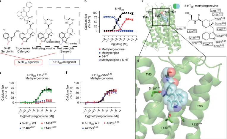
Structural insights into a 5-HT2B activation mechanism. Methysergide is rapidly N-demethylated in vivo to methylergono-vine, which is the major active metabolite that mediates methyser-gide’s anti-migraine actions in vivo27. Unlike methysergide, which is
a 5-HT2BR antagonist, methylergonovine is a potent 5-HT2BR ago-nist6 responsible for methysergide-induced VHD5. Because
methy-sergide differs from methylergonovine by a single ‘–CH3’ moiety (Fig. 1a), the pair represents a key ligand set to gain insight into the ligand-based structural features responsible for 5-HT2BR effi-cacy. Other unsubstituted N(1)-H ligands, such as methylergono-vine, LSD and ERG, are Gq partial agonists (Fig. 1b). By contrast, the
N(1)-methyl or alkyl ergoline ligands methysergide (Fig. 1b) and LY215840 (Supplementary Fig. 1a) are 5-HT2BR antagonists.
To identify residues critical for methylergonovine’s agonism, we obtained the crystal structure of the 5-HT2B R–methylergo-novine complex at a resolution of 2.9 Å (Fig. 1c and Table 1). Methylergonovine forms a salt bridge with Asp1353.32 in the pre-sumed orthosteric site, and the ergoline ring system forms an edge-to-face π -π stack with residues Phe3406.51 and Phe3416.52 in TM6—interactions that are commonly observed in aminergic20,23
and 5-HT13,17,19 structures. The binding mode of
methylergono-vine, when compared with those of LSD and ergotamine, bound to 5-HT2BR revealed a subtly different positioning of the indole
N(1)-H toward TM5 residues, with ERG being the deepest toward Ala2255.46 and LSD being the shallowest toward the backbone of Gly2215.42 (Supplementary Fig. 1b). This differential positioning is likely caused by a rotation around the axis of the ionic interac-tion between Asp3.32 and the protonated ergoline amine group13.
Similar to ERG, N(1)-H of methylergonovine points toward resi-dues Ala2255.46 and Thr1403.37 (Fig. 1d and Supplementary Fig. 1c).
Although the density of N(1)-H of methylergonovine was not resolved at 2.9-Å resolution, this hydrogen likely resides between residues Ala2255.46 and Thr1403.37.
To determine whether residues Thr1403.37 and Ala2255.46 were involved in methylergonovine’s agonism, we mutated the sequences encoding residues Thr1403.37 and Ala2255.46. Thr140Ala3.37 and Thr140Val3.37 substitutions substantially diminished methylergo-novine’s agonism (Fig. 1e) and binding affinity (Supplementary Table 1), despite similar surface expression levels relative to the wild-type receptor (Supplementary Fig. 1d). Although there was not an optimal angle for a hydrogen bonding between N(1)-H and residue Thr1403.37 in the 5-HT
2BR–methylergonovine structure (Supplementary Fig. 1e), the close distance suggests that at least an electrostatic interaction (van der Waals, vdW) between residue Thr1403.37 and N(1)-H may occur during activation. Indeed, 5-HT, which also contains an N(1)-H on the indole, displayed weak Gq activity at Thr140Ala3.37 and Thr140Val3.37 mutants (Supplementary Fig. 1f), indicating that Thr1403.37 is essential for receptor activa-tion. To recapitulate a favorable electrostatic interaction, we created Thr140Ser3.37 mutant receptor and found that methylergonovine’s Gq agonism (Fig. 1e) and binding affinity (Supplementary Table 1) were spared. By contrast, the substitutions Ala225Ser5.46 and Ala225Gly5.46 did not substantially affect methylergonovine’s G
q agonism (Fig. 1f) or binding affinity (Supplementary Table 1), and they spared 5-HT Gq activity (Supplementary Fig. 1g). Thus, methy-lergonovine’s interaction with Thr1403.37 is critical for 5-HT
2BR acti-vation.
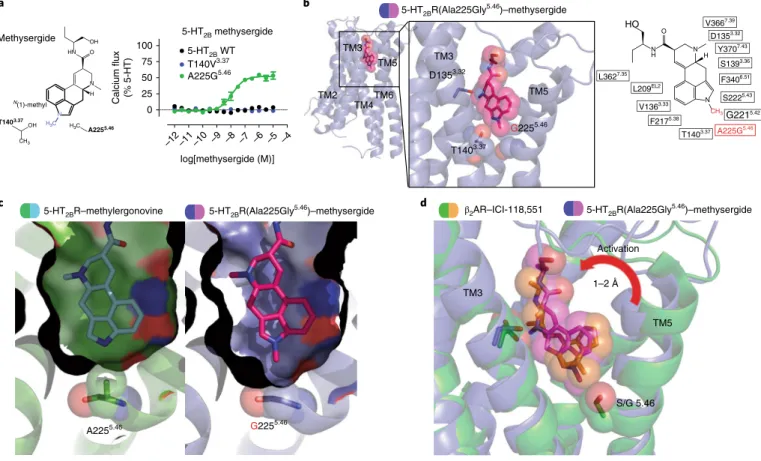
Structure of a 5-HT2BR Ala225Gly5.46 mutant designed to be activated by methysergide. We next sought to understand how the N(1)-methyl of methysergide causes 5-HT2BR antagonism. We hypothesized that methysergide’s N(1)-methyl would either lack interaction with Thr1403.37 and/or cause a steric clash with Ala2255.46. Methysergide failed to demonstrate any measurable Gq agonist activity with a Thr140Val3.37 mutant, where it was expected that the valine and N(1)-methyl could form additional vdW interactions to lead to activation (Fig. 2a). By contrast, methysergide demon-strated potent Gq partial agonist activity (Fig. 2a and Supplementary Fig. 2a) with the Ala225Gly5.46 mutant, which we hypothesized to introduce more bulk tolerance for methysergide’s N(1)-methyl—a notion consistent with TM5 engagement appearing to be important for G-protein-dependent agonism25.
To test this hypothesis, we obtained a structure for the 5-HT2BR(Ala225Gly5.46)–methysergide complex at 3.1-Å resolution (Fig. 2b, Table 1 and Supplementary Fig. 2b,c). Similar to what was seen in the structure of the 5-HT2BR–methylergonvine complex, methysergide made conserved contacts with Asp1353.32, Phe3406.51 and Phe3416.52. Of note, methysergide’s N(1)-methyl was pointed down into the space at the mutant Gly2255.46 residue. As predicted, removal of a methyl substituent from Ala2255.46 to form Gly2255.46 created ‘space’ (for example, bulk tolerance) for the methysergide’s N(1)-methyl at TM5 (Fig. 2c).
To obtain further insights into methysergide’s engagement at TM5, we compared the 5-HT2BR(Ala225Gly5.46)–methysergide structure to that of inactive-state β2AR bound to the antagonist ICI-118,55128. ICI-118,551, like methysergide, also contains a methyl
TM5 movement via the methyl substituent (Supplementary Fig. 2d). Our results thus reveal that residues Thr1403.37 and Ala2255.46 are essential for activation.
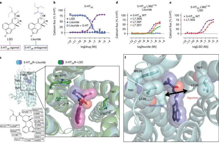
Structural basis for a 5-HT2B activation mechanism via the extended binding pocket. We next focused on the role of the EBP in receptor activation. We have previously shown that LSD’s dieth-ylamide, which is key for LSD’s potent hallucinogenic effects29,
con-tacts TM3 and TM7 within the EBP13. Furthermore, we found that
recognition of LSD in this region is stereo-selective, as LSD’s potent agonism was recapitulated by only the (S,S)-azetidide stereoisomer, a conformationally restricted diethylamide stereoisomer of LSD13.
Less clear, however, is the effect of opposing stereochemistry at the C8 position in antagonist versus agonist recognition. Here we sought to examine other ergoline ligands, such as lisuride, which has the same ergoline core scaffold as LSD yet possesses an (S )-diethylurea (Fig. 3a) and is a potent 5-HT2BR antagonist30 (Fig. 3b).
To identify the structural basis for lisuride’s antagonism, we solved the structure of the 5-HT2BR–lisuride complex to a resolution of 3.1 Å (Fig. 3c, Table 1 and Supplementary Fig. 3a,b). Alignment of the 5-HT2BR–LSD and 5-HT2BR–lisuride structures showed similar binding poses in the OBP, where the indole N(1)-H of both LSD and lisuride formed a hydrogen bond with the carbonyl backbone
at residue Gly2215.42 in TM5 (Supplementary Fig. 3c). Comparison of these poses at the OBP could not entirely explain the difference in pharmacological activity between LSD and lisuride. By contrast, comparison of the EBP poses revealed that the (S)-diethylurea of lisuride was exclusively wedged between residues Trp1313.28 and Leu1323.29 in a hydrophobic stack, making minimal contact with TM7 residues. This resulted in 1.2 Å less contraction of the binding pocket between residues Trp1313.28 and Leu3627.35 as compared to the distance in the LSD-bound structure. By contrast, LSD’s binding pose, in which the diethylamide contacts residue Leu3627.35 in TM7 (Fig. 3c), exhibited a more contracted binding pocket. This unex-pected difference in binding pose likely explains lisuride’s lack of agonism at 5-HT2BR, whereas contact with TM7 residue Leu3627.35 in the EBP appears essential for LSD’s agonism.
To test the hypothesis that ligand contact with TM7 in the EBP facilitates 5-HT2BR activation, we made substitutions at residue Leu3627.35, which is the closest residue in proximity to lisuride’s diethylurea in TM7, to facilitate an interaction between lisuride and TM7. Consistent with our hypothesis, substitution of Leu3627.35 with asparagine (Leu362Asn7.35), which we predicted would result in formation of a hydrogen bond with the backbone of lisuride’s diethylurea moiety, led to Gq partial agonism (half-maximal effec-tive concentration (EC50) = 395 nM, Emax= 41%; Fig. 3d). We also
N
H N O HN
OH
H N
NH2
H
HO
5-HT
Serotonin Methylergonovine N
H N O HN
OH
H3C
Metabolized N(1)-methyl
HN O N O
N
HO O
N
H N O
H
Methysergide (Sansert) Ergotamine
(Cafergot)
a
5-HT2B
–12 –11 –10 –9 –8 –7 –6 –5 –4
0 25 50 75 100
Methylergonovine Methysergide
log [drug (M)]
Calcium flux (% 5-HT)
Methysergide + 5-HT 5-HT
b
5-HT2B antagonist 5-HT2B agonists
5-HT2B–methylergonovine
TM5 D1353.32
T1403.37 A225 5.46
NH H N O
N H HO
TM3 5-HT2B T1403.37
Methylergonovine
–12 –11 –10 –9 –8 –7 –6 –5 –4
0 25 50 75 100
5-HT2B WT
log[methylergonovine (M)]
Calcium flux (% 5-HT)
T140A3.37 T140V3.37 T140S3.37
5-HT2B A2255.46 Methylergonovine
–12 –11 –10 –9 –8 –7 –6 –5 –4
0 25 50 75 100
5-HT2B WT
log[methylergonovine (M)]
Calcium flux (% 5-HT)
A225S5.46 A225G5.46
TM2 TM1 TM3
TM5
c
d
e f
TM6
L3627.35 L209EL2
V1363.33
F2175.38 T1403.37
A2255.46 G2215.42
S2225.43 F3406.51 S1393.36 Y3707.43 D1353.32 V3667.39
Fig. 1 |Structural insights into a 5-HT2B activation mechanism.a, Structure–activity relationship of 5-HT2BR ergoline ligands comparing unsubstituted
N(1)-H ligands (red) (such as 5-HT, ergotamine and methylergonovine) to N(1)-methyl methysergide (blue). b, 5-HT2BR Gq calcium flux activity by 5-HT (black; EC50 = 1.4 nM; Emax = 100%) or methylergonovine (red; EC50 = 31 nM; Emax = 66%) and lack of agonist activity by methysergide (blue, closed circles). Methysergide acts as a competitive antagonist (blue open circles; half-maximal inhibitory concentration (IC50) = 2.4 nM) in response to 5-HT. c, Structure of methylergonovine (blue) at 5-HT2BR (green) with 2D ligand plots of nearby residues (in single-letter designations) (PDB 6DRY). d, Close-up view of the methylergonovine binding pose in the binding pocket highlighting Asp1353.32 (D1353.32) interacting with the charged nitrogen of methysergide and the indole N(1)-H interacting with both Thr1403.37 (T1403.37) and Ala2255.46 (A2255.46) in the OBP. e, Methylergonovine G
q-mediated calcium flux agonist activity with Thr140Ala3.37 (red), Thr140Val3.37 (blue) and Thr140Ser3.37 (green) mutants (EC
50 = 18 nM; Emax = 64%) relative to wild-type (WT) 5-HT2BR (black; EC50 = 19 nM; Emax = 66%). f, Methylergonovine Gq-mediated calcium flux agonist activity in the Ala225Ser5.46 (red; EC50 = 15 nM; Emax = 58%) and Ala225Gly5.46 (blue; EC
substituted Leu3627.35 with either phenylalanine (Leu362Phe7.35) or tyrosine (Leu362Tyr7.35), which could facilitate either a hydropho-bic-aromatic interaction (phenylalanine or tyrosine) or a hydrogen bond (tyrosine) with the diethylurea of lisuride. With both mutants, lisuride was a potent partial agonist, with EC50 values as low as 77 nM (Fig. 3d) for the Leu362Phe7.35 mutant, a potency comparable to that of LSD with wild-type 5-HT2BR (EC50= 40 nM, Emax= 82%; Emax= % maximum efficacy compared with full agonist). Substitution of Leu3627.35 with alanine (Leu362Ala7.35) impaired LSD’s G
q agonist potency by tenfold (EC50= 401 nM, Emax= 79%; Fig. 3e), without altering 5-HT2BR surface expression levels (Supplementary Fig. 3d). Taken together, our results showing that LSD and lisuride occupy the OBP in a similar fashion yet exhibit different poses in the EBP indicate that ligand engagement with TM7, specifically with Leu3627.35, leads to an auxiliary mechanism of agonist activation via the EBP (Fig. 3f).
Divergent actions on β-arrestin recruitment by OBP versus EBP mutations. We previously reported that ergolines, such as ERG and LSD, display a preference for β -arrestin2 recruitment over Gq -mediated calcium flux at 5-HT2BR12 and that LSD’s recruitment of β -arrestin2 appears to be time dependent and a product of its slow off-rate from 5-HT2BR13. A key structural motif identified for LSD’s
potent β -arrestin2 recruitment is extracellular loop 2 (EL2); how-ever, other regions of the binding pocket that lead to β -arrestin2 recruitment at this receptor remain unexplored. Here we examined the roles of the EBP versus OBP regions for 5-HT2BR β -arrestin2 recruitment.
First, we examined the OBP mutants that were critical for Gq activity for methylergonovine and methysergide (at Thr1403.37 and Ala2255.46, respectively). With the Thr140Ala3.37 mutant, methy-lergonovine failed to recruit β -arrestin2 (Fig. 4a), indicating that the Thr140Ala3.37 substitution disrupts both G
q and β -arrestin2 agonism. Similarly, the Ala225Gly5.46 substitution, which restores methysergide’s Gq agonism, also restored methysergide’s β -arrestin2 recruitment (Fig. 4b). These results indicate that OBP activation via Thr1403.37 and Ala2255.46 leads to equal contributions for G
q activa-tion and β -arrestin2 recruitment, as observed for the endogenous ligand 5-HT (Supplementary Fig. 4a,b).
Unexpectedly, the EBP Leu362Phe7.35 substitution, which restored lisuride’s Gq agonism, did not restore lisuride’s β -arres-tin recruitment agonism (Fig. 4c). Although the Leu362Phe7.35 substitution did not affect LSD’s Gq agonism, it abolished LSD’s β -arrestin recruitment (Fig. 4d). Notably, impairment of β -arrestin2 recruitment by substitution at Leu3627.35 appeared to be dependent on the type of substitution—Leu362Phe7.35 showed the weakest
Table 1 | Data collection and refinement statistics
h5-HT2BR-BRiL-2– methylergonovine (PDB 6DRy)b
h5-HT2BR(Alal225gly5.46)-BRiL-3– methysergide (PDB 6DRZ)c
h5-HT2BR-BRiL-1– lisuride (PDB 6DRX)d
h5-HT2BR-BRiL-1– Ly266097 (PDB 6DS0)e
Data collection
Space group C2221 C2221 C2221 C2221
Cell dimensions
a, b, c (Å) 59.7, 119.5, 171.0 59.1, 119.3, 172.6 59.4, 118.6, 168.2 59.5, 120.1 169.5
α , β , γ (°) 90, 90, 90 90, 90, 90 90, 90, 90 90, 90, 90
Resolution (Å) 30.0–2.9 (2.99–2.90)a 30–3.1 (3.19–3.10) 30.0–3.1 (3.17–3.10) 30.0–3.2 (3.29–3.20)
Rmerge (%) 15.7 (71.8) 16.3 (84.5) 12.2 (95.0) 15.9 (128.0)
I/σ(I) 7.7 (1.4) 8.7 (1.2) 10.9 (1.1) 9.8 (1.2)
CC1/2 98.0 (56.0) 98.0 (61.2) 99.6 (46.2) 99.4 (39.1)
Completeness (%) 91.8 (81.1) 93.4 (94.4) 97.1 (97.1) 93.5 (96.5)
Redundancy 2.6 (2.1) 3.3 (3.3) 4.4 (4.3) 3.8 (3.8)
Refinement
Resolution (Å) 2.9 3.1 3.1 3.2
No. reflections 12,436 10,595 10,679 9,725
Rwork/Rfree 23.5/27.1 22.5/26.3 24.3/28.6 22.3/26.4
No. atoms
5-HT2BR 2,200 2,174 2,144 2,177
BRIL 733 622 506 637
Ligand 25 26 25 26
Lipid and other 82 79 53 54
B factors
5-HT2BR 72.5 73.8 81.7 82.7
BRIL 87.1 91.1 135.3 146.5
Ligand 70.2 57.7 77.1 81.7
Lipid and other 90.5 84.7 84.6 100.0
R.m.s. deviations
Bond lengths (Å) 0.003 0.004 0.002 0.002
Bond angles (°) 0.49 0.61 0.76 0.48
recruitment for β -arrestin with some restoration by Leu362Tyr7.35 (Supplementary Fig. 4c).
Further analysis of the Leu362Phe7.35 mutant revealed no defi-cits in Gq function as assessed by PI hydrolysis (Supplementary Fig. 4d) or Gq/γ1 dissociation, measured by bioluminescence reso-nance energy transfer (BRET; Supplementary Fig. 4e). By contrast, the Leu362Phe7.35 substitution attenuated LSD’s time-dependent recruitment of β -arrestin (Supplementary Fig. 4f), as observed previ-ously for the EL2 Leu209Ala substitution13. Accordingly, we directly
measured LSD’s dissociation rate with the Leu362Phe7.35 mutant, observing a > 10-fold faster dissociation rate than for wild-type 5-HT2BR (Fig. 4e). Thus, the Leu362Phe7.35 substitution decreases β -arrestin2 recruitment by accelerating dissociation, thereby con-tributing to β -arrestin2 recruitment. Taken together, ligand recog-nition in the OBP results in equivalent Gq and β -arrestin2 activity, whereas ligand recognition in the EBP, specifically at TM7, results in either Gq or β -arrestin2 recruitment activity and divergent effects on ligand bias (Fig. 4f).
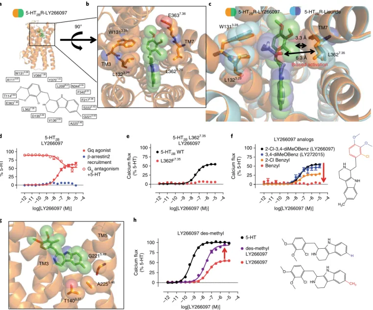
Structure of 5-HT2BR–LY266097 reveals TM7 as a trigger for biased signaling. Finally, we explored non-ergoline antagonists with distinct scaffolds to determine whether they displayed similar binding modes that were shared with other 5-HT2BR antagonists. Although lisuride is a 5-HT2BR antagonist, it lacks selectivity for
5-HT2BR, as is commonly observed for many ergolines19. LY266097, however, is a purported selective 5-HT2BR antagonist31 that contains a distinct tetrahydro-β -carboline pharmacophore, and determining the binding mode of LY266097 could illuminate novel structural determinants of 5-HT2BR selectivity.
Accordingly, we crystallized LY266097 in a BRIL-fused 5-HT2BR (BRIL; thermostabilized apocytochrome b562RIL) and solved the 5-HT2BR–LY266097 structure at 3.2-Å resolution (Fig. 5a, Table 1 and Supplementary Fig. 5a,b). Analysis of LY266097’s binding pose revealed that the tetrahydro-β -carboline core scaffold was oriented in the OBP with the charged nitrogen engaging Asp1353.32 in TM3 and the tetrahydro-β -carboline core engaging residues Phe3406.51 and Phe3416.52 in a π -π aromatic stack. Unexpectedly, when we compared the binding modes of LY266097 and lisuride in the EBP, we observed that LY266097’s 2-chloro-3,4-dimethoxybenzyl substituent was oriented much closer to TM7 than that of lisuride (Fig. 5b,c). On the basis of our previous finding that TM7 appears to be important for biased agonism, we hypothesized that LY266097 should show preference for either Gq or β -arrestin2 activity. We found LY266097 to be a modest Gq partial agonism without detect-able β -arrestin2 activity (Fig. 5d) and confirmed LY266097’s Gq par-tial agonism in an additional assay of Gq function via PI hydrolysis (Supplementary Fig. 5c). We also tested LY266097 in an orthologo-nal β -arrestin2 recruitment assay using BRET, which measured a
5-HT2B methysergide
–12 –11 –10 –9 –8 –7 –6 –5 –4
0 25 50 75
100 5-HT 2B WT
log[methysergide (M)]
Calcium flux (% 5-HT)
T140V3.37 A225G5.46
N
H N O HN
OH
H3C N(1)-methyl
Methysergide
OH
CH3
T1403.37
A2255.46 H3C
TM3
TM4 TM5
TM2
TM3
TM5 D1353.32
T1403.37
G2255.46
b
5-HT2BR–methylergonovine 5-HT2BR(Ala225Gly5.46)–methysergide
A2255.46 G2255.46
c β2AR–ICI-118,551
5-HT2BR(Ala225Gly5.46)–methysergide
5-HT2BR(Ala225Gly5.46)–methysergide
S/G 5.46 TM5 TM3
Activation
d
1–2 Å
N H N O
N H HO
CH3
TM6
L3627.35
L209EL2
V1363.33
F2175.38
T1403.37
G2215.42
S2225.43
F3406.51
S1393.36
Y3707.43
D1353.32
V3667.39
A225G5.46
Fig. 2 |Structure of a 5-HT2BR(Ala225gly5.46) mutant designed to be activated by methysergide.a, Left, design of the mutation to convert methysergide into an agonist by accommodating the N(1)-methyl (blue) with vdW interactions by Thr140Val3.37 or with space by Ala225Gly5.46. Right, 5-HT
β -arrestin2 recruitment in real time monitored across specific time points. At no tested time point did LY266097 display β -arrestin2 recruitment (Supplementary Fig. 5d); instead, it displayed potent β -arrestin2 antagonism (Supplementary Fig. 5e). These results indicate that LY266097 has a bias toward Gq.
On the basis of our previous results with lisuride and the Leu362Phe7.35 mutant, we hypothesized that the extent of G
q agonism by LY266097 is determined by ligand contact between 2-chloro-3,4-dimethoxybenzyl and Leu3627.35. To confirm that LY266097’s contact with TM7 resulted in selective Gq activa-tion, we tested LY266097 with the Leu362Phe7.35 mutant, which was previously found to restore lisuride’s Gq agonist activity. As expected, LY266097’s Gq agonist activity was abolished with the Leu362Phe7.35 mutant (Fig. 5e), suggesting that the bulkiness of the phenylalanine in the Leu362Phe7.35 mutant sterically clashed with the 2-chloro-3,4-dimethoxybenzyl moiety, resulting in a lack of agonism. To provide additional evidence that TM7 influ-ences activation via strict steric requirements, we synthesized analogs of LY266097 that lacked either a 2-chloro or a 3,4-dime-thoxy substituent on the benzyl moiety, which would be expected to show less TM7 interaction. We found a substituent-dependent decrease in Gq-mediated agonist potency, with the unsubstituted
benzyl analog showing no detectible Gq agonist activity, indicat-ing that the substitution pattern to LY266097’s scaffold conferred Gq agonism (Fig. 5f).
Similar to methysergide, LY266097 contains a methyl substituent on the tetrahydro-β -carboline scaffold that appears to ‘push’ on TM5 at position Gly2215.42 (Fig. 5g), a key residue implicated in confer-ring 5-HT2 subtype selectivity19. To determine whether LY266097’s methyl substituent was in part responsible for Gq partial agonism, we synthesized a des-methyl analog of LY266097 and found that it was nearly a full agonist (EC50= 20 nM; Emax= 93%; Fig. 5h). These results suggest that the methyl substituent on LY266097 and its interaction with TM5 impair agonism.
Discussion
Here leveraged by four new 5-HT2B crystal structures, including the structure of a mutant receptor (Ala225Gly5.46) designed to switch a ligand’s efficacy, we provide a comprehensive structural analysis of ligand-specific contacts that lead to GPCR activation. Notably, this study not only identifies mechanisms of 5-HT GPCR activation via the orthosteric site, but also identifies important determinants of β -arrestin recruitment via the EBP, thereby illuminating determi-nants of ligand bias that could apply to other GPCRs.
a
N H N
(R) O N
H
LSD Lisuride
NH
N H N
(S) N
H
O
C8 C8
N6 N6
5-HT2B
–12 –11 –10 –9 –8 –7 –6 –5 –4
0 25 50 75 100
LSD Lisuride
log[drug (M)]
Calcium flux (% 5-HT)
Lisuride + 5-HT
5-HT2B antagonist 5-HT2B agonist
5-HT2B L3627.35 Lisuride
–12 –11 –10 –9 –8 –7 –6 –5 –4
0 25 50 75
100 5-HT2B WT
log[lisuride (M)]
Calcium flux (% 5-HT)
L7.35N L7.35F L7.35Y
d
5-HT2B L3627.35 LSD
–12 –11 –10 –9 –8 –7 –6 –5 –4
0 25 50 75 100 5-HT
2B WT L7.35A
log[LSD (M)]
Calcium flux (% 5-HT)
b e
c
5-HT2BR–Lisuride 5-HT2BR–LSD
TM3 TM7
W1313.28
L1323.29
L3627.35 TM1
TM2 TM3
TM4 TM7
TM3 W3.28
L3.29
L7.35
Antagonism
Agonism
TM7
f
H N
NH H N N
O
L209EL2
V1363.33
F3416.52
T1403.37
G2215.42
S2225.43
F3406.51
S1393.36
Y3707.43
D1353.32
V3667.39
N3446.55
W1313.28
F2175.38
A2255.46
W3376.48
Fig. 3 |Structural basis for a 5-HT2B activation mechanism via the extended binding pocket.a, Comparison of the chemical structures of LSD and lisuride. LSD’s (R)-diethylamide (purple) and lisuride’s (S)-diethylurea (blue) lead to 5-HT2BR agonism and antagonism, respectively. b, 5-HT2BR Gq-mediated calcium flux activity. LSD shows Gq partial agonist activity (purple; EC50 = 40 nM; Emax = 82%), whereas lisuride lacks agonist activity (blue, closed circles). Lisuride instead shows competitive antagonism (blue, open circles; IC50 = 25 nM). c, Structure of lisuride (blue) at 5-HT2BR (light blue) in comparison to LSD (purple) bound to 5-HT2BR (green), showing lisuride’s (S)-diethylurea wedged between TM3 residues Trp1313.28 and Leu1323.29 and making no contact with TM7 Leu3627.35 (PDB 6DRX). d, Lisuride G
q-mediated calcium flux activity for the Leu362Asn7.35 (green; EC50 = 395 nM; Emax = 41%), Leu362Tyr7.35 (orange; EC50 = 465 nM; Emax = 69%) and Leu362Phe7.35 (purple; EC50 = 77 nM; Emax = 60%) mutants. e, LSD Gq-mediated calcium flux activity for the Leu362Alal7.35 mutant (red; EC
50 = 340 nM; Emax = 67%) relative to wild-type 5-HT2BR (purple; EC50 = 37 nM; Emax = 79%). Data in b, d and e represent the mean ± s.e.m. from n = 3 independent experiments, performed in triplicate. Source data are available online. f, Schematic illustrating that ligand contact with residue Leu3627.35 in TM7 in the EBP leads to 5-HT
Our results with the 5-HT2BR–methylergonovine and 5-HT2BR(Ala225Gly5.46)–methysergide structures illuminate an orthosteric activation mechanism that occurs via ligand engagement with residues Thr1403.37 and Ala2255.46. Although residue Ala2255.46 has been implicated as an important determinant of activation in β -adrenergic receptors21,32, the role of the highly conserved Thr1403.37 residue is less understood. In fact, crystallographic evidence that shows direct ligand engagement with residue Thr1403.37 in TM3 is scarce. In rhodopsin, however, the β -ionone ring of retinal, which is critical for receptor activation, is directly engaged with the Glu1223.37 residue. Retinal interaction with Glu1223.37 has been posited to lead to disruption of a hydrogen bond between Glu1223.37 and His2115.46, which causes an inward rotation of TM5, transducing disruption of
the ionic lock between the intracellular portions of TM3 and TM633.
Although here ligand engagement with Thr1403.37 appears to involve either a hydrogen bond or an electrostatic interaction, it remains to be seen whether this ligand contact occurs frequently with other ligands or whether its role is more indirect (and in concert with resi-due Ala2255.46) in the activation process, especially considering that residue Thr1403.37 is highly conserved across aminergic GPCRs. Our results do emphasize that ligand engagement with residue Ala2255.46 and TM5 movement is important for an orthosteric mechanism of class A aminergic GPCRs—both regions highly conserved in aminer-gic GPCRs that we have previously exploited to design biased ligands25.
To our knowledge, our results with the 5-HT2BR–lisuride struc-ture are the first to reveal a mechanism of activation that occurs
5-HT2B Gq calcium flux
–12 –11 –10 –9 –8 –7 –6 –5 –4
–12 –11 –10 –9 –8 –7 –6 –5 –4
1.0 1.5 2.0 2.5 3.0 3.5 4.0 4.5 5.0
5-HT2B WT
log[methylergonovine (M)] log[methylergonovine (M)]
–12 –11 –10 –9 –8 –7 –6 –5 –4 –12 –11 –10 –9 –8 –7 –6 –5 –4
Calcium flux
(fold-over-basal)
1.0 1.5 2.0 2.5 3.0 3.5 4.0 4.5 5.0
Calcium flux
(fold-over-basal)
T140A3.37
5-HT2B WT T140A3.37
5-HT2B β-arrestin2 recruitment
1.0 1.5 2.0 2.5
β
-arrestin2 recruitment (fold-over-basal)
1.0 1.5 2.0 2.5
β
-arrestin2 recruitment (fold-over-basal)
5-HT2B Gq calcium flux
5-HT2B Gq calcium flux 1.0
1.5 2.0 2.5 3.0 3.5 4.0 4.5 5.0
5-HT2B WT
log[methysergide (M)]
–12 –11 –10 –9 –8 –7 –6 –5 –4
log[methysergide (M)]
Calcium flux
(fold-over-basal)
1.0 1.5 2.0 2.5 3.0 3.5 4.0 4.5 5.0
Calcium flux
(fold-over-basal)
A225G5.46
5-HT2B WT A225G5.46
5-HT2B β-arrestin2 recruitment
5-HT2B β-arrestin2 recruitment 1.0
1.5 2.0 2.5
β
-arrestin2 recruitment (fold-over-basal)
1.0 1.5 2.0 2.5
β
-arrestin2 recruitment (fold-over-basal)
a MethylergonovineT140A3.37 MethysergideA225G5.46
5-HT2B Gq calcium flux
5-HT2B WT
log[lisuride (M)] –12 –11 –10 –9 –8 –7 –6 –5 –4log[lisuride (M)] –12 –11 –10 –9 –8 –7 –6 –5 –4 L362F7.35
5-HT2B β-arrestin2 recruitment
5-HT2B WT L362F7.35
5-HT2B WT L362F7.35
5-HT2B WT L362F7.35 Lisuride
L362F7.35
[3H]LSD dissociation
0 60 120 180 240 300 360 420 480 0
25 50 75
100 5-HT2B WT
L362F7.35
Time (min)
[
3H]LSD bound
(% specific binding)
log[LSD (M)] –12 –11 –10 –9 –8 –7 –6 –5 –4log[LSD (M)] LSD
L362F7.35
Orthosteric binding pocket
G protein and β-arrestin
Extended binding pocket
G protein or β-arrestin EL2
TM5
TM3 TM3
TM5
TM7 TM7
TM3 and TM5 TM7 and EL2
b
c d
e f
EL2
TM5 TM3
TM7 EL2
OBP
EBP
Fig. 4 |Divergent actions on β-arrestin2 recruitment by OBP versus eBP mutations.a, Left, methylergonovine Gq-mediated calcium flux comparing the Thr140Ala3.37 mutant (red) to wild-type 5-HT
2BR (black; EC50 = 21 nM). Right, β -arrestin2 recruitment comparing the Thr140Ala3.37 mutant (red) to wild-type 5-HT2BR (black; EC50 = 1.2 nM). b, Left, methysergide Gq-mediated calcium flux comparing the Ala225Gly5.46 mutant (green; EC50 = 33 nM) to wild-type 5-HT2BR (black). Right, β -arrestin2 recruitment comparing the Ala225Gly5.46 mutant (green; EC50 = 1.7 nM) to wild-type 5-HT2BR (black). c, Left, lisuride Gq-mediated calcium flux comparing the Leu362Phe7.35 mutant (purple; EC50 = 65 nM) to wild-type 5-HT2BR (black). Right, β -arrestin2 recruitment comparing the Leu362Phe7.35 mutant (purple) to wild-type 5-HT
2BR (black). d, Left, LSD Gq-mediated calcium flux comparing the Leu362Phe7.35 mutant (purple; EC50 = 40 nM) to wild-type 5-HT2BR (black; EC50 = 42 nM). Right, β -arrestin2 recruitment comparing the Leu362Phe7.35 mutant (purple) to wild-type 5-HT2BR (black; EC50 = 0.97 nM). Data in a–d are expressed as fold change relative to basal levels and represent the mean ± s.e.m. from n = 3 independent experiments, performed in triplicate. e, LSD dissociation comparing wild-type 5-HT2BR (black; koff = 0.015 min−1) to the Leu362Phe7.35 mutant (purple; koff = 0.240 min−1). Data represent the percentage specific binding, indicating the mean ± s.e.m. from n = 3 independent experiments, performed in duplicate. f, Schematic comparing the location of the EBP residues Leu209EL2 and Leu3627.35 (purple), which contribute to either G
q or β -arrestin2 recruitment preference, to the location of OBP residues Thr1403.37 and Ala2255.46 (green), which have equal contributions to G
via the EBP, specifically via ligand contact with TM7 at residue Leu3627.35. This extended binding region of the receptor is less con-served, allowing for ligand-specific engagement of non-conserved residues, and it likely explains the diverse pharmacological action that results from ligand-specific substituents that project from the core scaffold, as in the case of LSD, where the diethylamide projects away from the core ergoline scaffold. Of note, the same residues, Trp1313.28, Leu1323.29 and Leu3627.35, which are also involved in rec-ognition of LSD’s diethylamide and lisuride’s diethylurea, compose part of the allosteric site of muscarinic receptors34. This suggests
that ergoline ligands, such as LSD, access a potential allosteric site, leading to a diverse pharmacological profile (for example, antago-nism or biased agoantago-nism). However, it remains to be seen whether bona fide allosteric modulators can selectively target the EBP of 5-HT receptors, leading to bias.
Further study of the structural dynamics that lead to activation, either via the OBP or EPB, is needed to clarify trigger motifs (for example, NPxxY, P-I-F, DRY) involved in either balanced or biased activation. Although BRIL-fusion 5-HT2BR structures adopt an ‘active intermediate’ state, more ‘active-like’ 5-HT2BR structures will 5-HT2B
LY266097
–12 –11 –10 –9 –8 –7 –6 –5 –4
0 25 50 75 100
0 25 50 75 100 Gq agonist
β-arrestin2 recruitment
log[LY266097 (M)]
–12 –11 –10 –9 –8 –7 –6 –5 –4
log[LY266097 (M)]
–12 –11 –10 –9 –8 –7 –6 –5 –4
log[LY266097 (M)]
–12 –11 –10 –9 –8 –7 –6 –5 –4
log[LY266097 (M)]
% 5-HT
Gq antagonism +5-HT
5-HT2B L3627.35 LY266097 5-HT2B WT
L362F7.35
Calcium flux (% 5-HT)
0 25 50 75 100
Calcium flux (% 5-HT)
0 25 50 75 100
Calcium flux (% 5-HT)
a
5-HT2BR-LY266097
c
90°
W1313.28
W1313.28 A1112.61
T1142.64 E3637.36
L3627.35 D1353.32
V1363.33 A2255.46
G2215.42 S2225.43 F2175.38 F3406.51 N3446.55 L209EL2 Y3707.43
V3667.39 L1323.29
E3637.36
L3627.35 TM3
TM7
b
LY266097 analogs
2-Cl-3,4-diMeOBenz (LY266097) 3,4-diMeOBenz (LY272015) 2-Cl Benzyl
Benzyl
LY266097 des-methyl
LY266097 des-methyl LY266097 5-HT
H3C
H N
NH
O O
Cl
CH3
HN H N O
O Cl
g
d e
5-HT2BR-Lisuride 5-HT2BR-LY266097
3.3 Å
6.3 Å
f
G2215.42 TM5
A2255.46
T1403.37 TM3
h
H
HN H N O
O Cl TM7
L3627.35 W1313.28
L1323.29
Biased activation
Fig. 5 |Structure of the 5-HT2BR–Ly266097 complex reveals TM7 as a trigger for biased signaling. Structure of 5-HT2BR in complex with LY266097 reveals determinants of ligand bias via TM7. a, 5-HT2BR (orange) in complex with LY266097 (green) with a 2D ligand plot of nearby residues (PDB 6DS0).
b, View from the top of the receptor showing that the 2-chloro-3,4-dimethoxybenzyl substituent of LY266097 is oriented in close proximity to residue Leu3627.35 in TM7. c, Alignment of the 5-HT
2BR–LY266097 and 5-HT2BR–lisuride structures showing that LY266097’s 2-chloro-3,4-dimethoxybenzyl substituent is within 3.3 Å of Leu3627.35, whereas lisuride’s (S)-diethylurea is further away, at 6.3 Å from Leu3627.35. d, Profiling of LY266097 for ligand bias showing Gq partial agonist activity (red closed circles; EC50 = 37 nM; Emax = 62%) and partial antagonist activity (red open circles; IC50 = 78 nM), but no
β -arrestin2 recruitment activity (blue). e, LY266097 Gq-mediated calcium flux activity comparing the Leu362Phe7.35 mutant (red) to wild-type 5-HT2BR (black; EC50 = 41 nM; Emax = 54%). f, Gq-mediated calcium flux activity of benzyl-substituted LY266097 analogs 3,4-diMeOBenzyl (blue; EC50 = 24 nM;
likely shed light on key trigger motifs involved in G protein versus β -arrestin2 recruitment. Indeed, current inactive-state structures of 5-HT2C19 and 5-HT1B18, which have both been co-crystallized with inverse agonists, implicate the conserved P-I-F trigger as a motif essential for inactivation and biased signaling.
In our study here, Leu3627.35 in TM7 appears to be an important determinant of preference for G protein or β -arrestin2 recruitment, as was also implied for κ -opioid receptor biased signaling and for other GPCRs35,36,37. In the case of LY266097, which appears to make
no specific vdW or hydrogen bond contacts with residues in TM7, our results with Leu3627.35 mutants suggest that ligand engagement with TM7 may operate under strict steric constraints, as was simi-larly observed in the 5-HT2BR Ala225Gly5.46–methysergide struc-ture. Notably, hydrophobic contact with Leu3627.35, as in the case of LSD, leads to enhanced ligand residence time, and contributions from both TM7 and EL2 serve to stabilize ligand residence time. In fact, increased ligand residence time has been shown to lead to increased β -arrestin2 recruitment over time13, and it appears to
be a hallmark of β -arrestin-biased ligands at aminergic GPCRs25.
Further study, however, is required to illuminate subsequent sig-naling after arrestin recruitment, especially as it relates to G pro-tein dependence, as shown in one recent study8. Nevertheless, our
study illuminates that receptor recognition of small substitutions to core ligand scaffolds directly modulates G protein versus β -arrestin recruitment preferences37.
Identification of critical residue(s) for 5-HT2BR agonist versus antagonist recognition and encoding of efficacy is important for drug design strategies, which aim to avoid 5-HT2BR activation and VHD. On the basis of our previous structural and functional studies with 5-HT2BR, it is clear that 5-HT2BR represents an attractive recep-tor template to clarify the structural features necessary for biased signaling as they apply to other class A GPCRs. Insights into the structural basis of agonist versus antagonist action at GPCRs are essential for the design of safer and more effective medications.
Methods
Methods, including statements of data availability and any asso-ciated accession codes and references, are available at https://doi. org/10.1038/s41594-018-0116-7.
Received: 21 March 2018; Accepted: 2 July 2018; Published online: 20 August 2018
References
1. Berger, M., Gray, J. A. & Roth, B. L. The expanded biology of serotonin.
Annu. Rev. Med. 60, 355–366 (2009).
2. McCorvy, J. D. & Roth, B. L. Structure and function of serotonin G protein–coupled receptors. Pharmacol. Ther. 150, 129–142 (2015). 3. Allen, J. A. & Roth, B. L. Strategies to discover unexpected targets for drugs
active at G protein–coupled receptors. Annu. Rev. Pharmacol. Toxicol. 51, 117–144 (2011).
4. Vickers, S. P., Clifton, P. G., Dourish, C. T. & Tecott, L. H. Reduced satiating effect of d-fenfluramine in serotonin 5-HT2C receptor–mutant mice.
Psychopharmacology 143, 309–314 (1999).
5. Roth, B. L. Drugs and valvular heart disease. N. Engl. J. Med. 356, 6–9 (2007). 6. Rothman, R. B. et al. Evidence for possible involvement of 5-HT2B receptors
in the cardiac valvulopathy associated with fenfluramine and other serotonergic medications. Circulation 102, 2836–2841 (2000).
7. Zanettini, R. et al. Valvular heart disease and the use of dopamine agonists for Parkinson’s disease. N. Engl. J. Med. 356, 39–46 (2007).
8. Gustafsson, B. I., Hauso, O., Drozdov, I., Kidd, M. & Modlin, I. M. Carcinoid heart disease. Int. J. Cardiol. 129, 318–324 (2008).
9. Huang, X. P. et al. Parallel functional activity profiling reveals valvulopathogens are potent 5-hydroxytryptamine2B receptor agonists:
implications for drug safety assessment. Mol. Pharmacol. 76, 710–722 (2009). 10. Papoian, T. et al. Regulatory Forum Review*: utility of in vitro secondary
pharmacology data to assess risk of drug-induced valvular heart disease in humans: regulatory considerations. Toxicol. Pathol. 45, 381–388 (2017). 11. Hauso, Ø. et al. Long-term serotonin effects in the rat are prevented by
terguride. Regul. Pept. 143, 39–46 (2007).
12. Wacker, D. et al. Structural features for functional selectivity at serotonin receptors. Science 340, 615–619 (2013).
13. Wacker, D. et al. Crystal structure of an LSD-bound human serotonin receptor. Cell 168, 377–389 (2017).
14. Katritch, V., Cherezov, V. & Stevens, R. C. Diversity and modularity of G protein–coupled receptor structures. Trends Pharmacol. Sci. 33, 17–27 (2012). 15. Latorraca, N. R., Venkatakrishnan, A. J. & Dror, R. O. GPCR dynamics:
structures in motion. Chem. Rev. 117, 139–155 (2017).
16. Wacker, D., Stevens, R. C. & Roth, B. L. How ligands illuminate GPCR molecular pharmacology. Cell 170, 414–427 (2017).
17. Wang, C. et al. Structural basis for molecular recognition at serotonin receptors. Science 340, 610–614 (2013).
18. Yin, W. et al. Crystal structure of the human 5-HT1B serotonin receptor
bound to an inverse agonist. Cell Discov. 4, 12 (2018).
19. Peng, Y. et al. 5-HT2C receptor structures reveal the structural basis of GPCR
polypharmacology. Cell 172, 719–730 (2018).
20. Cherezov, V. et al. High-resolution crystal structure of an engineered human β2-adrenergic G protein–coupled receptor. Science 318, 1258–1265 (2007).
21. Ring, A. M. et al. Adrenaline-activated structure of β2-adrenoceptor stabilized
by an engineered nanobody. Nature 502, 575–579 (2013).
22. Ballesteros, J. A. & Weinstein, H. Integrated methods for the construction of three-dimensional models and computational probing of structure–function relations in G protein–coupled receptors. in Methods in Neurosciences Vol. 25 (ed. Sealfon, S. C.) 366–428 (Academic Press, San Diego, CA, USA, 1995). 23. Rasmussen, S. G. et al. Structure of a nanobody-stabilized active state of the
β2-adrenoceptor. Nature 469, 175–180 (2011).
24. Rasmussen, S. G. et al. Crystal structure of the β2-adrenergic receptor–Gs
protein complex. Nature 477, 549–555 (2011).
25. McCorvy, J. D. et al. Structure-inspired design of β -arrestin-biased ligands for aminergic GPCRs. Nat. Chem. Biol. 14, 126–134 (2018).
26. Stevens, R. C. et al. The GPCR Network: a large-scale collaboration to determine human GPCR structure and function. Nat. Rev. Drug Discov. 12, 25–34 (2013).
27. Bredberg, U., Eyjolfsdottir, G. S., Paalzow, L., Tfelt-Hansen, P. & Tfelt-Hansen, V. Pharmacokinetics of methysergide and its metabolite methylergometrine in man.
Eur. J. Clin. Pharmacol. 30, 75–77 (1986).
28. Wacker, D. et al. Conserved binding mode of human β2-adrenergic receptor
inverse agonists and antagonist revealed by X-ray crystallography. J. Am. Chem. Soc. 132, 11443–11445 (2010).
29. Nichols, D. E., Monte, A., Huang, X. & Marona-Lewicka, D. Stereoselective pharmacological effects of lysergic acid amides possessing chirality in the amide substituent. Behav. Brain Res. 73, 117–119 (1996).
30. Hofmann, C. et al. Lisuride, a dopamine receptor agonist with 5-HT2B
receptor antagonist properties: absence of cardiac valvulopathy adverse drug reaction reports supports the concept of a crucial role for 5-HT2B receptor
agonism in cardiac valvular fibrosis. Clin. Neuropharmacol. 29, 80–86 (2006). 31. Audia, J. E. et al. Potent, selective tetrahydro-β -carboline antagonists of the
serotonin 2B (5HT2B) contractile receptor in the rat stomach fundus. J. Med.
Chem. 39, 2773–2780 (1996).
32. Sato, T. et al. Pharmacological analysis and structure determination of 7-methylcyanopindolol-bound β1-adrenergic receptor. Mol. Pharmacol. 88,
1024–1034 (2015).
33. Ahuja, S. & Smith, S. O. Multiple switches in G protein–coupled receptor activation. Trends Pharmacol. Sci. 30, 494–502 (2009).
34. Kruse, A. C. et al. Activation and allosteric modulation of a muscarinic acetylcholine receptor. Nature 504, 101–106 (2013).
35. Nygaard, R. et al. The dynamic process of β2-adrenergic receptor activation.
Cell 152, 532–542 (2013).
36. Staus, D. P. et al. Allosteric nanobodies reveal the dynamic range and diverse mechanisms of G-protein-coupled receptor activation. Nature 535, 448–452 (2016).
37. Che, T. et al. Structure of the nanobody-stabilized active state of the κ opioid receptor. Cell 172, 55–67 (2018).
Acknowledgements
We thank R. Axel (Columbia University) for the HTLA cells expressing TEV-fused
β -arrestin2 and R. Fischetti and the staff of APS GM/CA for assistance in the
development and use of the minibeam and beam time at GM/CA-CAT beamline 23-ID at the Advanced Photon Source, which is supported by National Cancer Institute grant Y1-CO-1020 and National Institute of General Medical Sciences grant Y1-GM-1104. Use of the Advanced Photon Source was supported by the Office of Science of the US Department of Energy. This work was supported by US National Institutes of Health (NIH) grants R01MH61887 (B.L.R.), R01NS100930 (J.J.), U19MH82441 (J.J. and B.L.R.) and F31-NS093917 (R.H.J.O.), the NIMH Psychoactive Drug Screening Program Contract (B.L.R.) and the Michael Hooker Distinguished Chair of Pharmacology (B.L.R.).
Author contributions
protein, purified the receptor, optimized crystallization conditions, grew crystals for data collection, collected and processed diffraction data, supervised structure determination and assisted with preparing the manuscript; S.W. expressed protein, purified the receptor, optimized crystallization conditions, grew crystals for data collection, collected and processed diffraction data and assisted with preparing the manuscript; B.A. expressed protein, purified receptor, optimized crystallization conditions and grew crystals for data collection; J.L. designed and synthesized LY266097 analogs and performed analytical chemical analysis; K.L. assisted with performing PI hydrolysis signaling studies and
analyzed the data; A.R.T. assisted with performing β -arrestin recruitment experiments;
R.H.J.O. assisted with performing BRET experiments; T.C. assisted with binding studies; J.J. supervised ligand synthesis and edited the manuscript; B.L.R. was responsible for the overall project strategy and management and edited the manuscript.
Competing interests
The authors declare no competing interests.
Additional information
Supplementary information is available for this paper at https://doi.org/10.1038/ s41594-018-0116-7.
Tris-HCl pH 8.0, 100 mM sodium formate and 30% (vol/vol) polyethylene glycol 400 (PEG400) for h5-HT2BR-BRIL-1–LY266097; 100 mM Tris-HCl pH 7.4–7.7,
30–50 mM ammonium tartrate dibasic and 30% (vol/vol) PEG400 for h5-HT2B
R-BRIL-1–lisuride; 100 mM Tris-HCl pH 7.2–8.0, 170–190 mM potassium phosphate monobasic and 30% (vol/vol) PEG400 for h5-HT2BR-BRIL-2–methylergonovine;
and 100 mM Tris-HCl pH 7.3–7.5, 40–100 mM MgCl2 and 30% (vol/vol) PEG400
for h5-HT2BR(Ala225Gly5.46)-BRIL-3–methysergide. All crystals grew to a
maximum size of ~70 µ m × 30 µ m × 20 µ m within 3 d and were harvested directly from the LCP matrix using MiTeGen micromounts before flash-freezing and storage in liquid nitrogen.
Data collection, structure solution and refinement. X-ray data were collected at
the 23ID-B and 23ID-D beamline (GM/CA CAT) at the Advanced Photon Source (Argonne, IL, USA), using a 10-µ m minibeam at a wavelength of 1.0330 Å and either a MarMosaic 300 charge-coupled detector (CCD) or an Eiger-16m detector (Dectris). Diffraction data were collected by exposing the crystals for 1–3 s to unattenuated beam using primary oscillation. Full datasets for each complex were assembled from several crystals owing to the rapid onset of radiation decay at such high doses. Data were indexed, integrated, scaled and merged using HKL300041, and initial phases were obtained by molecular replacement in PHASER42 using the 5-HT2BR coordinates from PDB 4IB4 with the ligand, extracellular loops and
orthosteric residues removed. Refinement was performed with PHENIX43 and REFMAC and included extensive use of simulated annealing to remove model bias. Density was manually examined, and coordinates were rebuilt in COOT44 using |2Fo| – |Fc|, |Fo| – |Fc| and omit maps. After refinement, we did not observe
any Ramachandran outliers in any of the structures: 96.34% and 3.66% (5-HT2BR–
methylergonovine), 95.29% and 4.71% (5-HT2BR(Ala225Gly5.46)–methysergide),
and 96.7% and 3.3% (5-HT2BR–lisuride) as well as 97.47% and 2.53% (5-HT2BR–
LY266097) of residues were in favored and allowed regions, respectively, as defined by Ramachandran statistics. We further observed low Molprobity clash scores of 4.48, 4.94, 3.77 and 3.97 for 5-HT2BR–methylergonovine, 5-HT2BR(Ala225Gly5.46)–
methysergide, 5-HT2BR–lisuride and 5-HT2BR–LY266097, respectively.
Gq-mediated calcium flux FLIPR assays. Stable cell lines were generated for
wild-type 5-HT2BR and all of the 5-HT2BR mutants using the Flp-In 293 T-Rex
Tetracycline-inducible system (Invitrogen, mycoplasma-free). Cell lines were maintained in DMEM containing 10% FBS, 10 µ g/ml blasticidin (Invivogen) and 100 µ g/ml hygromycin B (KSE Scientific). The day before the FLIPR assay, tetracycline-induced cells were seeded into 384-well poly-(l-lysine)-coated black plates at a density of 10,000 cells/well in DMEM containing 1% dialyzed FBS. On the day of the assay, the cells were incubated with Fluo-4 Direct dye (Invitrogen, 20 µ l/well) for 1 h at 37 °C, which was reconstituted in drug buffer (20 mM HEPES-buffered Hank’s Buffered Salt Solution (HBSS), pH 7.4) containing 2.5 mM probenecid. After dye loading, cells were allowed to equilibrate to room temperature for 15 min and were then placed in a FLIPRTETRA fluorescence imaging
plate reader (Molecular Dynamics). Drug dilutions were prepared at 5 × final concentration in drug buffer (20 mM HEPES-buffered HBSS, pH 7.4) containing a final concentration of 0.1% BSA and 0.01% ascorbic acid. Drug dilutions were aliquotted into 384-well plastic plates and placed in the FLIPRTETRA for drug
stimulation. The drug solutions used for the FLIPR assay were exactly the same as those used for the Tango assay. The FLIPRTETRA was programmed to read baseline
fluorescence for 10 s (1 read/s), and afterward 5 µ l of drug/well was added and read for a total of 3 min (1 read/s). Fluorescence in each well was normalized to the average of the first ten reads for baseline fluorescence, and then the maximum fold increase was determined and calculated as fold change relative to basal levels (fold-over-basal). Fold-over-basal was plotted as a function of drug concentration, and data were normalized to percent 5-HT stimulation. Data were plotted, and nonlinear regression was performed using ‘log(agonist) versus response’ in GraphPad Prism 5.0 to yield Emax and EC50 parameter estimates.
β-arrestin2 recruitment Tango assays. Measurement of β -rrrestin2 recruitment at 5-HT2BR and its mutants used constructs that contained a TEV protease
cleavage site and the tetracycline transactivator (tTA) fused to the C terminus of the receptor. Assays were designed and performed as previously described45. HTLA cells expressing TEV-fused β -arrestin2 (derived from HEK cells and kindly provided by R. Axel) were grown and maintained in DMEM containing 10% FBS, 5 µ g/ml puromycin and 100 µ g/ml hygromycin B. On the day of transfection, medium was removed from the HTLA cells, the cells were washed with PBS, and DMEM containing 10% dialyzed FBS and no antibiotics was added. After at least 1 h, the cells were transfected with the 5-HT2BR- or mutant-encoding constructs
using a calcium-phosphate precipitation method46. After at least 24 h, medium was decanted, and the cells were washed with PBS and detached using trypsin. Cells were collected via centrifugation, resuspended in DMEM containing 1% dialyzed FBS and plated into poly-(l-lysine)-coated 384-well white clear-bottom cell culture plates at a density of 7,000–10,000 cells/well in a total of 40 µ l. The cells were incubated at 37 °C and 5% CO2 for at least 6 h before receiving drug stimulation.
Drug solutions were prepared in drug buffer (20 mM HEPES-buffered HBSS, pH 7.4) containing 0.1% BSA and 0.01% ascorbic acid (final concentrations). Drug stimulation was performed using the FLIPRTETRA system by dispensing 10 µ l/well.
Methods
Generation of 5-HT2B receptor constructs. Constructs encoding 5-HT2BR for
the generation of crystals were based on previously published 5-HT2BR constructs
in which thermostabilized apocytochrome b562 RIL (M7W, H102I, R106L) from
Escherichia coli—referred to as BRIL—was fused to intracellular loop 3 (ICL3)38,39 of the receptor12,13. The 5-HT2BR–LY266097 and 5-HT2BR–lisuride complexes were crystallized using a previously crystallized construct (h5-HT2BR-BRIL-1) that
lacked N-terminal residues 1–35 and C-terminal residues 406–481, contained a thermostabilizing Met144Trp3.41 substitution40 and possessed residues Ala1 to Leu106 of BRIL in place of receptor residues Tyr249 to Val313 of ICL338,39. The 5-HT2BR–methylergonovine complex was crystallized by using a different
previously crystallized construct13 (h5-HT
2BR-BRIL-2) that included Val313 but
that was otherwise identical to h5-HT2BR-BRIL-1. The 5-HT2BR(Ala225Gly5.46)–
methysergide complex (h5-HT2BR-BRIL-3) was generated by QuikChange
(Agilent) PCR, mutating the sequence for Ala2255.46 to glycine in the h5-HT 2B
R-BRIL-2 construct. All constructs also contained a hemagglutinin (HA) signal sequence followed by a FLAG tag at the N terminus and a PreScission protease site followed by a 10 × histidine tag (His tag) at the C terminus to enable purification by immobilized metal affinity chromatography.
Expression and purification of the 5-HT2B constructs. High-titer recombinant
baculovirus (> 109 viral particles/ml) was generated using the Bac-to-Bac
Baculovirus Expression System (Invitrogen). Recombinant baculovirus was obtained by transfecting ~5 µ g of recombinant bacmid into 5 × 10⁵ settled
Spodoptera frugiperda (Sf9) cells (Expression Systems) in a 12-well plate (Corning) using 3 µ l of Cellfectin II Reagent (Invitrogen). After 5–12 h, medium was exchanged for 1 ml of Sf-900 II SFM medium (Invitrogen), and the plates were incubated for 4–6 d at 27 °C. P0 viral stock was harvested as the supernatant and used to generate high-titer baculovirus stock by infection of 40–1,000 ml of 2 × 10⁶ Sf9 cells/ml and incubation for 3 d. Viral titers were determined by flow
cytometry analysis of cells that were stained with phycoerythrin (PE)-conjugated gp64 antibody (Expression Systems)38. Expression of 5-HT
2BR was carried out
by infection of Sf9 cells at a cell density of 2 × 10⁶ to 3 × 10⁶ cells/ml in ESF921
medium (Expression Systems) with P1 virus at a multiplicity of infection (MOI) of 3–5. Cells were harvested by centrifugation at 48 h after infection, washed in PBS and stored at –80 °C until use. Cells were disrupted by thawing frozen cell pellets in a hypotonic buffer (10 mM HEPES, 10 mM MgCl2, 20 mM KCl, pH 7.5)
containing protease inhibitors (500 µ M AEBSF, 1 µ M E-64, 1 µ M leupeptin, 150 nM aprotinin). Membranes were purified by repeated centrifugation in a high-osmotic buffer (containing 10 mM HEPES, 1 M NaCl, 10 mM MgCl2, 20 mM KCl, pH 7.5)
to remove soluble and membrane-associated proteins. Purified membranes were directly flash-frozen in liquid nitrogen and stored at –80 °C until protein purification. Purified membranes were resuspended in a buffer containing 10 mM HEPES, 10 mM MgCl2, 20 mM KCl, 150 mM NaCl (pH 7.5) and protease inhibitors
and were incubated for 1 h at room temperature with 50 µ M methylergonovine (Sigma, M2776), methysergide (Sigma, M137), lisuride (Tocris, 4052) or LY266097 (Tocris, 4081). After a 30-min incubation in the presence of 2 mg/ml iodoacetamide (Sigma), membranes were solubilized in 10 mM HEPES, 150 mM NaCl, pH 7.5, 1% (wt/vol) n-dodecyl-β -d-maltopyranoside (DDM, Anatrace), 0.2% (wt/vol) cholesteryl hemisuccinate (CHS, Sigma), 25 µ M of the indicated complex ligand and protease inhibitors for 2 h at 4 °C. Unsolubilized material was removed by centrifugation at 150,000g for 30 min, and 15 mM imidazole was added to the supernatant. Proteins were bound to TALON IMAC resin (Clontech) overnight at 4 °C using approximately 750 µ l of resin for protein purified from 2 liters of cells. The resin was then washed with 10 column volumes (CVs) of Wash Buffer I (50 mM HEPES, 800 mM NaCl, pH 7.5, 0.1% (wt/vol) DDM, 0.02% (wt/vol) CHS, 20 mM imidazole, 10% (vol/vol) glycerol and 20 µ M of the indicated complexing ligand), followed by 10 CVs of Wash Buffer II (25 mM HEPES, 150 mM NaCl, pH 7.5, 0.05% (wt/vol) DDM, 0.01% (wt/vol) CHS, 10% (vol/vol) glycerol and 20 µ M of the indicated complexing ligand). Proteins were eluted in 2.5 CVs of Wash Buffer II + 250 mM imidazole and concentrated in a Vivaspin 20 concentrator with a molecular weight cutoff of 100 kDa (Sartorius Stedim) to 500 µ l; imidazole was removed by desalting the protein over PD MiniTrap G-25 columns (GE Healthcare). The C-terminal 10 × His tag was removed by addition of His-tagged PreScission protease (GenScript) and incubation overnight at 4 °C. Protease, cleaved His tag and uncleaved protein were removed by passing the suspension through equilibrated TALON IMAC resin (Clontech) and collecting the flow-through. 5-HT2BR–ligand complexes were then concentrated to ~30 mg/ml using
a Vivaspin 500 centrifuge concentrator with a molecular weight cutoff of 100 kDa (Sartorius Stedim). Protein purity and monodispersity were tested by analytical size-exclusion chromatography.
Lipidic cubic phase crystallization. 5-HT2BR–ligand complexes were reconstituted