THREE-DIMENSIONAL EVALUATION OF MANDIBULAR CHANGES ASSOCIATED WITH HERBST TREATMENT IN GROWING CLASS II PATIENTS
Thomas R. Covington
A thesis submitted to the faculty at the University of North Carolina at Chapel Hill in partial fulfillment of the requirements for the degree of Master of Science in the School of Dentistry
(Orthodontics).
Chapel Hill 2015
Approved by:
Tung T. Nguyen
Ching Chang Ko
© 2015
ABSTRACT
Thomas R. Covington: Three-dimensional evaluation of mandibular changes associated with Herbst treatment in growing Class II Patients: Pilot Study
(Under the direction of Tung T. Nguyen)
Introduction: Herbst appliance treatment of Class II malocclusions is common in
orthodontic offices. Recent advances in 3D superimpositions have allowed semi-quantitative
assessment of skeletal and dental changes accompanied with Herbst treatment. The aim of this
study was to evaluate 3D skeletal and dental changes following Herbst removal. Methods: 7
consecutive Herbst patients had CBCTs taken pre-treatment (T1), post Herbst removal (T2), and
one year following Herbst treatment (T3). 3-D models were generated from CBCTs; anterior
cranial base registrations were performed to evaluate morphological changes of the maxillary
and mandibular skeleton. Mandibular registrations on the inner cortical symphysis were
performed to evaluate mandibular growth and displacement as well as mandibular dental
changes. Registered models were analyzed using color maps and vector based anatomical
point-to-point measurements. Results: During initial Herbst treatment, patients demonstrated anterior
translation of glenoid fossa and condyles and anterior projection of B point, however; variable
skeletal changes were observed one-year post Herbst removal. 4-patients demonstrated a
posterior displacement and 3-patients had continued anterior displacement of B-point
(0.88mm+/-2.54mm). The later three demonstrated inferior displacement of the glenoid
fossa/condyle complex leading to a counter clockwise rotation of the mandible one year post
one year post Herbst. While there was a significant maxillary restraining effect during Herbst
application, insignificant anterior/posterior changes were seen following Herbst removal.
Conclusions: Maxillary skeletal changes during Herbst treatment were maintained; however
anterior/posterior changes in mandibular position one year post Herbst removal were variable.
Condyle fossa complex moved anterior during Herbst treatment but was displaced posterior one
year following treatment. Inferior positioning of the glenoid fossa/condyle complex may have a
ACKNOWLEDGEMENTS
Thank you to my committee members, Dr. Nguyen, Dr. Ko and Dr. Paniagua for your
expertise, guidance, and advice throughout my project. Thank you to Cliff Wilson for your
assistance in the laboratory. Thank you to Dr. LeCornu for your introduction to the topic of 3D
imaging. Thank you to my beautiful wife Laura and family for your love and support. To my
son Carr, and daughter Ella, I am proud to be your father. Thank you to the Southern
TABLE OF CONTENTS
LIST OF TABLES ... vii
LIST OF FIGURES ... viii
LITERATURE REVIEW ...1
Cone Beam Computed Tomography to Evaluate Orthodontic Treatment ...1
Review of Mandibular Growth ...2
Class II Orthodontic Treatment ...3
Class II treatment with the Herbst Appliance ...5
Herbst Studies using CBCT Imaging ...11
Relapse with Herbst Treatment ...12
REFERENCES ...14
THREE-DIMENSIONAL EVALUATION OF MANDIBULAR CHANGES ASSOCIATED WITH HERBST TREATMENT IN GROWING CLASS II PATIENTS ...18
Introduction and Methods ...18
Results ...23
Discussion ...25
Conclusions ...29
Tables ...30
Figures...34
LIST OF TABLES
Table 1 – Patient Demographics treated with Herbst Appliance ...30
Table 2 – Mandibular Skeletal Changes (mm) ...30
Table 3 – Condylar Growth of the Mandible during and after Herbst Treatment (mm) ...31
Table 4 – Maxillary Skeletal Changes (mm) ...32
LIST OF FIGURES
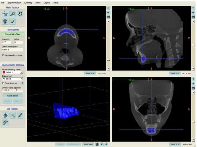
Figure 1 – Mandibular Region of Interest Mask for Superimposition ...34
Figure 2 - Patient Demonstrating Skeletal Relapse following Herbst Therapy ...35
Figure 3 – Patient Demonstrating continued Anterior projection
LITERATURE REVIEW
Cone Beam Computed Tomography to evaluate Orthodontic treatment
Three dimensional imaging combined with the ability to perform fully automated voxel
based registration on the anterior cranial base of growing patients has allowed accurate
observation of skeletal changes in adolescent patients1-3. These 3D imaging techniques overcome the inadequacies of 2D cephalometric imaging including magnification, distortion,
patient positioning errors and obstruction of critical landmarks4,5. There is also inherent bias in superimpositions with 2D images 5. There are different ways to register 3D segmentations2. The first is a point to point registration technique. With a point to point registration technique, the
researcher identifies landmarks on each time point and then overlays the two time points
registering on these two manually identified landmarks. The second 3D registration technique is
surface to surface. With surface to surface registration, two surfaces are identified on each time
point and the computer digitally overlays the surfaces. The computer algorithm finds a closest
approximation of the two surfaces by minimizing the distance between the two surfaces. The
third is a voxel based registration. This new registration technique selects thousands of voxels
and uses shape, volume and voxel intensity to demarcate between bone and soft tissue3. Maxillary and Mandibular adaptive and positional changes can be accurately examined and
measured relative to the anterior cranial base using these 3D superimposition techniques1-3. Imprecisions of the 2D registration technique are eliminated by an entirely computerized voxel
do not explain the complex 3-dimentional (3D) process of bone remodeling over time. If voxel
registration is completed on the anterior cranial base, the researcher is able to accurately examine
and measure maxillary and mandibular adaptive and positional changes. Once images are
segmented into appropriate skeletal regions, point measurements and visualization using color
maps are helpful to determine the true skeletal changes over time. While these observations in
maxillary and mandibular changes are progressive, there are still unknowns regarding regional
growth of the mandible that lead to anterior/posterior projection. To fully understand mandibular
growth and response to treatment, you need to combine cranial base superimpositions with
regional mandibular superimpositions.
Recently 3D voxel based mandibular regional superimpositions have been validated to be
accurate and reproducible. 3D mandibular regional superimpositions allow you to visualize and
quantitate mandibular growth in areas that are difficult to visualize on 2D images. With 3D
mandibular regional superimpositions you can measure growth in the condylar region,
mandibular surface remodeling, and dento-alveolar alterations. Cranial base superimpositions in
combination with mandibular regional superimpositions can define exact mandibular growth that
lead to an overall response.
Review of Mandibular Growth
Growth of the mandible occurs at the mandibular condyle and along the posterior surface
of the ramus with resorption of the anterior ramus; furthermore, the ramus is increased in height
by endochondral replacement at the condyle followed by surface remodeling6-10. The pattern of displacement of the condyles is superior and posterior leading to a forward and downward
condyle translates to the amount of vertical versus anterior growth each patient demonstrates.
Historically, growth of the mandible, in particular, rotation of the mandible dates back to 1955
when the idea that the mandibular corpus rotates during growth was first described6. Here Bjork said that shape is kept stable by associated substantial surface remodeling6. In 1983 Bjork published a 25 year longitudinal cephalometric implant study describing growth and remodeling
of the mandible. He concluded that there we individuals that had a forward rotation of the
mandible and others that experienced a backwards rotation of the mandible7. The rotation is based on how the inferior border of the mandible compares to the anterior cranial base. If these
lines converge forward to the face, it is forward rotation. This is a counter clock-wise rotation.
However, if there is a clock-wise rotation of the mandible, showing an increased MPA, then
Bjork described this as a backward rotational growth pattern7. Two distinct types of facial development create backwards rotation, Class II division I patients and people with pathology or
condylar fracture7. Bjork was also instrumental in describing surface remodeling of the
mandible with growth. The remodeling pattern is apposition below the symphysis and anterior
part of the lower boarder of the mandible as well as apposition at the posterior surface.
Resorption is experienced along the anterior surface of the ramus and at the lower boarder of the
angular region6-9. These studies also described the eruption of the mandibular dentition. There is marked growth in the height of alveolar process and the direction of eruption is forward
relative to the occlusal plane. Meaning there is a forward migration of the dentition relative to
mandibular corpus6-9. This leads to an increase in incisor proclination over time7.
Class II Orthodontic Treatment
The orthodontic profession is challenged with the treatment of malocclusions. Many
skeletal relationships are classified based on the amount of protrusion or retrusion of the maxilla
and mandible. Skeletal Class II relationships are commonly encountered in orthodontic practices
in the United States10. The etiology of a skeletal Class II malocclusion includes a prognatic maxilla, a retrognathic mandible or a combination of both. A study by McNamara in 1981
revealed that up to 85% of class II patients have some component of mandibular deficiency
underlying the skeletal class II discrepancy11. Treatment of the skeletal Class II patient is based on severity. Treatment ranges from dental compensation including camouflage with extractions
to surgical procedures targeted at moving the jaw at fault. In growing patients, growth
modification of the skeletal structures offers an intermediate treatment option. Growth
modification is appealing over camouflage because ideally, the skeletal discrepancy should be
addressed for optimal treatment results.
When evaluating treatment options for Class II patients, the extent of the skeletal
discrepancy and the skeletal maturity of the patient need to be considered. In non-growing
patients with a less severe skeletal discrepancy, class II camouflage may be appropriate.
However, if camouflage treatment is delivered to a patient with a relative severe skeletal class II
discrepancy, it can result in a poor esthetic outcomes12. Surgical treatment may be indicated for patients with extremely severe skeletal problems, or for patients with no growth potential
remaining. Most common surgical treatment involves mandibular bilateral sagittal split
osteotomy advancement, because a majority of the patients have some component of mandibular
deficiency13. However, maxillary set back can also be conducted as an isolated procedure or in conjunction with a mandibular advancement procedure13. Surgery is expensive and is associated with comorbidities including paraesthesia, anaesthesia, paralysis and potentially death. Because
fact, from 1984 to 1996, only 42% of the patients seen at the Dentofacial clinic at the University
of North Carolina for surgical correction of a class II skeletal problem accepted and completed
surgical treatment13. Alternatively, if the patient is intercepted when there is inherent growth remaining, growth modification can be attempted to correct the skeletal discrepancy.
Growth modification for skeletal Class II patients includes restricting forward (anterior)
growth of the maxilla and promoting forward growth/projection of the mandible. This takes
advantage of differential growth resulting is more anterior projection of the mandible. Timing
for growth modification is extremely important. Both human and animal orthopedic
investigations have recognized the ideal time for class II growth modification is during the
pubertal growth spurt14-17. This treatment window is during the peak pubertal growth spurt, which corresponds to CVM stage of CS3-CS418. From McNamara, we know a majority of class II patients have mandibular deficiency, thus, utilizing growth modification treatment modalities
that target the jaw at fault (i.e. the mandible) is ideal. Functional appliances are claimed to
increase mandibular projection. Orthodontic treatment with appliances like the Herbst, bionator,
twin block, or headgear can effectively achieve ideal overjet and class I dental relationships,
however a systematic review by Cozza and Baccetti published in 2006 revealed that the Herbst
appliance is the most effective at increasing mandibular projection19.
Class II treatment with the Herbst appliance
The Herbst appliance is a commonly used functional appliance for the correction of a
treatment modality. Herbst appliance design has evolved over the past 100 years; however, the
basic mechanism has remained unchanged. The device includes bilateral telescope mechanisms
that guide the mandible into an anterior position during rest, and all functional movements20. Current designs include crowns on the maxillary first molars and crowns on the mandibular first
molars with straight telescoping mechanisms. A cantilever extends mesial to the mandibular
molar crown to which the telescoping arms attach. These telescoping arms extend mesial of the
mandibular premolars applying a force in an anterior direction. It is often referred to as the
cantilever Herbst. The cantilever Herbst was initially designed for the mixed dentition prior to
the eruption of the mandibular canines or first pre-molars. This design also allows the
orthodontist to bond anterior teeth for increased anchorage and concurrent leveling/ aligning of
the mandibular arch.
The cantilever Herbst design requires extra consideration. Because of the long anterior
arm extension, the distance of the force to the center of rotation is very large and can lead to
significant mesial tipping of the mandibular molars. For this reason, an occlusal rest that extends
from the mesial of the mandibular molar to the occlusal of the 1st premolar is recommended. In addition, a rest from the distal of the mandibular first molars to the occlusal of the mandibular
second molars helps to prevent eruption of the second molar. Often, a lower lingual holding
(LLHA) arch is included in the design of the cantilever Herbst in order to prevent mesial crown
tip of the mandibular molars. From the LLHA a support bar that crosses the occlusion distal to
the mandibular canine can be added to preserve the transverse dimension. Preservation of the
transverse dimension protects the patient from adverse gingival damage of the cantilever arm.
In the maxilla, occlusal rests are extended from the distal of the first molars to the occlusal of the
inherent vertical force created by the telescoping arms, the maxillary occlusal rest also prevents
intrusion of the maxillary first molar and extrusion of the second molar.
An adverse effect of the Herbst appliance is proclination of the lower incisors21. Proclination of the lower incisors can be prevented in the cantilever Herbst with brackets and
rectangular wires that add negative root torque. Adding brackets to the lower incisors can also
help to control the cantilever forces exhibited on the molars by increasing anchorage. In
addition, adding brackets during Herbst treatment can allow leveling of the curve of spee that is
often present in Class II malocclusions.
The Herbst is a tooth supported appliance. As such, some studies suggest the effects of
the Herbst are primarily dentoalveolar22. Many studies report the Herbst, improves mandibular projection, consequently improving the underlying skeletal discrepancy22. A mild restraining effect in response to Herbst treatment has been noted by many studies, and the effect has been
shown to be statistically similar to the effect produced by headgear23-26. Meanwhile, some studies suggest the skeletal headgear effect displayed by the Herbst is negligible. Ultimately, available
data which examines the extent of skeletal verse dentoalveolar adaptation in that lead to the class
II correction when using the Herbst is controversial. The skeletal component of class II
correction has been reported to extend from 13% to 85%27-30.
Dentoalveolar effects of the Herbst provide large changes leading to class II correction.
In general, mandibular molars will move mesially (often tipping) between 0.5 and 5.5mm.
Maxillary molars may have up to 1 mm of intrusion, and distalize between 0.6 and 3.0 mm.
Distal tipping of the maxillary molars between 5.6° and 6.4° are also observed. The mandibular
negative overjet causes posterior disocclusion. The lower incisors will procline between 5.4° and
10.8° and will move mesially between 0.2mm and 4.0 mm. The occlusal plane rotates in a
clockwise direction due to intrusion of maxillary molars between 1.1° -5.5°.
Skeletal effects of the Herbst appliance have variances in the literature. It is partly due to
the discord in measurement methods used with 2D images. The method developed by Pancherz
utilizes a reference grid constructed from the occlusal line (OL) and the occlusal line
perpendicular (OLp)28,29. Maxillary measurements using this method are subject to patient positioning errors. Many studies use an angular measurement, SNA, to examine maxillary
changes. However, increases in the vertical dimension as seen with growth will mask the
anterior-posterior change when using these angular measurements5. Skeletal changes observed at A point, indubitably depend on the methodologies used.
Studies supporting a maxillary restraining effect of the Herbst theoretically make sense.
During treatment the Herbst appliance exerts an upward and posterior force that is similar to a
high-pull headgear. Studies report a restraining effect on the maxilla with decrease SNA ranging
from 0.4°-1.2°24,31. However, the SNA angle often relapses to preclinical values24. Authors using the grid system to evaluate maxillary restraint found 0.4 mm maxillary restraint to 2.8
mm21,29,32. It is important to understand that authors often found different effects on the maxilla depending on the method of analysis employed. Mild tipping of the palatal plane have also been
reported. Overall, many studies found no difference in the anterior-posterior projection of the
maxilla. These studies employed 2D technologies leaving image methodology as a variable in
the discrepancy. Other authors suggest that remaining growth potential after Herbst treatment is
cause for the relapse. Recent studies using 3D cone beam computed tomography demonstrate
the Herbst patients (1.20 +/- 0.53 mm vs -1.22 +/- 0.43 mm; P < 0.001)26. After a rigid, voxel based registration on the anterior cranial fossa was performed, this study used closed point
measurement between the two different time points.
Alteration of anterior-posterior projection of the mandible can be attributed to changes in
mandibular growth, changes in the direction of growth and/ or condylar/ fossa positional
changes. Previous studies report conflicting results with some showing increased mandibular
length with Herbst treatment14,19,24,30,33. While other studies show no significant increase in mandibular length21,30. Deviations in patient positioning, as well as differences in magnification ratios between the left and right sides of the mandible can affect 2-D measurements of
mandibular corpus length and ramus height.
Currently, most of the literature that evaluates mandibular growth following functional
appliance therapy use condylion, an arbitrary condylar point, or a proxy- point such as articulare.
Condylion landmark identification is associated with low reliability due to obstruction of the
overlying temporal bone. Utilizing an arbitrary condylar point, as in the method described by
Creekmoor, and used by Pancherz improves landmark identification. However, this method is
still subject to distortion, magnification, and mandibular regional registration errors during the
transfer process. Additionally, rotational deviations in patient positioning between T1 and T2
image capture, will have a large effect on the perceived mandibular corpus length regardless of
the measurement used. Any discrepancy in “tilt” or pitch positional errors will also affect vertical
measurement error. Lastly, using articulare as a proxy condylar point is going to present
significant measurement error. The position of articulare is dependent on vertical and
anterio-posterior changes of the glenoid fossa and condyle. Because articulare is dependent on growth, it
Despite these limitations, Baccetti et. al. found that class II subjects treated with a Herbst
achieved chin advancements from 2.5 mm to 5 mm greater than untreated class II patients and
had 2 mm to 4 mm greater chin advancements (determined by B point and pogonion) compared
to patients treated with head gear and class II elastics24. Increasing mandibular growth with Herbst therapy has also been reported by Pancherez et. al32. Meanwhile some studies show an increase in the anterior-posterior projection of the mandible without a statistically significant
increase in mandibular length. Long-term change in SNB angles are variable, with some studies
finding no difference, while other studies report increases of 0.3°-2.6°23,25,29,32,34. The ANB angle has been shown to decrease between 1.1° to 3.9°, and remains relatively stable. A
counterclockwise rotation secondary to dental effects was noted in a study by Papadopoulos.
This movement helps the class II skeletal relationship. The literature is in discord regarding
increases in posterior mandibular height. Again, these differences likely arise due to different
methodologies in measurement protocols.
In addition to increased length, alterations in growth pattern will also impact
anterior-posterior projection of the mandibular base. Opening of the gonial angle and anterior-posterior flexure of
the condyle are anatomical changes that can lead to more anterior mandibular positioning. Initial
placement of the Herbst causes the condyle to be placed anteriorly onto the articular eminence.
After 6-12 weeks, the condyles showed a more posterior position in the glenoid fossa, and the
posterior superior aspect of the condyle showed increased signal intensity on MRI35,36. Condylar osteogenesis during Herbst treatment has also been shown in animal studies. Sagittal condylar
growth has been reported to occur. The condyle moves between 1.5-3.1 mm superiorly and 2.1-
4.0 mm posteriorly. Interestingly, the direction of condylar osteogenesis occurs in the direction
In addition to redirection the growth pattern of the mandible, altering the growth process
of the glenoid fossa can also allow for increased mandibular projection. It is believed that there
are two sites in the temporal mandibular joint (TMJ) that adapt to the forces of the Herbst: 1)
condyle; and 2) glenoid fossa36. The condylar position changes within the fossa have also been proposed however this is not significantly confirmed in ether animal or human studies. Pancherz
et al looked at the size of the joint space pre- and post Herbst treatment. They found that there
was no statistical difference in the condylar position. However, there was great variation among
patients. It was revealed that post treatment condylar positions were on average slightly more
anterior than pretreatment positions.
Translation of the glenoid fossa, has been shown to contribute to mandibular positional
changes post Herbst treatment26. However, 2D imaging techniques used in human studies are greatly flawed when assessing for remodeling of the glenoid fossa. Human studies often rely on
an unchanged condyle-fossa relationship because they utilize the method described by Buschang
and Santos37. Ruf and Pancherz conducted an MRI study to evaluate effective condylar growth in Herbst patients36. They noted increase uptake in the T2-weighted sequences in the glenoid fossa and condyle. This was interpreted to be definitive areas of condyle and fossa remodeling.
However, because the incidence of capsulitis rises during Herbst treatment up to 100%, virtually
all patients would be expected to have increased T2 signal due to the amplified inflammatory
process. Differentiating inflammatory processes from the cellular cascade of skeletal remodeling
is difficult. Additionally, techniques to register and superimpose MRI scans to evaluate changes
critically from T1 to T2 have not been developed for the cranial base. Therefore, MRI scans
cannot be used to adequately examine skeletal adaptations until a proper registration and
Some studies have suggested remodeling may occur. However, these studies use
condylion or articulare as a proxy point to approximate the position of the fossa. Those
conclusions were not absolute due to imaging limitations and measurement errors. After
examining all of the condylar and fossa changes, they concluded overall the “effective condylar
growth” during Herbst treatment resulted in six-times more horizontal growth and four-times
more vertical growth when compared to Bolton Standards36. Recently a CBCT study registered on the anterior cranial base have demonstrated anterior glenoid fossa remodeling in a 1:1
relationship with the condyle26.
Herbst Studies using CBCT Imaging
Newer studies are using CBCT images to account for ambiguity in the methodology and
to avoid the limitations of 2D imaging. LeCornu et al. used 3D cranial base superimpositions to
describe skeletal changes immediately after Herbst removal26. They found that there was an anterior displacement of the condyles and glenoid fossa that lead to an increase in projection of
B-point in Herbst patients (2.62 +- 1.08mm vs 1.49 +/- 0.79mm; P < 0.05)26. This anterior translation of the glenoid fossa/condyle was compared to controls that have posterior
displacement in concordance with normal growth. They also confirmed the maxillary headgear
effect with mild maxillary restraint. Without mandibular regional registration they were unable
to account for the amount and direction of mandibular growth each patient demonstrated during
treatment. Growth of the mandible occurs at the mandibular condyle and along the posterior
surface of the ramus with resorption of the anterior ramus; furthermore, the ramus is increased in
was present. These, along with the cranial base superimpositions, demonstrate a more complete
picture of treatment response and mandibular growth patterns.
Relapse with Herbst Treatment
Occlusal results have been shown to be relatively stable with Herbst treatment. A recent
32 year follow up demonstrated that 86% of patients had acceptable overjet and overbite38. However, the amount of correction that was dentoalveolar versus skeletal in nature is still
controversial. The amount of favorable mandibular changes post Herbst treatment are disputed
due to 2D imaging methodology and continued growth of the patient. In 1991, Pancherz studied
15 relapse and 14 stable patients post Herbst treatment. It was stated that relapse in overjet and
sagittal molar relationship resulted mainly from post treatment maxillary and mandibular dental
changes28. However, Pancherz et al. in 2002 suggested that all the favorable condylar
(mandibular skeletal) changes and remodeling of the glenoid fossa reverted in a post treatment
period ranging from 6-12 months post Herbst removal. During this time the glenoid fossa was
displaced posteriorly; the amount of condylar growth and effective TMJ changes were reduced.
This would suggest any skeletal contribution was due to maxillary restraint or the headgear
effect. Wigal et al. suggested that after early treatment with the Herbst appliance there was a
continuous restraint of maxillary growth as well as dentoalveolar adaptations that contributed to
maintaining a portion of the correction23.
Demonstrated above, the literature reveals tremendous variation in the amount of skeletal
adaptation leading to improvement in the class II profile. This variation stems from the
limitations of 2D imaging. If a majority of skeletal Class II patients have a degree of mandibular
However, there is not data to suggest that we have any lasting effect on the mandible. Research
using novel three dimensional imaging techniques to clarify the skeletal response to the Herbst
appliance is suggestive of a maxillary headgear effect, anterior remodeling of the glenoid fossa,
and anterior projection of the condyle. This is a net combined anterior projection of B-point
without increasing corpus length of the mandible. However, there is 2D research showing that
the favorable changes in the TMJ do not last 1 year following Herbst removal. Research using
REFERENCES
1. Cevidanes LH, Oliveira AE, Grauer D, Styner M, Proffit WR. Clinical application of 3D imaging for assessment of treatment outcomes. Semin Orthod. 2011;17(1):72-80.
2. Cevidanes LH, Styner MA, Proffit WR. Image analysis and superimposition of 3-dimensional cone-beam computed tomography models. Am J Orthod Dentofacial Orthop. 2006;129(5):611-618.
3. Cevidanes LH, Heymann G, Cornelis MA, DeClerck HJ, Tulloch JF. Superimposition of 3-dimensional cone-beam computed tomography models of growing patients. Am J Orthod Dentofacial Orthop. 2009;136(1):94-99.
4. Houston WJ, Maher RE, McElroy D, Sherriff M. Sources of error in measurements from cephalometric radiographs. Eur J Orthod. 1986;8(3):149-151.
5. Spolyar JL. Head positioning error in cephalometric radiography--an implant study. Angle Orthod. 1987;57(1):77-88.
6. BJORK A. Facial growth in man, studied with the aid of metallic implants. Acta Odontol Scand. 1955;13(1):9-34.
7. Bjork A, Skieller V. Normal and abnormal growth of the mandible. A synthesis of
longitudinal cephalometric implant studies over a period of 25 years. Eur J Orthod. 1983;5(1):1-46.
8. Bjork A. Prediction of mandibular growth rotation. Am J Orthod. 1969;55(6):585-599.
9. BJORK A. Variations in the growth pattern of the human mandible: Longitudinal radiographic study by the implant method. J Dent Res. 1963;42(1)Pt 2:400-411.
10. Proffit W, Fields H, Sarver D, eds. Contemporary orthodontics. Fourth Edition ed. St. Louis, Mo: Mosby Elsevier; 2007.
11. McNamara JA,Jr. Components of class II malocclusion in children 8-10 years of age. Angle Orthod. 1981;51(3):177-202.
12. Ruf S, Pancherz H. Orthognathic surgery and dentofacial orthopedics in adult class II division 1 treatment: Mandibular sagittal split osteotomy versus herbst appliance. Am J Orthod Dentofacial Orthop. 2004;126(2):140-52.
13. Bell W, Proffit W, Wilson J, eds. Surgical correction of dentofacial deformities.
14. Ruf S, Pancherz H. Dentoskeletal effects and facial profile changes in young adults treated with the herbst appliance. Angle Orthod. 1999;69(3):239-246.
15. Ruf S, Pancherz H. Herbst/multibracket appliance treatment of class II division 1
malocclusions in early and late adulthood. a prospective cephalometric study of consecutively treated subjects. Eur J Orthod. 2006;28(4):352-360.
16. Bock N, Pancherz H. Herbst treatment of class II division 1 malocclusions in retrognathic and prognathic facial types. Angle Orthod. 2006;76(6):930-941.
17. Hagg U, Pancherz H. Dentofacial orthopaedics in relation to chronological age, growth period and skeletal development. an analysis of 72 male patients with class II division 1 malocclusion treated with the herbst appliance. Eur J Orthod. 1988;10(3):169-176.
18. Baccetti T, Franchi L, McNamara JA,Jr. An improved version of the cervical vertebral maturation (CVM) method for the assessment of mandibular growth. Angle Orthod. 2002;72(4):316-323.
19. Cozza P, Baccetti T, Franchi L, De Toffol L, McNamara JA,Jr. Mandibular changes produced by functional appliances in class II malocclusion: A systematic review. Am J Orthod Dentofacial Orthop. 2006;129(5):599.e1-12; discussion e1-6.
20. Pancherz H. History, background and development of the herbst appliance. Seminars in Orthodontics. 2003;9(1):3-11.
21. Barnett GA, Higgins DW, Major PW, Flores-Mir C. Immediate skeletal and dentoalveolar effects of the crown- or banded type herbst appliance on class II division 1 malocclusion. Angle Orthod. 2008;78(2):361-369.
22. von Bremen J, Pancherz H. Efficiency of early and late class II division 1 treatment. Am J Orthod Dentofacial Orthop. 2002;121(1):31-37.
23. Wigal TG, Dischinger T, Martin C, Razmus T, Gunel E, Ngan P. Stability of class II
treatment with an edgewise crowned herbst appliance in the early mixed dentition: Skeletal and dental changes. Am J Orthod Dentofacial Orthop. 2011;140(2):210-223.
24. Baccetti T, Franchi L, Stahl F. Comparison of 2 comprehensive class II treatment protocols including the bonded herbst and headgear appliances: A double-blind study of consecutively treated patients at puberty. Am J Orthod Dentofacial Orthop. 2009;135(6):698.
25. Valant JR, Sinclair PM. Treatment effects of the herbst appliance. Am J Orthod Dentofacial Orthop. 1989;95(2):138-147.
27. Nelson B, Hansen K, Hagg U. Class II correction in patients treated with class II elastics and with fixed functional appliances: A comparative study. Am J Orthod Dentofacial Orthop.
2000;118(2):142-149.
28. Pancherz H. The nature of class II relapse after herbst appliance treatment: A cephalometric long-term investigation. Am J Orthod Dentofacial Orthop. 1991;100(3):220-233.
29. Pancherz H, Anehus-Pancherz M. The headgear effect of the herbst appliance: A cephalometric long-term study. Am J Orthod Dentofacial Orthop. 1993;103(6):510-520.
30. Burkhardt DR, McNamara JA,Jr, Baccetti T. Maxillary molar distalization or mandibular enhancement: A cephalometric comparison of comprehensive orthodontic treatment including the pendulum and the herbst appliances. Am J Orthod Dentofacial Orthop. 2003;123(2):108-116.
31. Lai M, McNamara JA,Jr. An evaluation of two-phase treatment with the herbst appliance and preadjusted edgewise therapy. Semin Orthod. 1998;4(1):46-58.
32. Pancherz H. The effects, limitations, and long-term dentofacial adaptations to treatment with the herbst appliance. Semin Orthod. 1997;3(4):232-243.
33. Pancherz H. The mechanism of class II correction in herbst appliance treatment. A cephalometric investigation. Am J Orthod. 1982;82(2):104-113.
34. Pancherz H. Treatment of class II malocclusions by jumping the bite with the herbst appliance. A cephalometric investigation. Am J Orthod. 1979;76(4):423-442.
35. Baltromejus S, Ruf S, Pancherz H. Effective temporomandibular joint growth and chin position changes: Activator versus herbst treatment. A cephalometric roentgenographic study.
Eur J Orthod. 2002;24(6):627-637.
36. Ruf S, Pancherz H. Temporomandibular joint growth adaptation in herbst treatment: A prospective magnetic resonance imaging and cephalometric roentgenographic study. Eur J Orthod. 1998;20(4):375-388.
37. Buschang PH, Santos-Pinto A. Condylar growth and glenoid fossa displacement during childhood and adolescence. Am J Orthod Dentofacial Orthop. 1998;113(4):437-442.
38. Pancherz H, Bjerklin K, Lindskog-Stokland B, Hansen K. Thirty-two-year follow-up study of herbst therapy: A biometric dental cast analysis. Am J Orthod Dentofacial Orthop.
2014;145(1):15-27.
THREE-DIMENSIONAL EVALUATION OF MANDIBULAR GROWTH CHANGES ASSOCIATED WITH HERBST TREATMENT IN CLASS II PATIENTS
Introduction
Three dimensional imaging combined with the ability to perform fully automated voxel
based registration on the anterior cranial base of growing patients has allowed improved
observation of skeletal changes in adolescent patients1-3. These 3D imaging techniques overcome the inadequacies of 2D cephalometric imaging including magnification, distortion,
patient positioning errors and obstruction of critical landmarks4,5. Imprecisions of the 2D registration technique are eliminated by an entirely computerized voxel based registration
technique. The voxel registration technique selects thousands of voxels and uses shape, volume
and voxel intensity to demarcate between bone and soft tissue3. Maxillary and Mandibular adaptive and positional changes can be accurately examined and measured relative to the anterior
cranial base using these 3D superimposition techniques1-3. While these observations in maxillary and mandibular changes are progressive, there are still unknowns regarding regional growth of
the mandible that leads to anterior/posterior projection. To fully understand mandibular growth
and response to treatment, it is necessary combine cranial base superimpositions with regional
mandibular superimpositions.
3D mandibular regional superimpositions allow one to visualize and quantify mandibular
growth in areas that are difficult to visualize on 2D images. AP, vertical, and transverse changes
to the condylar region, lateral surfaces of the mandible, and dento-alveolar alterations in 3 planes
reproducible and accurate (Nguyen et al). Cranial base superimpositions in combination with
mandibular regional superimpositions can provide a better understanding of mandibular growth
patterns that lead to an overall anterior projection.
LeCornu et al. used 3D cranial base superimpositions to evaluate treatment response in
Class II patients treated with the Herbst appliance6. They found that there was an anterior displacement of the condyles and glenoid fossa that lead to an increased in projection of B-point
in Herbst patients6. However, they were unable to account for the amount and direction of mandibular growth each patient demonstrated during treatment. Growth of the mandible occurs
at the mandibular condyle and along the posterior surface of the ramus with resorption of the
anterior ramus; furthermore, the ramus is increased in height by endochondral replacement at the
condyle followed by surface remodeling7-11. The pattern of displacement of the condyles, relative to the cranial base, is superior and posterior leading to a forward and downward
movement of the chin11. The amount of superior versus posterior growth of the mandibular condyle translates to the amount of vertical versus anterior growth each patient demonstrates.
Thus, mandibular regional superimpositions are required to separate growth effect that leads to
forward projection of a patients chin.
Furthermore, investigators have explored the nature of Class II relapse following Herbst
treatment with two dimensional cephalometric radiographs with varying conclusions12-14. In a long term study Pancherz concluded that relapse of overjet and sagittal molar relationship
resulted from maxillary and mandibular dental changes and that skeletal contributions remained
The aim of this study is to evaluate the skeletal and dental changes associated with the
Herbst appliance using 3-D registrations and superimpositions, assess for skeletal and dental
relapse one year post Herbst removal, and evaluate mandibular growth patterns. Furthermore,
consider if posterior relapse in the glenoid fossa results in posterior movement of B-point.
Subjects and Methods
Skeletal Class II patients (ANB ≥ 4°) near the pubertal growth spurt (determined by
cervical vertebral maturation stages CS3-CS4) were identified at the University Of North
Carolina Department Of Orthodontics15. Seven consecutive patients, who met the inclusion criteria, were enrolled in a prospective pilot study to determine the skeletal effects of the Herbst
appliance (Table 1). The inclusion criteria included skeletal ANB ≥4°, Class II dental
relationship, and at/near the pubertal growth spurt defined by CVM stage CS3 or CS415. Patient were excluded if they were pre or post CVM CS3-4, had a history of prior orthodontic treatment,
trauma to the head and neck area, or a systemic medical condition known to effect growth.
Approval from the University of North Carolina Institutional review board was obtained.
Maxillary and mandibular models were obtained for fabrication of the Herbst appliance. The
Herbst appliance design included the mini-scope with telescoping arms (Allesee Orthodontic
Appliances, Sturtevant, WI). Crowns were placed on upper and lower first molars with a
cantilever extending mesial from the mandibular first molar (figure 1). Occlusal rests were
added on maxillary second molars and mandibular premolars. The appliance was initially
advanced to a Class I molar relationship. Fixed appliances were placed on maxillary first
premolar to first premolar and mandibular canine to canine. Mandibular incisor brackets had
advanced to upper and lower 19X25 CuNiTi. A crimp hook was added mesial to the Upper
molar and the arch wire was tied back during the Herbst treatment. The Herbst appliance was
advanced with 2mm shims to an overcorrected position (OJ = 0 to -1). The duration of the
advancement was 6-9 months with a 3-4 month retention period.
Cone Beam Computed Tomography (CBCT) scans were taken pre-treatment (T1), post
Herbst removal (T2), and post fixed appliance removal (T3). All CBCT scans were in place of
traditional orthodontic radiographs. Average treatment periods were 13 +/- 0.58 months from T1
– T2 and 15.4 +/- 1.6 months from T2 – T3. Premolar and molars were bonded on all Herbst
patients to complete orthodontic treatment (between T2 and T3). Scans were taken using the
New Tom 3G (Aperio Services LLC, Sarasota, FL) with a 12 inch field of view (FOV). All
patients were instructed to bite into maximum intercuspation during the scan. All scans were
evaluated to make sure the condyles were seated in the center of the fossa and patients were
excluded from study if condyles were postured. No subjects were excluded for posturing during
CBCT scan.
First, CBCT DICOM files were converted to an ITK compatible format (open-source
software http://www.itksnap.org). Then, 3-D virtual models were constructed of the cranial
base, maxilla, and mandible by highlighting each structure using ITK-SNAP. Cranial base
registration was completed by roughly approximating the segmentations of the anterior cranial
base for T1, T2, and T3 longitudinal scans on analogous landmarks in 3D Slicer (open-source
software, http://www.slicer.org). Then, a fully-automated voxel-wise rigid registration was
performed on the segmented anterior cranial fossa. This process uses voxel intensity and shape
of region of interest to find the best transformation to register each longitudinal scan on T1.
layer of the frontal bone, posteriorly by the anterior wall of sella, laterally including the lesser
wings of the speniod bone and superiorly including the frontal bone. The region includes the
cribiform plate and superior aspect of the ethmoid bone which are structures known to have
completed growth by age seven and are considered stable landmarks.
Mandibular registrations of T3 and T2 to T1 were then separately performed for all
Herbst patients. For each time point, a mandibular mask of the region of interest (ROI) was
generated in ITK SNAP and served as the volume to register and superimpose T3 and T2 to T1
(Figure 1). Note that the anterior surface on the bony chin above Pogonion and the inferior
surface at Menton were excluded because these regions exhibit active remodeling and bone
deposition during growth. The superior level of the mandibular registration mask was a line
intersecting B point and running parallel to the mandibular plane. The distal border was a plane
intersecting the mental foramen. T2 and T3 mandibles were roughly registered to T1 by
selecting equivalent landmarks; then, a voxel-based registration was performed using the ROI’s
to obtain a precise registration on the internal cortical bone of the mandibular symphysis using
3D Slicer. The mandibular registration process was recently validated by Nguyen et al. and
shown that including a 3rd molar crypt was not necessary because of the precise nature of voxel-wise registration.
Registered 3-D models were analyzed using closest point measurements from the
software 3DMeshMetric (http://www.nitrc.org/projects/meshmetric3d) and point-to-point
landmark identification using the software Vectra Analysis Model (Canfield Imaging Systems,
Fairfield, NJ). Quantitative evaluation of growth and treatment response were calculated for
landmarks on the mandible, fossa/condyle, and maxilla. For all measurements positive values
to pre-treatment scans (T1). For condylion, positive values indicate superior displacement and
negative values inferior displacement.
Each patient was evaluated with a focus on skeletal changes from the prospective of both
the cranial base and mandibular regional superimposition. Separate areas of interest were
assessed depending on the area of registration. The glenoid fossa/condyle, maxilla, and
mandible were evaluated on segmentations registered on the anterior cranial base. Specific
anatomical regions of interest included (1) anterior, superior, and posterior landmarks on the
glenoid fossa and condyle, (2) anterior surface of the maxilla including ANS and A-point, and
(3) anterior mandible including B-point and pogonion. When registered on the inner cortical
bone of the mandibular symphysis, skeletal changes in the condylar region and dento-alveolar
changes were analyzed.
Results
During Herbst treatment (T1-T2) variations in magnitude of skeletal changes occurred.
In the mandible, all patients showed an anterior projection of B-point (2.6 +/- 1.1mm) and
pogonion (4.4 +/- 2.3mm). In the maxilla, a skeletal headgear effect was also universally
observed with a posterior displacement of A-point (-1.2 +/- 0.4mm). Additionally, there was
anterior displacement of the condyle with forward remodeling of the glenoid fossa. Resorption
of bone on the anterior glenoid fossa and deposition on the posterior glenoid fossa was
witnessed. From a mandibular regional registration, all patients demonstrated superior and
posterior growth of the condyle and proclination of the mandibular incisors.
One year post Herbst removal, there were insignificant changes in the anterior/posterior
were translated anterior 3.1mm and 4.6mm respectively. A second patient demonstrated a
posterior displacement of A-point of -1.5mm. Table II shows individual skeletal changes for
each patient during Herbst treatment and one year following Herbst treatment.
In the mandible, one year following Herbst treatment, skeletal changes both in magnitude
and direction had greater variation as compared to the maxilla (Table III). B-point was moved in
a posterior direction in 57% of the Herbst patients however, 43% demonstrated continued
anterior projection of both B-point and pogonion (0.9mm +/- 2.5 and 1.3mm +/- 2.6
respectively). Figure II shows an individual mandibular response during Herbst treatment and
one year following Herbst treatment for a patient that experienced relapse of B-point and
pogonion. Figure 3 shows an individual mandibular response during Herbst treatment and one
year following Herbst treatment for a patient that demonstrated further anterior projection of
B-point and pogonion.
All patient had posterior displacement of the glenoid fossa and the condyle one year
following Herbst removal. Anterior condylion on the right and left was displaced posterior -0.6
+/- 0.6mm and -0.9 +/- 0.7mm respectively and posterior condylion also moved in a posterior
direction (-1.0 +/- 0.2mm on right and -0.9 +/- 0.5mm on left). Insignificant vertical changes in
the position of condylion were observed in 57% of patients, however, the remaining 43%
demonstrated inferior displacement of the glenoid fossa/condyle relationship. One patient
demonstrated inferior displacement of -3.2mm on right and -2.9mm on left condyle (figure same
as above).
Mandibular regional superimpositions showed that all patient had condylar growth in
+/- 1.0 and Left 2.1mm +/- 1.9) phase of treatment. Individual variations in vertical versus
posterior growth were observed.
Discussion
The intent of this study was to evaluate the skeletal and dental changes in patients treated
with the Herbst appliance using 3D image analysis. The combination of cranial base and
mandibular regional superimpositions allowed visualization of treatment response during Herbst
treatment and skeletal changes one year post Herbst treatment. Our focus was on maxillary,
mandibular, and glenoid fossa/condylar changes. LeCornu et al showed changes in maxilla and
glenoid fossa/ condylar region following Herbst application, but was unable to account for
mandibular growth at the condylar region because their study only incorporated cranial base
registrations6. Furthermore, their study had only pre-Herbst (T1) and immediately post-Herbst (T2) data and did not contain additional follow up data representing the end of comprehensive
treatment6. Numerous previous studies have explored skeletal and dental changes with Herbst treatment but have endured the limitation of 2D imaging16-19. Additionally, there were various methodologies used to measure these changes on 2D images also accounting for discord in the
literature. Hence, the present study using 3D images with reasonable follow up data post Herbst
removal using both cranial base and mandibular regional superimpositions was developed.
Maxillary Skeletal Changes
Many studies have reported a maxillary restraining effect, often referred to as the
headgear effect, produced by Herbst treatment 6,13,16,19-21. Our study indeed demonstrated this same result from T1 to T2. Of more interest are results one year post Herbst removal. Five of
showed a posterior movement of A-point -1.5mm as if additional “headgear” effect was
continued; yet, another patient showed an initial restraining effect during Herbst treatment and
then rebound of 3.1mm anterior movement of A-point one year later. Normal growth of the
maxilla is in a forward and downward pattern11. It seems likely that the majority of the patients had completed growth while the outlier had a larger amount of continued normal maxillary
growth. While this may be the case in the maxilla, all patients demonstrated mandibular growth
in the condylar region from T2 to T3 suggesting that there was still inherent craniofacial growth
during this time. A study by Pancherz suggested that most maxillary skeletal changes reverted
within 6 months following Herbst treatment22. While our results were variable, the majority of patients demonstrated retained maxillary restraint one year following Herbst treatment.
Mandibular Skeletal and Glenoid Fossa Changes
Anterior-posterior projection of the mandible is influenced by growth of the mandible in
the condylar region leading to downward and forward projection as well as condylar/fossa
positional changes. Translation of the glenoid fossa in an anterior direction has been shown to
contribute to favorable anterior positional changes during Herbst treatment 6,20,23-25. All patients demonstrated this expected downward and forward projection of B-point and pogonion during
Herbst treatment. The mandibular regional superimpositions confirmed that this was largely due
to mandibular condylar growth, however, there was also anterior translation of the glenoid fossa.
2D studies have suggested that this favorable anterior translation of the glenoid fossa/condyle
complex relapses12. Our study confirms that each patient had some degree of posterior
movement/relapse of the glenoid fossa/condyle complex from T2 to T3. However, it is not clear
if this was due to normal growth post Herbst removal. Buschang reported that normal condylar
base superimposition26. Kokich suggested that the temporal bone and glenoid fossa are displaced posteriorly during facial development which can have an effect on mandibular position27. Thus, the posterior displacement of the glenoid fossa/condyle complex from T2 to T3 could be a result
of remaining normal inherent growth. Additionally, when compared to previously published
Class II controls, these patient still have more anterior position of the glenoid fossa/ condyle
complex then if Herbst treatment was not initiated6.
To assess if this posterior movement of the glenoid fossa resulted in a posterior
movement of B-point, cranial base superimpositions from T2 to T3 were compared. B-point was
moved in a posterior direction in 57% of the patients even though there was continued growth in
the condyle region of these patients suggesting that posterior position of the condyle had a larger
impact on chin position than continued condylar growth one year post Herbst removal.
Continued mandibular growth did not overcome this relapse of chin position, due to the fact that
the direction of condylar growth was more vertical than horizontal. (Figure 2). Even though
there was posterior displacement in the glenoid fossa in all of the cases, 43% of the patients
experience anterior projection of B-point and pogonion. This patient exhibited an inferior
displacement of the glenoid fossa leading to a counter clockwise rotation of the mandible (Figure
3). Inferior displacement of the glenoid fossa has been documented in 2D studies in normal
growing children and adolescents26. However, 2D imaging techniques have intrinsic potential for errors when assessing remodeling of the glenoid fossa. Deviations in patient positioning, as
well as overlapping of the right and left sides of the glenoid fossa and condyle can effect 2D
measurements5. Also, these studies often use condylion and articulare as proxy points to
imaging that demonstrates the variation patient’s of growth in post-Herbst treatment as well as
the importance of inferior displacement in the glenoid fossa that leads to an increase in
mandibular projection.
Mandibular Dental Changes
Using 3D mandibular registrations and superimpositions, mandibular dentoalveolar
treatment responses to Herbst therapy were assessed. Studies have shown labial movement of
the lower incisors and/or proclination of the lower incisors during Herbst treatment mostly
thought to be due to a loss of anchorange18,19,28-30. All patients revealed proclination of mandibular incisors during Herbst treatment. However, from T2 – T3 there was favorable
relapse in incisor proclination that has not been described using 3D images before. In addition,
mesialization of molars during Herbst treatment showed relapse but Class I molar relationship
was still maintained due to a combination of mandibular growth and distalization of the
maxillary molars. Each patient finished in ideal over jet and Class I sagittal molar relationship
at the third time point.
This study was designed to observe skeletal and mandibular dental changes in patients
treated using the Herbst appliance. Limitations in sample size are well noted and a larger study
with long term follow up is recommended in light of the high variation in treatment response.
There are no normative 3D databases available to date, and changes seen in this pilot study could
represent those that the patient’s would experience with normal growth alone. Patient’s skeletal
changes were more uniform during Herbst treatment and the extreme variations in mandibular
position one year post Herbst removal demonstrate the complexity of growth in Class II
the glenoid fossa resulting in a counterclockwise rotation of the mandible, and greater projection
of the mandible post Herbst removal. However, this growth pattern was not seen in the Class II
treated controls6. Relapse potential for patients treated with the Herbst appliance is well documented 14,31-33. The ability to use CBCT’s to acquire norms and long term follow up data would be ideal, however, the increase in ionizing radiation exposure is not warranted. Our T3
data was a CBCT taken as a substitute for final orthodontic records which limited the increase in
radiation exposure. During this time, the patients were continuing with comprehensive treatment
from T2 to T3 striving to achieve an ideal occlusal relationship. True relapse data should be
acquired post comprehensive orthodontic treatment with similar retention protocols.
Conclusions
1. AP position of the mandible following Herbst treatment was variable
2. Condyle fossa complex moved anterior during Herbst treatment, but was displaced
posterior one year post Herbst removal
3. Inferior positioning of the fossa complex may have a larger change in the AP position of
the mandible
Tables
Table 1 – Patient Demographics.
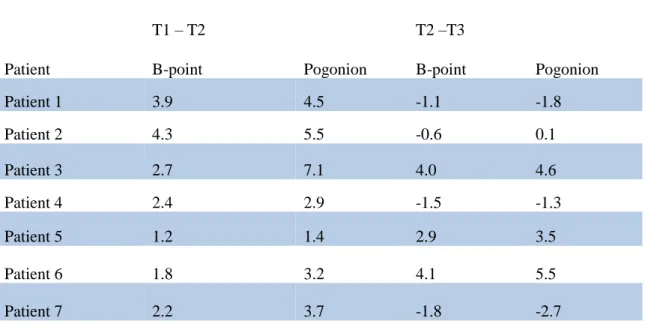
Table 2 – Mandibular Skeletal Changes measured from Cranial Base Superimpositions of B-point and Pogonion (mm).
Table II: Mandibular Skeletal Changes for each Herbst Subject (mm)
T1 – T2 T2 –T3
Patient B-point Pogonion B-point Pogonion
Patient 1 3.9 4.5 -1.1 -1.8
Patient 2 4.3 5.5 -0.6 0.1
Patient 3 2.7 7.1 4.0 4.6
Patient 4 2.4 2.9 -1.5 -1.3
Patient 5 1.2 1.4 2.9 3.5
Patient 6 1.8 3.2 4.1 5.5
Patient 7 2.2 3.7 -1.8 -2.7
Table I: Demographics for Herbst Subjects
Patient Sex (M/F) Age T1 Age T2 Age T3 ANB A-N Perp B-N Perp U1-SN MPA IMPA
Patient 1 M 13y 7m 14y 8m 16y 0m 4.9 2.1 -3.3 95,1 25.3 92.6
Patient 2 M 13y 11m 15y 1m 16y 2m 6.1 7.7 2.8 109.9 21.1 93.9
Patient 3 M 13y 7m 14y 9m 15y 11m 4.0 -2.6 -11.0 115.8 19.8 99.5
Patient 4 F 14y 1m 15y 2m 16y 3m 5.5 0.6 -7.8 91.3 26.4 100.5
Patient 5 F 13y 2m 14y 4m 15y 9m 4.0 1.9 -3.3 96.1 29.1 83.9
Patient 6 M 13y 1m 14y 10m 16y 4m 6.5 -1.6 -15.1 99.1 37.5 89.3
Table 3 – Mandibular Growth measured at condylion for subjects during Herbst treatment (T1-T2) and post Herbst Treatment (T2-T3). Measurements taken from Mandibular Regional Superimposition models.
Table III: Mandibular Growth measured at condylion (mm)
T1 – T2 T2 –T3
Patient Right Left Right Left
Patient 1 5.5 4.6 2.6 3.1
Patient 2 6.2 5.6 1.9 2.3
Patient 3 11.2 10.3 4.1 4.3
Patient 4 3.3 3.6 2.9 2.1
Patient 5 4 3.6 2.3 1.8
Patient 6 5.2 6.3 4.9 3.3
Table 4 – Maxillary Skeletal Changes measured from models registered on the Anterior Cranial Base during Herbst treatment and one year post Herbst treatment (mm).
Table IX: Maxillary Skeletal Changes for each Herbst Subject (mm)
T1 – T2 T2 –T3
Patient A-point ANS A-point ANS
Patient 1 -1.5 0.8 0.8 0.4
Patient 2 -1.7 -1.8 0.1 -0.1
Patient 3 -1.4 1.4 3.1 4.6
Patient 4 -0.4 -0.7 -0.1 -0.3
Patient 5 -1.3 0.6 0.6 1.4
Patient 6 -1.3 0.5 1.4 0.4
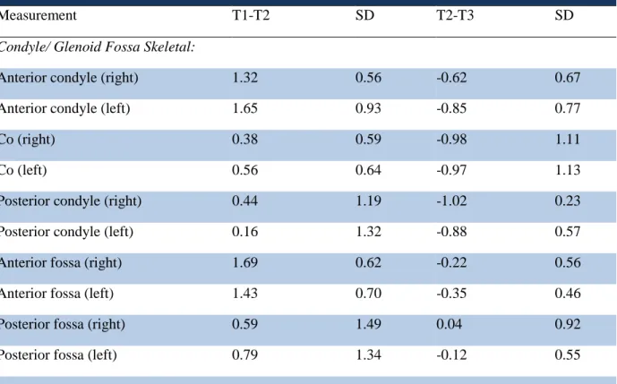
Table 5 – Positional Changes observed in the condyle and glenoid fossa of subjects during Herbst treatment and one year post Herbst treatment (mm).
Table V: Difference between T1-T2 and T2-T3 condyle/ fossa positional changes for Herbst subjects (mm)
Measurement T1-T2 SD T2-T3 SD
Condyle/ Glenoid Fossa Skeletal:
Anterior condyle (right) 1.32 0.56 -0.62 0.67
Anterior condyle (left) 1.65 0.93 -0.85 0.77
Co (right) 0.38 0.59 -0.98 1.11
Co (left) 0.56 0.64 -0.97 1.13
Posterior condyle (right) 0.44 1.19 -1.02 0.23
Posterior condyle (left) 0.16 1.32 -0.88 0.57
Anterior fossa (right) 1.69 0.62 -0.22 0.56
Anterior fossa (left) 1.43 0.70 -0.35 0.46
Posterior fossa (right) 0.59 1.49 0.04 0.92
Figures
Figure 1 - Mandibular Mask Region of Interest (ROI) segmentation. Mandibular regional voxel based registration was performed using a ROI created from each time point.
Figure 2 - A. T1 (white) to T2 (red) mandibular superimposition registered on the anterior cranial base showing anterior projection of B-point and pogonion. B. T2 to T3 (blue) mandibular
superimposition registered on the anterior cranial base demonstrating relapse at B-point and pogonion one year post Herbst removal. C. Condyles segmented from a cranial base
REFERENCES
1. Cevidanes LH, Oliveira AE, Grauer D, Styner M, Proffit WR. Clinical application of 3D imaging for assessment of treatment outcomes. Semin Orthod. 2011;17(1):72-80. doi: 10.1053/j.sodo.2010.08.012 [doi].
2. Cevidanes LH, Styner MA, Proffit WR. Image analysis and superimposition of 3-dimensional cone-beam computed tomography models. Am J Orthod Dentofacial Orthop. 2006;129(5):611-618. doi: S0889-5406(05)01275-8 [pii].
3. Cevidanes LH, Heymann G, Cornelis MA, DeClerck HJ, Tulloch JF. Superimposition of 3-dimensional cone-beam computed tomography models of growing patients. Am J Orthod Dentofacial Orthop. 2009;136(1):94-99. doi: 10.1016/j.ajodo.2009.01.018 [doi].
4. Houston WJ, Maher RE, McElroy D, Sherriff M. Sources of error in measurements from cephalometric radiographs. Eur J Orthod. 1986;8(3):149-151.
5. Spolyar JL. Head positioning error in cephalometric radiography--an implant study. Angle Orthod. 1987;57(1):77-88. doi: 10.1043/0003-3219(1987)057<0077:HPEICR>2.0.CO;2 [doi].
6. LeCornu M, Cevidanes LH, Zhu H, Wu CD, Larson B, Nguyen T. Three-dimensional treatment outcomes in class II patients treated with the herbst appliance: A pilot study. Am J Orthod Dentofacial Orthop. 2013;144(6):818-830. doi: 10.1016/j.ajodo.2013.07.014 [doi].
7. BJORK A. Variations in the growth pattern of the human mandible: Longitudinal radiographic study by the implant method. J Dent Res. 1963;42(1)Pt 2:400-411.
8. Bjork A. Prediction of mandibular growth rotation. Am J Orthod. 1969;55(6):585-599.
9. Bjork A, Skieller V. Normal and abnormal growth of the mandible. A synthesis of
longitudinal cephalometric implant studies over a period of 25 years. Eur J Orthod. 1983;5(1):1-46.
10. BJORK A. Facial growth in man, studied with the aid of metallic implants. Acta Odontol Scand. 1955;13(1):9-34.
11. Proffit W, Fields H, Sarver D, eds. Contemporary orthodontics. Fourth Edition ed. St. Louis, Mo: Mosby Elsevier; 2007.
13. Manfredi C, Cimino R, Trani A, Pancherz H. Skeletal changes of herbst appliance therapy investigated with more conventional cephalometrics and european norms. Angle Orthod. 2001;71(3):170-176. doi: 10.1043/0003-3219(2001)071<0170:SCOHAT>2.0.CO;2 [doi].
14. Pancherz H, Fischer S. Amount and direction of temporomandibular joint growth changes in herbst treatment: A cephalometric long-term investigation. Angle Orthod. 2003;73(5):493-501. doi: 10.1043/0003-3219(2003)073<0493:AADOTJ>2.0.CO;2 [doi].
15. Baccetti T, Franchi L, McNamara JA,Jr. An improved version of the cervical vertebral maturation (CVM) method for the assessment of mandibular growth. Angle Orthod. 2002;72(4):316-323. doi: 10.1043/0003-3219(2002)072<0316:AIVOTC>2.0.CO;2 [doi].
16. Pancherz H. The herbst appliance--its biologic effects and clinical use. Am J Orthod. 1985;87(1):1-20.
17. Pancherz H, Stickel A. Position changes of mandibular condyle in herbst treatment. radiographic study. Inf Orthod Kieferorthop. 1989;21(4):515-527.
18. Bock N, Pancherz H. Herbst treatment of class II division 1 malocclusions in retrognathic and prognathic facial types. Angle Orthod. 2006;76(6):930-941. doi: 10.2319/100605-352 [doi].
19. Pancherz H. The mechanism of class II correction in herbst appliance treatment. A cephalometric investigation. Am J Orthod. 1982;82(2):104-113.
20. Valant JR, Sinclair PM. Treatment effects of the herbst appliance. Am J Orthod Dentofacial Orthop. 1989;95(2):138-147. doi: 0889-5406(89)90392-2 [pii].
21. VanLaecken R, Martin CA, Dischinger T, Razmus T, Ngan P. Treatment effects of the edgewise herbst appliance: A cephalometric and tomographic investigation. Am J Orthod Dentofacial Orthop. 2006;130(5):582-593. doi: S0889-5406(06)00890-0 [pii].
22. Pancherz H, Anehus-Pancherz M. The headgear effect of the herbst appliance: A
cephalometric long-term study. Am J Orthod Dentofacial Orthop. 1993;103(6):510-520. doi: 0889-5406(93)70090-B [pii].
23. McNamara JA,Jr, Carlson DS. Quantitative analysis of temporomandibular joint adaptations to protrusive function. Am J Orthod. 1979;76(6):593-611.
24. Voudouris JC, Woodside DG, Altuna G, et al. Condyle-fossa modifications and muscle interactions during herbst treatment, part 2. results and conclusions. Am J Orthod Dentofacial Orthop. 2003;124(1):13-29. doi: 10.1016/S0889540603001501 [doi].
25. Baltromejus S, Ruf S, Pancherz H. Effective temporomandibular joint growth and chin position changes: Activator versus herbst treatment. A cephalometric roentgenographic study.
26. Buschang PH, Santos-Pinto A. Condylar growth and glenoid fossa displacement during childhood and adolescence. Am J Orthod Dentofacial Orthop. 1998;113(4):437-442. doi: S0889-5406(98)80016-4 [pii].
27. Agronin KJ, Kokich VG. Displacement of the glenoid fossa: A cephalometric evaluation of growth during treatment. Am J Orthod Dentofacial Orthop. 1987;91(1):42-48. doi: 0889-5406(87)90207-1 [pii].
28. Ruf S, Pancherz H. Herbst/multibracket appliance treatment of class II division 1
malocclusions in early and late adulthood. a prospective cephalometric study of consecutively treated subjects. Eur J Orthod. 2006;28(4):352-360. doi: cji116 [pii].
29. Pancherz H, Hansen K. Occlusal changes during and after herbst treatment: A cephalometric investigation. Eur J Orthod. 1986;8(4):215-228.
30. Pancherz H. Treatment of class II malocclusions by jumping the bite with the herbst appliance. A cephalometric investigation. Am J Orthod. 1979;76(4):423-442.
31. Nelson B, Hagg U, Hansen K, Bendeus M. A long-term follow-up study of class II
malocclusion correction after treatment with class II elastics or fixed functional appliances. Am J Orthod Dentofacial Orthop. 2007;132(4):499-503. doi: S0889-5406(07)00585-9 [pii].
32. McNamara JA,Jr, Bryan FA. Long-term mandibular adaptations to protrusive function: An experimental study in macaca mulatta. Am J Orthod Dentofacial Orthop. 1987;92(2):98-108.
33. Pancherz H, Bjerklin K, Lindskog-Stokland B, Hansen K. Thirty-two-year follow-up study of herbst therapy: A biometric dental cast analysis. Am J Orthod Dentofacial Orthop.