The Role of Membrane Carbohydrates in
Cell-Cell Recognition
Cells recognize each other by binding to surface
molecules, often containing carbohydrates, on the extracellular surface of the plasma membrane
Membrane carbohydrates may be covalently bonded
to lipids (forming glycolipids) or, more commonly, to
proteins (forming glycoproteins)
Carbohydrates on the external side of the plasma
CONCEPT 5.6: The plasma membrane plays a
key role in most cell signaling
In multicellular organisms, cell-to-cell communication
allows the cells of the body to coordinate their activities
Communication between cells is also essential for
Local and Long-Distance Signaling
Eukaryotic cells may communicate by direct contact
Animal and plant cells have junctions that directly
connect the cytoplasm of adjacent cells
These are called gap junctions (animal cells) and
plasmodesmata (plant cells)
The free passage of substances in the cytosol from
In many other cases of local signaling, messenger molecules are secreted by a signaling cell
These messenger molecules, called local
regulators, travel only short distances
One class of these, growth factors, stimulates
nearby cells to grow and divide
This type of local signaling in animal cells is called
Figure 5.19 Local regulator diffuses through extracellular fluid. Secreting cell Secretory vesicle Target cell
Local signaling Long-distance signaling
Target cell is stimulated.
Electrical signal along nerve cell triggers release of transmitter. Neurotransmitter diffuses across synapse.
(a) Paracrine signaling (b) Synaptic signaling
Endocrine cell
Target cell specifically binds hormone.
Figure 5.19a
Local regulator diffuses through Secreting
cell Secretory
vesicle Target cell
Another more specialized type of local signaling occurs in the animal nervous system
This synaptic signaling consists of an electrical signal
moving along a nerve cell that triggers secretion of neurotransmitter molecules
These diffuse across the space between the nerve
Figure 5.19b
Target cell
Electrical signal along nerve cell triggers release of
transmitter.
Neurotransmitter diffuses across synapse.
In long-distance signaling, plants and animals use
chemicals called hormones
In hormonal signaling in animals (called endocrine
signaling), specialized cells release hormone molecules that travel via the circulatory system
Figure 5.19c
Long-distance signaling
Endocrine cell
Target cell specifically binds
hormone.
Hormone travels in
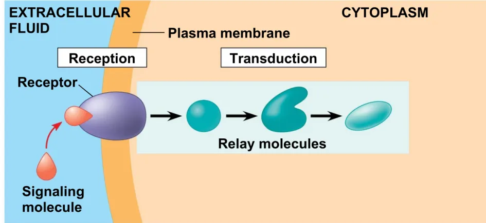
The Three Stages of Cell Signaling:
A Preview
Earl W. Sutherland discovered how the hormone
epinephrine acts on cells
Sutherland suggested that cells receiving signals
undergo three processes
Reception
Transduction
Figure 5.20-1
EXTRACELLULAR
FLUID Plasma membrane
Reception Receptor
Signaling molecule
Figure 5.20-2
EXTRACELLULAR FLUID
CYTOPLASM Plasma membrane
Reception Transduction
Relay molecules Receptor
Figure 5.20-3
EXTRACELLULAR FLUID
CYTOPLASM Plasma membrane
Response Reception Transduction
Relay molecules
Activation Receptor
Reception, the Binding of a Signaling Molecule to
a Receptor Protein
The binding between a signal molecule (ligand) and
receptor is highly specific
Ligand binding generally causes a shape change in
the receptor
Many receptors are directly activated by this shape
change
Receptors in the Plasma Membrane
Most water-soluble signal molecules bind to
specific sites on receptor proteins that span the plasma membrane
There are two main types of membrane receptors
G protein-coupled receptors
G protein-coupled receptors (GPCRs) are plasma
membrane receptors that work with the help of a G
protein
G proteins bind to the energy-rich molecule GTP
The G protein acts as an on-off switch: If GTP is
bound to the G protein, the G protein is inactive
Many G proteins are very similar in structure
Figure 5.21-1
CYTOPLASM
Plasma membrane
Activated G protein
Signaling molecule Inactiveenzyme Activated
Figure 5.21-2
CYTOPLASM
Plasma membrane
Activated G protein
Activated enzyme Signaling molecule Inactiveenzyme Activated
GPCR 1
A ligand-gated ion channel receptor acts as a “gate” for ions when the receptor changes shape
When a signal molecule binds as a ligand to the
receptor, the gate allows specific ions, such as Na+
or Ca2+, through a channel in the receptor
Ligand-gated ion channels are very important in the
nervous system
Figure 5.22-1
Ions
Plasma membrane Signaling
molecule (ligand) 1
Gate closed
Ligand-gated
Figure 5.22-2 Ions Plasma membrane Signaling molecule (ligand) 1 Cellular response Gate open Gate closed Ligand-gated
ion channel receptor
Figure 5.22-3 Ions Plasma membrane Signaling molecule (ligand) 1 Cellular response Gate open Gate closed Gate closed Ligand-gated
ion channel receptor
2
Intracellular Receptors
Intracellular receptor proteins are found in the cytosol
or nucleus of target cells
Small or hydrophobic chemical messengers can
readily cross the membrane and activate receptors
Examples of hydrophobic messengers are the steroid
Testosterone behaves similarly to other steroid hormones
Only cells that contain receptors for testosterone can
respond to it
The hormone binds the receptor protein and
activates it
The active form of the receptor enters the nucleus,
Figure 5.23
DNA
Plasma membrane
Hormone- receptor complex Receptor
protein Hormone (testosterone)
mRNA
EXTRA-CELLULAR
Figure 5.23a
Plasma membrane
Hormone- receptor complex Receptor
protein Hormone
(testosterone)
EXTRA-CELLULAR
FLUID
Figure 5.23b
DNA Hormone-
receptor complex
New protein mRNA
Transduction by Cascades of Molecular
Interactions
Signal transduction usually involves multiple steps
Multistep pathways can amplify a signal: A few
molecules can produce a large cellular response
Multistep pathways provide more opportunities for
The molecules that relay a signal from receptor to response are mostly proteins
Like falling dominoes, the receptor activates
another protein, which activates another, and so on, until the protein producing the response is activated
At each step, the signal is transduced into a
Protein Phosphorylation and Dephosphorylation
Phosphorylation and dephosphorylation are a
widespread cellular mechanism for regulating protein activity
Protein kinases transfer phosphates from ATP to protein, a process called phosphorylation
The addition of phosphate groups often changes the
Figure 5.24
Receptor
Signaling molecule
Activated relay molecule
Inactive protein
kinase 1 Ph
Figure 5.24a
Receptor
Signaling molecule
Activated relay molecule
Inactive protein kinase 1
Figure 5.24b
Inactive protein kinase 2
Active protein kinase 1
Figure 5.24c
Active protein kinase 2
Active protein Inactive
protein
Protein phosphatases remove the phosphates from proteins, a process called dephosphorylation
Phosphatases provide a mechanism for turning off
the signal transduction pathway
They also make protein kinases available for reuse,
Small Molecules and Ions as Second Messengers
The extracellular signal molecule (ligand) that binds
to the receptor is a pathway’s “first messenger”
Second messengers are small, nonprotein, water-soluble molecules or ions that spread throughout a cell by diffusion
Cyclic AMP and calcium ions are common second
Cyclic AMP (cAMP) is one of the most widely used second messengers
Adenylyl cyclase, an enzyme in the plasma
membrane, rapidly converts ATP to cAMP in response to a number of extracellular signals
The immediate effect of cAMP is usually the
Figure 5.25
G protein-coupled receptor
Protein kinase A Second messenger Adenylyl cyclase G protein
Response: Regulation of Transcription or
Cytoplasmic Activities
Ultimately, a signal transduction pathway leads to
regulation of one or more cellular activities
The response may occur in the cytoplasm or in the
nucleus
Many signaling pathways regulate the synthesis of
enzymes or other proteins, usually by turning genes on or off in the nucleus
Figure 5.26
Phosphorylation cascade
Inactive
transcription factor
CYTOPLASM
DNA
Active
transcription
factor Response
Reception
Transduction Growth factor
Figure 5.26a
Phosphorylation cascade
CYTOPLASM
Reception
Transduction Growth factor
Figure 5.26b
Phosphorylation cascade
Inactive
transcription factor
CYTOPLASM
DNA
Gene Active
transcription
factor Response
The Evolution of Cell Signaling
Biologists have discovered some universal
mechanisms of cellular regulation, evidence of the evolutionary relatedness of all life
Scientists think that signaling mechanisms first
evolved in ancient prokaryotes and single-celled eukaryotes
These mechanisms were adopted for new uses in
Figure 5.UN01 60 40 20 100 80
1-month-old guinea pig 15-day-old guinea pig
Glucose Uptake Over Time in Guinea Pig Red Blood Cells
Figure 5.UN02
Severe disease LDL receptor
Mild disease Normal
Figure 5.UN03
Channel
protein Carrierprotein
Figure 5.UN04
Figure 5.UN05
Signaling molecule
Activation of cellular response Relay molecules
Receptor
Reception Transduction Response