LECTURE PRESENTATIONS
For CAMPBELL BIOLOGY, NINTH EDITION
Jane B. Reece, Lisa A. Urry, Michael L. Cain, Steven A. Wasserman, Peter V. Minorsky, Robert B. Jackson
© 2011 Pearson Education, Inc.
Lectures by Erin Barley Kathleen Fitzpatrick
Cell Communication
Overview: Cellular Messaging
• Cell-to-cell communication is essential for both
multicellular and unicellular organisms
• Biologists have discovered some universal
mechanisms of cellular regulation
• Cells most often communicate with each other
via chemical signals
• For example, the fight-or-flight response is
triggered by a signaling molecule called epinephrine
Concept 11.1: External signals are
converted to responses within the cell
• Microbes provide a glimpse of the role of cell
signaling in the evolution of life
Evolution of Cell Signaling
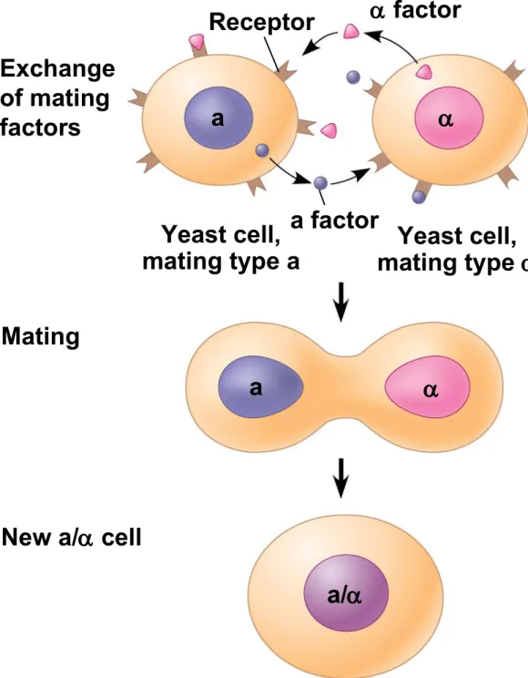
• The yeast, Saccharomyces cerevisiae, has two
mating types, a and
• Cells of different mating types locate each other
via secreted factors specific to each type
• A signal transduction pathway is a series of steps by which a signal on a cell’s surface is converted into a specific cellular response
• Signal transduction pathways convert signals on
a cell’s surface into cellular responses
Figure 11.2
Exchange of mating factors
Receptor factor
a factor Yeast cell,
mating type a mating type Yeast cell, Mating
New a/ cell
1
2
3
a
a
a/
• Pathway similarities suggest that ancestral
signaling molecules evolved in prokaryotes and were modified later in eukaryotes
• The concentration of signaling molecules allows
bacteria to sense local population density
Individual rod-shaped cells
Spore-forming structure
(fruiting body)
Aggregation in progress
Fruiting bodies
1
2
3
0.5 mm
2.5 mm
Figure 11.3a
Individual rod-shaped cells
Figure 11.3b
Aggregation in progress
Figure 11.3c
Spore-forming structure (fruiting body)
0.5 mm
Figure 11.3d
Fruiting bodies
Local and Long-Distance Signaling
• Cells in a multicellular organism communicate by
chemical messengers
• Animal and plant cells have cell junctions that
directly connect the cytoplasm of adjacent cells
• In local signaling, animal cells may communicate
by direct contact, or cell-cell recognition
Figure 11.4
Plasma membranes
Gap junctions between animal cells
Plasmodesmata between plant cells (a) Cell junctions
• In many other cases, animal cells communicate
using local regulators, messenger molecules that
travel only short distances
• In long-distance signaling, plants and animals use
chemicals called hormones
• The ability of a cell to respond to a signal depends
on whether or not it has a receptor specific to that signal
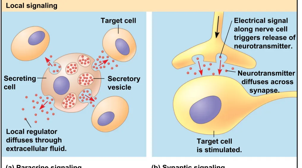
Figure 11.5
Local signaling Long-distance signaling Target cell
Secreting
cell Secretoryvesicle
Local regulator diffuses through extracellular fluid.
(a) Paracrine signaling (b) Synaptic signaling
Electrical signal along nerve cell triggers release of neurotransmitter. Neurotransmitter diffuses across synapse. Target cell is stimulated.
Endocrine cell Blood vessel Hormone travels in bloodstream. Target cell specifically binds hormone.
Figure 11.5a Local signaling Target cell Secreting cell Secretory vesicle Local regulator diffuses through extracellular fluid.
(a) Paracrine signaling (b) Synaptic signaling
Figure 11.5b
Long-distance signaling
Endocrine cell Blood vessel
Hormone travels in bloodstream.
Target cell specifically binds
hormone.
The Three Stages of Cell Signaling:
A Preview
• Earl W. Sutherland discovered how the hormone
epinephrine acts on cells
• Sutherland suggested that cells receiving signals
went through three processes
– Reception
– Transduction – Response
© 2011 Pearson Education, Inc.
Animation: Overview of Cell Signaling
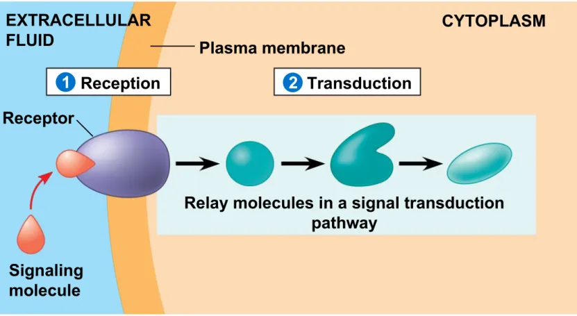
Figure 11.6-1
Plasma membrane EXTRACELLULAR
FLUID
CYTOPLASM
Reception
Receptor
Signaling molecule
Figure 11.6-2
Plasma membrane EXTRACELLULAR
FLUID
CYTOPLASM
Reception Transduction
Receptor
Signaling molecule
Relay molecules in a signal transduction pathway
Figure 11.6-3
Plasma membrane EXTRACELLULAR
FLUID
CYTOPLASM
Reception Transduction Response
Receptor
Signaling molecule
Activation of cellular response Relay molecules in a signal transduction
pathway
3 2
Concept 11.2: Reception: A signaling
molecule binds to a receptor protein, causing
it to change shape
• The binding between a signal molecule (ligand)
and receptor is highly specific
• A shape change in a receptor is often the initial
transduction of the signal
• Most signal receptors are plasma membrane
proteins
Receptors in the Plasma Membrane
• Most water-soluble signal molecules bind to
specific sites on receptor proteins that span the plasma membrane
• There are three main types of membrane
receptors
– G protein-coupled receptors – Receptor tyrosine kinases – Ion channel receptors
• G protein-coupled receptors (GPCRs) are the largest family of cell-surface receptors
• A GPCR is a plasma membrane receptor that
works with the help of a G protein
• The G protein acts as an on/off switch: If GDP is
bound to the G protein, the G protein is inactive
Figure 11.7a
G protein-coupled receptor
Signaling molecule binding site
Figure 11.7b G protein-coupled receptor 2 1 3 4 Plasma membrane G protein (inactive) CYTOPLASM Enzyme Activated
receptor Signalingmolecule Inactiveenzyme
Activated enzyme Cellular response GDP GTP GDP GTP GTP
P i GDP
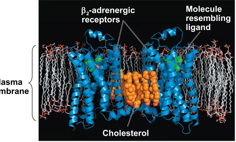
Figure 11.8
Plasma membrane
Cholesterol
2-adrenergic receptors
• Receptor tyrosine kinases (RTKs) are
membrane receptors that attach phosphates to tyrosines
• A receptor tyrosine kinase can trigger multiple
signal transduction pathways at once
• Abnormal functioning of RTKs is associated with
many types of cancers
Figure 11.7c Signaling molecule (ligand) 2 1 3 4 Ligand-binding site
helix in the membrane
Tyrosines
CYTOPLASM Receptor tyrosine
kinase proteins (inactive monomers) Signaling molecule Dimer Tyr Tyr Tyr Tyr Tyr Tyr Tyr Tyr Tyr Tyr Tyr Tyr Tyr Tyr Tyr Tyr Tyr Tyr Tyr Tyr Tyr Tyr Tyr Tyr Tyr Tyr Tyr Tyr Tyr Tyr Tyr Tyr Tyr Tyr Tyr Tyr P P P P P P P P P P P P Activated tyrosine kinase regions (unphosphorylated dimer) Fully activated receptor tyrosine kinase (phosphorylated dimer) Activated relay proteins Cellular response 1 Cellular response 2 Inactive relay proteins
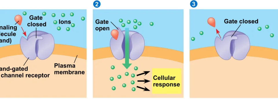
• A ligand-gated ion channel receptor acts as a gate when the receptor changes shape
• When a signal molecule binds as a ligand to the
receptor, the gate allows specific ions, such as
Na+ or Ca2+, through a channel in the receptor
Figure 11.7d
Signaling molecule (ligand)
2
1 3
Gate
closed Ions
Ligand-gated
ion channel receptor
Plasma membrane
Gate open
Cellular response
Intracellular Receptors
• Intracellular receptor proteins are found in the
cytosol or nucleus of target cells
• Small or hydrophobic chemical messengers can
readily cross the membrane and activate receptors
• Examples of hydrophobic messengers are the
steroid and thyroid hormones of animals
• An activated hormone-receptor complex can act
as a transcription factor, turning on specific genes
Figure 11.9-1
Hormone (testosterone)
Receptor protein
Plasma membrane
DNA
NUCLEUS
CYTOPLASM
Figure 11.9-2
Hormone (testosterone)
Receptor protein
Plasma membrane
Hormone-receptor complex
DNA
NUCLEUS
CYTOPLASM
Figure 11.9-3
Hormone (testosterone)
Receptor protein
Plasma membrane
Hormone-receptor complex
DNA
NUCLEUS
CYTOPLASM
Figure 11.9-4
Hormone (testosterone)
Receptor protein
Plasma membrane
Hormone-receptor complex
DNA
mRNA
NUCLEUS
CYTOPLASM
Figure 11.9-5
Hormone (testosterone)
Receptor protein
Plasma membrane EXTRACELLULAR FLUID
Hormone-receptor complex
DNA
mRNA
NUCLEUS
CYTOPLASM
Concept 11.3: Transduction: Cascades of
molecular interactions relay signals from
receptors to target molecules in the cell
• Signal transduction usually involves multiple steps
• Multistep pathways can amplify a signal: A few
molecules can produce a large cellular response
• Multistep pathways provide more opportunities for
coordination and regulation of the cellular response
Signal Transduction Pathways
• The molecules that relay a signal from receptor to
response are mostly proteins
• Like falling dominoes, the receptor activates
another protein, which activates another, and so on, until the protein producing the response is activated
• At each step, the signal is transduced into a
different form, usually a shape change in a protein
Protein Phosphorylation and
Dephosphorylation
• In many pathways, the signal is transmitted by a
cascade of protein phosphorylations
• Protein kinases transfer phosphates from ATP to protein, a process called phosphorylation
• Protein phosphatases remove the phosphates from proteins, a process called dephosphorylation
• This phosphorylation and dephosphorylation
system acts as a molecular switch, turning
activities on and off or up or down, as required
Receptor Signaling molecule Activated relay molecule Ph os ph or yla tio n c as ca de Inactive protein kinase 1 Active protein kinase 1 Active protein kinase 2 Active protein kinase 3 Inactive protein kinase 2 Inactive protein kinase 3 Inactive protein Active
protein Cellularresponse
ATP ADP ATP ADP ATP ADP PP PP PP P P P P i
P i
P i
Activated relay molecule Ph os ph or yla tio n c as ca de Inactive protein kinase 1 Active protein kinase 1 Active protein kinase 2 Active protein kinase 3 Inactive protein kinase 2 Inactive protein kinase 3 Inactive protein Active protein ATP ADP ATP ADP ATP ADP PP PP PP P P P i
P i
P i
P
Small Molecules and Ions as Second
Messengers
• The extracellular signal molecule (ligand) that
binds to the receptor is a pathway’s “first messenger”
• Second messengers are small, nonprotein, water-soluble molecules or ions that spread throughout a cell by diffusion
• Second messengers participate in pathways
initiated by GPCRs and RTKs
• Cyclic AMP and calcium ions are common second
messengers
Cyclic AMP
• Cyclic AMP (cAMP) is one of the most widely used second messengers
• Adenylyl cyclase, an enzyme in the plasma
membrane, converts ATP to cAMP in response to an extracellular signal
Figure 11.11
Adenylyl cyclase Phosphodiesterase
Pyrophosphate
AMP
H2O
ATP
P i P
Figure 11.11a
Adenylyl cyclase
Pyrophosphate
ATP
P i
P
Figure 11.11b
Phosphodiesterase
AMP H2O
cAMP
• Many signal molecules trigger formation of cAMP
• Other components of cAMP pathways are G
proteins, G protein-coupled receptors, and protein kinases
• cAMP usually activates protein kinase A, which
phosphorylates various other proteins
• Further regulation of cell metabolism is provided
by G-protein systems that inhibit adenylyl cyclase
Figure 11.12
G protein First messenger (signaling molecule such as epinephrine)
G protein-coupled receptor
Adenylyl cyclase
Second messenger
Cellular responses Protein kinase A GTP
ATP
Calcium Ions and Inositol Triphosphate (IP
3)
• Calcium ions (Ca2+) act as a second messenger in
many pathways
• Calcium is an important second messenger
because cells can regulate its concentration
Figure 11.13
Mitochondrion EXTRACELLULAR
FLUID Plasmamembrane
Ca2
pump
Nucleus CYTOSOL
Ca2
pump Ca2
pump Endoplasmic reticulum (ER) ATP ATP
Low [Ca2]
High [Ca2]
• A signal relayed by a signal transduction pathway may trigger an increase in calcium in the cytosol
• Pathways leading to the release of calcium involve
inositol triphosphate (IP3) and diacylglycerol (DAG) as additional second messengers
© 2011 Pearson Education, Inc.
Animation: Signal Transduction Pathways
G protein EXTRA-CELLULAR FLUID Signaling molecule (first messenger) G protein-coupled
receptor Phospholipase C
DAG
PIP2
IP3
(second messenger)
IP3-gated
calcium channel
Endoplasmic reticulum (ER)
CYTOSOL
Ca2
GTP
Figure 11.14-2 G protein EXTRA-CELLULAR FLUID Signaling molecule (first messenger) G protein-coupled
receptor Phospholipase C
DAG
PIP2
IP3
(second messenger)
IP3-gated
calcium channel
Endoplasmic reticulum (ER)
CYTOSOL
Ca2
(second messenger) Ca2
Figure 11.14-3 G protein EXTRA-CELLULAR FLUID Signaling molecule (first messenger) G protein-coupled
receptor Phospholipase C
DAG PIP2
IP3
(second messenger)
IP3-gated
calcium channel Endoplasmic reticulum (ER) CYTOSOL Various proteins activated Cellular responses Ca2
(second messenger) Ca2
Concept 11.4: Response: Cell signaling leads
to regulation of transcription or cytoplasmic
activities
• The cell’s response to an extracellular signal is
sometimes called the “output response”
Nuclear and Cytoplasmic Responses
• Ultimately, a signal transduction pathway leads to
regulation of one or more cellular activities
• The response may occur in the cytoplasm or in the
nucleus
• Many signaling pathways regulate the synthesis of
enzymes or other proteins, usually by turning genes on or off in the nucleus
• The final activated molecule in the signaling
pathway may function as a transcription factor
• Other pathways regulate the activity of enzymes rather than their synthesis
Figure 11.16
Reception
Transduction
Response
Binding of epinephrine to G protein-coupled receptor (1 molecule)
Inactive G protein
Active G protein (102 molecules)
Inactive adenylyl cyclase
Active adenylyl cyclase (102)
ATP
Cyclic AMP (104)
Inactive protein kinase A
Active protein kinase A (104)
Inactive phosphorylase kinase
Active phosphorylase kinase (105)
Inactive glycogen phosphorylase
Active glycogen phosphorylase (106)
Glycogen
•
Signaling pathways can also affect the
overall behavior of a cell, for example,
changes in cell shape
Wild type (with shmoos) Fus3 formin Mating factor activates receptor. Mating
factor G protein-coupled receptor
Shmoo projection forming
Formin
G protein binds GTP and becomes activated. 2 1 3 4 5 P P P P Formin Formin Fus3 Fus3 Fus3 GDP GTP lation cascade Microfilament Actin subunit Phosphorylation cascade activates Fus3, which moves to plasma membrane.
Fus3 phos-phorylates formin,
activating it.
Formin initiates growth of microfilaments that form the shmoo projections.
RESULTS
CONCLUSION
Figure 11.17a
Figure 11.17b
Figure 11.17c
Fine-Tuning of the Response
• There are four aspects of fine-tuning to consider
– Amplification of the signal (and thus the response) – Specificity of the response
– Overall efficiency of response, enhanced by scaffolding proteins
– Termination of the signal
Signal Amplification
• Enzyme cascades amplify the cell’s response
• At each step, the number of activated products is
much greater than in the preceding step
The Specificity of Cell Signaling and
Coordination of the Response
• Different kinds of cells have different collections of
proteins
• These different proteins allow cells to detect and
respond to different signals
• Even the same signal can have different effects in
cells with different proteins and pathways
• Pathway branching and “cross-talk” further help
the cell coordinate incoming signals
Figure 11.18 Signaling molecule Receptor Relay molecules Response 1
Cell A. Pathway leads to a single response.
Response 2 Response 3 Response 4 Response 5
Activation or inhibition
Cell B. Pathway branches, leading to two responses.
Cell C. Cross-talk occurs between two pathways.
Cell D. Different receptor leads to a different
Signaling molecule
Receptor
Relay molecules
Response 1
Cell A. Pathway leads to a single response.
Response 2 Response 3
Response 4 Response 5 Activation
or inhibition
Cell C. Cross-talk occurs between two pathways.
Cell D. Different receptor leads to a different
Signaling Efficiency: Scaffolding Proteins
and Signaling Complexes
• Scaffolding proteins are large relay proteins to which other relay proteins are attached
• Scaffolding proteins can increase the signal
transduction efficiency by grouping together
different proteins involved in the same pathway
• In some cases, scaffolding proteins may also help
activate some of the relay proteins
Figure 11.19
Signaling molecule
Receptor
Plasma
membrane
Scaffolding protein
Termination of the Signal
• Inactivation mechanisms are an essential aspect
of cell signaling
• If ligand concentration falls, fewer receptors will be
bound
• Unbound receptors revert to an inactive state
Concept 11.5: Apoptosis integrates multiple
cell-signaling pathways
• Apoptosis is programmed or controlled cell suicide
• Components of the cell are chopped up and
packaged into vesicles that are digested by scavenger cells
• Apoptosis prevents enzymes from leaking out of a
dying cell and damaging neighboring cells
Figure 11.20
Apoptosis in the Soil Worm
Caenorhabditis
elegans
• Apoptosis is important in shaping an organism
during embryonic development
• The role of apoptosis in embryonic development
was studied in Caenorhabditis elegans
• In C. elegans, apoptosis results when proteins that “accelerate” apoptosis override those that “put the brakes” on apoptosis
Figure 11.21
Mitochondrion Ced-9
protein (active)
inhibits Ced-4 activity Receptor for death-signaling molecule Ced-4 Ced-3 Inactive proteins
(a) No death signal
Death-signaling molecule
Ced-9
(inactive) Cellforms
blebs
Active
Ced-4 ActiveCed-3 Otherproteases
Nucleases Activation
cascade
Figure 11.21a
Mitochondrion Ced-9
protein (active)
inhibits Ced-4 activity
Receptor for death-signaling molecule
Ced-4 Ced-3
Inactive proteins
Death-signaling molecule
Ced-9
(inactive) Cellforms
blebs
Active
Ced-4 ActiveCed-3 Otherproteases
Nucleases Activation
cascade
(b) Death signal
Apoptotic Pathways and the Signals That
Trigger Them
• Caspases are the main proteases (enzymes that
cut up proteins) that carry out apoptosis
• Apoptosis can be triggered by
– An extracellular death-signaling ligand – DNA damage in the nucleus
– Protein misfolding in the endoplasmic reticulum
• Apoptosis evolved early in animal evolution and is essential for the development and maintenance of all animals
• Apoptosis may be involved in some diseases (for
example, Parkinson’s and Alzheimer’s);
interference with apoptosis may contribute to some cancers
Figure 11.22
Interdigital tissue
Cells undergoing
Figure 11.22a
Figure 11.22b
Figure 11.22c
Space between digits
Figure 11.UN01
Reception
1 2 Transduction 3 Response
Receptor
Signaling molecule
Relay molecules