Azizi Miskon*and Jatendra Uslama
Faculty of Electrical & Electronic Engineering, UniversitiTun Hussein Onn Malaysia, 86400 Parit Raja, Batu Pahat Johor Malaysia
* Corresponding author.
Abstract— The objective of this paper is to provide the funda-mental mathematical formula to predict the effect of magnetic fields (MF) on stem cell differentiation. The data were re-viewed from journals related to the effects of magnetic fields on stem cell differentiation. These data were given a value for differentiation which is related to their MF strength with these conditions; MF strength that does not affect the stem cell dif-ferentiation given the value of zero, MF strength that results in stem cells death given the value of 1, and the MF strength that affects stem cells The differentiation given the value between 0.1 and 0.9. graph was plotted according to these data and the mathematical equation is designed from the graph. From this review, we suggest that the intensity of MF that can affect the
stem cell differentiation is between 600µT and 9.4T in which
the cell differentiation will not occur with intensity of less than
10µT and intensity of more than 12T will cause the death of
stem cells. We also suggest that the limit of MF effects on stem
cell differentiation lies between 10 µT and 600 µT, and the
limit of MF strength that can lead to the death of stem cells lies between 9.4 T and 12 T. It can be concluded that the exposure of MF on stem cell differentiation depends not only on the MF intensity, but also on the period of exposure.
Keywords— Differentiation, Ion Resonance Frequency, Magnetic Fields, Stem Cells, Tissue Engineering.
I. INTRODUCTION
Stem cells are primitive cells, which are present in all or-ganisms and have the ability to divide and give rise to more stem cells, or switch to become more specialized cells in human body like cells in brain, heart, muscle, and kidney [1]. There are two types of stem cell; embryonic stem (ES) cell and adult stem cell. ES cells are pluripotent and har-vested from inner cell mass of blastocyst and possess the ability to differentiate into all of the specialized embryonic tissues [1], [2]. ES cells also may open the door to the rap-idly progressing field of therapeutic cell transplantation [3]. The adult stem cells are multipotent with capacity to differ-entiate or transdifferdiffer-entiate into cell types other than their tissue of origin [1]. Adult stem cells and progenitor cells can be found in the adult tissue. Both of these cells act as a repair system for body, replenishing the specialized cells,
and maintaining the normal regenerative of organs, such as blood, skin, or intestinal tissues.
Magnetic fields (MF) produced by moving electric charge and exist all around us like earth MF and manmade MF sources. Numerous static and alternating MF arising from man-made sources have possible biological effect [4]. There are many biological functions that are modulated by ex-tremely low frequency magnetic field (ELF-MF) [5]-[7]. However, there is not enough evidence that the ELF-MF could endanger the human health [8]. ELF-MF is MF with a range of frequency of 3 to 30Hz. Even so, MF has been shown to affect proliferation and growth factor expression in cultured cells [9]-[11] and also interfere with endorphinergic and cholinergic system [12]-[14]. Other than MF, electrical fields (EF) also have biological effects that can influence neural growth and orientation in vitro [15], and have been applied for the treatment of spinal cord injuries in recent clinical trials [16]. The response of cells to the EF was essen-tially passive and determined by the physical properties of the cell, however cells can also actively respond to EF [17]. Electromagnetic fields (EMF) are produced when electric current flows through an electrical conductor like power line [18]. Like MF and EF, EMF also has biological effects such as altered rate of cell growth [5], [19], altered quantities of RNA transcript and proteins [20], altered cell surface proper-ties [21] and effect on development [22]. However EMF-based technologies have not progressed to clinical translation and the reason for this is the scepticism due to differences in experimental exposure protocols and static MF (SMF) varia-tion applied in experiment [23].
differentiation or vice versa. Therefore, we may avoid un-necessary failure during the experimental works.
II. METHOD
The data from several journals related to the effects of MF on stem cells differentiation that were published in various journals from the year 2004 until 2010 were re-viewed [23]-[29] as shown in Table 1. Since EF and EMF can also affect the stem cells differentiation, this review only focuses on the effect of MF and EMF on the stem cells differentiation. The data in Table 1 were revalued according to the MF strength. The value for differentiation of related MF strength were given to these conditions (Table 2); MF strength that does not affect the stem cells differentiation was given the value of zero and MF strength that results in stem cells death was given the value of 1. For the MF strength that affects stem cells differentiation, the value was given between 0.1 and 0.9. The given value for MF group that affect the stem cells differentiation was done with the assumptions that the smaller or higher the value, the lesser the MF effect on differentiation. The data were plotted into graph by using Microsoft Office Excel 2010 software and the result of project presented in Graphical User Interface (GUI) design by using the Microsoft Visual Studio Ultimate 2010.
Table 1 MF strengths that were used in reviewed journals and classified into their corresponding group; with group 0 for MF strength does not affect the stem cells differentiation, group 1 for MF strength that effects the stem cells differentiation, and group 2 for MF strength that kill the stem cells
0)LQWHQVLW\7 *URXS
P
P
P
III. RESULT
From Table 2, we design the graph using Microsoft Of-fice Excel 2010 as shown in Fig. 1. From Table 2, we ob-served that the effect of MF on stem cell differentiation still
occur in 600µT but the differentiation is not observed in
10µT. This may indicate that the minimum strength of MF to influence the stem cell differentiation in frequency of 50Hz lies somewhere between 10µT and 600µT. We also observed that the effect of MF on differentiation of stem cells still occur in MF strength of 9.4T. However, such high field strength magnet like 9.4T is not easily available and such studies may not be translated into the clinical study because the current limits for magnetic field strengths ap-proved by U.S. Food and drug Administration (FDA) is 3T [24]. This is the reason that many of the research works were carried out using the magnetic fields less than 3T. The stem cells death will occur at the exposure of 12T for more than 24h. Therefore, the minimum exposure of MF before resulting in stem cells death lies between 9.4T and 12T.
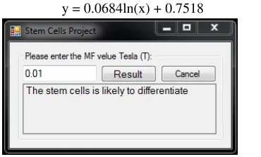
We revalued the data in Table 1 into Table 2, and as a result, the graph was plotted as depicted in Fig. 1. The graph is rea-sonable as the trait of MF effect on the stem cells is almost resembling the growth model. The closer the strength of MF to the lower asymptotes or upper asymptotes, the lesser the sig-nificance the differentiation is observed. The smaller the inten-sity of MF used, the lesser the possibility of MF to affect the stem cell differentiation. Whereas, the higher the intensity of MF used, the more possibilities that differentiation will not occur since stem cells were more likely to die. From the loga-rithmic graph, we design the mathematical function as shown in equation (1). From this equation, we can predict the strength of MF which is able to influence the stem cells differentiation ability. Referring to the equation (1), the value of ‘y’ is as-sumed to be ‘y=0’ for value of ‘x’ less than 10µT and ‘y=1’ for ‘x’ value more than 12T. The logarithmic function is inserted in GUI code shown in Fig. 2 so that it can be used in any future in vitro experiment.
y = 0.0684ln(x) + 0.7518 (1)
Fig. 2 GUI for predicting the MF effects on stem cells
IV. DISCUSSION
Cardiosperes (CDps) and cardiospere-derived cells (CDCs) exposed to extremely low frequency magnetic fields (ELF-MF) (10µT, 18Hz) did not affect the expression of cardiac and vascular markers (cTnl, Nkx 2.5, MHC, VEGF, KDR, and SMA) during the experiment, thus this may suggest an evidence that the stem cells did not differen-tiate with MF of 10µT [23]. Human Mesenchymal stem cells (hMSC) are multipotential cells and possess high rep-lication capacity, and there are potentials to differentiate into different lineages of mesenchymal tissue such as bone, cartilage, muscle, fat, and marrow stroma [30]. The expo-sure of human mesenchymal stem cells (hMSC) to 600µT enables the MSC to differentiate into adipogenic cells as indicated by enhanced expression of lipoprotein lipase and peroxisome proliferatoractivated receptor gamma [24]. In addition, the immediate exposure of MSC to MF has enhanced the cells differentiation. This research was ac-complished to investigate the effect of MF on hMSC and labelled with super magnetic particles of iron oxide (SPIO). This was done by using SPIO to label the cells, and detect ion was done by using Magnetic Resonance Imaging (MRI). The viability or differentiation potential of the cells has not been affected by SPIOlabelling of MSC [31], [32], but it has an impact on the iron metabolism, the migration capacity, and the colony formation of MSC [32]-[34]. The principle function of MRI is the exposure of cells to high MF and magnetic force that can direct the iron labelled stem cells in vitro and in vivo [35]-[37], guided localization of iron la-belled stem cells to the desired region [35], [38], for the seeding of scaffolds with stem cells [36], [39], or for the engineering of 3D tissues by stem cells [37]. Therefore, the effect of MF strength in vitro experiment should be carried out in order to verify the equation (1).
The stem cell is able to differentiate in a sinusoidal MF of 800µT with frequency of 50Hz. Real-time quantitative reverse transcriptase-polymerase chain reaction (RT-PCR)
analysis revealed a remarkably increased of GATA-4 and Nkx-2.5 mRNA expression for both embryoid bodies (EBs) and puromycinselected cardiomyocytes [25]. Hence, the result of exposing GTR1 embryonic stem cells with this MF was the differentiation into cardiac specific cells, and this experiment was done without an aid of gene transfer tech-nology [25]. As for information, GATA-4 and Nkx-2.5 mRNA encode respectively for a zinc finger containing transcription factor and homeodomain, and both of these have been shown to be essential for cardiogenesis in differ-ent animal species [40], [41], and human [42 ].
The effects of MF were also done on P19 embryonal car-cinoma cells (P19 cells) [26]. Differentiation was detected by exposing the P19 cells to 1mT of MF with frequency of 50Hz, however the analysis result was not very significant. By exposing P19 cells into more intense MF with the strength of 10mT, P19 cells were differentiated into neu-ronal cells [26]. The effects of ELF-MF after neuneu-ronal dif-ferentiation were evaluated by morphological analysis, immunochemical analysis (MAP2 and GFAP), and devel-opmental neuronal network activities recorded by the mi-cro-electrode arrays (MEAs). From the results, the percent-age of MAP2 positive cells and the spike frequencies had increased, but the percentage of GFAP positive cells has reduced. These results suggest that an exposure to 10mT ELF-MF would affect the characteristic of neuronal differ-entiation and functional neuronal properties [26]. These results also may suggest that the effect of MF on stem cell differentiation will become less significant with lower in-tensity of MF strength as verified by using our equation in Fig 1.
The stem cells are also able to differentiate in MF inten-sity of 1.1mT as demonstrated in previous work using the bone morrow stem cells (BMSC) [27]. It results in the dif-ferentiation of BMSC into osteogenesis and the increase of intracellular Ca2+ after MF stimulation. From this result,
they postulated that the elevated Ca2+ is possibly the
under-lying biochemical mechanism which is responsible for the induction of terminal differentiation [27].
As mentioned above, most of the experiments were done to investigate the effect of MF on stem cells ability to dif-ferentiate, and were performed with the intensity less than 3T. Even so, there are also experiments done with the MF intensity above 3T. The MF intensity between 4.7T and 9.4T were found to affect the stem cells differentiation [28]. Therefore, our range for cells differentiation was limited at 9.4T.
[image:3.595.90.275.87.198.2]of the osteogenesis of hMSC [29]. Therefore, we assumed that 12T and 16T still affect the cell differentiation for a short period of exposure. This response may be due to the synergistic effect of high magnetic force and MG existed in their experiment modelling systems [43]-[46]. Hence, from the data reviewed, we suggest that the MF intensity between 9.4T and 12T could lead to stem cell death.
As stated in the result section, the MF was classified into three groups according to their effects on stem cell differen-tiation. From these groups, we know that MF less than 600µT or more than 9.4T do not lead to stem cell differen-tiation. Also, the MF effect on stem cells differentiation will not occur at MF of 10µT and below. However, MF of 12T and above will result in stem cell death.
The effects to stem cells differentiation by MF ranged from 600µT to 9.4T vary according to the MF strength itself. It is likely that the MF of 10mT affects more cells differentiation as compared with 600µT or 9.4T. By using the mathematical equation (1), not only we can predict whether the strength of MF is able to affect the stem cells differentiation, but can also predict the strength of MF on stem cell differentiation. This is because the mathematical formula was designed after considering the analysis of the reviewed data.
Stem cells have been used in several pre-clinical models of disease [47]-[49], and are currently applied in clinical trials of phase I-III [50]-[53]. For example, neuronal stem cells (NSC) can differentiate into neuronal or glial cells and also express trophic factors, which may be used to rescue dysfunction brain tissue [54]-[58]. Stem cells are also used in tissue transplantation, and the optimal cell type to be transplanted should have these characters: (a) spontaneous disposition to integrate with the target tissue without induc-tion of immune reacinduc-tion; (b) differentiate into specific cells commitment; (c) have the capacity to develop gap junctions with host cells; and (d) have some degree of resistance to ischaemia, in order to avoid massive apoptosis, which is currently observed during cell transfer [23], [59]. The cur-rent findings of MF effect on stem cell diffecur-rentiation may open a new prospective, in particular the use of MF to direct the differentiation processes of stem cells into a specific cellular phenotype without the aid of gene transfer tech-nologies [25]. On the other hand, there are increasing public interests of possible health risk associated with ELF-MF [8], [18], [60]. Therefore, fundamental studies are necessary to ensure the safe method to differentiate stem cell into specific cell through MF exposure.
In conclusion, we suggest that the intensity of MF that can affect stem cell differentiation is between 600µT and 9.4T. Also the differentiation will not occur if the intensity is less than 10µT and with intensity of more than 12T, it will cause death of stem cells. We also suggest that the
range of MF effects on stem cell differentiation lies between 10µT and 600µT. The limit of MF strength that can lead towards the death of stem cells lies between 9.4T and 12T. We conclude that the result of the exposure of MF on stem cells differentiation depends not only on the MF intensity, but also on the period of exposure.
REFERENCES
[1] R. Passier, C. Mummery,“Origin and use of embryonic and adult stem cells in differentiation and tissue repair,”Cardiovascular Re-search 2003, vol. 58, pp. 324-335.
[2] C.M. Metallo, S.M. Azarin, Lin ji, et al.,“Engineering tissue from human embryonic stem cells,” J. Cell Mol Med 2008, vol. 12(3), pp. 709-729.
[3] S.G. Nir, R. David, M. Zaruba, et al.,“Human embryonic stem cells for cardiovascular repair,” Cardiovascular Research 2003, vol. 58 (2), pp. 313-323.
[4] W.T. Kaune,“Introduction to power-frequency electric and magnetic fields,” Environmental Health Perspectives Supplements 1993, vol. 101, pp. 73-81.
[5] A.R. Liboff, T. Williams Jr., D.M. Strong,et al., “Time-varying magnetic fields: effects on DNA synthesis,”Science, 1984, vol. 223, pp. 818-820.
[6] J. Jajte, M. Zmyslony, J. Palus, et al., “Protective effects of mela-tonin against in vitro iron ions and 7 mT, 50 Hz, magnetic fields-induced DNA damage in rat lymphocytes,”Mutat Res 2001, vol. 483, pp. 57-64.
[7] S. Falone., A. Mirabilio, M.C. Carbone, et al., “Chronic exposure to 50Hz magnetic fields causes a significant weakening of antioxidant defence systems in aged rat brain,”Int J Biochem Cell Biol 2008, vol. 12, pp. 2762-2770.
[8] D.W. Zipse, “Health effects of extremely low frequency (50 and 60 Hertz) electric and magnetic fields,” IEEE 1991, vol. PCIC-91-09. [9] A. Cossarizza, D. Monti, F. Bersani, et al. “Extremely low frequency
pulsed electromagnetic fields increase cell proliferation in lympho-cytes from young and aged subjects,”Biochem. Biophys. Res. Com-mun. 1989, vol. 160, pp. 692-698.
[10] R. Cadossi, F. Bersani, A. Cossarizza, et al. “Lymphocytes and low-frequency electromagnetic fields,” FASEB J. 1992, vol. 6, pp. 2667– 2674.
[11] A. Cossarizza, , S. Angioni, , F. Petraglia, , et al. “Exposure to low-frequency pulsed electromagnetic fields increases interleukin-1 and interleukin-6 production by human peripheral blood mononuclear cells,” Exp. Cell Res. 1993, vol. 204, pp. 385–387.
[12] A.W. Thomas, M. Kavaliers, F.S. Prato, et al. “Pulsed magnetic field induced “analgesia” in the land snail, Cepaeanemoralis, and the ef-fects of mu, delta, and kappa opioid receptor agonists/antagonists” Peptides 1997, vol. 18, pp. 703–709.
[13] V.V. Vorobyov, E.A. Sosunov, N.I. Kukushkin, et al.“Weak com-bined magnetic field affects basic and morphine-induced rat’s EEG,” Brain Res. 1998, vol. 781, pp. 182–187.
[14] H. Lai, M.A. Carino, A. Horita, et al.“Effects of a 60 Hz magnetic field on central cholinergic systems of the rat,”Bioelectromagnetics1993, vol. 14, pp. 5–15.
[15] N.B. Patel, M.M. Poo, “Perturbation of the direction of neurite growth by pulsed and focal electric fields,” J Neurosci, 1984, vol. 4, pp. 2939-47.
[17] H.M. Gerard,“The use of electric fields in tissue engineering,” Or-ganogenesis 2004, vol. 4, pp. 11-17.
[18] R. Goodman, Y. Chizmadzhew, A. Shirley- Hender-son,“Electromagnetic fields and cells” Journal of Cellular Biochem-istry 1993, vol. 51, pp. 436-441.
[19] K. Takahashi, I. Kaneko, M. Date, et al.,“Effect of pulsing electro-magnetic fields on DNA synthesis in mammalian cells in cul-ture,”Experimentia 1986, vol. 42, pp. 185- 186.
[20] E. Czerska, H. Al-Baranzi, J. Casamento, et al.,“Comparison of the effect of elf fields on cnayc oncogene expression in normal and trans-formed human cells,” In Transection of the Bioelectromagnetic Soci-ety, Thirteenth Annual Meeting 1991. Salt Lake City, UT, B-2-14. [21] M.T. Marron, E.M. Goodman, P.T. Sharpe, et al.,“Low frequency
electric and magnetic fields have different effects on the cell sur-face,” FEBS Lett 1988, vol. 230, pp. 13-16.
[22] M.R. Delgado, J. Leal, J.L. Monteagudo, et al.,“Embryological changes induced by weak, extremely low frequency electromagnetic fields,” J Anat 1982, vol. 134, pp. 533-551.
[23] G. Roberto, L. Mario, B. Lucio, et al.,“Differentiation of human adult cardiac stem cells exposed to extremely low-frequency electromag-netic fields,” Cardiovascular Research 2009, vol. 82, pp. 411-420. [24] S. Richard, B. Rudiger, K. Rainer, et al.,“Functional investigation on
human mesenchymal stem cells exposed to magnetic fields and la-beled with clinically approved iron nanoparticles,” BMC Cell Biol-ogy 2010, vol. 11, pp. 22
[25] C. Ventura, M. Maioli, Y. Asara, , et al.,“Turning on stem cell car-diogenesis with extremely low frequency magnetic fields,” FASEB J. 2004, vol. 19, pp. 155–157.
[26] S. Atsushi, T. Yuzo, M. Hiroyuki, et al. “Developmental effects of low frequency magnetic fields on P19-Derived Neuronal Cells,” IEEE EMBS 31st Annual International Conference, September 2009.
[27] W. Zhao, W. Ma, H. Wu.,“The effects of magnetic fields on the dif-ferentiation and intracellular free calcium of bone marrow mesen-chymal stem cells,” IEEE WAC 2008.
[28] S. Magnisky, R.M. Walton, J.H. Wolfe, et al., “Magnetic resonance imaging detects differences in migration between primary and im-mortalized neural stem cells,” AcadRadiol 2008, vol. 15(10), pp. 1269-1281.
[29] D. Shi, R. Meng, W. Deng, et al., “Effects of Microgravity Modeled by Large Gradient High Magnetic Field on the Osteogenic Initiation of Human Mesenchymal Stem Cells,” Stem Cell Rev and Rep 2010. [30] M.F. Pittenger, A.M. Beck S.C. Mackay, et al.,“Multilineage poten-tial of adult human mesenchymal stem cells,” Science 1999, vol. 284, pp. 143–147.
[31] C. Bos, Y. Delmas, A. Desmouliere, et al., “In vivo MR imaging of intravascularly injected magnetically labeledmesenchymal stem cells in rat kidney and liver,” Radiology 2004, vol. 233, pp. 781-789. [32] R. Schäfer, R. Kehlbach, M. Muller, et al.,“Labeling of human
mes-enchymal stromal cells with superparamagnetic iron oxide leads to adecrease in migration capacity and colony formation abil-ity,”Cytotherapy2009, vol. 11, pp. 68-78.
[33] E. Pawelczyk, A.S. Arbab, S. Pandit, et al.,“Expression of transfer-rin receptor and ferritin following ferumoxides-protamine sulfatela-beling of cells: implications for cellular magnetic resonance imag-ing,” NMR Biomed 2006, vol. 19, pp. 581-592.
[34] R. Schäfer, R. Kehlbach, J. Wiskirchen, et al., “Transferrin Receptor Upregulation: In Vitro Labeling of Rat Mesenchymal Stem Cells with Superparamagnetic Iron Oxide,” Radiology 2007, vol. 244, pp. 514-523.
[35] S.V. Pislaru, A. Harbuzariu, G. Agarwal, et al.,“Magnetic forces en-able rapid endothelialization of synthetic vascular grafts,” Circula-tion 2006, vol. 114, pp. I314-I318.
[36] K. Shimizu, A. Ito, H. Honda,“Mag-seeding of rat bone marrow stromal cells into porous hydroxyapatite scaffolds for bone tissue engineering,” J BiosciBioeng 2007, vol. 104, pp. 171-177
[37] K. Shimizu, A. Ito, T. Yoshida, et al.,“Bone tissue engineering with human mesenchymal stem cell sheets constructed using magnetite nanoparticles and magnetic force,” J Biomed Mater Res B ApplBiomater 2007, vol. 82, pp. 471-480.
[38] S.V. Pislaru, A. Harbuzariu, R. Gulati, et al.,“Magnetically targeted endothelial cell localization in stented vessels,” J Am CollCardiol 2006, vol. 48, pp. 1839-1845.
[39] D. Robert, D. Fayol, C.L. Visage, et al.,“Magnetic micro-manipulations to probe the local physical properties of porous scaf-folds and to confine stem cells,” Biomaterials 2010, vol. 21, pp. 1586- 1595.
[40] C. Biben, R.P. Harvey,“Homeodomain factor Nkx-2.5 controls left/right asymmetric expression of bHLH gene eHand during heart development,” Genes Dev. 1997, vol. 11, pp. 1357–1369.
[41] T.J. Lints, L.M. Parsons, L. Hartley, et al., “Nkx- 2.5: a novel murine homeobox gene expressed in early heart progenitor cells and their myogenic descendants,” Development 1993, vol. 119, pp. 419–431. [42] D.W. Benson, G.M. Silberbach, A. Kavanaugh- McHugh, et
al.,“Mutations in the cardiac transcription factor Nkx-2.5 affect di-verse cardiac developmental pathways,” J. Clin. Invest. 1999, vol. 104, pp. 1567–1573
[43] Q. Airong, Z. Wei, W. Yuanyuan, et al.,“Gravitational environment produced by superconducting magnet affects osteoblast morphology and functions,”ActaAstronautica, 2008, vol. 63, pp. 929–946. [44] P.W. Neurath,“High gradient magnetic field inhibits embryonic
de-velopment of frogs,” Nature 1968, vol. 219, pp. 1358–1359. [45] Q. Airong, D. Shengmeng, G. Xiang, et al.,“cDNA microarray
re-veals the alterations of cytoskeleton-related genes in osteoblast un-der high magneto-gravitational environment,”ActaBiochimica et BiophysicaSinica (Shanghai)2009, vol. 41, pp. 561–577.
[46] Q. Airong, H. Lifang, G. Xiang, et al.,“Large gradient high magnetic field affects the association of MACF1 with actin and microtubule cytoskeleton,” Bioelectromagnetics 2009, vol. 30, pp. 545–555. [47] P. Hematti, P. Obrtlikova, D.S. Kaufman, “Nonhuman primate
em-bryonic stem cells as a preclinical model for hematopoietic and vas-cular repair,” Experimental Hematology. 2005, vol. 33(9), pp. 980–6. [48] G. Martino, S. Pluchino,“The therapeutic potential of neural stem
cells,” Nature Reviews Neuroscience 2006, vol. 7(5), pp. 395–406. [49] R.L. Zhang, Z.G. Zhang, M. Chopp,“Neurogenesis in the adult
ischemic brain: generation, migration, survival, and restorative ther-apy,” Neuroscientist 2005, vol. 11(5), pp. 408–16.
[50] S.S. Kuthiala, G.H. Lyman, O.F. Ballester, “Randomized clinical trials for hematopoietic stem cell transplantation: lessons to be learned from the European experience,” Bone Marrow Transplanta-tion 2006, vol. 37(2), pp. 219–21.
[51] R.A. Nash, J.D. Bowen, P.A. McSweeney, et al., “High-dose immu-nosuppressive therapy and autologous peripheral blood stem cell transplantation for severe multiple sclerosis,” Blood 2003, vol. 102(7), pp. 2364–72.
[52] H. Okada, I.F. Pollack, F. Lieberman, et al., “Gene therapy of ma-lignant gliomas: a pilot study of vaccination with irradiated autolo-gous glioma and dendritic cells admixed with IL-4 transduced fibro-blasts to elicit an immune response,” Human Gene Therapy 2001, vol. 12(5), pp. 575–95.
[54] E.Y. Snyder, D.L. Deitcher, C. Walsh, et al., “Multipotent neural cell lines can engraft and participate in development of mouse cere-bellum,” Cell. 1992, vol. 68(1), pp. 33–51.
[55] E.Y. Snyder, R.M. Taylor, J.H. Wolfe, “Neural progenitor cell en-graftment corrects lysosomal storage throughout the MPS VII mouse brain,” Nature 1995, vol. 374(6520), pp. 367–70.
[56] J.D. Flax, S. Aurora, C. Yang, et al.,“Engraftable human neural stem cells respond to developmental cues, replace neurons, and express foreign genes,” Nature Biotechnology. 1998, vol. 16(11), pp. 1033–9.
[57] R.A. Fricker, M.K. Carpenter, C. Winkler, et al., “Site-specific mi-gration and neuronal differentiation of human neural progenitor cells after transplantation in the adult rat brain,” Journal of Neuroscience 1999, vol. 19(14), pp. 5990–6005.
[58] J. Ourednik, V. Ourednik, W.P. Lynch, et al., “Neural stem cells display an inherent mechanism for rescuing dysfunctional neurons,” Nature Biotechnology. 2002, vol. 20(11), pp. 1103–10.
[59] H.J. Cho, N. Lee, J.Y. Lee, et al., “Role of host tissues for sustained humoral effects after endothelial progenitor cell transplantation into the ischemic heart,” J Exp Med 2007, vol. 204, pp. 3257-3269. [60] V. Hartwig, G. Giovannetti, N. Vanello, et al., “Biological Effects