Takahiro Takano, Maiken Nedergaard
J Clin Invest.
2009;
119(1)
:16-19.
https://doi.org/10.1172/JCI38051
.
Migraine is an episodic headache disorder affecting as many as 10% of people worldwide.
Familial hemiplegic migraine (FHM) is an autosomal dominant subtype of severe migraine
accompanied by visual disturbances known as aura. Migrainous aura is caused by cortical
spreading depression (CSD) — a slowly advancing wave of tissue depolarization in the
cortex. More than half of FHM cases are caused by mutations in the
CACNA1A
gene, which
encodes a neuronal Ca
v2.1 Ca
2+channel, resulting in increased Ca
2+flow into dendrites
and excessive release of the excitatory neurotransmitter glutamate. In this issue of the JCI,
Eikermann-Haerter et al. show that transgenic mice with FHM-associated mutations
inCacna1a have increased susceptibility to CSD compared with wild-type animals, likely
due to augmentation of excitatory neurotransmission (see the related article beginning on
page 99). Additional as-yet-undefined channel mutations may similarly render the migraine
brain more susceptible to the initiation of CSD, with implications not only for the genesis of
migraine but also for the hypoxic injury that accompanies its worst manifestation,
complicated migraine.
Commentary
Find the latest version:
Deciphering migraine
Takahiro Takano and Maiken Nedergaard
Division of Glial Disease and Therapeutics, Center for Translational Neuromedicine, Department of Neurosurgery, University of Rochester, Rochester, New York, USA.
Migraine is an episodic headache disorder affecting as many as 10% of people
worldwide. Familial hemiplegic migraine (FHM) is an autosomal dominant
subtype of severe migraine accompanied by visual disturbances known as
aura. Migrainous aura is caused by cortical spreading depression (CSD) — a
slowly advancing wave of tissue depolarization in the cortex. More than half
of FHM cases are caused by mutations in the
CACNA1A
gene, which encodes
a neuronal Cav2.1 Ca
2+channel, resulting in increased Ca
2+flow into
den-drites and excessive release of the excitatory neurotransmitter glutamate. In
this issue of the
JCI
, Eikermann-Haerter et al. show that transgenic mice with
FHM-associated mutations in
Cacna1a
have increased susceptibility to CSD
compared with wild-type animals, likely due to augmentation of excitatory
neurotransmission (see the related article, doi:10.1172/JCI36059).
Addition-al as-yet-undefined channel mutations may similarly render the migraine
brain more susceptible to the initiation of CSD, with implications not only
for the genesis of migraine but also for the hypoxic injury that accompanies
its worst manifestation, complicated migraine.
Cortical spreading depression as a trigger of migraine pain
Written accounts of migraine are nearly as old as writing itself. Descriptions of head-aches, dating to roughly 3000 BCE, have been found in the ruins of the ancient Sumerian civilization. When people lacked the understanding of the human body that modern medicine grants us, migraine pain was ascribed to the will of evil spirits or malevolent gods — doctors of the time recommended cranial trepanation as a way to release “unholy forces.” In the early sev-enteenth century, European clinicians first proposed the vascular hypothesis, which long dominated our views of migraine. Patients’ descriptions of the pulsating char-acter of migraine pain led to the concept that the vasculature might play a central role in
amplitude and is followed by a prolonged period of suppressed neural activity (2). Milner saw the connection between this and the migraine aura, because it was known that the scintillating scotomata (often manifesting as flashes of light in a geometric pattern that slowly move across the visual field) that precede and accom-pany migraine pain propagate at a rate of about 3 mm/min through the visual cortex (3). It is now generally accepted that CSD constitutes the biological basis of most, if not all, types of migraine. Approximately 30% of migraine patients experience this aura (4). The idea that CSD is the endog-enous trigger of migraine was initially met with considerable skepticism; one article of the time called this theory “ingenious, if absurd” (quoted in ref. 5). One obvious
Additional pathways, including those involving gap junctions, cytokines, and NO release, may contribute to the activa-tion of trigeminal ganglion nerve afferents and to the prolonged pain associated with migraine headaches (7, 8). Migraine with-out aura may result from advancement of CSD waves in regions (such as cerebellum) where the tissue depolarization is not per-ceived by the patient.
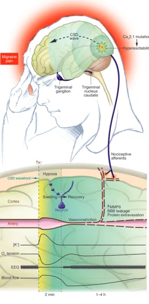
into the exposed cortex, electrical stimula-Figure 1
The link between CSD and migraine pain. CSD is most often initiated in the occipital cor-tex of patients with visual migraine aura. It is believed that CSD is ignited by local elevation
of extracellular K+ levels in pockets of intense
excitatory transmission. When K+ levels reach a
critical threshold of 10–12 mM, a self-propagat-ing CSD wave is initiated and advances across the cortex with a slow velocity of 3 mm/min. The threshold for CSD initiation is reduced
in FHM patients with mutations in the Cav2.1
Ca2+ channel because the higher Ca2+ level
in dendrites facilitates glutamate release and
thereby increases the likelihood that K+ levels
will reach the CSD threshold. A combination of stress and food intake may be sufficient to ignite CSD in patients with FHM, whereas stronger stimulation is required in the rest of the population. The lower diagram depicts the cortical events linking CSD to migraine
pain. The high extracellular K+ level at the
edge of the CSD wavefront is key for wave
propagation. K+ is normalized within
min-utes, but the restoration of normal membrane potential of neurons and glial cells is a high energy–demanding process. The cortical tis-sue experiences a minutes-lasting period of
severe reduction of tissue O2 tension
(hypox-ia) during CSD because O2 consumption
tran-siently exceeds the vascular supply of O2. This
[image:3.585.56.364.99.726.2]Strong genetic component of migraine
Analysis of genetic data from patients with familial hemiplegic migraine (FHM), an autosomal dominant subtype of migraine with aura associated with hemiparesis (weakening or paralysis of one side of the body), has in recent years proven to be a powerful tool to determine why CSD typi-cally manifests in patients with migraines. FHM is a heterogeneous genetic disease, but up to 50% of the patients have a mutation in the CACNA1A gene, which encodes the
a1A subunit of the neuronal, voltage-gated Cav2.1 Ca2+ channel (14). Cav2.1 channels are primarily located in presynaptic termi- nals and are important regulators of neu-rotransmitter release in excitatory synapses (7). An analysis of the single-channel prop-erties of 8 types of mutant Cav2.1 channels reported in individuals with FHM showed that channel activation in cerebellar gran-ule cells was shifted to lower voltages as a result of all mutations, resulting in a greater influx of Ca2+ in cerebellar granule cells (15). Although the functional significance of these changes in the intact brain is not completely understood, a likely scenario is that the increased Ca2+ influx augments release of the neurotransmitter glutamate from excitatory neurons in FHM patients, thus increasing the likelihood of triggering CSD. In fact, in a knock-in mouse model carrying the R192Q mutation in Cacna1a (the same mutation observed in individuals with FHM), the animals exhibited a reduced threshold for experimentally elicited CSD; in addition, the waves propagated faster once evoked (16).
The study by Eikermann-Haerter and coworkers in this issue of the JCI sheds light on several key aspects of the basic biol-ogy of migraine headaches (17). The authors describe what is believed to be a novel phe- channel inactivation, but the S218L muta- tion was associated with more severe altera- tions in both channel activity and neuro-logical deficits, compared with changes associated with the R192Q mutation. Inter-estingly, corticostriatal propagation of CSD was observed in the majority of mutant mice, and more serious and prolonged post-CSD neurological deficits followed, compared with those observed in wild-type mice. More-over, the higher prevalence of migraine in females was replicated in the mutant mice. Female mutant mice were more susceptible to CSD, and their neurological deficits mani-fested more severely than those in their male equivalents. The sex difference was elimi-nated by ovariectomy and partially restored by estrogen replacement, suggesting that differences in CSD susceptibility between males and females involve the effect of ovar-ian hormones. Like any important study, this new work raises a number of questions: Only experimentally induced CSD was stud-ied, and it is tempting to ask whether the mutant mice experience spontaneous waves of CSD. Moreover, FHM is a rare disease, and the current study does not directly address the pathogenesis of migraine with or with- out aura. Common migraine is a multifacto- rial polygenetic disease, and a genetic com-ponent can only been identified in about 50% of individuals with migraine. In these patients, recent genome-wide screens have pointed to several susceptibility loci, but no causative genes have been identified (7).
Long-term effect of migraine
Pain is an unpleasant and not easily over- ridden sensation of potential or actual dan-ger to the body. Does migraine pain signify that the brain is in potential danger? Per- haps. Recent studies show that cortical tis-sue experiences a short-lasting episode of hypoxia during CSD, which may be similar CSD (19). The changes in neuronal structure are reversible, and neurons do recover, but whether repeated episodes of migraine might cause permanent neuronal damage remains an unanswered question. Several prospective studies have shown that migraine patients do not experience cognitive decline (20, 21). It is interesting to note, however, that all studies published to date document that long-term migraineurs consistently score lower than matched controls in the processing of visual information (22, 23). Thus, visual aura, which is initiated and advances through the occipi- tal cortex, may impair cortical network func-tion locally. Alternatively, the impairment of visual processing and the lower threshold for CSD might be somehow coassociated through a common etiology.
1. Dodick, D.W., and Gargus, J.J. 2008. Why migraines strike. Sci. Am. 299:56–63.
2. Leao, A. 1944. Spreading depression of activity in cerebral cortex. J. Neurophysiol. 7:359–390.
3. Milner, P.M. 1958. Note on a possible correspon-dence between the scotomas of migraine and spreading depression of Leao. Electroencephalogr. Clin. Neurophysiol. 10:705.
4. Lashley, K. 1941. Patterns of cerebral integration indicated by scotomas of migraine. Arch. Neurol. Psychiat. 46:331–339.
5. Sacks, O. 1970. Migraine: the evolution of a common disorder. Faber & Faber. London, United Kingdom. 298 pp.
6. Gursoy-Ozdemir, Y., et al. 2004. Cortical spread-ing depression activates and upregulates MMP-9.
J. Clin. Invest. 113:1447–1455.
7. Pietrobon, D. 2005. Migraine: new molecular mechanisms. Neuroscientist. 11:373–386. 8. Grafstein, B., Liu, S., Cotrina, M.L., Goldman, S.A.,
and Nedergaard, M. 2000. Meningeal cells can communicate with astrocytes by calcium signaling.
Ann. Neurol. 47:18–25.
9. Hadjikhani, N., et al. 2001. Mechanisms of migraine aura revealed by functional MRI in human visual cortex. Proc. Natl. Acad. Sci. U. S. A. 98:4687–4692. 10. Bowyer, S.M., Aurora, K.S., Moran, J.E., Tepley, N.,
and Welch, K.M. 2001. Magnetoencephalographic fields from patients with spontaneous and induced migraine aura. Ann. Neurol. 50:582–587.
11. Lauritzen, M. 2001. Cortical spreading depression in migraine. Cephalalgia. 21:757–760.
12. Strong, A.J., et al. 2002. Spreading and synchronous depressions of cortical activity in acutely injured human brain. Stroke. 33:2738–2743.
13. Nedergaard, M., and Astrup, J. 1986. Infarct rim: effect of hyperglycemia on direct current potential and [14C]2-deoxyglucose phosphorylation. J. Cereb. Blood Flow Metab. 6:607–615.
14. Ophoff, R.A., et al. 1996. Familial hemiplegic migraine and episodic ataxia type-2 are caused by mutations in the Ca2+ channel gene CACNL1A4.
Cell. 87:543–552.
15. Hans, M., et al. 1999. Functional consequences of mutations in the human alpha1A calcium channel subunit linked to familial hemiplegic migraine.
J. Neurosci. 19:1610–1619.
16. van den Maagdenberg, A.M., et al. 2004. A Cacna1a knockin migraine mouse model with increased sus-ceptibility to cortical spreading depression. Neuron. 41:701–710.
17. Eikermann-Haerter, K., et al. 2009. Genetic and hormonal factors modulate spreading depression and transient hemiparesis in mouse models of familial hemiplegic migraine type 1. J. Clin. Invest. 119:99–109.
18. Kors, E.E., et al. 2001. Delayed cerebral edema and fatal coma after minor head trauma: role of the CACNA1A calcium channel subunit gene and rela-tionship with familial hemiplegic migraine. Ann.
Neurol. 49:753–760.
19. Takano, T., et al. 2007. Cortical spreading depres-sion causes and coincides with tissue hypoxia. Nat. Neurosci. 10:754–762.
20. Waldie, K.E., Hausmann, M., Milne, B.J., and Poul-ton, R. 2002. Migraine and cognitive function: a life-course study. Neurology. 59:904–908. 21. Riva, D., et al. 2006. Cognitive and behavioural
effects of migraine in childhood and adolescence.
Cephalalgia. 26:596–603.
22. McKendrick, A.M., Badcock, D.R., Badcock, J.C., and Gurgone, M. 2006. Motion perception in migraineurs: abnormalities are not related to attention.
Cephalalgia. 26:1131–1136.
23. Yenice, O., et al. 2007. Assessment of spatial-con- trast function and short-wavelength sensitivity def-icits in patients with migraine. Eye. 21:218–223. 24. Kalaydjian, A., Zandi, P.P., Swartz, K.L., Eaton,
W.W., and Lyketsos, C. 2007. How migraines impact cognitive function: findings from the Bal-timore ECA. Neurology. 68:1417–1424.
25. Matsushima, K., Hogan, M.J., and Hakim, A.M. 1996. Cortical spreading depression protects against subsequent focal cerebral ischemia in rats.
J. Cereb. Blood Flow Metab. 16:221–226.