R E V I E W
Open Access
Vascular and plaque imaging with ultrasmall
superparamagnetic particles of iron oxide
Shirjel R. Alam
1,2*, Colin Stirrat
1,2, Jennifer Richards
1, Saeed Mirsadraee
3,4, Scott I. K. Semple
3, George Tse
5,
Peter Henriksen
1,2†and David E. Newby
1,2†Abstract
Cardiovascular Magnetic Resonance (CMR) has become a primary tool for non-invasive assessment of cardiovascular
anatomy, pathology and function. Existing contrast agents have been utilised for the identification of infarction, fibrosis,
perfusion deficits and for angiography. Novel ultrasmall superparamagnetic particles of iron oxide (USPIO) contrast
agents that are taken up by inflammatory cells can detect cellular inflammation non-invasively using CMR, potentially
aiding the diagnosis of inflammatory medical conditions, guiding their treatment and giving insight into their
pathophysiology. In this review we describe the utilization of USPIO as a novel contrast agent in vascular disease.
Keywords:
USPIO, Nanoparticles, CMR, Cardiovascular imaging, Inflammation, Macrophage
Introduction
Inflammation is central to many cardiovascular
patho-physiological processes including atherosclerosis,
myo-cardial infarction and heart failure. Macrophages are key
mediators of these inflammatory pathways, initiating
both destructive and reparative processes [1].
Quantifi-cation and characterization of tissue macrophage activity
may therefore assist in our understanding of the
patho-genesis of cardiovascular disease and help determine
dis-ease severity and prognosis, as well as providing a
biomarker to assess the efficacy of established or novel
therapeutic interventions.
Cardiovascular magnetic resonance (CMR) is a
well-established clinical imaging modality offering excellent
soft tissue contrast and spatial resolution, whilst
avoid-ing ionizavoid-ing radiation. Standard gadolinium-based
con-trast agents are paramagnetic and are infused into the
blood pool with variable organ extraction rates, although
subsequent extravasation and redistribution can be used
to identify the interstitial and extracellular spaces.
Gado-linium is commonly used as an CMR contrast agent
after acute myocardial infarction (MI) to identify areas
of tissue infarction and fibrosis [2, 3]. Tissue oedema
and rupture of cell membranes with consequent
diffu-sion of gadolinium into the inter- and intra-cellular
spaces [2] results in a
“
delayed gadolinium
enhance-ment
”
effect in infarcted regions. Recent interest has
turned to novel agents that provide additional structural
and functional cellular information. Such
‘
smart
’
contrast
agents include iron oxide nanoparticles.
Iron oxide nano-particles
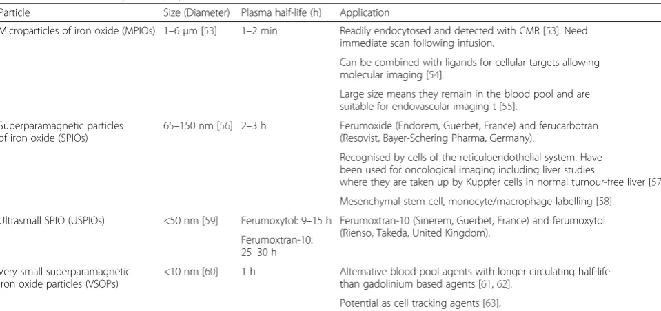
Particles of iron oxide are divided into classes based on
their size (Table 1). In this review, we will focus on
ultra-small superparamagnetic particles of iron oxide (USPIOs)
that consist of nanoparticles with a diameter of <50 nm
and include ferumoxtran-10 (Sinerem, Guerbet) and
feru-moxytol (Rienso, Takeda; Feraheme, AMAG
Pharmaceuti-cals). Although Rienso had been authorised for use in
European Union, Takeda since has withdrawn it. However
Feraheme is clinically available in the United States for the
treatment of iron deficiency anemia in adult patients with
chronic kidney disease (CKD).
Ferumoxytol is well tolerated by patients with chronic
kidney disease and iron deficiency anaemia, and had a
similar overall treatment-related adverse event rate to
oral iron [4]. This safety data is further supported by
additional retrospective observational data from three
* Correspondence:[email protected]†Equal contributors 1
Centre for Cardiovascular Science, The University of Edinburgh, The Chancellor’s Building, Little France Crescent, Edinburgh EH16 5SA, UK
2
Department of Cardiology, Royal Infirmary of Edinburgh, Edinburgh EH16 5SA, UK
Full list of author information is available at the end of the article
large haemodialysis clinics in the United States involving
more than 8600 patients and more than 33,300
adminis-tered doses of ferumoxytol [5, 6]. The only
contraindica-tions to use are known hypersensitivity or iron overload.
Therefore there is little to limit widespread clinical use
as an imaging agent.
USPIOs can be used as a blood pool contrast agent
but it is their ability to be taken up by inflammatory cells
that has distinguished them [7]. Cellular uptake of
USPIOs occurs through a variety of mechanisms.
Phago-cytosis and receptor-mediated endoPhago-cytosis are important
for uptake of larger particles, whilst smaller particles are
internalized by pinocytosis. Although the avidity of
macrophage uptake is strongly influenced by particle size
and charge, the surface coating is particularly important
[8, 9]. As a result of their smaller size, USPIOs are less
readily recognized by phagocytic cells and persist in the
circulation for longer than other iron particles (plasma
half-life 14
–
30 h in humans) [10, 11]. They are capable
of passing through capillary walls, to be taken up by
tissue-resident macrophages and neutrophils (Fig. 1)
[12
–
14]. These characteristics allow USPIOs to detect
and highlight cellular inflammation within tissues using
CMR.
Imaging methodology
USPIOs induce local magnetic field inhomogeneities that
shorten T2 and T2* relaxations times resulting in a
sig-nal deficit on magnetic resonance images. USPIOs also
have a T1 shortening effect, particularly at low
concen-trations, and appear bright on T1 weighted images. The
T1 shortening effect mainly depends on the strength of
the magnetic field, and is higher in lower field strength.
A range of approaches have been used to evaluate
USPIO accumulation in tissues. Most simply, images
may be qualitatively assessed for signal deficits. However
this approach is subjective, and signal deficits due to
cal-cification or other artefacts may be misinterpreted.
Manually drawn regions of interest have been used to
allow comparison of signal intensity of the target tissue
with that of control tissue although discrete focal areas
of USPIO accumulation, and thus focal inflammation,
may be missed.
[image:2.595.58.538.98.324.2]Tissue properties, such as the presence of oedema or
haemorrhage, can alter image intensities on T2*
se-quences, and so pre- and post-contrast images need to
be compared to delineate the impact of USPIO
accumu-lation. This requires accurate co-registration of these
paired scans and adjustments for differences in baseline
intensity. A specific region of interest (ROI) map can be
Table 1
Iron oxide nanoparticle preparations
Particle Size (Diameter) Plasma half-life (h) Application
Microparticles of iron oxide (MPIOs) 1–6μm [53] 1–2 min Readily endocytosed and detected with CMR [53]. Need immediate scan following infusion.
Can be combined with ligands for cellular targets allowing molecular imaging [54].
Large size means they remain in the blood pool and are suitable for endovascular imaging t [55].
Superparamagnetic particles of iron oxide (SPIOs)
65–150 nm [56] 2–3 h Ferumoxide (Endorem, Guerbet, France) and ferucarbotran (Resovist, Bayer-Schering Pharma, Germany).
Recognised by cells of the reticuloendothelial system. Have been used for oncological imaging including liver studies
where they are taken up by Kuppfer cells in normal tumour-free liver [57]. Mesenchymal stem cell, monocyte/macrophage labelling [58].
Ultrasmall SPIO (USPIOs) <50 nm [59] Ferumoxytol: 9–15 h Ferumoxtran-10 (Sinerem, Guerbet, France) and ferumoxytol (Rienso, Takeda, United Kingdom).
Ferumoxtran-10: 25–30 h
Very small superparamagnetic iron oxide particles (VSOPs)
<10 nm [60] 1 h Alternative blood pool agents with longer circulating half-life than gadolinium based agents [61,62].
Potential as cell tracking agents [63].
[image:2.595.306.539.556.696.2]drawn and subsequently transferred to each subsequent
co-registered image, thus ensuring the signal intensity
can be compared for identical sample regions in
differ-ent scans from the same patidiffer-ent.
Rather than assessing focal image brightness at a single
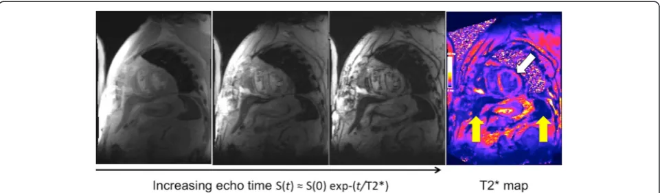
echo time, the T2* time constant can be calculated from
the exponential decay curve using multiple echo times
(Fig. 2). This method provides greater reproducibility,
broad applicability throughout the field of view, and
inde-pendence from T1 effects and a range of imaging
vari-ables. In the presence of USPIO, the T2* relaxation rate is
increased thus giving a lower T2* value, or higher R2*
value (R2* is the inverse of T2*, R2* = 1/T2*). Calculation
of these values permits the generation of T2* or R2* maps
indicative of USPIO accumulation (Fig. 3).
Various authors have used different techniques to
calcu-late USPIO uptake in tissues, and have reported results
using T2, T2* or R2*. This can cause confusion since higher
values infer diminished USPIO uptake in T2/T2* weighted
images, but higher uptake in R2* maps. For the purposes of
this paper, imaging techniques will be described but results
reported in terms of
“
increased USPIO uptake.
”
In order to
account for native R2* values, various authors have used
the delta increase in R2* value from successive scans, or
factor increase. When pre-USPIO scans have not been
per-formed, it must be assumed that non-inflamed tissue has
similar R2* values to pre-USPIO native R2* values.
Finally, it must be noted that USPIO imaging can be
affected by artefact. USPIO also shorten T1, and so
cause signal enhancement of T1 weighted imaging [15].
However, at high concentration USPIO can cause signal
loss with such imaging limiting its use with T1 weighted
sequences [16]. The superparamagnetic nature of the
particles means that they generate strong local magnetic
field inhomogeneities, and it is this magnetic
susceptibil-ity that is being imaged by CMR. However this can
cause loss of distinction of anatomical borders and
dis-tort normal tissues (
“
blooming artefact
”
). USPIO will
ac-cumulate in the reticulo-endothelial system including
the liver and spleen. This accumulation can affect
neigh-bouring structures, and care must be taken not mistake
blooming artefact for USPIO uptake.
Cardiovascular applications
Atherosclerotic plaque
Given the central role of macrophage biology in the
patho-genesis of atherosclerosis, USPIOs have an obvious
applica-tion in the investigaapplica-tion of atherosclerotic disease. In
pre-clinical studies, uptake of USPIOs is demonstrable within
numerous atherosclerotic models including aortic plaques
of hyperlipidemic rabbits [17, 18] and mice [19] as well as
the neointimal hyperplasia following balloon injury [12, 20],
and is proportional to plaque macrophage content.
Modulation of inflammation within atherosclerotic
pla-ques can be assessed by USPIO imaging [21]. P38
Mitogen-activated protein kinase (MAPK) is an
inflamma-tory signalling pathway activated by angiotensin II in
vari-ous vascular cell types [22, 23]. Angiotensin II infusion
[image:3.595.57.539.456.673.2]leads to macrophage accumulation and UPSIO uptake
in atherosclerotic plaques of mice [21, 24] that can be
inhibited by co-administration of a p38 MAPK pathway
inhibitor. Interestingly, this effect was predominantly
manifested by a reduction in USPIO uptake by
macro-phages rather than a reduction in macrophage
num-bers, suggesting an effect on macrophage activity rather
than recruitment. In contrast, the angiotensin II type 1
receptor antagonist, irbesartan, decreased both USPIO
uptake and macrophage content in the apolipoprotein
E deficient mouse model [25].
USPIO uptake occurs in human carotid atherosclerotic
plaques and appears to correlate with the number of
iron-laden macrophages on histology [26]. Consistent with the
inflammatory cell infiltrate associated with vulnerable
pla-ques, 75 % of ruptured or rupture-prone lesions show
USPIO uptake compared to only 7 % of apparently stable
lesions. Determining the overall macrophage burden is
challenging because of a number of factors. There is a
curvilinear relationship between area of signal intensity
re-duction and USPIO concentration. Signal intensity is also
dependent on density of particle accumulation, and a
heterogeneous population of macrophages would be
ex-pected to have differing degrees of USPIO uptake. The
amount of USPIO infused, and by extrapolation
perfu-sion of target tissue, will also determine the magnitude
of CMR changes [27].
USPIO uptake and inflammation does not correlate
with plaque volume or the degree of luminal stenosis
[28], and as already stated USPIO maybe useful in
inves-tigating USPIO activity in contrast to concentration [24].
This raises the possibility of using USPIO uptake to
monitor disease activity in carotid stenosis rather than
using conventional anatomical measurements. For
in-stance, statins reduce inflammation within
atheroscler-otic plaques as well as systemic markers of inflammation
[29, 30] and this has been assessed using USPIO uptake.
The ATHEROMA study (Atorvastatin THerapy: Effects
on Reduction Of Macrophage Activity) compared the
ef-fect of high-dose (80 mg daily) versus low-dose (10 mg
daily) atorvastatin on plaque inflammation [31]. Patients
underwent UPSIO-enhanced CMR at baseline, 6 weeks
and 12 weeks of therapy. Although there were no
differ-ences in USPIO uptake over the course of the study in the
low-dose group, there was a reduction in USPIO uptake in
the high-dose group at both 6 and 12 weeks. This
corre-lated with a reduction in LDL cholesterol and a reduction
in micro-emboli count on trans-cranial Doppler [32].
Abdominal aortic aneurysms
Macrophages are intimately involved in the development,
expansion and ultimately rupture of abdominal aortic
an-eurysms. Preliminary evidence of USPIO uptake in human
abdominal aortic aneurysms (AAA) has been described
[33, 34]. In a pilot study, we demonstrated that just under
half of patients with AAA had focal mural uptake of
USPIOs. The aneurysm expansion rate was three-fold
higher in patients with USPIO uptake in the aneurysm
wall (0.66
versus
0.22 cm/year) [35]. Histology of tissue
excised at the time of elective surgical repair confirmed
co-localization of USPIOs with CD68 immunostaining for
macrophages. Thus USPIO-enhanced CMR appears to
identify those patients with more rapid disease progression
requiring earlier preventative surgical or endovascular
intervention to prevent rupture.
Cerebrovascular disease
[image:4.595.58.539.89.230.2]of iron-containing macrophages in the infarcted tissue
consistent with migration of macrophages from the
pen-umbra. Further work has indicated that in the setting of
established stroke, USPIO leaks through the injured
blood
–
brain barrier accounting for the initial
accumula-tion at the periphery of the infarct and intravascular
trap-ping rather than macrophage uptake [40]. In addition
there is widespread uptake resulting from leakage of
USPIO into the cerebro-spinal fluid with delivery of
nano-particals to more remote areas. Thus the application of
USPIO in such settings is limited although it is possible to
track focal USPIO uptake associated with macrophage/
microglial infiltration 6 days after cerebral ischaemia,
iden-tifying a subacute pathological process [41, 42].
Clinical studies have utilized ferumoxtran-10 in patients
4
–
5 days after stroke, with imaging at 24
–
36 h and
re-peated 48
–
72 h later [43]. T1- and T2/T2*-weighted
im-aging reveals parenchymal enhancement that increases
between the 2 scans, corresponding to the expected
macrophage distribution. These USPIO induced changes
do not correspond to conventional gadolinium-enhanced
changes, suggesting they occurred independently of
blood
–
brain-barrier breakdown. It could be speculated
that these changes may have been due to differences in
blood pooling effects due to perfusion changes rather than
USPIO inflammatory cell uptake. It would be expected
that ischemic volume would correlate with inflammatory
burden and CMR changes if USPIOs were being taken up
by inflammatory cells. Nighoghossian et al. found no such
correlation six days after stroke [44] although the study
had a number of limitations including imperfect timing of
the scans and the completion of only 5 patients using the
more sensitive T2* imaging protocol.
Despite these limitations, a pre-clinical model of the
investigation of anti-inflammatory medication in the
treatment of stroke has major potential [45]. Using a
murine model, the anti-inflammatory agent minocycline
can be evaluated after middle cerebral artery occlusion
[45]. Minocycline treatment reduced USPIO uptake
within the infarct, and was associated with reductions in
infarct size, blood
–
brain barrier permeability and
micro-glia/macrophage counts.
Future applications
The application of USPIOs to study myocardial
inflam-mation has translational application where the pathology
involves substantial monocyte influx into the plaque or
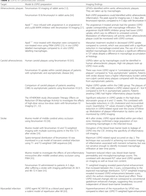
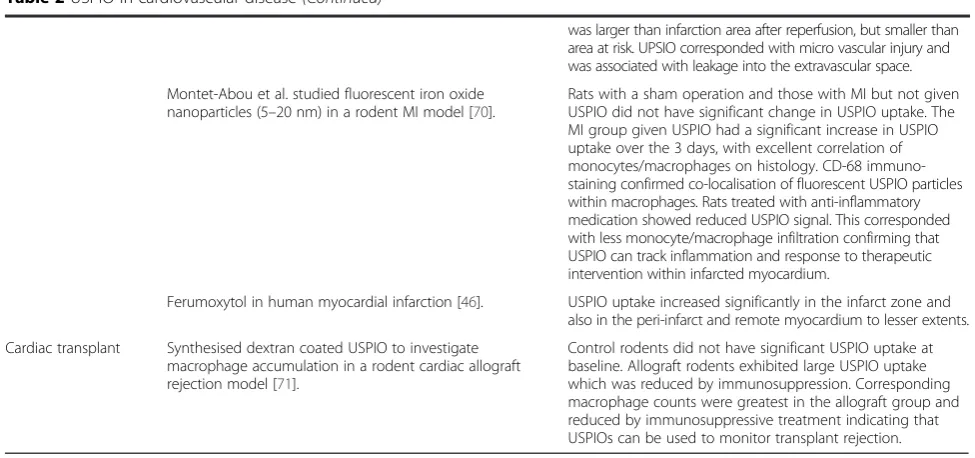
tissue [46] (Table 2).
Targeted iron oxide particles
Conjugating iron oxide particles with antibodies allows
targeted imaging. Pre-clinical imaging to date has
employed 9.4 Tesla CMR. This would be more sensitive
in detecting USPIO than clinical CMR systems (1.5 or
3-tesla). In addition, injected unconjugated USPIOs
injected directly into the blood stream concentrate
within macrophages resulting in high local distribution.
It remains to be seen if antibody-labelled USPIOs will
be sufficiently concentrated at their target site to allow
detection in clinical CMR systems. Specific subsets of
monocytes or other cell types could be tracked with
successful application of this method. This would allow
delineation of the temporal dynamics of cellular and
immunological processes by repeated scanning. This
has been demonstrated in a pre-clinical model of cerebral
ischaemia using USPIOs grafted with a specific peptide
tar-geting vascular cellular adhesion molecule-1 (VCAM-1).
This study indicated the potential of VCAM-1 to assess
vascular injury.
E-selectin is an adhesion molecule between the
endo-thelium and leukocytes that plays a critical role in the
pathogenesis of inflammation [47]. An E-selectin
mono-clonal antibody-USPIO conjugate has been used to track
vascular inflammation in a murine model of contact
hypersensitivity [48]. More recently, USPIOs have been
conjugated with a scavenger receptor to identify
inflam-mation in atherosclerotic plaques [49].
Another potential confounding factor is that
macro-phages of different subsets or with different activation
status take up USPIO at different rates. This could result
in false positive or negative CMR enhancement. Direct
labeling of cells with USPIO would avoid this error but
published data are limited. Although directly labeling of
macrophages with USPIO and delivery through the
ca-rotid artery has been successful in producing T2*
hypo-enhancement after transient ischaemia, it is associated
with increased mortality in a rat model [50].
USPIO cell labelling and monitoring cell trafficking
The ability to track cells non-invasively
in vivo
would be a
valuable technique with a number of potential applications
that include inflammatory cell tracking and evaluation of
engraftment of cells administered as part of cell-based
therapies.
USPIOs can be used to label cells
in vitro
for
subse-quent
in vivo
tracking. Smooth muscle cells labelled with
iron nanoparticles can be imaged when directly injected
into either healthy or infarcted myocardium in a
pre-clinical model [51]. This technique can be utilized to
label human aortic smooth muscle cells incorporated
into tissue engineered vascular grafts and implanted into
mice [52]. We have also demonstrated that cell tracking
can be achieved in vivo in humans using similar
ap-proaches with the larger SPIOs [53].
Summary
Table 2
USPIO in cardiovascular disease
Target Model & USPIO preparation Imaging findings
Atherosclerotic plaques Ferumoxtran-10 imaging of rabbit aorta [12]. UPSIOs identified within aortic atherosclerotic plaques. They are taken up by macrophages.
Ferumoxtran-10 & ferumoxytol in rabbit aorta [64]. Both USPIO preparations could lidentified within atherosclerotic inflammation. The peak signal for imaging was 2–3 days after ferumoxytol injection, compared to 4–5 days with ferumoxtran-10.
ApoE−/−mice infused with angiotensin II, or angiotensin II and a p38 MAPK inhibitor with ferumoxtran-10 imaging [21].
The angiotensin II treated animals had the greatest USPIO uptake corresponding with macrophage infiltration. The angiotensin II/p38 MAPK inhibitor group had lower USPIO uptake, which was no different to untreated controls. Modulation of inflammatory cell activity within atherosclerotic plaque could be monitored with USPIO contrast.
ApoE−/−mice treated with irbesartan were compared to non-treated mice using P904 USPIO [25].in vivoUSPIO labelled macrophages compared toin vitroUSPIO labelling macrophages.
Irbesartan treatment resulted in decreased USPIO uptake compared to controls, which was associated with a significant reduction in macrophage-covered area. The use ofin vitro
labelled macrophages did not produce a significant difference in T2* values despite a difference in macrophage accumulation at histology.
Carotid atherosclerosis Human carotid plaques using ferumoxtran-10 [65]. USPIOs taken up by macrophages could be identified in human atherosclerotic plaques. High risk plaques took up USPIO more avidly.
Ferumoxtran-10 uptake within carotid plaques of patients with symptomatic and asymptomatic disease [66].
There was more USPIO signal in“contralateral asymptomatic plaques”compared to“truly asymptomatic”patients. Patients with stroke disease have a higher inflammatory burden within non-culprit carotid artery plaques compared with the plaques from asymptomatic patients.
Comparison of carotid plaques of patients awaiting CABG to asymptomatic patients using ferumoxtran-10 [67].
Higher USPIO uptake within the CABG group. The plaques of the CABG patients exhibited a USPIO related signal of i 16.4 % compared to 8.4 % in asymptomatic patients. Patients awaiting CABG had higher inflammatory plaque burden.
The ATHEROMA study (Atorvastatin THerapy: Effects on Reduction Of Macrophage Activity) to investigate the effects of high-dose versus low-dose statin with ferumoxtran-10 imaging [31,32].
Significant reduction in USPIO uptake in the high-dose atorvastatin group at 6 and 12 weeks. This correlated with favourable reductions in LDL cholesterol and micro-emboli count. Quantitative T2* values showed a highly significant reduction in USPIO-related signal over the course of follow-up. Modulation of plaque inflammation by statins can be monitored by USPIO imaging.
Stroke Murine model of middle cerebral artery occlusion using ferumoxtran-10 [38].
48 h after stroke, USPIO signal identified within peri-infarct zone. Histology confirmed a large population of iron containing macrophages in the infarcted tissue.
Murine model with ferumoxtran-10 and T2-weighted imaging with multiple scanning points in the first 72 h after stroke [39].
Disruption of the blood brain barrier leads to leakage of USPIO into the CSF, limiting the specificity of inflammatory cell imaging.
Spatio-temporal distribution of ferumoxtran-10 was monitored in a rat model of transient cerebral infarction using T1- and T2-weighted CMR sequences [68].
Maximum USPIO related signal occurred on day 2. The technique was successful in achieving non-invasive imaging of inflammation associated with transient ischaemia, but was not sensitive enough to identify increased macrophage infiltration at later time points.
Murine model to investigate the effects of anti-inflammatory minocycline after middle cerebral artery occlusion using P904 [45].
Treatment reduced infarct size, blood–brain barrier permeability and microglia/macrophage counts. This correlated with decreased R2* value (and USPIO uptake) on imaging as well as tissue iron content.
Ferumoxtran-10 administered to patients 4–5 days after suffering a stroke with imaging performed 24–36 h and 48–72 h later [43].
T1 weighted imaging revealed parenchymal enhancement that increased between the 2 scans, corresponding to the expected macrophage distribution. T2/T2* weighted imaging revealed increased USPIO enhancement between scans, which the authors interpreted as blood pool effect. These USPIO induced changes did not correspond to conventional gadolinium enhanced changes, suggesting they occurred independent of blood–brain-barrier breakdown.
Myocardial infarction USPIO agent NC100150 as a blood pool agent in a rodent model of reperfusion after MI [69].
scanning provide a sensitive method of assessing USPIO
accumulation.
USPIO-enhanced magnetic resonance imaging is a
promising method for assessing inflammatory processes
associated with a range of cardiovascular diseases
includ-ing those affectinclud-ing the atherosclerotic plaque and large
ar-teries. Potential clinical applications are under evaluation
and include assessing the effects of novel pharmacological
agents and
in vivo
cell tracking to determine the fate of
cells administered as part of cell therapy.
Abbreviations
CABG:Coronary artery bypass grafting; CMR: Cardovascular magnetic resonance; FDG-PET: Fluorodeoxyglucose-positron emission tomography; MAPK: Mitogen-activated protein kinase; MCP-1: Monocyte chemotactic protein-1; MI: Myocardial infarction; RAS: Renin-angiotensin system; ROI: Region of interest; SPIO: Super-paramagnetic iron oxide; TNF-α: Tumour necrosis factor alpha; USPIO: Ultrasmall super-paramagnetic iron oxide; VCAM-1: Vascular cell adhesion molecule 1.
Competing interests
The authors declare that they have no competing interests.
Authors’contributions
[image:7.595.54.542.95.324.2]SA conceived and wrote the review paper. GT provided Fig. 1, SS provided Fig. 2. CA, JR, SM,PH and DN reviewed and edited the paper. All authors read and approved the manuscript.
Acknowledgements
The authors are supported by grants from the British Heart Foundation (RE/08/001 and FS/12/83), Medical Research Council (G1001339), National Institute for Health Research (EME 11/20/03), Chest Heart and Stroke Scotland (R11/A135) and Chief Scientist Office (ETM/266). DEN is supported by the British Heart Foundation (CH/09/002). PAH is supported by NHS Research Scotland Fellowship.
Support
Dr Shirjel Alam was supported by a Scholarship grant from the British Heart Foundation.
Author details 1
Centre for Cardiovascular Science, The University of Edinburgh, The Chancellor’s Building, Little France Crescent, Edinburgh EH16 5SA, UK.
2
Department of Cardiology, Royal Infirmary of Edinburgh, Edinburgh EH16 5SA, UK.3Clinical Research Imaging Centre, University of Edinburgh,
Edinburgh EH16 5SA, UK.4Department of Radiology, Royal Infirmary of Edinburgh, Edinburgh EH16 5SA, UK.5MRC Centre for Inflammation Research,
The University of Edinburgh, Edinburgh EH16 5SA, UK.
Received: 20 November 2014 Accepted: 16 August 2015
References
1. Nahrendorf M, Pittet MJ, Swirski FK. Monocytes: protagonists of infarct inflammation and repair after myocardial infarction. Circulation. 2010;121:2437.
2. Lima JA, Judd RM, Bazille A, Schulman SP, Atalar E, Zerhouni EA. Regional heterogeneity of human myocardial infarcts demonstrated by contrast-enhanced MRI. Potential mechanisms. Circulation. 1995;92:1117–25. 3. Schelbert EB, Hsu LY, Anderson SA, Mohanty BD, Karim SM, Kellman P, et al.
Late gadolinium-enhancement cardiac magnetic resonance identifies postinfarction myocardial fibrosis and the border zone at the near cellular level in ex vivo rat heart. Circ Cardiovasc Imaging. 2010;3:743–52. 4. Spinowitz BS, Kausz AT, Baptista J, Noble SD, Sothinathan R, Bernardo MV,
et al. Ferumoxytol for treating iron deficiency anemia in CKD. J Am Soc Nephrol. 2008;19:1599–605.
5. Schiller B, Bhat P, Sharma A, Li Z, Fortin G, McLaughlin J, et al. Safety of Feraheme¬Æ(Ferumoxytol) in hemodialysis patients at 3 dialysis chains over a 1-year period. J Am Soc Nephrol. 2011;22:477A–8.
6. Sharma A, Bhat P, Schiller B, Fortin G, McLaughlin J, Li Z, et al. Efficacy of Feraheme®(Ferumoxytol) administration on target hemoglobin levels and other iron parameters across 3 dialysis chains. J Am Soc Nephrol. 2011;22:485A. 7. Christen T, Ni W, Qiu D, Schmiedeskamp H, Bammer R, Moseley M, et al.
High-resolution cerebral blood volume imaging in humans using the blood pool contrast agent ferumoxytol. Magn Reson Med. 2013; 70(3):705-10. 8. Saito S, Tsugeno M, Koto D, Mori Y, Yoshioka Y, Nohara S, et al. Impact of
surface coating and particle size on the uptake of small and ultrasmall superparamagnetic iron oxide nanoparticles by macrophages. Int J Nanomedicine. 2012;7:5415–21.
[image:7.595.302.538.468.741.2]9. Tsuchiya K, Nitta N, Sonoda A, Otani H, Takahashi M, Murata K, et al. Atherosclerotic imaging using 4 types of superparamagnetic iron oxides: new possibilities for mannan-coated particles. Eur J Radiol. 2013;82:1919–25.
Table 2
USPIO in cardiovascular disease
(Continued)
was larger than infarction area after reperfusion, but smaller than area at risk. UPSIO corresponded with micro vascular injury and was associated with leakage into the extravascular space.
Montet-Abou et al. studied fluorescent iron oxide nanoparticles (5–20 nm) in a rodent MI model [70].
Rats with a sham operation and those with MI but not given USPIO did not have significant change in USPIO uptake. The MI group given USPIO had a significant increase in USPIO uptake over the 3 days, with excellent correlation of monocytes/macrophages on histology. CD-68 immuno-staining confirmed co-localisation of fluorescent USPIO particles within macrophages. Rats treated with anti-inflammatory medication showed reduced USPIO signal. This corresponded with less monocyte/macrophage infiltration confirming that USPIO can track inflammation and response to therapeutic intervention within infarcted myocardium.
Ferumoxytol in human myocardial infarction [46]. USPIO uptake increased significantly in the infarct zone and also in the peri-infarct and remote myocardium to lesser extents.
Cardiac transplant Synthesised dextran coated USPIO to investigate macrophage accumulation in a rodent cardiac allograft rejection model [71].
10. Landry R, Jacobs PM, Davis R, Shenouda M, Bolton WK. Pharmacokinetic study of ferumoxytol: a new iron replacement therapy in normal subjects and hemodialysis patients. Am J Nephrol. 2005;25:400–10.
11. Hunt MA, Bago AG, Neuwelt EA. Single-dose contrast agent for intraoperative MR imaging of intrinsic brain tumors by using ferumoxtran-10. AJNR Am J Neuroradiol. 2005;26:1084–8.
12. Ruehm SG, Corot C, Vogt P, Kolb S, Debatin JF. Magnetic resonance imaging of atherosclerotic plaque with ultrasmall superparamagnetic particles of iron oxide in hyperlipidemic rabbits. Circulation. 2001;103:415–22. 13. Dousset V, Delalande C, Ballarino L, Quesson B, Seilhan D, Coussemacq M, et al. In vivo macrophage activity imaging in the central nervous system detected by magnetic resonance. Magn Reson Med. 1999;41:329–33. 14. Gellissen J, Axmann C, Prescher A, Bohndorf K, Lodemann KP. Extra- and
intracellular accumulation of ultrasmall superparamagnetic iron oxides (USPIO) in experimentally induced abscesses of the peripheral soft tissues and their effects on magnetic resonance imaging. Magn Reson Imaging. 1999;17:557–67.
15. Small WC, Nelson RC, Bernardino ME. Dual contrast enhancement of both T1- and T2-weighted sequences using ultrasmall superparamagnetic iron oxide. Magn Reson Imaging. 1993;11:645–54.
16. Fananapazir G, Marin D, Suhocki PV, Kim CY, Bashir MR. Vascular artifact mimicking thrombosis on MR imaging using ferumoxytol as a contrast agent in abdominal vascular assessment. J Vasc Interv Radiol. 2014;25:969–76. 17. Schmitz SA, Coupland SE, Gust R, Winterhalter S, Wagner S, Kresse M, et al. Superparamagnetic iron oxide-enhanced MRI of atherosclerotic plaques in Watanabe hereditable hyperlipidemic rabbits. Invest Radiol. 2000;35:460–71. 18. Schmitz SA, Taupitz M, Wagner S, Coupland SE, Gust R, Nikolova A, et al.
Iron-oxide-enhanced magnetic resonance imaging of atherosclerotic plaques: postmortem analysis of accuracy, inter-observer agreement, and pitfalls. Invest Radiol. 2002;37:405–11.
19. Sigovan M, Bessaad A, Alsaid H, Lancelot E, Corot C, Neyran B, et al. Assessment of age modulated vascular inflammation in ApoE−/−mice by USPIO-enhanced magnetic resonance imaging. Invest Radiol. 2010;45:702–7. 20. Hyafil F, Laissy JP, Mazighi M, Tchetche D, Louedec L, Adle-Biassette H, et al.
Ferumoxtran-10-enhanced MRI of the hypercholesterolemic rabbit aorta: relationship between signal loss and macrophage infiltration. Arterioscler Thromb Vasc Biol. 2006;26:176–81.
21. Morris JB, Olzinski AR, Bernard RE, Aravindhan K, Mirabile RC, Boyce R, et al. p38 MAPK inhibition reduces aortic ultrasmall superparamagnetic iron oxide uptake in a mouse model of atherosclerosis: MRI assessment. Arterioscler Thromb Vasc Biol. 2008;28:265–71.
22. Herlaar E, Brown Z. p38 MAPK signalling cascades in inflammatory disease. Mol Med Today. 1999;5:439–47.
23. Ju H, Nerurkar S, Sauermelch CF, Olzinski AR, Mirabile R, Zimmerman D, et al. Sustained activation of p38 mitogen-activated protein kinase contributes to the vascular response to injury. J Pharmacol Exp Ther. 2002;301:15–20. 24. Brasier AR, Recinos A, 3rd, Eledrisi MS. Vascular inflammation and the
renin-angiotensin system. Arterioscler Thromb Vasc Biol. 2002;22:1257–66. 25. Sigovan M, Kaye E, Lancelot E, Corot C, Provost N, Majd Z, et al.
Anti-inflammatory drug evaluation in ApoE−/−mice by ultrasmall superparamagnetic iron oxide-enhanced magnetic resonance imaging. Invest Radiol. 2012;47:546–52.
26. Trivedi RA, Mallawarachi C, JM UK-I, Graves MJ, Horsley J, Goddard MJ, et al. Identifying inflamed carotid plaques using in vivo USPIO-enhanced MR imaging to label plaque macrophages. Arterioscler Thromb Vasc Biol. 2006;26:1601–6.
27. Lutz AM, Weishaupt D, Persohn E, Goepfert K, Froehlich J, Sasse B, et al. Imaging of macrophages in soft-tissue infection in rats: relationship between ultrasmall superparamagnetic iron oxide dose and MR signal characteristics. Radiology. 2005;234:765–75.
28. Tang TY, Howarth SP, Miller SR, Graves MJ, JM UK-I, Li ZY, et al. Correlation of carotid atheromatous plaque inflammation using USPIO-enhanced MR imaging with degree of luminal stenosis. Stroke. 2008;39:2144–7.
29. Ridker PM, Rifai N, Clearfield M, Downs JR, Weis SE, Miles JS, et al. Measurement of C-reactive protein for the targeting of statin therapy in the primary prevention of acute coronary events. N Engl J Med. 2001;344:1959–65. 30. Tahara N, Kai H, Ishibashi M, Nakaura H, Kaida H, Baba K, et al. Simvastatin
attenuates plaque inflammation: evaluation by fluorodeoxyglucose positron emission tomography. J Am Coll Cardiol. 2006;48:1825–31.
31. Tang TY, Howarth SP, Miller SR, Graves MJ, Patterson AJ, JM UK-I, et al. The ATHEROMA (Atorvastatin Therapy: Effects on Reduction of Macrophage
Activity) Study. Evaluation using ultrasmall superparamagnetic iron oxide-enhanced magnetic resonance imaging in carotid disease. J Am Coll Cardiol. 2009;53:2039–50.
32. Patterson AJ, Tang TY, Graves MJ, Muller KH, Gillard JH. In vivo carotid plaque MRI using quantitative T2* measurements with ultrasmall superparamagnetic iron oxide particles: a dose–response study to statin therapy. NMR Biomed. 2011;24:89–95.
33. Sadat U, Taviani V, Patterson AJ, Young VE, Graves MJ, Teng Z, et al. Ultrasmall superparamagnetic iron oxide-enhanced magnetic resonance imaging of abdominal aortic aneurysms–a feasibility study. Eur J Vasc Endovasc Surg. 2011;41:167–74.
34. Howarth SP, Tang TY, Graves MJ, JM UK-I, Li ZY, Walsh SR, et al. Non-invasive MR imaging of inflammation in a patient with both asymptomatic carotid atheroma and an abdominal aortic aneurysm: a case report. Ann Surg Innov Res. 2007;1:4.
35. Richards JM, Semple SI, MacGillivray TJ, Gray C, Langrish JP, Williams M, et al. Abdominal aortic aneurysm growth predicted by uptake of ultrasmall superparamagnetic particles of iron oxide: a pilot study. Circ Cardiovasc Imaging. 2011;4:274–81.
36. Braun JS, Jander S, Schroeter M, Witte OW, Stoll G. Spatiotemporal relationship of apoptotic cell death to lymphomonocytic infiltration in photochemically induced focal ischemia of the rat cerebral cortex. Acta Neuropathol. 1996;92:255–63.
37. del Zoppo G, Ginis I, Hallenbeck JM, Iadecola C, Wang X, Feuerstein GZ. Inflammation and stroke: putative role for cytokines, adhesion molecules and iNOS in brain response to ischemia. Brain Pathol. 2000;10:95–112. 38. Rausch M, Sauter A, Frohlich J, Neubacher U, Radu EW, Rudin M. Dynamic
patterns of USPIO enhancement can be observed in macrophages after ischemic brain damage. Magn Reson Med. 2001;46:1018–22.
39. Wiart M, Davoust N, Pialat JB, Desestret V, Moucharrafie S, Cho TH, et al. MRI monitoring of neuroinflammation in mouse focal ischemia. Stroke. 2007;38:131–7. 40. Desestret V, Brisset JC, Moucharrafie S, Devillard E, Nataf S, Honnorat J, et al.
Early-stage investigations of ultrasmall superparamagnetic iron oxide-induced signal change after permanent middle cerebral artery occlusion in mice. Stroke. 2009;40:1834–41.
41. Yang YM, Feng X, Yin LK, Li CC, Jia J, Du ZG. Comparison of USPIO-enhanced MRI and Gd-DTPA enhancement during the subacute stage of focal cerebral ischemia in rats. Acta Radiol. 2014;55(7)864–73.
42. Yang YM, Feng XY, Yin le K, Li CC, Li AN, Jia J, et al. In vivo USPIO-enhanced MR signal characteristics of secondary degeneration in the ipsilateral substantia nigra after middle cerebral artery occlusion at 3 T. J Neuroradiol. 2013;40:198–203.
43. Saleh A, Schroeter M, Jonkmanns C, Hartung HP, Modder U, Jander S. In vivo MRI of brain inflammation in human ischaemic stroke. Brain. 2004;127:1670–7. 44. Nighoghossian N, Wiart M, Cakmak S, Berthezene Y, Derex L, Cho TH, et al. Inflammatory response after ischemic stroke: a USPIO-enhanced MRI study in patients. Stroke. 2007;38:303–7.
45. Marinescu M, Chauveau F, Durand A, Riou A, Cho TH, Dencausse A, et al. Monitoring therapeutic effects in experimental stroke by serial USPIO-enhanced MRI. Eur Radiol. 2013;23(1):37–47.
46. Alam SR, Shah AS, Richards J, Lang NN, Barnes G, Joshi N, et al. Ultrasmall superparamagnetic particles of iron oxide in patients with acute myocardial infarction: early clinical experience. Circ Cardiovasc Imaging. 2012;5:559–65. 47. Kansas GS. Selectins and their ligands: current concepts and controversies.
Blood. 1996;88:3259–87.
48. Reynolds PR, Larkman DJ, Haskard DO, Hajnal JV, Kennea NL, George AJ, et al. Detection of vascular expression of E-selectin in vivo with MR imaging. Radiology. 2006;241:469–76.
49. Segers FM, den Adel B, Bot I, van der Graaf LM, van der Veer EP, Gonzalez W, et al. Scavenger receptor-AI-targeted iron oxide nanoparticles for in vivo MRI detection of atherosclerotic lesions. Arterioscler Thromb Vasc Biol. 2013;33:1812–9.
50. Riou A, Chauveau F, Cho TH, Marinescu M, Nataf S, Nighoghossian N, et al. MRI assessment of the intra-carotid route for macrophage delivery after transient cerebral ischemia. NMR Biomed. 2013;26(2):115-23.
51. Riviere C, Boudghene FP, Gazeau F, Roger J, Pons JN, Laissy JP, et al. Iron oxide nanoparticle-labeled rat smooth muscle cells: cardiac MR imaging for cell graft monitoring and quantitation. Radiology. 2005;235:959–67. 52. Nelson GN, Roh JD, Mirensky TL, Wang Y, Yi T, Tellides G, et al. Initial evaluation
53. Richards JM, Shaw CA, Lang NN, Williams MC, Semple SI, MacGillivray TJ, et al. In vivo mononuclear cell tracking using superparamagnetic particles of iron oxide: feasibility and safety in humans. Circ Cardiovasc Imaging. 2012; 5(4): 509-17. 54. Shapiro EM, Skrtic S, Koretsky AP. Sizing it up: cellular MRI using
micron-sized iron oxide particles. Magn Reson Med. 2005;53:329–38.
55. McAteer MA, von Zur Muhlen C, Anthony DC, Sibson NR, Choudhury RP. Magnetic resonance imaging of brain inflammation using microparticles of iron oxide. Methods Mol Biol. 2011;680:103–15.
56. Yang Y, Yanasak N, Schumacher A, Hu TC. Temporal and noninvasive monitoring of inflammatory-cell infiltration to myocardial infarction sites using micrometer-sized iron oxide particles. Magn Reson Med. 2010;63:33–40. 57. Boutry S, Brunin S, Mahieu I, Laurent S, Vander Elst L, Muller RN. Magnetic
labeling of non-phagocytic adherent cells with iron oxide nanoparticles: a comprehensive study. Contrast Media Mol Imaging. 2008;3:223–32. 58. Margolis DJ, Hoffman JM, Herfkens RJ, Jeffrey RB, Quon A, Gambhir SS.
Molecular imaging techniques in body imaging. Radiology. 2007;245:333–56. 59. Di Marco M, Sadun C, Port M, Guilbert I, Couvreur P, Dubernet C.
Physicochemical characterization of ultrasmall superparamagnetic iron oxide particles (USPIO) for biomedical application as MRI contrast agents. Int J Nanomedicine. 2007;2:609–22.
60. Taupitz M, Wagner S, Schnorr J, Kravec I, Pilgrimm H, Bergmann-Fritsch H, et al. Phase I clinical evaluation of citrate-coated monocrystalline very small superparamagnetic iron oxide particles as a new contrast medium for magnetic resonance imaging. Invest Radiol. 2004;39:394–405.
61. Schnorr J, Taupitz M, Schellenberger EA, Warmuth C, Fahlenkamp UL, Wagner S, et al. Cardiac magnetic resonance angiography using blood-pool contrast agents: comparison of citrate-coated very small superparamagnetic iron oxide particles with gadofosveset trisodium in pigs. Rofo. 2012;184:105–12. 62. Wagner M, Wagner S, Schnorr J, Schellenberger E, Kivelitz D, Krug L, et al.
Coronary MR angiography using citrate-coated very small
superparamagnetic iron oxide particles as blood-pool contrast agent: initial experience in humans. J Magn Reson Imaging. 2011;34:816–23.
63. Ludwig A, Poller WC, Westphal K, Minkwitz S, Lattig-Tunnemann G, Metzkow S, et al. Rapid binding of electrostatically stabilized iron oxide nanoparticles to THP-1 monocytic cells via interaction with
glycosaminoglycans. Basic Res Cardiol. 2013;108:328.
64. Herborn CU, Vogt FM, Lauenstein TC, Dirsch O, Corot C, Robert P, et al. Magnetic resonance imaging of experimental atherosclerotic plaque: comparison of two ultrasmall superparamagnetic particles of iron oxide. J Magn Reson Imaging. 2006;24:388–93.
65. Kooi ME, Cappendijk VC, Cleutjens KB, Kessels AG, Kitslaar PJ, Borgers M, et al. Accumulation of ultrasmall superparamagnetic particles of iron oxide in human atherosclerotic plaques can be detected by in vivo magnetic resonance imaging. Circulation. 2003;107:2453–8.
66. Tang TY, Howarth SP, Miller SR, Graves MJ, JM UK-I, Trivedi RA, et al. Comparison of the inflammatory burden of truly asymptomatic carotid atheroma with atherosclerotic plaques contralateral to symptomatic carotid stenosis: an ultra small superparamagnetic iron oxide enhanced magnetic resonance study. J Neurol Neurosurg Psychiatry. 2007;78:1337–43. 67. Tang TY, Howarth SP, Miller SR, Graves MJ, JM UK-I, Li ZY, et al. Comparison
of the inflammatory burden of truly asymptomatic carotid atheroma with atherosclerotic plaques in patients with asymptomatic carotid stenosis undergoing coronary artery bypass grafting: an ultrasmall
superparamagnetic iron oxide enhanced magnetic resonance study. Eur J Vasc Endovasc Surg. 2008;35:392–8.
68. Rausch M, Baumann D, Neubacher U, Rudin M. In-vivo visualization of phagocytotic cells in rat brains after transient ischemia by USPIO. NMR Biomed. 2002;15:278–83.
69. Krombach GA, Wendland MF, Higgins CB, Saeed M. MR imaging of spatial extent of microvascular injury in reperfused ischemically injured rat myocardium: value of blood pool ultrasmall superparamagnetic particles of iron oxide. Radiology. 2002;225:479–86.
70. Montet-Abou K, Daire JL, Hyacinthe JN, Jorge-Costa M, Grosdemange K, Mach F, et al. In vivo labelling of resting monocytes in the
reticuloendothelial system with fluorescent iron oxide nanoparticles prior to injury reveals that they are mobilized to infarcted myocardium. Eur Heart J. 2010;31:1410–20.
71. Kanno S, Wu YJ, Lee PC, Dodd SJ, Williams M, Griffith BP, et al. Macrophage accumulation associated with rat cardiac allograft rejection detected by magnetic resonance imaging with ultrasmall superparamagnetic iron oxide particles. Circulation. 2001;104:934–8.
Submit your next manuscript to BioMed Central
and take full advantage of:
• Convenient online submission
• Thorough peer review
• No space constraints or color figure charges
• Immediate publication on acceptance
• Inclusion in PubMed, CAS, Scopus and Google Scholar
• Research which is freely available for redistribution