ScholarWorks @ Georgia State University
ScholarWorks @ Georgia State University
Biology Dissertations Department of Biology
Spring 5-10-2014
Extracellular Pyruvate Kinase M2 regulates tumor angiogenesis
Extracellular Pyruvate Kinase M2 regulates tumor angiogenesis
Liangwei Li
Georgia State University
Follow this and additional works at: https://scholarworks.gsu.edu/biology_diss
Recommended Citation Recommended Citation
Li, Liangwei, "Extracellular Pyruvate Kinase M2 regulates tumor angiogenesis." Dissertation, Georgia State University, 2014.
https://scholarworks.gsu.edu/biology_diss/141
by
LIANGWEI LI
Under the Direction of Zhi-Ren Liu
ABSTRACT
Pyruvate kinase M2 (PKM2) has been studied for decades on its role in cancer metabolism.
Re-cently, PKM2 is highlighted again for its new function: promoting gene transcription by acting as a
pro-tein kinase. Moreover, the PKM2 levels in patient circulation have been used as a diagnostic marker for
many types of cancers. However, it remains unclear whether PKM2 in blood circulation has any
physio-logical or pathophysio-logical function. In my dissertation, I demonstrate that PKM2 released from cancer cells
facilitates tumor growth by promoting tumor angiogenesis. Our experiments show that PKM2 promotes
endothelial cell proliferation, migration and survival. Only the dimeric PKM2, not the tetrameric PKM2
possesses the activity in angiogenesis promotion. Our results further indicate that PKM2 regulates
angi-ogenesis by integrin αvβ3 activation and integrin redistribution. I also found that PKM2 enhances drug
resistance of cancer cells expressing integrin αvβ3.
by
LIANGWEI LI
A Dissertation Submitted in Partial Fulfillment of the Requirements for the Degree of
Doctor of Philosophy
in the College of Arts and Sciences
Georgia State University
Copyright by Liangwei Li
by
LIANGWEI LI
Committee Chair: Zhi-Ren Liu
Committee: Yuan Liu
Ritu Aneja
Electronic Version Approved: Gabriela Ares Mcnicoll
Office of Graduate Studies
College of Arts and Sciences
Georgia State University
DEDICATION
At this very moment, I want to dedicate my dissertation to my grandfather: Deyuan Fang. He
was a strict person and a great man in my memory. He taught me a lot when I was a little kid. I am the
eldest among my cousins. He taught me how to become an honest man, a respectful man and a man
with a sense of responsibility. He was not a talkative man himself and he actually showed me in person
how to treat people well and how to deal with difficulty and adversity. In the last several hours in his
life, he held my hands and tried to say something to me. But Parkinson’s symptoms denied this. I knew
he wanted me to take care of all the younger cousins in my family when I grow up. I promised him. From
that moment, I also made a promise in my mind that I will try to understand those diseases and how to
cure them. By doing that, grandparents or parents would live longer with their children. Here I am,
Grandpa. I am approaching to my promise step by step. I sincerely hope my grandfather is backing me
up in heaven. He lives in my memory forever.
I also want to dedicate my dissertation to my parents: my mother, Chuanqin Fang and my
ACKNOWLEDGEMENTS
I would like to thank my advisor Dr. Zhi-Ren Liu. He is very knowledgeable. More importantly, he
always generates great ideas and encourages us to discover more interesting things. Also he helped me
a lot on scientific writing. Dr. Jenny Yang, Dr. Yuan Liu and Dr. Ritu Aneja gave me a lot of suggestion
from their expertise and kindly let me use their lab resourses; Dr. Delon Barfuss gave me a lot of
sugges-tions and help on my early GSU study; Dr. Siming Wang for Mass Spectrometry consulting. I want to
ex-press my great appreciation. I would like to give my special thanks to my wife, Dr. Jina, Jingjuan Qiao.
She helped me a lot on my research and also takes care of me in daily life. I thank my lab members who
are involved in PKM2 projects: Dr. Xueliang Gao, Dr. Haizhen Wang and Yinwei and all other members:
Lucy, Heena, Lixia, Christie, Lillian, Huiwen, Wangda, Xiaowei, Bing and Ravi. I want to thank all Cancer
Joint meeting members from Dr. Yang’s, Dr. Aneja’s and Dr. Jun Yin’s labs.
I thank Mr. Latesha Morrison Warren. She helped me a lot during my Ph.D. study. And thank Dr.
Francisco Cruz and Dr. Therese Poole for the help when I was teaching Bio 3810.
I also thank GSU Biology core facility, animal facility and store room, CDT fellowship and NIH
TABLE OF CONTENTS
ACKNOWLEDGEMENTS ... v
LIST OF FIGURES xiii CHAPTER 1 GENERAL BACKGROUND ...1
1.1 Cancer Metabolism and Warburg effect ... 1
1.2 Pyruvate kinase ... 2
1.2.1 Pyruvate kinase transcription and expression ... 2
1.2.2 Pyruvate kinase structure ... 3
1.2.3 PKM2 localization ... 4
1.2.4 PKM2 biological functions ... 6
1.2.4.1 Pyruvate kinase M2 acts as a gene transcription activator ... 7
1.2.4.2 PKM2 is also a protein kinase ... 7
1.2.4.3 Extracellular PKM2 ... 8
1.2.5 PKM2 inhibitor and activator in cancer therapy ... 9
1.3 Angiogenesis ... 9
1.3.1 Angiogenesis and its regulation ... 10
1.3.2 Angiogenesis and cancer therapy ... 11
1.3.2.1 VEGF-dependent and VEGF-independent angiogenesis ... 11
1.4 Integrins ... 14
1.4.1 Cell adhesion ... 14
1.4.1.1 Cell-matrix interaction ... 15
1.4.2 Integrin structure ... 16
1.4.2.1 Integrin conformation change ... 16
1.4.2.2 Integrin ligand ... 17
1.4.2.3 Metal binding vs. Ligand binding ... 19
1.4.3 Integrin functions ... 20
1.4.3.1 Integrin activation ... 20
1.4.3.2 Integrin endocytosis and recycling activation ... 21
1.4.3.3 Integrin regulates cell adhesion ... 22
1.4.3.4 Integrin dictates cell migration and invasion ... 22
1.4.3.5 Integrin signaling mediates cell proliferation and cell survival ... 23
1.5 Figures ... 26
CHAPTER 2 Pyruvate Kinase M2 in Blood Circulation Facilitates Tumor Growth by Promoting Angiogenesis 32 2.1 Abstract ... 33
2.2 Introduction ... 34
2.3 Results ... 35
2.3.2 PKM2 promotes angiogenesis ... 36
2.3.3 Dimer PKM2 promotes angiogenesis ... 38
2.3.4 PKM2 facilitates endothelial cell migration ... 39
2.4 Discussion ... 40
2.5 Materials and Methods ... 42
2.5.1 Reagents, cell lines, antibodies, and protein expression/purifications ... 42
2.5.2 Mice xenografts and treatments ... 42
2.5.3 Boyden chamber and cell proliferation assays ... 43
2.5.4 Endothelial tube formation and cell attachment assays ... 43
2.5.5 Size-exclusion chromatography ... 44
2.5.6 Pyruvate kinase activity ... 44
2.5.7 Biacore binding analyses ... 44
2.6 Conflict of interest ... 45
2.7 Acknowledgments ... 45
2.8 References ... 46
CHAPTER 3 Circulative pyruvate Kinase M2 mediate cancer drug resistance by activation of integrin αvβ3 signaling ... 94
3.1 Abstract ... 95
3.2 Introduction ... 96
3.4 Discussion ... 100
3.5 Materials and Methods ... 101
3.5.1 Cells, reagents, and antibodies ... 101
3.5.2 Plasmids for integrins and transient transfection ... 102
3.5.3 Cell viability and TUNEL assays ... 102
3.5.4 Mice xenografts and treatments ... 102
3.6 Acknowledgments ... 103
3.7 Reference ... 104
CHAPTER 4 1-[(6, 7-substituted alkoxyquinoxalinyl)aminocarbonyl]-4-(hetero)arylpiperazine Interrupts the interaction between the Y593 phosphor-p68 and β-catenin ... 118
4.1 Abstract ... 119
4.2 Introduction ... 120
4.3 Results and discussion ... 121
4.4 Materials and Methods ... 125
4.4.1 Cell Culture and antibodies ... 125
4.4.2 Protein Expression & Purification and in vitro phosphorylation ... 126
4.4.3 Identification of RX-5902 binding proteins by DARTS method ... 127
4.4.4 ATPase Assay ... 127
4.5 References ... 130
5.1 PKM2 is a multi-functional protein ... 136
5.2 PKM2 regulates tumor growth in vivo by promoting angiogenesis ... 137
5.3 PKM2 promotes endothelial tube formation ... 139
5.4 PKM2 promotes endothelial cell proliferation, migration and survival ... 140
5.5 PKM2 interacts with Integrin family proteins ... 141
5.6 How does PKM2 bind to integrins and activate integrins... 144
5.7 PKM2 binds integrins and activates integrin signaling pathways and cellular respons-es……….145
5.8 PKM2 dimeric and tetrameric forms ... 147
5.9 PKM2 secretion mechanism ... 148
5.10 PKM2 is involved in cancer cell drug resistance ... 150
CHAPTER 6 METHODOLOGY ... 152
6.1 Molecular techniques ... 152
6.1.1 Polymerase Chain Reaction (PCR) ... 152
6.1.2 Agarose gel electrophoresis ... 152
6.1.3 PCR gel extraction ... 152
6.1.4 Restriction Digestion ... 153
6.1.5 PCR products phosphorylation ... 153
6.1.6 DNA ligation ... 154
6.1.8 Plasmid miniprep ... 154
6.1.9 Recombinant protein expression ... 155
6.1.10 Recombinant protein purification ... 155
6.1.11 Protein concentration measurement... 156
6.1.12 Gel filtration ... 156
6.1.13 Pyruvate kinase enzymatic activity assy ... 157
6.1.14 SDS-PAGE ... 157
6.1.15 Coomassie brilliant blue staining... 158
6.1.16 Gelcode gel staining ... 158
6.1.17 Protein phosphorylation assay of pyrvuate kinase ... 159
6.1.18 Western blot ... 159
6.1.19 Antibody generation ... 160
6.1.20 Antibody purification ... 160
6.1.21 PKM2 specific antibody purification ... 161
6.1.22 PKM2 purification from cell culture medium ... 161
6.2 Cellular techniques ... 162
6.2.1 Mammalian Cell culture ... 162
6.2.2 Subculture ... 162
6.2.3 Cell storage ... 163
6.2.5 Immunoprecipitation ... 163
6.2.6 DNA precipitation ... 164
6.2.7 Cell transfection ... 164
6.2.8 Immunofluorescence ... 164
6.2.9 Cell attachment assay ... 165
6.2.10 Cell migration assay ... 165
6.2.11 Cell proliferation assay ... 166
6.2.12 Culture medium collection ... 167
6.2.13 Tumor cell preparation for animal experiment ... 167
6.3 Animal experiments ... 167
6.3.1 Nude mice tumor model ... 167
6.3.2 Breast Cancer Orthotopic model... 168
6.3.3 Frozen tissue sectioning ... 168
6.3.4 Matrigel plug assay ... 168
6.4 Statistics ... 169
6.5 Materials ... 169
LIST OF FIGURES
Figure 1-1 PKM1 and PKM2 sequence alignment ... 26
Figure 1-2 Dimeric and tetrameric PKM2 regulates glycolysis ... 27
Figure 1-3 Pyruvate kinase M2 structure ... 28
Figure 1-4 Angiogenesis balance... 29
Figure 1-5 Integrin activation ... 30
Figure 1-6 Microenvironment influences cell fate via integrins ... 31
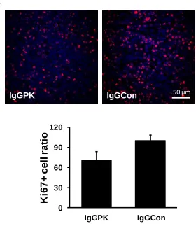
Figure 2-1 Antibody against PKM2 inhibits tumor growth. ... 49
Figure 2-2 Recombinant PKM2 promotes SW620 tumor growth. ... 55
Figure 2-3 Recombinant PKM2 promotes PC-3 tumor growth. ... 61
Figure 2-4 PKM2 and anti-PKM2 antibody does not affect cell proliferation of SW620 and PC-3 cells. ... 66
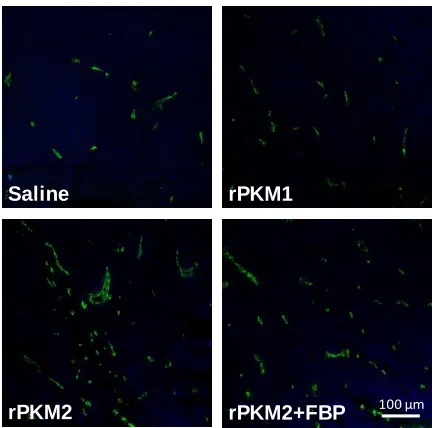
Figure 2-5 PKM2 affects endothelial tube formation, migration, and extracellular matrix attachment... 67
Figure 2-6 Tetramer or dimer of PKM2 and PKM1. ... 71
Figure 2-7 PKM2 interacts with integrins to activate integrin signaling. ... 75
Figure 2-8 PKM2 was actively released or secreted to cell culture medium. ... 80
Figure 2-9 PKM2 secreted by SW620 cell promotes enthelial cell tube formation. ... 87
Figure 2-10 FBP does not convert circulating PKM2 to tetramer. ... 88
Figure 2-11 PKM2 does not affect SW620 and PC-3 cell migration. ... 89
Figure 2-12 PKM2 dimer is required for promoting endothelial cell attaching to ECM. ... 93
Figure 3-1 integrin expression and PKM2 secretion in SK-MEL-28 and SW620 cells. ... 106
Figure 3-3 Apoptosis resistance mediated by extracellular PKM2 is integrin αvβ3 dependent. 111
Figure 3-4 The effects of antibody against PKM2 on tumor drug treatment. ... 113
Figure 3-5 Extracellular PKM2 activates FAK via integrin αvβ3. ... 115
Figure 3-6 AKT inhibitor abolishes PKM2 induced oxaliplatin resistance. ... 117
Figure 4-1 RX-5902 interacts with p68 RNA helicase. ... 132
Figure 4-2 Effects of RX-5902 on ATPase activity of p68: ... 133
Figure 4-3 RX-5902 abrogates the p68 and β-catenin interaction. ... 134
CHAPTER 1 GENERAL BACKGROUND
1.1 Cancer Metabolism and Warburg effect
Cancer progression requires high division rate. To accommodate themselves to this change,
can-cer cells produce more metabolic intermediates to synthesize cell components such as nucleotides, cell
skeletons and other bio-macromolecules. Normal cells utilize glucose to produce
three-carbon-intermediate Acetyl-CoA, which is transported to mitochondria for further metabolism, TCA cycle and
oxidative phosphorylation. Major ATP producing sources are citric acid cycle (Three Carbon Acid cycle or
TCA cycle) and oxidative phosphorylation. This occurs in normal cells with sufficient oxygen supply.
However, cancer cells modify these metabolic pathways. Cancer cells elevate glucose and glutamine
up-takes and produce ATP from glycolysis instead of TCA cycle and oxidative phosphorylation in
mitochon-dria. Pyruvate produced by glycolysis is further metabolized to lactate in cytoplasm. Cancer cells
pro-duce ATP via glycolysis even when oxygen supply is sufficient (Hsu and Sabatini 2008). This pathological
phenomenon was termed as Warburg effect, named after Nobel Laureate Dr. Otto Heinrich Warburg.
Warburg effect is one of the manifest characteristics of cancer cells (Warburg 1956). Glycolysis only
pro-duces two moles of ATP per mole of glucose used, while TCA cycle propro-duces 34 molecules of ATP. Why
do cancer cells rely on glycolysis, a so inefficient pathway to produce energy? The most popular and
ac-ceptable explanation is that: cancer cells require huge amount of metabolic intermediates such as amino
acids, nucleic acid and lipids to synthesize proteins, DNA/RNA and cell membrane for cancer rapid cell
division (Soga 2013). Glycolytic intermediates provide the sources for synthesis of these
bio-macromolecules. For instance, Fructose-6-phosphate can be used to synthesize nucleic acid via Pentose
Phosphate Pathway (PPP); 3-phosphoglycerate is used to synthesize amino acid such as Glycine,
ki-nase is impaired, all intermediates from glycolysis are retained for macromolecule biosynthesis. Another
explanation for Warburg effect is that glycolysis is much faster than TCA cycle in producing ATPs. But
this requires cancer cells to provide more glucose for glycolysis. The direct evidence for this is that
can-cer cells uptake more glucose than normal cells do. (Cairns, Harris et al. 2011). Warburg effect is caused
by many metabolism alterations. For instance, cancer cells express Pyruvate Kinase M2 instead of
Py-ruvate Kinase M1 by alternative splicing; c-Myc and HIF-1 were reported to induce the expression of
GLUT, a glucose transporter, on the cell membrane to facilitate glucose uptake; constitutive activation of
signaling pathways in cancer cells manipulates metabolic enzymes and regulates their enzymatic
activi-ties. In conclusion, cancer cells modify metabolic pathways to support their rapid cell division.
1.2 Pyruvate kinase
1.2.1 Pyruvate kinase transcription and expression
Glycolysis is a critical metabolic pathway to convert glucose to pyruvate, which occurs in the
cy-tosol. The last and rate-limiting step of glycolysis is catalyzed by an enzyme called pyruvate kinase to
convert PEP and ADP to pyruvate and ATP. Pyruvate kinase family contains four isoenzymes in
mam-mals: M2, M1, L and R types. PKL is expressed in liver, while PKR in red blood cells. PKM1 is expressed in
most other cell types in adult tissues, mainly in muscle. PKM2 and PKM1 are both gene products of M
gene (Noguchi, Inoue et al. 1986). M gene contains 12 exons and 11 introns. Alternative splicing leads to
a 23 amino acid variation between PKM1 and PKM2 (between residues 378-434). Similarly, PKLR gene
encodes both PKL and PKR. PKM2 is expressed in fetal tissues, and gradually replaced by other three
isotypes during fetus maturation. It was noted for decades that PKM2 is expressed not only in
embryon-ic cells but also in other highly proliferating cell types, such as cancer cells and immortal cell lines,
MCF10A cells for example (Christofk, Vander Heiden et al. 2008). C-Myc has been reported to induce the
and tumor progression. It was also reported that cancer cells can be converted to ‘normal-like’ or benign
cells and that Warburg effect is partially reversed by knockdown of PKM2 and re-expression of PKM1 in
cancer cells.
1.2.2 Pyruvate kinase structure
Since they are encoded by the same genes with one exon difference, human PKM1 and PKM2
share 96% identity in their amino acid sequences (Fig 1-1). Pyruvate kinases in different species are very
conservative at the enzymatic active site, especially Glu271 and Asp295 residues (Larsen, Laughlin et al.
1994). Therefore, the major difference is from their quaternary structure. PKM1 forms homo-tetramers
and each monomer is composed of four domains (N, A, B, C), while PKM2 has two quaternary
confor-mations: dimers and tetramers. PKM2 tetrameric conformation possesses high binding affinity to its
substrate phosphoenolpyruvate (PEP), but dimeric PKM2 has low affinity to PEP (Christofk, Vander
Heiden et al. 2008) (Fig 1-2). The interfaces between each monomer can be defined as A-A’ (strong
in-teraction interface) and C-C’ interface (weak inin-teraction interface). The differences between the amino
acid sequences of PKM1 and PKM2 locate in C-C’ interface and in FBP binding site (Dombrauckas,
Santarsiero et al. 2005) (Fig 1-3).
PKM2 forms both homodimer and homotetramer conformations and the equilibrium of dimer
and tetramer is regulated by numerous allosteric cofactors. Fructose 1, 6-Bisphosphate (FBP) has been
reported to allosterically regulate PKM2 enzymatic activity (Ashizawa, McPhie et al. 1991). FBP binding
site is located at a loop region from residue 402 to 407 and binding of FBP orientates the loop to an
or-dered structure (Jurica, Mesecar et al. 1998). In the absence of FBP, C domains from two subunits expel
each other; upon the binding to PKM2, FBP induces a huge conformational change of C domains at the
interface and exposes charged residues to form salt bridges interaction. The binding of FBP to PKM2
en-zymatic activity. Besides FBP, metabolic intermediates such as amino acids Alanine, Leucine and
L-Proline are also able to regulate PKM2 enzymatic activity. On the contrary to FBP, these factors inhibit
PEP binding to PKM2 and suppress PKM2 activity physiologically (Mazurek, Boschek et al. 1997).
Besides the physiological regulations, cancer cells regulate PKM2 dimer/tetramer in different
ways. Oncoprotein pp60v-src (Presek, Reinacher et al. 1988) and E7 of the human papilloma virus
(Mazurek, Zwerschke et al. 2001) phosphorylates PKM2 and stabilizes dimeric PKM2. However, the
de-tailed mechanism remains unclear in their studies. Recently, a number of studies have highlighted novel
mechanisms of regulation of enzymatic activity of PKM2. PKM2 was also converted to dimer
confor-mation by binding to certain synthesized phosphotyrosine peptides screened from a peptide library
(Christofk, Vander Heiden et al. 2008). This peptide however does not belong to any existing protein as
they discussed in their study, however, it is possible that peptide with similar sequence but not identical
sequence could bind to PKM2. Other studies have confirmed this result that PKM2 binds to certain
ki-nases in the cells. PKM2 is phosphorylated at tyrosine 105 residue by Fibroblast Growth Factor Receptor
(FGFR) after ligand binding and phosphorylated PKM2 is induced to dimer conformation (Hitosugi, Kang
et al. 2009). All the studies indicated that PKM2 is converted to dimers by different pathways in various
types of cancers. We also reported that 399R mutated to Glutamic acid residue (E) in PKM2 mimics
mer conformation of PKM2. This mutation breaks down the salt bridge interactions between two
di-mers.
1.2.3 PKM2 localization
Glycolysis takes place in perinulear region in cytosol. Pyruvate produced by glycolysis is
trans-ported to mitochondria and further oxidized to CO2 by in TCA and produce more ATPs. Pyruvate kinase is
associated with glycolytic enzyme complex with other glycolytic enzymes located in cytosol; therefore,
However, non-canonical localizations of PKM2 have been reported in recent studies. Firstly,
PKM2 translocates to nucleus. First report is that Interleukin-3 induces PKM2 nuclear localization which
regulates cell proliferation (Hoshino, Hirst et al. 2007). Our lab also revealed that dimeric PKM2 has
more nuclear localization than tetrameric PKM2 has (Gao, Wang et al. 2012). Our results indicated that
mutation of residue Lysine 399 to Glutamic acid (R399E) converts recombinant PKM2 (rPKM2) from
te-tramer to dimer. Dimer rPKM2 translocates to nucleus to phosphorylate STAT3. Phosphorylated STAT3
further activates MEK5 gene transcription to promote cancer cell proliferation. PKM2 nuclear
transloca-tion might be regulated by ERK2. The detailed mechanism is studied by Yang and colleagues that
acti-vated ERK2 phosphoryaltes Pyruvate kinase M2 at Serine 37 residue. PIN1 recruites importin α5 binding
to phosphorylated PKM2 and leads to PKM2 nuclear translocation (Yang and Lu 2013).
Furthermore, PKM2 is detected in body fluids from cancer patients. PKM2 has been used as a
cancer biomarker especially for colorectal cancer, gastric cancer and lung cancer diagnosis. PKM2 is
de-tected by diagnosis kit (ELISA, Enzyme-Linked ImmunoSorbent Assay kit) in patients’ blood sera or feces
samples (Kumar, Tapuria et al. 2007). In vitro, PKM2 is released from colorectal carcinoma cell line
colo205 and SW480 to culture medium (Wu, Chen et al. 2008). However, it remains unresolved how
PKM2 is secreted and whether circulation PKM2 possesses any function. Many researchers have
at-tempted to investigate the potential physiological or pathological functions of PKM2. Shin et al. found
that PKM2 released by cancer cells is related with 5-fluorouracil resistance in colon cancer cell lines
(Shin, Yoo et al. 2009). 5-fluorouracil resistance cell line secretes more PKM2 to the culture medium
than 5-fluorouracil non-resistance cell line does.
PKM2 in cell culture medium is possibly released from exosomes or microvesicles (Meckes,
Gunawardena et al. 2013). Exosomes and microvesicles are small vascular compartments and secreted
by many cell types such as B lymphocytes, dendritic cells and cancer cells. The physiological roles of
com-munication or cell-matrix modification. Pyruvate kinase was reported to be present in exosomes and
microvesicles identified by Mass spectrum. PKM2 was also reported to be included in neutrophil
gran-ules, which suggested another potential pathway of PKM2 secretion in immune cells (Lominadze, Powell
et al. 2005). Our unpublished data also supported that PKM2 is secreted by neutrophils. In summary,
non-canonical localizations of PKM2 are related to its novel biological functions from the enzymatic
ac-tivity.
1.2.4 PKM2 biological functions
Pyruvate kinase catalyzes the last step of glycolysis to convert PEP to pyruvate and generate one
mole of ATP. The active site locates in a pocket between the interface of A and B domains. Mg2+, K+ and
H2O molecules are required for PKM2 catalytic activity (Larsen, Benning et al. 1998). When oxygen is
supplied, glucose is metabolized through glycolysis and followed by TCA cycle in mitochondria. Glucose
is converted to pyruvate and lactate when oxygen in the cell environment is low. Cancer cells, however,
produce lactate even with sufficient oxygen supply (Mazurek, Boschek et al. 1997). Metabolic enzymes
form a protein complex termed as glycolytic enzyme complex, containing enolase, lactate
dehydrogen-ase, pyruvate kinase and other glycolysis enzymes (Mazurek, Michel et al. 1997). Once PKM2 forms
di-mers, the quaternary structure changes and the binding affinity to its substrate PEP decreases.
Mean-while, dimeric PKM2 dissociates from the glycolyitc enzyme complex. Due to these changes, metabolic
intermediates in glycolysis are accumulated and utilized to synthesize amino acids, nucleotides and
phospholipids for cancer cells (Mazurek, Boschek et al. 2005). PKM2 enzymatic activity is fully
manipu-lated in cancer cells. Firstly, switch of PKM1 to PKM2 expression is one of strategies that cancer cells use
to meet the demand for their high proliferation rate. As mentioned above, phosphorylation of PKM2 at
several residues was reported to enhance PKM2 dimer formation and promote tumorigenesis.
activ-ity (Anastasiou, Poulogiannis et al. 2011). The metabolic pathways, manipulated by cancer cells,
down-regulate PKM2 enzymatic activity and support cancer cells to achieve Warburg effect. .
1.2.4.1 Pyruvate kinase M2 acts as a gene transcription activator
Early in 2008, Lee et al. reported that PKM2 interacts with transcription factor Oct-4 in vitro. This
PKM2 and Oct-4 interaction was analyzed by affinity purification and mass spectrometry (Lee, Kim et al.
2008). The authors speculated that the interaction plays a role in regulation of gene transcription.
Moreover, hypoxia inducible factor 1 (HIF-1) is well-known to be responsible for oncogene transcription
activation. PKM2 has also been shown to enhance HIF-1α transcription activity by binding with prolyl
hydroxylase 3 (PHD3) (Luo, Hu et al. 2011). The interaction is critical for regulation of HIF-1α
transcrip-tion activity, thus it was proposed that PKM2 coordinates with HIF-1α in manipulating hypoxia response
in cancer cells. PKM2 also plays an important role in β-catenin transactivation in tumor proliferation and
malignancy. In quiescent cells, β-catenin forms complex with Axin, APC and GS3K. Β-catenin is
phos-phorylated by GS3K and degraded through proteasome. Once cells are activated by Wnt ligand,
β-catenin dissociates from the complex and translocates to nucleus to activated
tumorigenesis-related-gene activation. PKM2 binds Y333 phosphorylated β-catenin and co-activates β-catenin downstream
gene transcription (Yang, Xia et al. 2011). Our lab also reported that PKM2 regulates MEK5 gene
tran-scription resulting in promoting cancer cell proliferation (Gao, Wang et al. 2012). It remains unclear that
how PKM2 binds to those gene transcription factors. We proposed that PKM2 acts as a protein kinase to
phosphorylate STAT3.
1.2.4.2 PKM2 is also a protein kinase
Small molecules PEP and ADP are the substrates of PKM2. The enzymatic active sites are only
able to accommodate small molecules. However, proteins were also found as the substrates of PKM2.
H1 is phosphorylated by utilizing PEP as phosphate donor (Ignacak and Stachurska 2003).
Multifunction-al protein prothymosin Multifunction-alpha (ProTα) is another target, which is phosphorylated by PKM2 (Diaz-Jullien,
Moreira et al. 2011). Furthermore, Dr. Zhimin Lu’s group recently reported that PKM2 phosphorylated
Histone H3 and this modification led to gene transcription and further promoted tumor growth. Our lab
data also demonstrated that PKM2 phosphorylates STAT3. Transcription factor STAT3 is phosphorylated
at Y705 by PKM2 using PEP as phosphate donor. Phosphorylated STAT3 leads to the enhanced tumor
cell proliferation. However, PKM1 lacks this protein kinase activity (Gao, Wang et al. 2012). FBP
regu-lates PKM2 protein kinase activity by promoting tetramer formation. It is likely that PKM2 protein kinase
activity requires the conformational change because PKM2 crystal structure indicated that the
enzymat-ic activity pocket does not provide enough space for protein substrates. Dimerization might induce
con-formational change to expose the enzymatic pocket to improve accessibility for protein substrates.
1.2.4.3 Extracellular PKM2
PKM2 in patients’ serum and stool samples was used as a biomarker in some inflammatory
dis-eases as well as malignant tumors (Staib, Hoffmann et al. 2006; Aloysius, Zaitoun et al. 2009). PKM2 is a
potential fecal marker in Crohn’s disease patients (Day, Judd et al. 2012). However, it was also reported
that PKM2 level in inflammatory bowel disease (IBD) patients serum is not as high as in various cancer
patients (Meng, Zhu et al. 2012). Colorectal cancer has elevated PKM2 concentration in blood serum
(5-7 times higher than normal individuals); two-fold increase of serum PKM2 is in adenoma compared to
health population. PKM2 secreted by cancer cells was reported to correlate with 5-fluorouracil
re-sistance in colon cancer cell lines (Shin, Yoo et al. 2009). PKM2 in the culture medium is up-regulated in
the cancer cell line with 5-fluorouracil resistance compared to the cell line without 5-FU resistance.
However, the mechanism needs to be further studied. Similarly, another glycolytic enzyme:
regulates angiogenesis (Lay, Jiang et al. 2000). In our study, we found that PKM2 enhances angiogenesis
by promoting endothelial cell migration, adhesion and survival via interaction with integrin αvβ3.
1.2.5 PKM2 inhibitor and activator in cancer therapy
Since PKM2 is universally expressed in various types of cancers, researchers tried to develop small
compounds to inhibit PKM2 and induce cell apoptosis (Vander Heiden, Christofk et al. 2010). However,
cancer cell induces tetrameric PKM2 to form dimers either through binding phosphorylated peptide or
through being phosphorylated by tyrosine kinase. Decrease in enzymatic activity of pyruvate kinase
fa-vors the accumulation of metabolic intermediates in cancer cells. Therefore, PKM2 inhibitor may not be
successful due to this end. More recently studies focused on reversing the process by promote PKM2
enzymatic activity. Substitution of PKM2 by PKM1 decreases tumor sizes in mice model (Christofk,
Vander Heiden et al. 2008). PKM2 allosteric co-activator, FBP acts as an activator to promote PKM2
te-tramer formation. However, it was demonstrated that FBP has no effect on the PKM2 phosphorylated
by FGFR in cancer cells (Hitosugi, Kang et al. 2009). Anastasiou and colleagues synthesized compounds
which specifically recognize PKM2 and convert dimeric PKM2 to tetrameric PKM2. These compounds
were termed as PKM2 activators (Anastasiou, Yu et al. 2012). PKM2 enzymatic activity was up-regulated
by PKM2 activators and these activators have been shown to inhibit mice tumorigenesis.
1.3 Angiogenesis
The closed blood circulation system is unique in vertebrates. Blood vessel growth is a vital
physio-logical process in embryonic development and wound healing. Blood vessels are formed by tube forming
endothelial cells, called endothelium. Endothelium is surrounded and covered by smooth muscle cell
layer. Elastic tissues and connective tissues provide support and protection to the blood vessel from
stress and physical damage (Adair and Montani 2010). Blood vessels without vessel wall or with thin wall
ves-sels are formed by two stages: vasculogenesis and angiogenesis. Vasculogenesis is the process that
blood vessels start growth from scratch. It is related to the differentiation of blood vessel precursor cells
such as angioblasts (Patan 2004). It has been well-studied that blood vessel precursor cells differentiate
into endothelial cells in response to growth factor or extracellular matrix, such as bFGF and VEGF
(Kazemi, Wenzel et al. 2002); while angiogenesis is the process that blood vessels or capillaries grow or
extend from the existing vessels. Angiogenesis involves endothelial cell proliferation, migration, survival
and tube-like structure formation.
1.3.1 Angiogenesis and its regulation
Angiogenesis is tightly regulated in the body. Loss of the control of angiogenesis is related to a
series of pathological conditions. Insufficient angiogenesis leads to Alzheimer’s disease, diabetes,
chron-ic wound healing and etc. On the contrary, excessive angiogenesis also impairs the physiologchron-ical orders
and is involved in more than 20 diseases including cancer. Cancer is one of the most important disorders
highly related to angiogenesis (Griffioen and Molema 2000). Angiogenesis balance is maintained by two
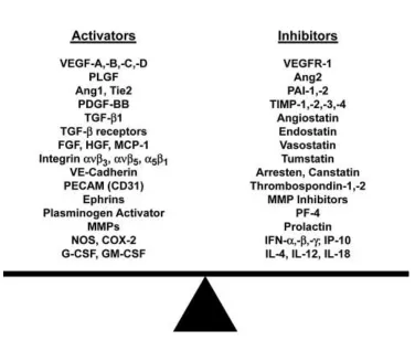
protein families: angiogenic activators and inhibitors (Mundel and Kalluri 2007) (Fig 1-4). Both
angiogenic activators and inhibitors collaborate to regulate physiological angiogenesis. Angiogenesis
activator family contains various growth factors (such as fibroblast growth factor, placental growth
fac-tor and vascular endothelial growth facfac-tor), angiopoietin 1, interleukin 8 and etc. Angiogenesis inhibifac-tor
family consists of a group of proteins or peptides which specifically inhibit angiogenesis, such as
endostatin, interferon and thrombospondin 1 (Zetter 2008). The high proliferation rate of cancer cells
requires enhanced nutrients transportation. Initial tumor growth depends on diffused oxygen and
nutri-ents from adjacent stroma cells; while larger tumors need blood vessels to support their further growth
(Folkman, Long et al. 1963). In clinical cases, tumor size is limited to 1-2mm in diameter without
1.3.2 Angiogenesis and cancer therapy
1.3.2.1 VEGF-dependent and VEGF-independent angiogenesis
Vascular endothelial growth factor (VEGF) is the most important angiogenesis activator, which
strongly stimulates vasculogenesis and angiogenesis. VEGF was identified in early 90s (Senger, Galli et al.
1983) and belongs to platelet derived growth factors VEGF/PDGF superfamily (Trelles, Leon et al. 2002).
VEGF has at least 5 members: VEGF-A, B, C, D, E (Shibuya 2008). VEGF is a homodimeric glycosylated
protein and contains two intermonomer disulfide bonds and three intramonomer disulfide bonds. VEGF
has 5 isotypes: VEGF121, 145, 165, 189 and 206. All those isotypes are encoded by VEGF gene and
pro-duced by alternative splicing. VEGF has two subcellular forms: secreted VEGF and membrane-bound
VEGF (Salvador, Li et al. 2008). VEGF is secreted by many types of cells, such as endothelial, fibroblasts
and activated immune cells. VEGF is also expressed and secreted by cancer cells themselves or by cancer
cell infiltrated stroma cells.
VEGF induced cellular response is mainly via VEGF receptors (VEGFRs). VEGFR is a tyrosine kinase
receptor, which contains an extracellular domain, a transmembrane domain and a cytoplasmic tail.
VEGFR family has 3 members, VEGFR-1 (flt-1), VEGFR-2 (flk-1 or KDR) and VEGFR-3 (flt-4). VEGFR-1
main-ly is the receptor for VEGF-A and VEGF-B; VEGFR-2 is receptor for VEGF-A, C, D and E; VEGFR-3 is for
VEGF-C and D. VEGFR-1 and VEGFR-2 is responsible for vasculogenesis and angiogenesis signaling
trans-duction; while VEGFR-3 is conducting the signaling pathways related to lymph-angiogenesis (Hicklin and
Ellis 2005). Binding VEGF to their receptors dimerizes VEGFR VEGFR c-terminal undergoes
autophosphorylation. Phosphorylation of tyrosine residues leads to the activation of various signal
pathways such as Ras, PLC, p38 and PI3K. Those downstream kinases regulate cell functions related to
It is necessary to understand angiogenesis mechanism better in order to treat or diagnose cancer
and other diseases with deregulated angiogenesis. Besides VEGF-dependent angiogenesis,
VEGFR-independent angiogenesis is equally crucial. Other growth factors than vascular endothelial growth
fac-tor also regulate angiogenesis. It was reported that platelet-derived growth facfac-tor (PDGF) regulates
an-giogenesis through PDGF receptor by promoting endothelial cell proliferation and tube formation
(Battegay, Rupp et al. 1994). EGF was also reported to modulate angiogenesis by increasing endothelial
cell migration (Mehta and Besner 2007). Moreover, Angiopoietin regulates angiogenesis through Tie
receptors. Angiopoietin family has four members: Ang-1, Ang-2, Ang-3 and Ang-4. Among all the
mem-bers, Ang-1, Ang-3 and Ang-4 bind Tie2 receptor, while Ang-2 binds Tie1 receptor. After binding to
re-ceptors, Ang dimerizes Tie receptor and then autophosphorylates the intracellular domain of the
recep-tor. Receptor activation phosphorylates their downstream signaling cascades, mainly through PI3K/Akt
signaling pathway and STAT to regulate gene transduction of cell survival and cell proliferation
(Mochizuki, Nakamura et al. 2002).
1.3.2.2 Angiogenesis inhibitor
Increase in VEGF secretion and/or VEGF receptor expression induces angiogenesis in cancer. Tumor
vessels are different from vessels in normal tissues (Shojaei 2012). -Tumor blood vessels are not as
or-ganized as the normal vessels due to the defective vascular structure in cancer. Repressing angiogenesis
is one of the important ways for cancer therapy. Different strategies have been explored for
angiogene-sis related cancer therapy.
1. Targeting VEGF. VEGF antibody was developed to specifically target VEGF and decreased the
se-rum level of VEGF. Bevacizumab (Avastin) is the most successful case and was approved for clinical use
in 2004 by U.S. Food and Drug Administration (FDA). Bevacizumab is also the first FDA approved
radio-therapy chemicals or cocktailed with chemoradio-therapy drugs to bring more benefits to cancer patients
(Bhuvaneswari, Yuen et al. 2007; Jayson 2011).
2. VEGFR inhibitors. Two distinct inhibitors for VEGFR were exploited to inhibit angiogenesis. The
first type of VEGFR inhibitor is anti-VEGFR antibody, which were developed to inhibit VEGF ligand
bind-ing to its receptor. DC101, an anti-VEGFR2 monoclonal antibody potentially inhibits prostate tumor
growth and metastasis (Sweeney, Karashima et al. 2002). DC101 has also been shown to be used to
en-hance paclitaxel or radiology treatment (Inoue, Slaton et al. 2000; Verhoeff, Stalpers et al. 2009).
Anti-bodies anti-VEGFR extracellular domain also induce cancer cell apoptosis (Sweeney, Karashima et al.
2002). The second type inhibitor is small compounds. Virtual Screening and computational modeling
were used to design and screen the compounds for inhibiting VEGFR dimerization (Elgamacy, Shalaby et
al. 2011).
3. VEGFR signaling pathway inhibitors. VEGF receptor is one of the receptor tyrosine kinase family
members. Therefore, the most common strategy to inhibit angiogenesis is to repress VEGFR signal
transduction. Plentiful tyrosine kinase inhibitors (TKIs) are developed and utilized to treat different
tu-mor types. Pazopanib is an FDA-approved medication for treatment of advanced renal cell carcinoma
(Sloan and Scheinfeld 2008). Pazopanib is a synthetic chemical compound which specifically targets and
inhibits tyrosine kinase sites of VEGFR and platelet-derived growth factor receptor (PDGFR).
Generally, VEGF-VEGF receptor induced angiogenesis has been well studied. However, inhibition of
angiogenesis through VEGF-VEGFR pathway is not sufficient to treat cancer. Some important drawbacks
cannot be ignored. Firstly, VEGF-VEGFR is not the only signaling pathway to regulate angiogenesis
(Shibuya 2008). Secondly, inhibition of VEGFR pathway causes disease-related side-effects such as
dys-function or failure of major organs: heart, liver and renal system (Chen and Cleck 2009); repression on
themselves cannot inhibit tumor growth and it is required to combine VEGF with other anti-cancer drugs
for better clinical outcomes.
1.4 Integrins
Eukaryotic cells are constantly communicating with their microenvironment through both
cell-matrix adhesion and cell-cell adhesion. Numerous molecules are involved in cell adhesion. Integrin
fami-ly is one of the most important proteins involved in cell adhesion and migration. Specific integrin
expres-sion pattern is correlated to endothelial cell proliferation, spreading, migration and invaexpres-sion. Integrins
activate intracellular signaling pathways after interacting with extracellular ligands such as extracellular
matrix proteins (ECM).
1.4.1 Cell adhesion
Cells interact with surrounding cells to form tissue layers or to transfer nutrient and cellular
sig-nals. Four major cell-cell adhesion complexes exist in epithelial and endothelial cells: tight junction,
adherens junction, gap junction and desmosome.
Tight junction functions at anchoring adjacent cell together tightly and prohibiting fluids to
dif-fuse through the gaps between epithelial/endothelial cells. Tight junction also maintains the polarity of
epithelia or endothelia layer. Tight junction is composed of a series of transmembrane proteins such as
claudin, occludin and tetraspanins. The interaction between these proteins from adjacent cells provides
the molecular bases for tight junction (Gonzalez-Mariscal, Betanzos et al. 2003).
Adherens junction is mainly for holding epithelial and endothelial cells together and supporting
the cell architecture. Cadherin and catenin family proteins are responsible for adherens junction
for-mation. E-cadherin and N- cadherin are calcium binding transmembrane proteins and interact with
cad-herin molecules from adjacent cells. After two cadcad-herin molecules interact, α- and β-catenins are
Gap junction is involved in cytoplasm component exchange. Gap junction is a hexamer formed
by connexin family proteins. Gap junction close and open conformation switch is regulated by either
calcium (Lurtz and Louis 2007)or post-translational regulation such as phosphorylation (Warn-Cramer
and Lau 2004).
The last but not the least type of cell-cell junction is desmosome. Desmosome exists mainly in
epithelial cells other than endothelial cells, but desmosome protein desmoplakin is expressed in
cul-tured endothelial cells (Valiron, Chevrier et al. 1996). Desmoplakin interacts with cadherin or vimentin
to regulate endothelial cell intercellular junction. In epithelial cells, the function of desmosome is to
sus-tain the cells against shearing force. Desmosome complex forms plaque structure on the membrane
across cells and the complex is constructed by a series of cadherin family proteins such as Desmoplakin,
Desmocollin and Plakoglobin. The complex attaches to intermediate filaments inside of the cells, such as
keratin (Bornslaeger, Corcoran et al. 1996).
Both epithelial cells and endothelial cells rely on the four types of cell junctions to interact or
communicate with adjacent counterpart cell, to maintain and protect epithelia and endothelia tissues
and to prohibit random diffuse. Besides the cell-cell interactions, epithelial and endothelial cells require
the interaction with the extracellular matrix to survive and to perform various physiological functions.
1.4.1.1 Cell-matrix interaction
Basement membranes are thin layers under epithelia or surrounding endothelia. Basement
membranes have fiber structure and are composed of various extracellular matrix proteins such as
col-lagen, laminin and fibronectin. Basement membranes provide supports to epithelial and endothelial cell
layers. Integrin and hemidesmosome are the two major receptors for cell-matrix interaction.
Hemidesmosome shares the similar structure and components as the desmosome complex however,
cells together while hemidesmosome is to attach epithelial cells to basement membranes or
extracellu-lar matrix. Hemidesmosome attaches to intermediate filament in the cells to regulate celluextracellu-lar functions
(Zhang and Labouesse 2010). The other protein family for epithelial and endothelial attachment to
basement membranes is the integrin. Integrins are heterodimeric transmembrane proteins, which
regu-late various types of cell functions such as cell adhesion, migration and proliferation through either
in-side-out or outside-in activation. Integrins are also involved in many pathological conditions such as
an-giogenesis. Since integrins are important molecules in my dissertation, I will discuss it in details.
1.4.2 Integrin structure
Integrins are membrane proteins with single alpha-helices transmembrane domain. Integrins
are heterodimers formed by α subunits and β subunits. More than 20 different heterodimeric integrins
are formed by 18 α subunits and 8 β subunits in mammals (Margadant, Monsuur et al. 2011).
Extracellu-lar domains of integrins are glycosylated and form functional domains to interact with ligands or other
proteins. Integrin family can be divided into two subgroups by their structures difference: with or
with-out an extra von Willebrand factor type A domain (αA or αI domain). Integrins can also be divided into
four groups based on their ligands or binding partners: collagen-binding integrins; laminin-binding
integrins; RGD dependent integrins and Leukocyte integrins. Each integrin subunit contains a bunch of
specific domains. Integrin heterodimers are illustrated as a headpiece and ‘leg’ part (Springer 2002).
In-tegrin ligand binding site is located in the headpiece.
1.4.2.1 Integrin conformation change
Integrins have two conformational structures: close and open conformation. In the close or
‘bent’ conformation, the headpiece of integrin heterodimers bends over to the ‘leg’ part. The ligand
binding site is facing to the cell membrane, which results in the low binding affinity of integrin
un-derstand the relationship between integrin conformation change and its function. Upon the binding of
integrin to its ligand, integrin undergoes dramatic conformational change in its extracellular domain.
This change makes integrins skewed towards clustering with adjacent integrin. The conformational
change in the extracellular domain of integrins also induces a structure change in integrin
transmembrane domain and intracellular domain (ICD), which separates integrin c-terminals of α and β
subunits. C-terminal separation of integrin α and β subunits leads to the assembly of focal adhesion
complex and activation of FAK and its further downstream signaling pathways (Askari, Buckley et al.
2009).
1.4.2.2 Integrin ligand
RGD-containing ligand is the largest protein family which binds integrins including αvβ3, α5β1,
αIIbβ3, and etc. This integrin subfamily recognizes a consensus binding sequence: three amino acids
RGD (R: Arginine, G: Glycine, D: Aspartic acid). Based on the analysis of the crystal structure, the binding
site for RGD-containing ligand locates at the interface of integrin α and β subunits (Xiong, Stehle et al.
2002). Lysine residue interacts with β-propeller domain in α subunit while aspartic acid binds to von
Willebrand factor A (vWF) domain in β subunit. Many extracellular matrix (ECM) components contain
RGD sequence including fibronectin and vitronectin. Certain ECM proteins do not have accessible RGD
sequences on the protein surface, but expose their cryptic RGD sequence to interact with RGD-specific
integrins after being denatured by temperature or cleavage by the matrix metalloproteinase
(Yamamoto, Yamato et al. 1995). For instance, Integrin α5β1 and αvβ3 are the typical RGD dependent
integrins. But collagen I does not bind to either integrin α5β1 or αvβ3 due to the absence of any solvent
accessible RGD sequence. However, after being partially denatured by increasing temperature or
de-graded by extracellular proteases (MMP-2 or MMP-9), collagen I unfolds and exposes its RGD sequence
related to cancer angiogenesis and metastasis (Eliceiri and Cheresh 1999). GRGDSP is a sequence from
fibronectin and used as an angiogenesis inhibitor as to compete with RGD-containing native ligands.
In addition, some non-ECM proteins containing RGD sequence were also reported to interact
with integrins. The human immunodeficiency virus (HIV-1) protein, transactivating factor (TAT) activates
FAK through integrin αvβ3 and promote angiogenesis (Urbinati, Mitola et al. 2005). TAT interaction with
integrin is inhibited by both RGD peptide competitive inhibitor and blocking antibody LM609, which
in-dicates that the interaction is via RGD sequence in TAT protein. Another example is CD40 ligand or
CD40L. Soluble CD40 ligand was reported to bind to integrin α5β1 and induce signaling transduction by
activating MAPK and its downstream ERK1/2 in endothelial cells (Leveille, Bouillon et al. 2007). This
in-teraction is through RGD sequence on the CD40L (Andre, Prasad et al. 2002).
Besides RGD sequence, KGD (Lysine-Glycine-Aspartic acid) is also recognized by integrins in
cer-tain cases. Integrin β3 is the major type of integrin for interacting with KGD, especially αIIbβ3. KGD plays
important role in disintegrin binding to integrin. Disintegrin is a family of proteins in viper venom.
Disintegrin inhibits platelet aggregation and causes hemorrhage through the interaction of KGD
se-quence with αIIbβ3 integrin (Lu, Lu et al. 2005).
A synergetic site might be important for integrin binding to RGD. RGD peptides are used for
can-cer therapy to target cancan-cer angiogenesis. RGD peptide such as cilengitide a cyclic RGD pentapeptide has
been applied as an angiogenesis inhibitor. However, the affinity of RGD peptide is 1000 fold lower than
their natural ligand. One of the most important reason is that integrin ligand contains not only RGD
se-quence but also synergetic site. Pro–His–Ser–Arg–Asn (PHSRN) is a synergetic binding site in fibronectin
for integrin α5β1 and this synergy site facilitates HUVEC cell attachment and spreading in vitro
(Ochsenhirt, Kokkoli et al. 2006).
Other than RGD sequence, LDV is another major binding sequence for integrins. LDV mainly
such as IDA or REDV (Mould and Humphries 1991). As mentioned earlier, a subgroup of integrins contain
α domain, α1β1, α2β1, α10β1 and α11β1. Those integrin binds to specific residues other than RGD and
LDV. GFOGER is the critical sequence on collagen to be recognized by αA-containing integrins. The
glu-tamic acid residue in the recognition sequence is crucial to interact with the metal in the integrins
(Emsley, Knight et al. 2000).
1.4.2.3 Metal binding vs. Ligand binding
Magnesium and calcium metal ions are crucial to maintain integrin proper structure and ligand
binding activity. As described above, integrin can be divided into two subfamilies based on their
struc-ture similarity: αA integrins and non-αA integrins. αA integrins contain a von Willebrand factor type A
domain. αA is responsible for mediating integrins binding to their ligands and α domain contains an ion
binding site, named metal-ion-dependent adhesion site (MIDAS) (Michishita, Videm et al. 1993). MIDAS
coordinates one divalent cation, usually Magnesium and MIDAS domain accepts one interaction from an
acidic amino acid from integrin ligand (Lee, Rieu et al. 1995). MIDAS has even higher binding affinity for
Manganese (Li, Rieu et al. 1998). However, Manganese is not a physiological ion. Although structure
dif-ferent exists between αA-containing and α-lacking Integrins, integrins without αA domain shows similar
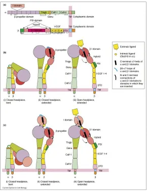
strategy for ligand binding. Integrin αvβ3 is an important αA-lacking integrin. αvβ3 contains an αA-like
domain or βA/βI domain (Fig 1-5)(Luo and Springer 2006). Aspartic acid in RGD tripeptide attaches to
the ion located at MIDAS site, while Arginine (R) residue interacts with a couple of negative charged
res-idues at β propeller on αsubunit. MIDAS on integrin β subunit and β propeller domain from α subunit
forms the basic ligand binding pocket. Besides MIDAS at αA domain or A-like domain on integrin β
subu-nit, other two metal binding sites locates adjacent to MIDAS: the ADMIDAS (adjacent to MIDAS) and
LIMBS (ligand-associated metal binding site). Those sites favor calcium ion and provide additional
transmembrane protein, which highly expresses in tumor endothelium.TEM8 also contains an MIDAS
site and this MIDAS was reported to be involved in binding to the anthrax toxin antigen (Ramey, Villareal
et al. 2010). The interaction of TEM8 and its ligand is much similar to the binding of integrin and RGD
peptide. It was reported that PKM2 also could bind to TEM8 in vitro (Duan, Hu et al. 2007). This
interac-tion indicates the possibility that PKM2 also binds to MIDAS on the integrin.
Integrin α subunit also contains 5 Calcium binding sites. 4 of them are in beta propeller loop
re-gion and the other one is at ‘genu’ rere-gion, which acts as a ‘knee’ for integrin conformational change
(Arnaout, Goodman et al. 2007). Metal ions are required for integrin structure and integrin ligand
bind-ing. EDTA completely inhibits integrin function and detaches cells.
1.4.3 Integrin functions
Integrins transmit outside-in or inside-out signals astransmembrane proteins. The major
physio-logical function of integrins is involved in cell adhesion and migration. Integrins are the main receptors
for extracellular matrix proteins such as collagen, fibronectin and laminin. Binding of integrins with ECM
proteins anchors the cells to basement membrane. After binding to ECM components, integrin recruits
paxillin and talin to form focal adhesion complex in cytosol. Focal adhesion complex attaches to
cyto-skeleton: actin filament (Brakebusch and Fassler 2003). Ligand-bound integrins also activated Focal
Ad-hesion Kinase (FAK). FAK phosphorylated and activated various downstream pathways, for example Src
and PI3K/Akt pathways, which were well-studied to regulate cell proliferation, survival and migration
(Yano, Mazaki et al. 2004; Caron-Lormier and Berry 2005).
1.4.3.1 Integrin activation
Integrins are activated via two different ways: out and outside-in pathways. The
inside-out activation of integrin has been well studied. Withinside-out stimuli, integrins rest in the inactive (bent
in-teract with each other. When the cells receive stimulation from growth factors such as VEGF or EGF,
cells initiate signal transduction. One important regulator, talin is activated by phosphorylation. The
de-tailed mechanism of talin activation is not fully understood. Activated talin exposes its FERM domain
(Pearson, Reczek et al. 2000) and binds to integrin intracellular domain. Binding of talin to integrin
c-terminus induces a conformational change of cytoplasm domain and transmembrane domain of
integrins. This change leads to a further conformational change of integrin extracellular domain: integrin
ECD switch from bent form (low affinity to ligand) to extended form (high affinity to ligand). Inside-out
activated integrin facilitates cell to attach to ECM (Qin, Vinogradova et al. 2004). The conformational
change of bent and extended form is visualized by electron microscopy (Weisel, Nagaswami et al. 1992).
Outside-in activation is more straightforward. Ligands bind to integrin ECDs and cause integrins
conformational change, which leads to integrin activation. Ligand binding also induces integirn clustering
to enlarge integrin activation. Activated integrins (extended or open conformation) cluster together on
the cytoplasm membrane to form a patch-like structure. Integrin clustering provides stronger binding
capability of integrins to ECM by holding a bunch of integrins at one single spot. Those structures then
form focal adhesion (Kinashi 2005). Upon focal adhesion complex assembled, integrins regulate cell
at-tachment and migration through this outside-in manner.
1.4.3.2 Integrin endocytosis and recycling activation
In order to attach to or migrate on ECM proteins or basement membrane, cells require assembly
and disassembly of focal adhesion complex rapidly. Therefore, integrins need to be transported from the
rear region to the migrative leading edge. This is achieved by integrin endocytosis and recycling. Integrin
endocytosis is initiated by binding to integrin ligand or synthetic peptide such as RGD peptide Cilengitide
(Reynolds, Hart et al. 2009). Growth factors could also induce integrin endocytosis and recycling. After
mem-brane for reuse (Bretscher 1996). Integrins retrieved from the rear of the moving cells is mediated by
Rab4 and Rab5; while integrin delivered to the frontier of the cell is mediated by Rab11 and Arf6
(Margadant, Monsuur et al. 2011).
1.4.3.3 Integrin regulates cell adhesion
Cell adhesion regulates various physiological events, such as embryonic development, cancer
metastasis, inflammation, homeostasis and wound healing (Edelman and Crossin 1991). Endothelial cells
and epithelial cells have to line on the extracellular matrix to form tissues. Integrin family members
show specificity to different ECM substrates. For example, integrin α5β1 has highest affinity to
fibronectin, αvβ3 has higher affinity to vitronectin and α1β1 and α2β1 mostly bind to collagens and
laminins. Upon the binding of integrins to their ECM, integrins convert from bent form to extended form
and cluster together. Integrin activation and clustering recruit a group of focal adhesion proteins such as
talin, paxillin and vinculin. Focal adhesion complex attaches to actin filaments to regulate cell
attach-ment to ECM. Other than integrins, desmosome and other proteoglycans also provide cell-matrix
adhe-sion. However, integrins are the most dominant and universal adhesion molecules. Cancer cells alter
integrin patterns to dysfunction cell adhesion and deregulate cell migration.
1.4.3.4 Integrin dictates cell migration and invasion
Migration is described as directional cell movement. This movement is generally divided into
several steps including (1) Lamellipodium extension at the leading edge; (2) detachment on the rear side
of moving cells; (3) make new contact at the front side; (4) cell contract to move forward. Integrins play
vital roles at both front and rear sides of moving cells. Integrin endocytosis and recycling is responsible
for redistribution of integrins. The intracellular domain of integrins contains a motif for
clathrin-dependent endocytosis (Gawaz, Besta et al. 2001). After endocytosis, Rab GTPase family proteins such
short-loop and long short-loop. Rab4 GTPase is involved in short-short-loop recycling and integrins enter early endosomes
and are rapidly transported to the plasma membrane. On the other hand, long-loop recycling is
mediat-ed by Rab11 small GTPase. Integrins are translocatmediat-ed to perinuclear recycling compartment (PNRC) first
and then transported back to plasma membrane (Caswell and Norman 2006).
It was also reported that integrin α4β1 regulates SPARC activity, which regulates extracellular
matrix remodeling (Gerson, Shearstone et al. 2012). MMP-2 and MMP-9 were commonly studied to
regulate cancer cell invasion by degrading ECM components. Both MMP-2 and MMP-9 were reported to
be modulated by integrin directly and indirectly (Chen, Wei et al. 2009; Morozevich, Kozlova et al. 2009).
Cancer cells frequently mutate integrin genes to facilitate cell migration.
1.4.3.5 Integrin signaling mediates cell proliferation and cell survival
In early 1990’s, Integrins were proved to be responsible for cell proliferation upon cell
attach-ment on the fibronectin-coated surface. The proliferation is associated to Mitogen-activated protein
kinase activation, ERK1 and ERK2 (Zhu and Assoian 1995). Various types of integrin expressions were
found to be associated with endothelial and cancer cell proliferation. The main mechanism is that Iigand
binding to integrins activates FAK and its downstream signaling pathway. Meanwhile, it was also
report-ed that integrin α5β1 regulates epithelial cell proliferation through interaction with epidermal growth
factor receptor (Kuwada and Li 2000). This suggested an alternative pathway for integrins to regulate
cell proliferation. Another membrane-bound receptor, Urokinase-type plasminogen activator receptor
(uPAR) was reported to interact with intergrins to regulate cell proliferation (Ossowski and
Aguirre-Ghiso 2000).
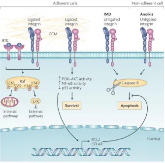
In certain extracellular environment, integrins were shown to be able to promote cell survival or
induce cell apoptosis (Desgrosellier and Cheresh 2010) (Fig 1-6). Integrin activation caused the
sur-vival signaling pathways, such as PI3K-Akt, NF-κB and Bcl-2. Among them, Bcl-2 is the member of
anti-apoptotic Bcl family. Bcl-2 binds Bax or Bak in resting cells and BH-3 only proteins compete with Bcl-2 to
release Bax or Bak. Bax or Bak forms pores on the mitochondria membrane leading to the release of
Cy-tochrome c to cytosol. It was reported that activation of integrin α5β1 and αvβ3 increased Bcl-2 on both
mRNA and protein level (Matter and Ruoslahti 2001). This increase is FAK-activation and Src-activation
dependent. Immobilized vitronectin (VN, an ECM component and secreted by cancer cells) conferred
resistance of cancer cells to drug treatments (Uhm, Dooley et al. 1999). This is due to the increase of
Bcl-2 and Bcl-XL induced by α5β1 and αvβ3 activation. Recently, PKM2 was also suggested to be related
to drug resistance (5-FU resistance) (Shin, Yoo et al. 2009). This might be explained by the fact that
PKM2 binds and activated integrins, and increased anti-apoptotic protein expression. Similar result was
reported that integrins prevents breast cancer cells from apoptosis by activating PI3K-Akt signaling
pathway (Aoudjit and Vuori 2001). It was also reported that integrin activates Scr/Akt pathway and
me-diates anti-cancer drug resistance in cancer cells (Kanda, Kawahara et al. 2013). Scatena and colleagues
had shown that NF-κB is responsible for integrin suppression of endothelial cell apoptosis induced by
integrin αvβ3 during angiogenesis (Scatena, Almeida et al. 1998).
Pyruvate Kinase M2 has been studied for decades on its role in cancer metabolism. Recently,
PKM2 is highlighted again for its new function: promotion gene transcription by acting as a protein
ki-nase. Moreover, it is well known that PKM2 is released into the circulation of cancer patients. The PKM2
levels in patient circulation have been exploited as a diagnostic marker for many types of cancers.
How-ever, it is not known how the glycolytic enzyme is released into circulation, and whether the circulative
PKM2 has any physiological function(s) in tumor progression. In my dissertation, I demonstrated that
PKM2 in the blood circulation facilitates tumor growth by promoting tumor angiogenesis. My data have
shown that PKM2 promotes tumor angiogenesis by increasing endothelial cell proliferation, migration
PKM2 increased FAK phosphorylation and activated focal adhesion complex. I also found that PKM2
re-distributed integrin to the leading edge of endothelial cells, which might be involved in endothelial cell
migration. In my dissertation, I also discussed PKM2 dimer/tetramer issue. Dimer PKM2, other than
tetrameric PKM2 possesses the activity in promoting tumor angiogenesis. I screened several PKM2
monoclonal antibodies, one of which has been proved to prevent PKM2 from binding to integrin and
repress PKM2-induced angiogenesis response. Our data suggested that PKM2 in blood circulation could
be used as a therapeutic target for cancer treatment. In addition, I found that PKM2 enhances drug
re-sistance of cancer cells with high integrin αvβ3 expression. I also discussed a cooperative study on
inves-tigating a compound, RX-5902 which specifically interrupts the interaction of phosphorylated p68 RNA
1.5 Figures
‘+’ indicates similar charged residues. Blank indicated completely different residues. Human PKM1 and
PKM2 has two isoforms: dimer and tetramer. Tetrameric PKM2 (also termed as Tumor M2-PK) has high
affinity to its substrate PEP, while dimeric PKM2 has relatively low affinity. In cancer cells, oncoproteins
binding or modification leads to the conversion of tetramer PKM2 to dimer PKM2. Therefore, all
inter-mediates above pyruvate kinase are accumulated and utilized for other macromolecules synthesis,
[image:43.612.109.503.160.453.2]which facilitates cancer cell proliferation.
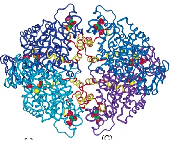
Figure 1-3 Pyruvate kinase M2 structure
Human pyruvate kinase crystal structure was illustrated. Pyruvate kinase M1 and M2 difference was
Figure 1-4 Angiogenesis balance
Angiogenesis balance is maintained by levels of angiogenesis activator and angiogenesis inhibitor.
Im-propriate amount of activator or inhibitor leads to dysfunction of angiogenesis and angiogenesis related
a. Functional domain of integrin α and βchains
b. Activation of integrin with I-domain
[image:46.612.167.457.117.494.2]c. Activation of integrin without I-domain
Figure 1-6 Microenvironment influences cell fate via integrins
Integrin regulate cell survival signals upon binding to ECM. Integrin & ECM interaction activates PI3k-Akt
and NFκB signaling pathways and its downstream survival response. Unligated integrins induce cell
CHAPTER 2 Pyruvate Kinase M2 in Blood Circulation Facilitates Tumor Growth by Promoting An-giogenesis
Liangwei Li1,Yinwei Zhang1, Jingjuan Qiao2, Jenny J. Yang2, and Zhi-Ren Liu1*
1
Department of Biology, 2Department of Chemistry
Georgia State University, Atlanta, GA 30303, USA
*Corresponding Author:
Zhi-Ren Liu, Ph.D. Department of Biology Georgia State University University Plaza
Atlanta, GA 30303 USA ([email protected])
Key words: Pyruvate kinase M2, angiogenesis, endothelial, cell migration, cell adhesion, glycolysis
Runing title: PKM2 promotes angiogenesis