AUTHENTICATION OF CLASSES OF TULSI LEAF FOR THE
EXTRACTION OF MEDICINAL QUALITIES USING IMAGE
PROCESSING TECHNIQUE
Vijayashree T.
1and
A. Gopal
21Department of Electronics and Control Engineering, Faculty of Electrical and Electronics Engineering, Sathyabama University, Chennai,
Tamil Nadu, India
2CSIR, CEERI, Madras Complex, Taramani, Chennai, India
E-Mail: [email protected]
ABSTRACT
Leaf plays a major role in plant species. There are certain leaves which have medicinal qualities. Identification of leaf with look-alike is becoming a major task in day to day life. In order to overcome that computer vision technique is used which includes image processing algorithm. In this technique the features and texture of the leaf are extracted and the closest match is taken and identified to which class it belongs to. This paper deals with the algorithm of morphological processing to which the signature parameter is considered as the vein. Morphological processing includes structuring element which is a process of dilation and erosion. Authenticating an image with its signature parameter will be the most efficient method of identification to give highest accuracy than other methods.
Keywords: tulsi leaf, morphological processing, Gray level co-occurrence matrix (GLCM), zernike moments, entropy, inverse
difference moment (IDM).
1. INTRODUCTION
Plants play a major role in environment. These plants have a very good medicinal feature from root to leaf. Different plants have different medicinal qualities and each leaf is characterized by its medicinal features. Herbal leaves like Pudina, Krpuravalli, Tulsi, etc., are used in our day to day life. Certain leaf has its own medicinal properties like skin ailment, cold, blood purifier and for increasing the memory level.
The brief discussion about the further topics is described in this section. This section will be the introduction to each section for simple the next section, section II describes about the prior and related work. In this section a summary of related work is described elaborately with the disadvantage of the method used. Section III describes about the proposed methodology. In this section the methodology used for the authentication of leaf is explained in a step by step process. Section IV deals about the feature extraction and Texture analysis. Section V deals about the experimental results and discussion obtained by the method used. Section VI gives the conclusion of the work with accuracy specified.
This section clearly portraits the drawback of the proposed system, Section VII gives a brief description about the work to be carried out in future. This section gives the explanation of the drawback and how it can be overcome.
2. PRIOR AND RELATED WORK
Forming a database is the important task for identifying a leaf. Computer vision and pattern recognition plays a major role in the creation of database. On using Zernike moments for extracting the features by Probabilistic Neural Network (PNN) training. The drawback on using this method was that for complex image this technique did not work properly [1]. Enhancement
technique is used for using a clear vision of the image. While considering image enhancement contrast stretching is a normal parameter for consideration. The identified drawback of this method is that evaluation time is large and it depends on the memory size of the image. Multiple noises cannot be removed simultaneously [2]. Ant colony algorithm is considered which can identify only primary and secondary venation. Tertiary venation identification is a major drawback in this method [3]. Leaf vein and feature extraction was done which is done by histogram processing on rotating the image to 180degrees. The point of maximum histogram is considered for extraction. Fast Fourier Transform (FFT) method is used based on distance calculated from centroid. Accuracy was not up to the expected level. Only the main vein of the leaf is taken in to account [4]. Apart from these techniques various methods are used for identifying the medicinal leaves. On feature based approach only mature leaf can be identified [5]. An Intelligent Scissor (IS) method is also used for identifying the vein characteristics. Expected accuracy was not achieved [4]. By extracting the leaf vein based on gray-scale morphology the image was scanned and then fed to the computer. Here the background of the image was close to raw image and hence accuracy was less [7].
3. PROPOSED METHODOLOGY
after edge detection is feature extraction. Gray level co-occurrence matrix (GLCM), is used for extraction the features. Here eight features are considered. It will be explained in next section. The major step is morphological processing. It is done for extracting the vein of the leaf which is considered as the signature parameter. Each leaf differs with its own character; they are distinguished only based on their structure. Vein plays the major role in a leaf. The final step is creating the database with all medicinal herbal leaves based on its features.
Figure-1. Proposed methodology.
Feature extraction and texture analysis
Features extraction and texture analysis is the important task in authentication of leaf.
A. Feature extraction
For the authentication of leaf 8 features are considered. The features are extracted using GLCM technique.
1) Contrast
It returns a measure of intensity contrast between pixel and its neighborhood over the whole image. Contrast will be zero for a constant image. It is given by,
Contrast=∑│i-j│2p(i,j) (1)
2) Correlation
It returns a measure of how correlated a pixel is to its neighbor over the whole image. The range of this is fronm [-1,1]. Correlation is -1 or 1 for a perfectly positively or negatively correlated image. Correlation is NaN for a constant image.It is given by,
Correlation = ∑(i-µi)(j-µj)p(i,j)/σiσj (2)
3) Energy
It returns the sum of squared element in GLCM. It is also called as uniformity or uniformity of energy or angular secondary moment. Energy is one for a constant image.
4) Homogeneity
It returns a value that measures the closeness of the distribution of element in GLCM to GLCM diagonal. It ranges from [0 1]. Homogeneity is 1 for a diagonal GLCM. It is given by
Homogeneity=∑│p(i,j)│/(1+│i-j│) (3)
5) Inverse difference moment (IDM)
This is another feature influenced by the homogeneity of the image. It contains a weighting factor resulting in smaller contributions from inhomogeneous areas. So lack of homogeneity in images causes a low value of Inverse Difference Moment.
IDM=∑ 1/1+(i−j) P (i,j) (4)
6) Entropy
Frist order entropy is dependent on the homogeneity of the image. Homogeneity lowers the entropy of an image.
Entropy= -∑∑p (i, j) log (p (i,j)) (5)
Sum Entropy= -∑px+y (i) log (px+y (i)) (6)
Sum Average= ∑ipx+y (i) (7)
Sum Variance= [i+∑px+y (i) log (px+y (i))]2px+y (i) (8)
B. Texture analysis
Textures are complex visual patterns composed of entities, or sub patterns, which have characteristic brightness, colour, slope, size, etc. Thus texture can be regarded as a similarity grouping in an image. The local sub pattern properties give rise to the perceived lightness, uniformity, density, roughness, regularity, linearity, frequency, phase, directionality, coarseness, randomness, fineness, smoothness, granulation, etc., of the texture as a whole
There are four major issues in texture analysis: 1) Feature extraction: to compute a characteristic of a digital image able to numerically describe its texture properties;
2) Texture discrimination: to partition a textured image into regions, each corresponding to a perceptually homogeneous texture (leads to image segmentation);
3) Texture classification: to determine to which of a finite number of physically defined classes (such as normal and abnormal tissue) a homogeneous texture region belongs;
4) Shape from texture: to reconstruct 3D surface geometry from texture information. Feature extraction is the first stage of image texture analysis. Results obtained from this stage are used for texture discrimination, texture classification or object shape determination.
Texture analysis is categorized into:
Structural- Provides a good symbolic description of image. Powerful tool for structural texture is provided by mathematical morphology. It is based on the
Feature extraction Image Preprocessing
Morphological Processing
arrangement image primitives such as description of texture based on regularly spaced parallel lines.
Statistical- It is a quantitative measure of arrangement. The approach is based on multidimensional co-occurrence matrix. The statistical approach is used to characterize the texture of the region as: smooth, coarse and grainy. For considering the statistical moment on texture analysis the moment, mean uniformity, entropy is taken.
Model based- It is not suitable for describing local image structure.
Transform Method- It uses Fourier descriptors, Gabor descriptors and wavelet transform. Gabor filter provides better spatial localization.
Texture content is an important quantity used to describe a region.
4. EXPERIMENTAL RESULTS AND DISCUSSIONS
[image:3.612.318.534.184.349.2]The experimental results are based on the partial work done. The original image is shown in Figure 2.
Figure-2. Original Tulsi
image.
Figure-3. Converted
image.
[image:3.612.77.296.325.477.2]The converted image is shown in Figure-3. The color image is converted into gray level image for equal distribution of pixels and also for easy processing. The second stage of preprocessing is edge detection; here perwitt mask and sobel mask are used for detecting the edge.
Figure-4. Perwitt edge
detection. Figure-5.detection. Sobel edge
On comparing Perwitt with Sobel edge detection it has been identified that Sobel mask is more clear. Since sobel mask is having high signal-to-noise ratio (SNR), it is better to use sobel mask rather than perwitt mask. The next step is the dilation of image. The gray scale image is dilated with the structuring element which is nothing but the binary image.
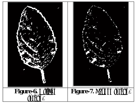
Figure 6 shows the dilated image.
Figure-6. Dilated
image. Figure-7. Eroded image.
[image:3.612.317.539.419.651.2]Erosion is another step in morphological processing. It is the process of removing zeros in an image. Figure-7 shows the eroded image. After identifying the process of erosion the image is again resized for cropping. It is shown in Figure-8 and Figure-9.
Figure-8. Resized image.
Figure-9. Cropped image.
[image:3.612.73.296.562.726.2]values in the table show the various kinds of tulsi leaves
used. Here three varieties of tulsi leaf are used for the creation of database. They are classified as Karun tulsi, Krishna tulsi and Rama Tulsi.
Table-1. Values of GLCM properties.
S. No. Leaf name Energy Homogeneity Correlation Contrast
1. Krishna
Tulsi 0.3360 0.9410 0.9795 0.1301
2. Karun Tulsi
0.3303 0.9461 0.9914 0.1379
3. Rama Tulsi 0.3497 0.9631 0.9907 0.0902
The table shown above gives features of GLCM. Energy, Homogeneity, Correlation and contrast are calculated for tulsi leaves alone.
5. CONCLUSIONS AND FUTURE WORK
The main aim of this work is to find the exact match of the leaf with test parameters. Here of 8 features, 4 features are taken initially. The texture analysis is done with the morphological processing which is nothing but the structuring elements of binary images. To take the vein characteristics, the image is eroded. The features are extracted with the GLCM technique. The future work is to form the database with all herbal medicinal leaves. After the database is created it is to be compared and to identify the closest match. The identified leaf is to be labeled. Later Arduino processor is to be used the identification is to be implemented which makes the system even more user-friendly.
ACKNOWLEDGMENT
I would like to express sincere thanks to Colonel Dr. JEPPIAAR for his constant encouragement and motivating a lot in submitting this paper. I would like to thank our beloved Directors Dr. Marie Johnson and Dr. Mariazeena Johnson Sathyabama University for their support in submission of our paper. I would like to thank my supervisor Dr. A.Gopal for his support throughout the work and helping a lot for the preparation of this paper. My sincere thanks to Dr.N.M.Nandhitha, Research Head, Sathyabama University for helping me with the initial step in carrying this work. A sincere thanks to her for always helping me during the need and explaining clearly how the work has to be carried out. I also express my thanks to my colleague Mr. R.Immanuel Rajkumar for helping to create the database with MySQL. Finally I would like to thank my family members for the support throughout in submitting this paper. My special thanks to my son Pranav Navajath and my husband T.S.Navajath for motivating me and allowing me to do my work without any disturbance. Finally I would like to thank God for blessing me always in all means.
REFERENCES
[1] Pallavi. P, V.S.Veena Devi, “Leaf Recognition based on feature extraction and Zernike moments”, International Journal of Innovative Research in
Computer and Communication Engineering, Vol. 2, Special Issue 2, May 2014.
[2] N. Valliammai and Dr.S.N. Geethalakshmi, “A Hybrid method for enhancement of plant leaf recognition”, World of Computer Science and Information Technology Journal, ISSN: 2221-0741, Vol. 1, No 9, 370-375, 2011. A. Ehsanarid, “Plant classification based on leaf recognition”, International Journal of Computer Science & Information Security, vol. 8, no 4, pp. 78-81, 2010.
[3] James S Cope, Paolo Remagnino, Sarah Barman and Paul Wilkin, “The Extraction of venation from leaf images by evolve vein classifiers and ant colony”, Digital imaging Research centre, Kingston University, London U.K .
[4] Kue-Bum Lee and Kwang-Seok Hong,“An
implementation of Leaf Recognition System using leaf Vein and Shape” , International Journal on Bio-Science and Bio-Technology, Vol 5, No 2 April 2013. [5]. Sandeep Kumar E, “Leaf Volor, Area and Edge Features based approach for Identificationnof Indian Medicinal Plants”, Indian Journal of Computer Science and Engineering, Vol. 3 No. 3, Jun-Jul 2012, ISSN:0976-5166.
[5] Abdul Kadir, Lukito Edi Nugroho, Adhi Susanta, Paulus Insap Santosa, “Leaf Classification using shape, color and texture features”, International Journal of Computer Trends and Technology July to Aug issue 2011, ISSN:2231-2803.
[6] Xiaodong Zheng and Xiaojie Wang, “ Leaf Vein Extraction based on Gray-scale Morphology”, I.J. Image Graphics and Signal Processing, 2010, 2, 25-31.
[7] P.Hiremath and J.Pujari, “Content based image retrieval based on color, texture and shape features using image and its complement”, International Journal of Computer Science and Security, vol. 1(4), pp. 44-50, 2011.
[9] T. Beghin, J.S. Cope, P. Remagrino and S. Barman, “Shape and Texture based plant leaf classification”, ACIVS (2), 2010, pp. 345-353.
[10]Pande Ankita V, Prof. Shandilya V.K., “Digital Image Processing Approach for Fruit and Flower Leaf Identification and Recognition”, International Journal of Engineering and Computer Science, ISSN: 2319-7242, vol 2, Issue 4, April 2013, pp. 1280-1285.
[11]Ekshinge Sandip Sambhaji, D.B.Andor, “Leaf recognition algorithm using neural network based image processing, Asian Journal of Engineering and Technology, ISSN: 2347-738, 21-03-2014. pp. 10-16.
[12]C.Ananthi, Azha. Periyasamy, S.Muruganand, “ Pattern Recognition of medicinal leaves using image processing techniques, Journal of nanoscience and nanotechnology, vol. 2, Issue 2, Spring edition DOI: feb 2014, pp 214-218, ISSN: 2279-0381.
[13]Pallavi. P, V.S. Veena Devi, “Leaf Recognition based on feature extraction and Zernike moments”, International Journal of Innovative Research in Computer and Communication Engineering, Vol. 2, Special Issue 2, May 2014.
[14]Abdolvahab Ehsanirad and Sharath Kumar. sY.H, “Leaf recognition for plant classification using GLCM and PCA methods”, Oriental Journal of Computer Science & Technology, vol. 3(1), 31-36 2010.
[15]S.G. Wu, F.S.Bao, E.Y. Xu, Y.Wang, Y.F.Chang and Q.L. Xiang, “A leaf recognition algorithm for plant classification using probabilistic neural network”, CoRR, vol. abs/0707, 4289, 2007.