INTRODUCTION
Craniofacial development begins when the cranial neural crest cells (CNCCs), migratory multipotent precursors that contribute to most of the face, delaminate from the dorsal brain and migrate ventrolaterally to form the ectomesenchyme of facial primordia known as the frontonasal prominence (FNP) and branchial arches (BAs) (see Fig. S1A in the supplementary material) (Noden, 1978; Couly et al., 1993; Osumi-Yamashita et al., 1994; Köntges and Lumsden, 1996; Chai et al., 2000). Subsequently, the FNP becomes the mid- and upper face, while the first branchial arch (BA1) develops into most of the jaw, the lateral skull, palate and the middle ear (Köntges and Lumsden, 1996). BA1 is further divided into maxillary arch (mxBA1, prospective upper jaw) on the proximal half, and mandibular arch (mdBA1, prospective lower jaw) on the distal half (see Fig. S1A,B in the supplementary material). The second branchial arch (BA2) mainly contributes to the ear and neck skeleton.
How CNCCs recognize their positional information and develop accordingly is beginning to be understood (reviewed by Depew et al., 2002a; Santagati and Rijli, 2003; Chai and Maxon, 2006). Interactions of CNCCs with the neighboring tissues result in the
expression of a diverse set of transcription factors in CNCCs, and the specific combination of transcription factors provides a positional identity to the cells.
The vertebrate Dlx genes are homologs of Drosophila Distal-less; they encode homeodomain transcription factors (Panganiban and Rubenstein, 2002). Mice have six Dlx genes, which are organized as three linked pairs in the genome (Dlx1/2, Dlx3/4and Dlx5/6) (Porteus et al., 1991; Price et al., 1991; Robinson and Mahon, 1994; Simeone et al., 1994; McGuinness et al., 1996; Nakamura et al., 1996; Liu et al., 1997).
During craniofacial development, mouse Dlx genes are regionally expressed within BAs as well as in olfactory and otic placodes (see Fig. S1A,C in the supplementary material) (Dolle et al., 1992; Bulfone et al., 1993; Robinson and Mahon, 1994; Simeone et al., 1994; Qiu et al., 1997; Depew et al., 2002b). In the ectomesenchyme of BA1 in mid-gestation stage embryos, Dlx1/2are expressed in both mxBA1 and mdBA1, whereas Dlx5/6are expressed in mdBA1 only. Dlx3/4expression is further restricted to a narrow domain within mdBA1. The same proximodistal arrangement is also found in BA2. Since Dlx3/4expression is dependent on Dlx5/6(Depew et al., 2002b) (this study), essentially two different combinations of Dlx partition much of BA1: Dlx1/2for mxBA1 and Dlx1/2+5/6for mdBA1. The functional importance of this ‘Dlx code’ in BA patterning has been investigated using mouse loss-of-function mutants. Owing to the tight linkage in the genome, the double mutations of Dlx1/2and Dlx5/6pairs were achieved by deleting both genes in one allele (Qiu et al., 1997; Merlo et al., 2002; Depew et al., 2002b). Inactivation of Dlx1 and/or Dlx2 (Dlx1–/–, Dlx2–/–and Dlx1/2–/–) caused abnormalities in upper jaw skeleton with little effect on the lower jaw (Qiu et al., 1995; Qiu et al., 1997; Depew et al., 2005). By contrast, Dlx5–/– exhibited defects in lower jaw development (Depew et al., 1999). Most strikingly, the simultaneous inactivation of Dlx5 and Dlx6 (Dlx5/6–/–) resulted in homeotic
Dlx genes pattern mammalian jaw primordium by regulating
both lower jaw-specific and upper jaw-specific genetic
programs
Juhee Jeong1,*, Xue Li2, Robert J. McEvilly3, Michael G. Rosenfeld3, Thomas Lufkin4and John L. R. Rubenstein1,*
Dlx transcription factors are implicated in patterning the mammalian jaw, based on their nested expression patterns in the first branchial arch (primordium for jaw) and mutant phenotypes; inactivation ofDlx1and Dlx2(Dlx1/2–/–) causes defects in the upper jaw, whereas Dlx5/6–/–results in homeotic transformation of the lower jaw into upper jaw. Therefore, the ‘Dlx codes’ appear to regionalize the jaw primordium such that Dlx1/2regulate upper jaw development, while Dlx5/6confer the lower jaw fate. Towards identifying the genetic pathways downstream of Dlx5/6, we compared the gene expression profiles of the wild-type and Dlx5/6–/– mouse mandibular arch (prospective lower jaw). We identified 20 previously unrecognized Dlx5/6-downstream genes, of which 12 were downregulated and 8 upregulated in the mutant. We found a Dlx-regulated transcriptional enhancer in close proximity to Gbx2, one of the Dlx5/6-downstream genes, strongly suggesting that Gbx2is a direct target of Dlx5/6. We also showed that Pou3f3 is normally expressed in the maxillary (prospective upper jaw) but not mandibular arch, is upregulated in the mandibular arch of Dlx5/6–/–, and is essential for formation of some of the maxillary arch-derived skeleton. A comparative analysis of the morphological
and molecular phenotypes of various Dlx single and double mutants revealed that Dlx1, 2, 5 and 6act both partially redundantly and antagonistically to direct differential expression of downstream genes in each domain of the first branchial arch. We propose a new model for Dlx-mediated mammalian jaw patterning.
KEY WORDS: Dlx, Gbx2, Pou3f3, Craniofacial, Branchial arch, Jaw, Mouse
Development 135, 2905-2916 (2008) doi:10.1242/dev.019778
1Department of Psychiatry, Nina Ireland Laboratory of Developmental Neurobiology, University of California San Francisco, 1550 4th street, San Francisco, CA 94158, USA. 2Department of Surgery/Urology and Department of Pathology, Children’s Hospital of Boston, Harvard Medical School, 300 Longwood Avenue, Boston, MA 02115, USA. 3Howard Hughes Medical Institute, Department of Medicine, University of California, San Diego, School of Medicine, 9500 Gilman Drive, La Jolla, CA 92093, USA. 4Genome Institute of Singapore, 60 Biopolis Street, Singapore 138672, Singapore.
*Authors for correspondence (emails: [email protected]; [email protected])
Accepted 26 June 2008
D
E
V
E
LO
P
M
E
N
transformation of the lower jaw into upper jaw (Beverdam et al., 2002; Depew et al., 2002b). Therefore, the differential expression of Dlx genes along the proximodistal axis is important for the regional specification of BA1; Dlx1/2 are necessary for the proper development of mxBA1, whereas Dlx5/6confer mdBA1 identity.
Our current work addresses three important issues on how the Dlx genes regulate BA patterning. First, the mechanism through which Dlx5/6specify lower jaw fate needs to be understood. To this end, we performed a genome-wide transcriptional profiling and obtained a comprehensive list of genes with altered expression in Dlx5/6–/– mdBA1. Second, we provide the first evidence of the upper jaw-specific genetic program and show that it is partly regulated by Dlx genes. Prior to this study, the abundance of mdBA1-specific markers but the lack of any known mxBA1-specific markers has been compatible with the idea that the upper jaw is the default state upon which the lower jaw fate is imposed. Finally, we investigated the functional relationship of different Dlx genes expressed in BA1 by comparing the morphological and molecular phenotypes of various combinations of Dlx single and double mutants. We found that Dlx1, 2, 5 and 6 act both partially redundantly and antagonistically, depending on the context, to achieve differential expression of their downstream genes in mxBA1 and mdBA1.
MATERIALS AND METHODS Animals
All experiments using mice were performed following UCSF institutional regulations on the care and use of laboratory animals.
Transcriptional profiling using DNA microarrays
Dlx5/6–/– and Dlx5/6+/+ littermates were collected from Dlx5/6+/–
intercrosses at E10.5. The mdBA1s were dissected, flash-frozen and stored in liquid nitrogen until the day of RNA extraction. The tissue was homogenized in Trizol reagent (Invitrogen) using Pellet Pestle (Kontes). RNA was extracted using chloroform and then concentrated by isopropanol precipitation. After a rinse with 80% ethanol, the RNA pellet was dissolved in water and purified using the RNeasy Mini Kit (Qiagen). From 13
Dlx5/6–/–and 13 Dlx5/6+/+E10.5 embryos, we recovered 14.7 μg and 13 μg of mdBA1 total RNA, respectively.
All subsequent steps of the microarray experiment were performed by the Translational Genomics Research Institute (TGen, Phoenix, AZ), through the NIH Neuroscience Microarray Consortium. The RNA sample from each genotype was hybridized onto GeneChip Mouse Genome 430 2.0 arrays (Affymetrix) in triplicate. Data acquisition and analysis employed GeneChip Operating Software (GCOS, Affymetrix) version 1.2.
In situ hybridization and skeletal preparation
Whole-mount and section in situ hybridizations were performed using digoxigenin-labeled RNA probes as described (Jeong et al., 2004; Jeong and
McMahon, 2005), except that 20 μm sections were used for the section in
situ hybridization. The control and mutant embryos were stage-matched using a combination of several morphological criteria, including the size and shape of the limb buds, morphogenesis of the eye, and somite numbers. Skeletons of E18.5 or P0 animals were stained with Alcian Blue and Alizarin Red as described (Jeong et al., 2004). For the in situ hybridization and
skeletal preparations shown in Figs 1-4, embryos of +/+ and +/–genotypes
were used indiscriminately and are referred to as ‘wild type’. For Figs 5 and
6, ‘wild type’ refers to +/+ for all the genes in question, or Dlx1+/–.
DNA templates for in situ hybridization probes were obtained by PCR from a wild-type mouse E10.5 BA1 cDNA library or from adult tail genomic DNA, purchased from companies, or kindly provided by other investigators. Further information on the probes is available upon request.
Luciferase reporter activation assay
The 1 kb putative Gbx2enhancer (see Fig. 3A) was amplified by PCR from
mouse tail genomic DNA using primers 5⬘ACACCTCGAGAGAGGAT
-GA CAGC-GAGCTTCG-3⬘and 5⬘GTGTAAGCTTGAGCA AACATT CC
-AGTTTTAATGC-3⬘, and cloned into XhoI-HindIII sites of pGL4.23
(Promega). pGL4.23 contains a minimal promoter and the firefly luciferase coding sequence. pCAGGS-Dlx5 (Stuhmer et al., 2002) was used to express Dlx5 protein. pGL4.73 (Promega), a plasmid that constitutively expresses
Renillaluciferase, was used as a control for variations in transfection efficiency. 3T3 cells were transfected with FuGene 6 (Roche), and 40 hours later the cells were lysed and analyzed for luciferase activity using the Dual Luciferase Reporter Assay System (Promega). The experiments were performed in triplicate and the results combined for statistical analysis.
Generation of Dlx6mutant allele
To generate the Dlx6-lacZ(Dlx6–) allele, a 4.3 kb SpeI-SnaBI genomic
fragment spanning Dlx6exon 3 (which encodes the homeodomain) was
subcloned into the SpeI-SmaI sites of pBS KS+. Site-directed mutagenesis
was performed to engineer a unique NruI site immediately following the
219th amino acid from the N-terminus of the Dlx6 protein (F of the sequence VKIWFQNKRS). Flanking genomic sequences were added between the
unique 5⬘XhoI site (5.8 kb 5⬘homology arm) and the 3⬘NotI site (3.5 kb 3⬘
homology arm), and the reporter cassette IRES-lacZ-PGKneo (Robledo et
al., 2002) was cloned into the unique NruI site to generate the final targeting
construct. The Dlx6-lacZ allele therefore interrupts the Dlx6 protein
immediately following the amino acid F as described above. ES cell culture, screening, chimera generation and testing were as previously described (Robledo et al., 2002).
RESULTS
Genome-wide transcriptional profiling identifies changes in RNA expression in the mdBA1 of Dlx5/6–/–mutants
To understand the molecular changes underlying Dlx5/6–/–jaw phenotypes, we compared transcriptional profiles of the wild-type and mutant mdBA1 at E10.5 using Affymetrix GeneChip Mouse Genome 430 2.0 array. We chose E10.5 for our analysis because E9.0-10.5 is when the Dlx genes exhibit proximodistally nested expression in BA1 (Qiu et al., 1997; Acampora et al., 1999) (see Fig. S1 in the supplementary material), and the wild-type and mutant BA1 still appear grossly comparable in size and morphology at E10.5 (see also Fig. S2 in the supplementary material). The Dlx5/6–/– mutant exhibited downregulation of 39 genes (see Table S1 in the supplementary material) and upregulation of 24 genes (see Table S2 in the supplementary material) with greater than a 2-fold change. Our results included most, but not all, of the genes that were previously shown to be dysregulated in the Dlx5/6–/–mutant (Depew et al., 1999; Depew et al., 2002b). Thus, although it is a robust procedure, technical limitations exist (see Tables S1 and S2 in the supplementary material).
The complete set of raw data from our transcriptional profiling experiment is available at NIH Neuroscience Microarray Consortium data repository (http://arrayconsortium.tgen.org) under accession ruben-affy-mouse-187820, and at GEO (accession number GSE 4774).
Genes encoding transcription factors, non-coding RNAs and signaling molecules exhibit decreased expression in Dlx5/6–/–mdBA1
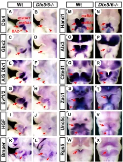
The Dlx3, Hand2 and Alx4 transcription factors were previously identified as being downstream of Dlx5/6(Depew et al., 2002b). Our screen found that their close relatives, Dlx4, Hand1 and Alx3, are also downregulated in Dlx5/6–/– mdBA1 and BA2 (Fig. 1A,B,M-P). In the otic vesicle, Dlx5/6function is required for the expression of Gbx2, a homeodomain transcription factor (Robledo and Lufkin, 2006). We confirmed the same regulatory relationship in mdBA1 (Fig. 1C,D). The transcription factors Cited1 (Msg1) (Shioda et al., 1996) and Zac1 (Lot1, Plagl1) (Abdollahi et al.,
D
E
V
E
LO
P
M
E
N
1997; Spengler et al., 1997) are expressed at high levels in the medial mdBA1 and BA2; their expression was downregulated in Dlx5/6–/–(Fig. 1Q-T).
Mouse Dlx1 locus encodes several putative non-coding RNA (ncRNA) transcripts (A/S Dlx1; see Fig. S3A in the supplementary material) (McGuiness et al., 1996; Liu et al., 1997). In BA1 and BA2, the expression of A/S Dlx1was restricted to the distal region (Fig. 1E), and was completely abolished in Dlx5/6–/–(Fig. 1F). Evf1/2, the two splicing variants of a ncRNA gene (Dlx6os1– Mouse Genome Informatics), map adjacent to Dlx5/6; their brain expression depends on Dlx1/2 function (see Fig. S3B in the supplementary material) (Faedo et al., 2004; Kohtz and Fishell, 2004). Evf1/2expression was greatly reduced in both brain and BAs of Dlx5/6–/– mutants (Fig. 1G,H). It should be noted that the promoter and the first exon of Evf2fall within the deletion in the Dlx5/6null allele (Robledo et al., 2002), and thus the decreased Evf1/2expression in Dlx5/6–/–could simply be due to the loss of the Evf2promoter. However, we found reduced expression of Evf1/2 also in Dlx5–/–and Dlx6–/–mutants (see below), in which both Evf1 and Evf2sequences are intact. This result, and the fact that Dlx1/2 activity is necessary for the expression of Evf1/2in the brain, argue that Dlx5/6regulate Evf1/2transcription, directly or indirectly.
A secreted signaling molecule, hepatocyte growth factor (Hgf, also known as scatter factor, SF) (Stoker et al., 1987; Birchmeier and Gherardi, 1998), is expressed in BA1 and BA2; this expression was
dependent on Dlx5/6function (Fig. 1I,J). Unc5cencodes one of the receptors for the axon guidance molecule netrin 1 (Ackerman et al., 1997). Unc5cwas expressed in the medial domain of mdBA1 and BA2, and was severely downregulated in Dlx5/6–/–(Fig. 1U,V). BMP-binding endothelial regulator (Bmper, also known as crossveinless-2, Cv2) is a secreted molecule that binds to and enhances the signaling of bone morphogenetic proteins (BMPs) (Coffinier et al., 2002; Moser et al., 2003; Coles et al., 2004; Ikeya et al., 2006). Bmperexpression in distal BA1 and BA2 was dependent on Dlx5/6(Fig. 1K,L). The regulator of G-protein signaling 5 (Rgs5) gene is expressed only at the rostromedial tip of mdBA1; this expression was lost in Dlx5/6–/–(Fig. 1W,X). Rgs proteins are GTPase-activating proteins that attenuate G-protein-coupled receptor signaling (Chen et al., 1997; Xie and Palmer, 2007).
Since Dlx3/4expression is lost in Dlx5/6–/–mdBA1 (Depew et al., 2002b) (Fig. 1A,B), it is possible that at least some of the gene expression changes in Dlx5/6–/–mutants are due to the loss of Dlx3/4 activity.
Pou3f3, Foxl2and uncharacterized transcripts linked to them, are strongly repressed by Dlx5/6 in mdBA1
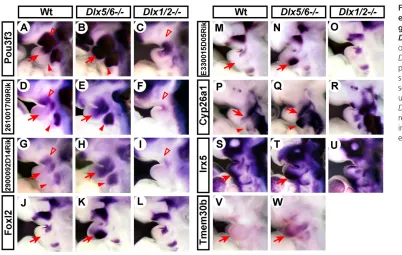
Pou3f3 (Brn1), a POU-domain transcription factor (Hara et al., 1992), was highly expressed in the entire mxBA1 and maxillary-mandibular junction, but was absent from most of mdBA1 (Fig. 2A). It was also confined to the proximal region in BA2. In Dlx5/6–/–, Pou3f3expression expanded into mdBA1 and distal BA2 with the same intensity as in its normal expression domains (Fig. 2B).
We identified two Riken cDNA clones (26100017I09Rik, 2900092D14Rik), both suspected to be non-coding, that are closely linked to and have very similar expression patterns as Pou3f3, in both wild-type and Dlx5/6–/–embryos (Fig. 2D,E,G,H; see Fig. S3C in the supplementary material). Because the proximal BA expression of Pou3f3, 2610017I09Rikand 2900092D14Rikoverlaps with that of Dlx1/2(see Fig. S1C in the supplementary material), we tested whether the expression of these genes is dependent on the activity of Dlx1/2 in the mxBA1. We found downregulation of all three genes in BA1 and BA2 of Dlx1/2–/–, demonstrating that Dlx1/2are necessary for their normal expression (Fig. 2C,F,I).
Foxl2 encodes a winged helix/forkhead transcription factor (Crisponi et al., 2001). It was expressed strongly in the dorsal mxBA1 just below the eye, in addition to a small domain at the maxillary-mandibular junction (Fig. 2J). In Dlx5/6–/–, the mxBA1 pattern of Foxl2expression was duplicated in mdBA1 (Fig. 2K). Riken cDNA E330015D05Rikis a poorly characterized gene closely linked to Foxl2 (see Fig. S3D in the supplementary material). Its expression pattern, both in wild type and Dlx5/6–/–, was identical to that of Foxl2 (Fig. 2M,N).
[image:3.612.50.300.58.386.2]Cyp26a1, a retinoic acid-metabolizing enzyme cytochrome P450 (White et al., 1996; Fujii et al., 1997; Ray et al., 1997), is normally expressed along the border of BA1 and BA2 with higher expression in mxBA1 than in mdBA1 (Fig. 2P). In BA2, Cyp26a1expression was also strong proximally and weak distally. Removal of Dlx5/6 activity lead to upregulation of Cyp26a1in distal BA1 and BA2 to the same intensity as in the proximal domains (Fig. 2Q). Irx5, which encodes an Iroquios-related homeodomain transcription factor (Bosse et al., 2000; Cohen et al., 2000), was expressed strongly in the dorsal mxBA1 and weakly in the ventral mdBA1 (Fig. 2S). The latter expression was moderately increased in Dlx5/6–/–(Fig. 2T). Unlike Pou3f3, 2610017I09Rikand 2900092D14Rik, the mxBA1 expression of Foxl2, E330015D05Rik, Cyp26a1and Irx5was not dependent on Dlx1/2 (Fig. 2L,O,R,U).
Fig. 1. Branchial arch expression patterns of the genes downregulated in Dlx5/6–/–.Lateral views (A-L) or frontal views (M-X) of wild-type and Dlx5/6–/–E10.5 mouse embryos processed by
whole-mount in situ hybridization. Arrows and arrowheads indicate changes in gene expression in mdBA1 and BA2, respectively.
D
E
V
E
LO
P
M
E
N
The genes described above in this section are normally expressed in mxBA1 and upregulated in the Dlx5/6–/–mdBA1, making the mutant mdBA1 molecularly similar to mxBA1. By contrast, transmembrane protein 30b (Tmem30b), a homolog of yeast endosomal protein Cdc50 (Katoh and Katoh, 2004), was barely detectable in either mxBA1 or mdBA1 in wild-type embryos (Fig. 2V), but was strongly upregulated in Dlx5/6–/–mdBA1, making the mutant mdBA1 molecularly different from mxBA1 (Fig. 2W).
Identification of a Dlx-regulated transcriptional
enhancer upstream of Gbx2
An important question about the Dlx5/6-downstream genes identified by our microarray analysis is whether Dlx5/6 directly regulate them. There is already evidence that three of the genes listed in Table S1 (see Table S1 in the supplementary material) are direct targets of Dlx5/6. Charite et al. (Charite et al., 2001) showed that Dlx6 binds to, and thus most likely directly regulates, a Hand2BA enhancer. Sumiyama and Ruddle (Sumiyama and Ruddle, 2003) showed that the BA enhancer for Dlx3/4contains a consensus Dlx-binding motif, and thus it is highly likely that Dlx5/6 directly regulate Dlx3/4expression in the BA. Thus, the fact that Hand2, Dlx3and Dlx4were identified by our microarray analysis supports the validity of our approach, and suggests that additional genes listed in Table S1 (see Table S1 in the supplementary material) are directly regulated by Dlx5 and Dlx6 proteins.
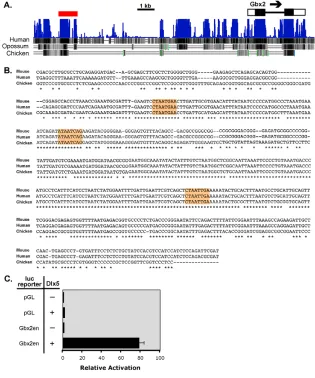
To identify direct targets of Dlx5/6, we performed in silico analyses of the genomic sequences surrounding Dlx5/6-downstream genes. Previously, a systematic in vitro binding assay determined that (A/C/G)TAATT(G/A)(C/G) is a consensus binding motif for Dlx proteins (Feledy et al., 1999). In addition, various researchers have analyzed cis-regulatory elements from several Dlx target genes, discovering 13 sequences to which Dlx proteins bind directly, including some that do not conform to (A/C/G)TAATT(G/A)(C/G) (see Table S3 in the supplementary material). Using rVISTA (Loots and Ovcharenko, 2004), we searched genomic sequences flanking several of the genes listed in Table S1 (see Table S1 in the supplementary material) for all the known Dlx-binding motifs that
are conserved between mouse and human. This analysis identified ~0.6 kb of a highly conserved region (conserved down to chick) with a cluster of three putative Dlx-binding sites located ~10 kb upstream of Gbx2 (Fig. 3A,B). We tested a 1 kb fragment containing this region (red bar in Fig. 3A, including the adjacent 0.25 kb sequence conserved down to opossum) for transcriptional enhancer activity in 3T3 cells. Co-transfection with a Dlx5 expression vector resulted in a >70-fold activation (Fig. 3C), demonstrating that the fragment contains a transcriptional enhancer regulated by Dlx proteins.
Pou3f3is required for the formation of the zygomatic arch and the maxillary component of the jaw joint
To our knowledge, Pou3f3, Foxl2, 26100017I09Rik, 2900092D14Rikand E330015D05are the first examples of genes that are expressed specifically in the maxillary domain of BA1 at any stage of mouse development. Therefore, they provide evidence of an upper jaw-specific genetic program (see Discussion).
Among the five genes, Pou3f3has the strongest and broadest expression in mxBA1 (Fig. 2), and thus we performed further analysis on its expression and function during upper jaw development (Fig. 4). Section in situ hybridization at E10.5 revealed that Pou3f3is expressed only in the mesenchyme (Fig. 4A,B). At E12.5 (data not shown) and E13.5 (Fig. 4C-E), Pou3f3expression was found in both upper and lower jaw; however, its expression in the condensed dental mesenchyme was restricted to upper molars (Fig. 4D). In addition, Pou3f3was expressed in the caudal, but not rostral, palatal shelves (PS) (Fig. 4C,E).
[image:4.612.52.454.56.311.2]Pou3f3–/– (McEvilly et al., 2002) mutant skull revealed the essential role of this gene in the development of a part of the upper jaw; mxBA1-derived squamosal bone (SQ) normally articulates with mdBA1-derived dentary to make the functioning jaw joint in mammals. InPou3f3–/–, the squamosal bone is largely missing (except the retrotympanic process, rt), and thus the jaw joint does not exist (Fig. 4F-I,L,M). Jugal bone (JG), which forms the zygomatic arch on the lateral skull together with the maxilla and squamosal bone, was also lost in the mutant (Fig. 4H,I,L,M). In the
Fig. 2. Branchial arch expression patterns of the genes upregulated in
Dlx5/6–/–.(A-W) Lateral views of wild-type, Dlx5/6–/–and Dlx1/2–/–E10.5 mouse embryos
processed by whole-mount in situ hybridization. Arrows and solid arrowheads indicate upregulation of expression in Dlx5/6–/–mdBA1 and distal BA2,
respectively; open arrowheads indicate downregulation of expression in Dlx1/2–/–mxBA1.
D
E
V
E
LO
P
M
E
N
mutant middle ear, the malleus (mdBA1-derived) appeared normal, whereas the incus (mxBA1-derived) had a slightly elongated short crus (Fig. 4J,K). In addition, the BA2-derived stapes was fused to the styloid process (Fig. 4J,K). Other bones in the skull, and the teeth and palate, appeared unaffected in the Pou3f3–/–mutant (data not shown).
Dlx6–/–exhibits BA1-associated phenotypes that
are very similar to those of Dlx5–/–but far less severe than those of Dlx5/6–/–
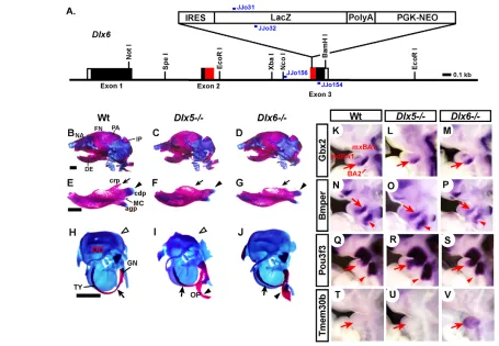
Simultaneous inactivation of Dlx5/6 results in the homeotic transformation of the lower jaw into upper jaw, whereas Dlx5single mutants have relatively mild defects in lower jaw morphogenesis (Depew et al., 1999; Acampora et al., 1999; Beverdam et al., 2002; Depew et al., 2002b). Based on these results alone, it was not clear whether Dlx5and Dlx6are functionally redundant, or whether Dlx6 has a more prominent role. To address this, we generated an allele that inactivated Dlx6alone, by inserting an IRES-lacZ-neomycin resistance cassette within the Dlx6 homeobox coding sequence (Fig. 5A). This insertion prevents translation of one-third of the homeodomain and the entire C-terminal domain, including the nuclear localization signal [amino acids 220-228 (Cokol et al., 2000)]; thus, this allele is likely to be null.
Dlx6–/– mice were born alive but died within a day with aerophagia, as reported for Dlx2–/–,Dlx1/2–/–and Dlx5–/–(Qiu et al., 1995; Qiu et al., 1997; Acampora et al., 1999; Depew et al., 1999).
The head skeleton of Dlx6–/–neonates had several abnormalities that are also found in Dlx5–/–animals (Acampora et al., 1999; Depew et al., 1999). The mutant had a slightly reduced mandible (Fig. 5B,D), in which the dentary lacked the coronoid process and had a hypoplastic condylar process (Fig. 5E-G, arrows and arrowheads). The ectotympanic of the mutant ear was shortened (Fig. 5H-J, arrows), and the gonial bone was attached to an ectopic piece of bone [named os paradoxicum (Depew et al., 1999)] extending toward the ala temporalis on the skull base (Figs 5H-J; see S4A-C, arrowheads, in the supplementary material). The skeletal elements mentioned thus far are thought to be derivatives of BA1. Therefore, allowing for some individual variations in morphological details, Dlx6–/–mice have BA1-associated defects that are very similar to those of Dlx5–/–, but are much less severe than the homeotic transformation observed in Dlx5/6–/–. This result establishes that Dlx5and Dlx6are in large part functionally redundant in lower jaw development.
[image:5.612.49.364.63.441.2]In addition to the BAs, Dlx5/6 were expressed in otic and olfactory placodes (see Fig. S1A,C in the supplementary material) and, as a result, Dlx5–/–animals have dorsally deficient otic capsule (Fig. 5H,I, open arrowheads) and hypoplastic nasal cartilage (see Fig. S4D,E, arrowheads, in the supplementary material) (Acampora et al., 1999; Depew et al., 1999). However, both structures appeared normal in Dlx6–/– (Fig. 5J; see Fig. S4F in the supplementary material), which suggests that Dlx6is less important than Dlx5in otic and olfactory placode development.
Fig. 3. Identification of a Dlx-regulated enhancer upstream of Gbx2. (A) Evolutionary conservation of the genomic sequence upstream of Gbx2analyzed using 30-way multiz alignment (Blanchette et al., 2004). Image generated using University of California Santa Cruz Genome Browser. Blue, degree of conservation among mammals; black vertical bars, conservation in each species as indicated; red bar, the Gbx2enhancer used for the reporter assay in C; black box, coding region of an exon; white box, untranslated region. (B) ClustalW alignment (Larkin et al., 2007) of the 0.6 kb region of the Gbx2enhancer conserved down to chicken. *, conserved nucleotides; putative Dlx-binding sites are highlighted in orange. (C) Results of the luciferase reporter activation assay. pGL, minimal promoter-reporter plasmid without an enhancer; Gbx2en, pGL plasmid with mouseGbx2 enhancer; – Dlx5, co-transfected with empty expression vector; + Dlx5, co-transfected with Dlx5 expression vector.
D
E
V
E
LO
P
M
E
N
Next, we examined the expression of the molecular markers that are affected in Dlx5/6–/–mdBA1 (Figs 1 and 2) in Dlx5–/–and Dlx6–/– single mutants. Most of the genes showed no, or moderate, changes in either mutant, indicating that they are directly or indirectly regulated by both Dlx5and Dlx6 to similar degrees (see Fig. S5A-d in the supplementary material). Surprisingly, however, several genes were differentially changed in Dlx5–/–and Dlx6–/–; Gbx2, Bmper, A/S Dlx1, Dlx4and Evf1/2were all severely downregulated in Dlx5/6–/– BAs (Fig. 1), but Gbx2, Dlx4and Evf1/2were more downregulated in Dlx5–/–than in Dlx6–/–, whereas Bmperand A/S Dlx1were more affected in Dlx6–/–than in Dlx5–/–(Fig. 5K-P; see Fig. S4G-I and Fig. S5e-j in the supplementary material). Similarly, for the genes upregulated in Dlx5/6–/–, Pou3f3, Foxl2, 2610017I09Rik and E330015D05Rikshowed greater changes in Dlx5–/–than in Dlx6–/–, whereas Tmem30bwas upregulated only in Dlx6–/–(Fig. 5Q-V; see Fig. S4J-L and Fig. S5k-p in the supplementary material). These results suggest that there are some differences between the transcriptional activities of Dlx5and Dlx6, even though inactivation of each gene results in similar morphological consequences.
Dlx6activity in lower jaw development is shared
by Dlx1and Dlx2
Although Dlx1/2–/–mice have no significant defects in the lower jaw (Qiu et al., 1995; Qiu et al., 1997; Depew et al., 2005), Dlx1/2 can contribute to mdBA1 development, as the lower jaw phenotypes of Dlx5–/– are greatly exacerbated in Dlx1–/–;5–/– and Dlx2–/–;5–/– mutants (Depew et al., 2005).
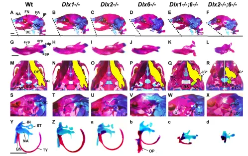
We tested whether Dlx6 is also at least in part functionally redundant with Dlx1/2by generating Dlx1–/–;6–/–and Dlx2–/–;6–/– double mutants. The compound mutants exhibited far greater craniofacial defects than those found in any of the single mutants. The dentaries of Dlx1–/–;6–/– and Dlx2–/–;6–/– were shortened, fragmented, and bifurcated to become bones resembling the maxilla, jugal and pterygoid of the upper jaw (Fig. 6A-L; see S6A-F in the supplementary material). The ventrolateral side of the skull of
Dlx1–/–;6–/–and Dlx2–/–;6–/–had what appears to be a duplicate lamina obturans juxtaposed to the endogenous one (Fig. 6S-X; LO* in Fig. 6W,X). In the ear, the ectotympanic and middle ear ossicles were progressively reduced or lost in Dlx1–/–;6–/–and Dlx2–/–;6–/– (Fig. 6Y-d). The basihyoid and lesser horn of the hyoid were also progressively more affected in Dlx1–/–;6–/–and Dlx2–/–;6–/–(see Fig. S6G-L in the supplementary material).
The phenotypes of Dlx1–/–;6–/–and Dlx2–/–;6–/–are reminiscent of those reported for Dlx1–/–;5–/–and Dlx2–/–;5–/–(Depew et al., 2005). In all four cases, the mdBA1-derivatives are reduced in size and/or transformed into structures resembling upper jaw elements, although the extent of transformation is less than in Dlx5/6–/–.
Dlx1/2 function partially redundantly with Dlx5/6 in regulating mdBA1 gene expression
To elucidate the mechanisms underlying the morphological changes in Dlx1–/–;6–/–, Dlx2–/–;6–/–and Dlx2–/–;5–/–, we examined the gene expression patterns in their mdBA1.
[image:6.612.177.563.61.256.2]The expression of Gbx2and Bmperin mdBA1 was minimally or moderately downregulated in Dlx5–/– and Dlx6–/– (Fig. 5). However, their expression is severely reduced or completely abolished in Dlx1–/–;6–/–, Dlx2–/–;6–/–and Dlx2–/–;5–/–(Fig. 7A-H). Pou3f3 was repressed by Dlx5/6in mdBA1, and was moderately upregulated in Dlx5–/–and Dlx6–/–(Fig. 5). Further removing Dlx1 or Dlx2 activity expanded and intensified Pou3f3overexpression in mdBA1 (compare Fig. 7J,K with Fig. 5S, and Fig. 7L with Fig. 5R). Therefore, even though Dlx1/2 activity is required for the normal expression of Pou3f3in mxBA1 (Fig. 2A,C), Dlx1/2 apparently share the repressive effects of Dlx5/6on Pou3f3in mdBA1. Hand2was severely downregulated in Dlx5/6–/–, but unaffected in Dlx5–/–and Dlx6–/–(Depew et al., 2002b) (see Fig. S5A-D in the supplementary material). Hand2 expression appeared normal in Dlx1–/–;6–/–, but was gradually reduced and became restricted to caudomedial mdBA1 in Dlx2–/–;6–/– and Dlx2–/–;5–/–(Fig. 7M-P).
Fig. 4. Expression of Pou3f3RNA during jaw development and craniofacial skeletal defects in
Pou3f3–/–mutants.(A-E) In situ hybridization for Pou3f3on the coronal sections of E10.5 (A,B) and E13.5 (C-E) wild-type heads. B and D are high-magnification views of the boxed areas in A and C, respectively. C and E are from the same head, but C is rostral to E. Arrow in C, mdBA1 expression of Pou3f3. (F-M) Head skeleton of E18.5 wild-type (F,H,J,L) and Pou3f3–/–(G,I,K,M) mice stained
with Alcian Blue (cartilage) and Alizarin Red (bone). H and I are enlargements of the boxed areas in F and G, respectively. Asterisks in I indicate the absence of jugal and squamosal bone in the mutant. (J,K) Otic capsule and middle ear ossicles.
Open arrowheads, incus phenotype of the mutant; solid arrowheads, abnormal attachment of stapes and styloid process in the mutant. L and M are the same pictures as H and I, but with individual skeletal elements highlighted by color: yellow, zygomatic process of maxilla; green, jugal; dark green, zygomatic process of squamosal; orange, squamosal; gray, lamina obturans. DE, dentary; FN, frontal bone; IN, incus; JG, jugal; LM, lower molar; LO, lamina obturans; MA, maleus; PA, parietal bone; PS, palatal shelf; rt, retrotympanic process of squamosal bone; SP, styloid process; SQ, squamosal bone; ST, stapes; TO, tongue; UM, upper molar; zpm, zygomatic process of maxilla; zps, zygomatic process of squamosal bone. Scale bars: 0.1 mm in A; 0.5 mm in C,E; 1 mm in F,J.
D
E
V
E
LO
P
M
E
N
The expression changes of Gbx2, Bmper and Pou3f3 in Dlx1–/–;6–/–, Dlx2–/–;6–/– and Dlx2–/–;5–/– are either similar or identical to the changes in Dlx5/6–/–(Figs 1 and 2), in line with the overall similar morphological defects of these four mutants. By contrast, Hand2expression was more severely reduced in Dlx5/6–/– than in any of Dlx1–/–;6–/–, Dlx2–/–;6–/–or Dlx2–/–;5–/–(see Fig. S5D in the supplementary material). This could explain why the morphological transformations of lower jaw into upper jaw in the latter three mutants are incomplete compared with Dlx5/6–/–.
DISCUSSION
Genes downstream of Dlx5/6in mouse jaw
development
Our genome-scale expression profiling experiment greatly expanded the number of potential targets of Dlx5/6in mdBA1 (see Tables S1 and S2 in the supplementary material), and we confirmed 20 novel Dlx5/6-downstream genes by in situ hybridization (Figs 1 and 2). They include genes encoding transcription factors, signaling molecules, ncRNAs or unclassifiable products. Some of the Dlx5/6-downstream genes have been implicated in craniofacial development based on their mutant analysis in mice (summarized in Table 1); our study added Pou3f3 to this list (Fig. 4). However, the reported mutant
phenotypes of several genes identified from our screen do not provide an obvious connection to craniofacial development (Table 1). The possibilities are: (1) BA1 expression of these genes has no biological function; (2) the mutants of these genes do have craniofacial phenotypes but the previous studies did not examine/detect them; or (3) these genes contribute to BA1 development redundantly with others, so that the individual mutation does not result in defects. Finally, the rest of the genes from our screen await mutant generation and analysis to reveal their role in facial development.
A recent study showed that Evf2ncRNA and Dlx2 protein form a complex in embryonic tissues and that Evf2 functions as a transcriptional coactivator of Dlx2 in tissue culture cells (Feng et al., 2006). Therefore, it is possible that Evf2 similarly controls the activities of Dlx proteins in mdBA1 and thus the downregulation of Evf1/2 expression in Dlx5/6–/– (Fig. 1) contributes to the gene expression changes observed in Dlx5/6–/– mutants.
[image:7.612.51.506.58.373.2]To date, none of the Dlx5/6-downstream genes has been shown to recapitulate the phenotypes of Dlx5/6–/–when mutated in mice. Given that Dlx5/6directly or indirectly regulate the expression of dozens of genes, it is likely that Dlx5/6 achieve their function through the combined efforts of many genes.
Fig. 5. Comparison of Dlx5–/–and Dlx6–/–head skeletal phenotypes and branchial arch gene expression changes. (A) Structure of the
Dlx6-lacZ(Dlx6–) allele. Black boxes, exon coding region; white boxes, untranslated region; red boxes, homeodomain; blue bars, PCR primers for
genotyping. (B-J) Head skeleton and skeletal elements of E18.5-P0 mice stained with Alcian Blue (cartilage) and Alizarin Red (bone). (B-D) Lateral views of the whole head. (E-G) Dentaries. (H-J) Otic capsules and associated skeletal elements. Arrows and arrowheads indicate the skeletal abnormalities of the mutants; see text for details. (K-V) Lateral views of E10.5 mouse embryos processed by whole-mount in situ hybridization. Arrows and arrowheads, downregulation (K-P) or upregulation (Q-V) of expression in the mutant mdBA1 and BA2, respectively. agp, angular process; cdp, condylar process; crp, coronoid process; GN, gonial; IP, interparietal; MC, Meckel’s cartilage; NA, nasal bone; OC, otic capsule; OP, os paradoxicum; TY, ectotympanic; for remainder, see legend to Fig. 4. Scale bar: 1 mm.
D
E
V
E
LO
P
M
E
N
Among the Dlx5/6-downstream genes listed in Tables S1 and S2 (see Tables S1 and S2 in the supplementary material), BA enhancers have been characterized for only three (Hand2 and Dlx3/4), all of which showed some evidence of direct regulation by Dlx proteins (Charite et al., 2001; Sumiyama and Ruddle, 2003). Our identification of a Dlx5-regulated enhancer near Gbx2 suggests that Gbx2 might also be a direct target of Dlx5/6. Since Gbx2 is downstream of Dlx5/6in BA1 and the otic vesicle, this enhancer is likely to function in one or both of these tissues.
Upper jaw-specific developmental program
Our screen discovered several genes, including Pou3f3and Foxl2, the expression of which is largely restricted to mxBA1. Furthermore, we demonstrated that Pou3f3 is essential for the normal development of a part of the upper jaw (Fig. 4). These results provide the first evidence for the genetic program that specifically regulates upper jaw development, and argue against the idea that upper jaw fate is the default state of BA1, whereas lower jaw fate requires specification.
We found that the normal level of Pou3f3 expression in mxBA1 requires Dlx1/2activity, whereas Foxl2expression does not (Fig. 2). Therefore, the upper jaw-specific program in mxBA1 has both Dlx-dependent and Dlx-independent components. In addition, the mxBA1-specific genes that we identified show altered expression
in Dlx5/6–/– mdBA1. It is possible that there are genes, the expression of which is restricted to mxBA1 but which is not upregulated in Dlx5/6–/–mdBA1; our screen was not designed to identify these.
Functional comparison of different Dlx genes in jaw patterning
[image:8.612.53.551.59.374.2]Morphological analysis of the Dlx5 and Dlx6 single mutants indicates that they have very similar roles in mdBA1 development, despite the intriguing differences in their transcriptional activities (Fig 5; see Fig. S4 in the supplementary material). In addition, the analysis of Dlx1–/–;5–/–, Dlx2–/–;5–/– (Depew et al., 2005), Dlx1–/–;6–/–and Dlx2–/–;6–/– (Figs 6 and 7) establishes that the activity of Dlx5/6to specify lower jaw fate is shared by Dlx1/2. However, Dlx1/2are clearly less important than Dlx5/6in this process because lower jaw development is essentially normal in Dlx1/2–/–mutants (Qiu et al., 1997; Depew et al., 2005). Also, amongDlx1and Dlx2, Dlx2appears to have a greater influence on lower jaw development because the defects seen inDlx2–/–;5–/–and Dlx2–/–;6–/– are more severe than those in Dlx1–/–;5–/– and Dlx1–/–;6–/–, respectively (Depew et al., 2005) (this study). These differences could be due to the molecular properties of each Dlx protein (as determined by amino acid sequence), but they could also be owing to differences in expression level, pattern or timing.
Fig. 6. Dlx1–/–;6–/–and Dlx2–/–;6–/–head skeleton phenotypes. Head skeleton and skeletal elements of E18.5-P0 mice stained with Alcian Blue (cartilage) and Alizarin Red (bone). (A-F) Lateral views of the whole head. (G-L) Dentaries. (M-R) Skull base views; the right half is a mirror image of the left half with individual skeletal components highlighted by color. (S-X) Oblique lateral views of the head after removing dentary. (Y-d) Middle ear ossicles, ectotympanic and gonial. Note that os paradoxicum (OP) of Dlx1–/–;6–/–and Dlx2–/–;6–/–has been integrated into the skull base (see Fig.
S6E,F in the supplementary material), and thus excluded from c and d. avp, alveolar process; BS, basisphenoid; E, eye; PT, pterygoid; for remainder, see legends to Figs 4 and 5. *, duplicates found in the mutants. Scale bars: 1 mm.
D
E
V
E
LO
P
M
E
N
Another important conclusion from our analysis of Dlx1–/–;6–/– and Dlx2–/–;6–/–mutants concerns the functional relevance of the previous classification of Dlx genes based on their sequence homology (Stock et al., 1996) into type A (Dlx2, 3, 5) and type B (Dlx1, 4, 6). If two Dlx genes of the same type were functionally closer than those of different types, then removing one gene of each type would result in milder phenotypes than removing two genes of the same type, owing to compensation. Whereas Dlx1–/–;5–/–has milder phenotypes than Dlx2–/–;5–/– (Depew et al., 2005), Dlx2–/–;6–/–has more severe phenotypes than Dlx1–/–;6–/–(Figs 6 and 7), contrary to the prediction. Therefore, it appears that the greater sequence divergence between Dlx genes of different types does not result in more-dissimilar functions.
Proximodistal patterning of the mammalian jaw by the Dlx code
The Dlx code model proposed in previous studies (Qiu et al., 1995; Qiu et al., 1997; Depew et al., 2002b) is compatible with two different hypotheses on the nature of the codes: the qualitative hypothesis invokes the unique activities of Dlx1/2 versus Dlx5/6 proteins in specifying each domain of BA1, whereas the quantitative hypothesis proposes that the higher level of total Dlx protein in mdBA1 differentiates it from mxBA1. Although we do not have positive proof for the quantitative theory, a growing body of data has accumulated that cannot be explained by the qualitative theory, at least on its own. First of all, a recent study (Depew et al., 2005) and the present work (Fig. 6) showed that both Dlx1/2and Dlx5/6 can regulate lower jaw development, and that Dlx1/2and
Table 1. Mouse mutant phenotypes for the genes downstream of Dlx5/6
Gene Mouse mutant phenotype References
Those with known craniofacial defects†
Alx3, Alx4* Cleft nose, distal truncation and midline fusion of the dentary,
hypoplastic skull vault
(Beverdam et al., 2001)
Gsc* Malformed nose and ear, hypoplastic dentary and malleus, absence of
ectotympanic, abnormal tongue musculature
(Rivera-Perez et al., 1995; Yamada et al., 1995)
Hand1,
Hand2*
Reduced mandible, ectotympanic and gonial, cleft palate. Distal truncation and midline fusion of the dentary, fusion of the lower incisors
(Yanagisawa et al., 2003; Barbosa et al., 2007)
Pitx1* Reduced mandible, bifurcate tongue, cleft palate, reduced ectotympanic
and missing gonial
(Lanctot et al., 1999)
Gbx2 Hypoplastic otic capsule and middle ear ossicles. Reduced mandible
(Gbx2–/–;Fgf8+/–) (Byrd and Meyers, 2005)
Dlx3* Hypoplasia of proximal dentary, dysmorphic incus and Meckel's cartilage,
truncated ectotympanic (Dlx3+/–;Dlx5–/–)
(Depew et al., 2005)
Hgf Impaired ingression of muscle precursors and motor axons into the
tongue
(Bladt et al., 1995; Dietrich et al., 1999; Caton et al., 2000)
Bmper Reduced or missing laryngeal cartilages, hypoplastic skull vault, a cavity in basisphenoid, reduced squamosal bone
(Ikeya et al., 2006)
Foxl2 Eye lid malformation (Crisponi et al., 2001; Uda et al., 2004)
Pou3f3 Loss of jugal and most of squamosal bone, fusion of stapes and styloid
process
This study
Those without known craniofacial defects
Cited1 Placenta defects and embryonic growth restriction, neonatal lethality (Rodriguez et al., 2004)
Zac1 Embryonic growth restriction, reduced ossification in vertebrae and limb,
neonatal lethality
(Varrault et al., 2006)
Unc5c Abnormal neuronal migration in cerebellum (Ackerman et al., 1997)
Irx5 Defects in retina development and cardiac function (Cheng et al., 2005; Costantini et al., 2005)
Cyp26a1 Posterior axis truncation, spina bifida, internal organ defects,
abnormalities in vertebrae and hindbrain, mid-late gestation lethality
(Abu-Abed et al., 2001)
[image:9.612.53.296.57.295.2]*Genes identified to be downstream of Dlx5/6 in previous studies (Depew et al., 1999; Depew et al., 2002b). †Only craniofacial phenotypes are listed for these genes.
Fig. 7. Gene expression phenotypes in Dlx1–/–;6–/–, Dlx2–/–;6–/–and Dlx2–/–;5–/–mdBA1.Lateral views (A-L) and frontal views (M-P) of E10.5 mouse embryos processed by whole-mount in situ hybridization. Note that the samples in M-P are each one half of a hemisected face, but are digitally modified into a full face to help visualization. Arrows, expression changes in mdBA1.
D
E
V
E
LO
P
M
E
N
[image:9.612.50.577.414.731.2]Dlx5/6co-regulate several genes in mdBA1, directly or indirectly (Fig. 7). More importantly, Dlx1/2perform opposing roles in mxBA1 versus in mdBA1: in mxBA1, Dlx1/2positively regulate Pou3f3 and other mxBA1-specific genes to guide upper jaw development (Fig. 2); whereas in mdBA1, Dlx1/2act partially redundantly with Dlx5/6to repress Pou3f3(and mxBA1 fate), and upregulate mdBA1-specific genes to promote lower jaw fate (Fig. 7). These results suggest that the patterning activity of a particular Dlx protein is context-dependent. For example, the activities of Dlx1/2 proteins might be modulated by some factors that are unevenly distributed between mxBA1 and mdBA1. Alternatively, Pou3f3might be induced by the moderate level of Dlx proteins found in mxBA1, but repressed by the high level of Dlx proteins in mdBA1.
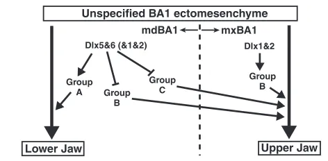
Fig. 8 summarizes our hypothesis on the regionalization of BA1 by Dlx genes. During early stages of craniofacial development, the BA1 ectomesenchyme forms without its regional identity. Subsequently, through a poorly understood mechanism that involves endothelin signaling and Mef2c (Ozeki et al., 2004; Ruest et al., 2004; Miller et al., 2007; Verzi et al., 2007), Dlx genes are expressed such that Dlx5/6are restricted to mdBA1, while Dlx1/2 are in both mdBA1 and mxBA1. Dlx5/6induce and/or maintain expression of the genes that promote the development of the lower jaw (Group A; see Fig. 8 legend). At the same time, Dlx5/6repress other sets of genes (Group B and Group C) so that their expression is mostly confined to mxBA1. Here, Group B and Group C genes promote upper jaw development. Dlx1/2 participate in BA1 patterning by inducing and/or maintaining Group B genes in mxBA1. By contrast, in mdBA1, Dlx1/2positively regulate Group A genes and repress Group B genes, directly or indirectly, to specify lower jaw fate.
We thank Winnie Liang for microarray analysis; Ayako Kuroda for ES cell blastocyst injection; Ugo Borello for help with manuscript preparation; Ugo Borello and Pooja Agarwal for help with the luciferase reporter experiment; and Gail Martin, Maria Barna, Ross Metzger and members of the Rubenstein and Martin laboratories for helpful discussions. We are grateful to Ken Weiss, Walter Birchmeier, Cam Patterson, Maxime Bouchard and Stephen Gold for
providing plasmids for in situ hybridization probes. This work was supported by grants to J.L.R.R. from Nina Ireland, Hillblom Foundation, NIH/NIDCD R01 DC005667 and March of Dimes.
Supplementary material
Supplementary material for this article is available at http://dev.biologists.org/cgi/content/full/135/17/2905/DC1
References
Abdollahi, A., Roberts, D., Godwin, A. K., Schultz, D. C., Sonoda, G., Testa, J. R. and Hamilton, T. C.(1997). Identification of a zinc-finger gene at 6q25: a chromosomal region implicated in development of many solid tumors.
Oncogene 14, 1973-1979.
Abu-Abed, S., Dolle, P., Metzger, D., Beckett, B., Chambon, P. and Petkovich, M.(2001). The retinoic acid-metabolizing enzyme, CYP26A1, is essential for normal hindbrain patterning, vertebral identity, and development of posterior structures. Genes Dev. 15, 226-240.
Acampora, D., Merlo, G. R., Paleari, L., Zerega, B., Pia Postiglione, M., Mantero, S., Bober, E., Barbieri, O., Simeone, A. and Levi, G.(1999). Craniofacial, vestibular and bone defects in mice lacking the Distal-less related gene Dlx5. Development 126, 3795-3809.
Ackerman, S. L., Kozak, L. P., Przyborski, S. A., Rund, L. A., Boyer, B. B. and Knowles, B. B.(1997). The mouse rostral cerebellar malformation gene encodes an UNC-5-like protein. Nature386, 838-842.
Barbosa, A. C., Funato, N., Chapman, S., McKee, M. D., Richardson, J. A., Olson, E. N. and Yanagisawa, H.(2007). Hand transcription factors cooperatively regulate development of the distal midline mesenchyme. Dev. Biol. 310, 154-168.
Beverdam, A., Brouwer, A., Reijnen, M., Korving, J. and Meijlink, F.(2001). Severe nasal clefting and abnormal embryonic apoptosis in Alx3/Alx4 double mutant mice. Development128, 3975-3986.
Beverdam, A., Merlo, G. R., Paleari, L., Mantero, S., Genova, F., Barbieri, O., Janvier, P. and Levi, G.(2002). Jaw transformation with gain of symmetry after Dlx5/Dlx6 inactivation: mirror of the past? Genesis 34, 221-227.
Birchmeier, C. and Gherardi, E.(1998). Developmental roles of HGF/SF and its receptor, the c-Met tyrosin kinase. Trends Cell Biol. 8, 404-410.
Bladt, F., Riethmacher, D., Isenmann, S., Aguzzi, A. and Birchmeier, C.(1995). Essential role for the c-met receptor in the migration of myogenic precursor cells into the limb bud. Nature376, 768-771.
Blanchette, M, Kent, W. J., Riemer, C, Elnitski, L, Smit, A. F., Roskin, K. M., Baertsch, R., Rosenbloom, K., Clawson, H., Green, E. D. et al.(2004). Aligning multiple genomic sequences with the threaded blockset aligner.
Genome Res. 14, 708-715.
Bosse, A., Stoykova, A., Nieselt-Struwe, K., Chowdhury, K., Copeland, N. G., Jenkins, N. A. and Gruss, P.(2000). Identification of a novel mouse Iroquois homeobox gene, Irx5, and chromosomal localisation of all members of the mouse Iroquois gene family. Dev. Dyn. 218, 160-174.
Bulfone, A., Kim, H.-J., Puelles, L., Porteus, M. H., Grippo, J. E. and Rubenstein, J. L. R.(1993). The mouse Dlx-2 (Tes-1) gene is expressed in spatially restricted domains of the forebrain, face and limbs in midgestation mouse embryos. Mech. Dev. 40, 129-140.
Byrd, N. A. and Meyers, E. N.(2005). Loss of Gbx2 results in neural crest cell patterning and pharyngeal arch artery defects in the mouse embryo. Dev. Biol. 284, 233-245.
Caton, A., Hacker, A., Naeem, A., Livet, J., Maina, F., Bladt, F., Klein, R., Birchmeier, C. and Guthrie, S.(2000). The branchial arches and HGF are growth-promoting and chemoattractant for cranial motor axons. Development 127, 1751-1760.
Chai, Y. and Maxon, Jr, R. E.(2006). Recent advances in craniofacial morphogenesis. Dev. Dyn. 235, 2353-2375.
Chai, Y., Jiang, X., Ito, Y., Bringas, P., Jr, Han, J., Rowitch, D. H., Soriano, P., McMahon, A. P. and Sucov, H. M.(2000). Fate of the mammalian cranial neural crest during tooth and mandibular morphogenesis. Development127, 1671-1679.
Charite, J., McFadden, D. G., Merlo, G., Levi, G., Clouthier, D. E., Yanagisawa, M., Richardson, J. A. and Olson, E. N.(2001). Role of Dlx6 in regulation of an endothelin-1-dependent, dHAND branchial arch enhancer.
Genes Dev. 15, 3039-3049.
Chen, C., Zheng, B., Han, J. and Lin, S. C.(1997). Characterization of a novel mammalian RGS protein that binds to G alpha proteins and inhibits pheromone signaling in yeast. J. Biol. Chem. 272, 8679-8685.
Cheng, C. W., Chow, R. L., Lebel, M., Sakuma, R., Cheung, H. O.-L., Thanabalasingham, V., Zhang, X., Bruneau, B. G., Birch, D. G., Hui, C.-C. et al.(2005). The Iroquoishomeobox gene, Irx5, is required for retinal cone bipolar cell development. Dev. Biol. 287, 48-60.
Coffinier, C., Ketpura, N., Tran, U., Geissert, D. and De Robertis, E. M.(2002). Mouse Crossveinless-2 is the vertebrate homolog of a Drosophila extracellular regulator of BMP signaling. Mech. Dev. 119S,S179-S184.
Unspecified BA1 ectomesenchyme
[image:10.612.51.287.59.175.2]Upper Jaw Lower Jaw Dlx1&2 Dlx5&6 (&1&2) Group A Group B Group C Group B mdBA1 mxBA1
Fig. 8. A model for the mechanism of jaw patterning by Dlx genes. In BA1 mesenchyme, Dlx5/6are only expressed in mdBA1, whereas Dlx1/2are expressed in both mdBA1 and mxBA1. Dlx5/6 induce and/or maintain expression of Group A genes (Dlx3, Dlx4, Hand1, Hand2, Gbx2, Gsc, Alx3, Alx4, Bmper, Cited1, Zac1, Unc5c, Hgf, Rgs5, A/S Dlx1and Evf1/2) in mdBA1, while repressing Group B (Pou3f3, 2610016I09Rikand 2900092D14Rik) and Group C (Foxl2, E330015D05Rik, Cyp26a1and Irx5) genes so that their expression is mostly confined to mxBA1. Dlx1/2induce and/or maintain Group B genes in mxBA1. By contrast, in mdBA1, Dlx1/2induce and/or maintain Group A genes and repress Group B genes. Presumably, Group A genes promote lower jaw development, whereas Group B and Group C genes promote upper jaw development.
Cohen, D. R., Cheng, C. W., Cheng, S. H. and Hui, C. C.(2000). Expression of two novel mouse Iroquois homeobox genes during neurogenesis. Mech. Dev. 91, 317-321.
Cokol, M., Nair, R. and Rost, B.(2000). Finding nuclear localization signals.
EMBO Rep. 1, 411-415.
Coles, E., Christiansen, J., Economou, A., Bronner-Fraser, M. and Wilkinson, D. G.(2004). A vertebrate crossveinless 2 homologue modulates BMP activity and neural crest cell migration. Development 131, 5309-5317.
Costantini, D. L., Arruda, E. P., Agarwal, P., Kim, K.-H., Zhu, Y., Zhu, W., Lebel, M., Cheng, C. W., Park, C. Y., Pierce, S. A. et al.(2005). The homeodomain transcription factor Irx5 establishes the mouse cardiac ventricular repolarization gradient. Cell 123, 347-358.
Couly, G. F., Coltey, P. M. and Le Douarin, N. M.(1993). The triple origin of skull in higher vertebrates: a study in quail-chick chimeras. Development 117, 409-429.
Crisponi, L., Deiana, M., Loi, A., Chiappe, F., Uda, M., Amati, P., Bisceglia, L., Zelante, L., Nagaraja, R., Porcu, S. et al.(2001). The putative forkhead transcription factor FOXL2 is mutated in blepharophimosis/ptosis/epicanthus inversus syndrome. Nat. Genet. 27, 159-166.
Depew, M. J., Liu, J. K., Long, J. E., Presley, R., Meneses, J. J., Pedersen, R. A. and Rubenstein, J. L. R.(1999). Dlx5regulates regional development of the branchial arches and sensory capsules. Development126, 3831-3846. Depew, M. J., Tucker, A. S. and Sharpe, P. T.(2002a). Craniofacial Development.
In Mouse Development, Patterning, Morphogenesis, and Organogenesis(ed. J. Rossant and P. P. L. Tam), pp. 421-498. Academic Press, San Diego, CA. Depew, M. J., Lufkin, T. and Rubenstein, J. L. R.(2002b). Specification of jaw
subdivisions by Dlxgenes.Science298, 381-385.
Depew, M. J., Simpson, C. A., Morasso, M. and Rubenstein, J. L. R.(2005). Reassessing the Dlx code: the genetic regulation of branchial arch skeletal pattern and development. J. Anat. 207, 501-561.
Dietrich, S., Abou-Rebyeh, F., Brohmann, H., Bladt, F., Sonnenberg-Riethmacher, E., Yamaai, T., Lumsden, A., Brand-Saberi, B. and Birchmeier, C.(1999). The role of SF/HGF and c-Met in the development of skeletal muscle.
Development126, 1621-1629.
Dolle, P., Price, M. and Duboule, D.(1992). Expression of the murine Dlx-1 homeobox during facial, ocular, and limb development. Differentiation49, 93-99.
Faedo, A., Quinn, J. C., Stoney, P., Long, J. E., Dye, C., Zollo, M., Rubenstein, J. R., Price, D. J. and Bulfone, A.(2004). Identification and characterization of a novel transcript down-regulated in Dlx1/Dlx2 and upregulated in Pax6 mutant telencephalon. Dev. Dyn. 231, 614-620.
Feledy, J. A., Morasso, M. I., Jang, S.-I. and Sargent, T. D.(1999). Transcriptional activation by the homeodomain protein distal-less 3. Nucleic Acids Res.27, 764-770.
Feng, J., Bi, C., Clark, B. S., Mady, R., Shah, P. and Kohtz, J. D.(2006). The Evf-2 noncoding RNA is transcribed from the Dlx-5/6 ultraconserved region and functions as a Dlx-2 transcriptional coactivator. Genes Dev. 20, 1470-1484. Fujii, H., Sato, T., Kaneko, S., Gotoh, O., Fujii-Kuriyama, Y., Osawa, K., Kato,
S. and Hamada, H.(1997). Metabolic inactivation of retinoic acid by a novel P450 differentially expressed in developing mouse embryos. EMBO J. 16, 4163-4173.
Hara, Y., Rovescalli, A. C., Kim, Y. and Nirenberg, M.(1992). Structure and evolution of four POU domain genes expressed in mouse brain.Proc. Natl. Acad. Sci. USA 89, 3280-3284.
Ikeya, M., Kawada, M., Kiyonari, H., Sasai, N., Nakao, K., Furuta, Y. and Sasai, Y.(2006). Essential pro-Bmp roles of crossveinless 2 in mouse organogenesis. Development 133, 4463-4473.
Jeong, J. and McMahon, A. P.(2005). Growth and pattern of the mammalian neural rube are governed by partially overlapping feedback activities of the hedgehog antagonists patched 1 and Hhip1. Development132, 143-154. Jeong, J., Mao, J., Tenzen, T., Kottmann, A. H. and McMahon, A. P.(2004).
Hedgehog signaling in the neural crest cells regulate the patterning and growth of facial primordia. Genes Dev. 18, 937-951.
Katoh, Y. and Katoh, M.(2004). Identification and characterization of CDC50A, CDC50B and CDC50C genes in silico. Oncol. Rep. 12, 939-943.
Kohtz, J. D. and Fishell, G.(2004). Developmental regulation of EVF-1, a novel non-coding RNA transcribed upstream of the mouse Dlx6 gene. Gene Expr. Patterns4, 407-412.
Köntges, G. and Lumsden, A.(1996). Rhombencephalic neural crest
segmentation is preserved throughout craniofacial ontogeny. Development122, 3229-3242.
Lanctot, C., Moreau, A., Chamberland, M., Tremblay, M. L. and Drouin, J. (1999). Hindlimb patterning and mandible development require the Ptx1 gene.
Development 126, 1805-1810.
Larkin, M. A., Blackshields, G., Brown, N. P., Chenna, R., McGettigan, P. A., McWilliam, H., Valentin, F., Wallace, I. M., Wilm, A., Lopez, R. et al.(2007). ClustalW2 and ClustalX version 2. Bioinformatics 23, 2947-2948.
Liu, J.-K., Ghattas, I., Liu, S., Chen, S. and Rubenstein, J. L. R.(1997). Dlx genes encode DNA-binding proteins that are expressed in an overlapping and sequential pattern during basal ganglia differentiation. Dev. Dyn. 210, 498-512.
Loots, G. and Ovcharenko, I.(2004). rVISTA 2.0: evolutionary analysis of transcription factor binding sites. Nucleic Acids Res. 32, W217-W221. McEvilly, R. J., Ortiz de Diaz, M., Schonemann, M. D., Hooshmand, F. and
Rosenfeld, M. G.(2002). Transcriptional regulation of cortical neuron migration by POU domain factors. Science295, 1528-1532.
McGuinness, T., Porteus, M. H., Smiga, S., Bulfone, A., Kingsley, C., Qiu, M., Liu, J. K., Long, J. E., Xu, D. and Rubenstein, J. L.(1996). Sequence, organization, and transcription of the Dlx-1 and Dlx-2 locus. Genomics 35, 473-485.
Merlo, G. R., Paleari, L., Mantero, S., Genova, F., Beverdam, A., Palmisano, G. L., Barbieri, O. and Levi, G.(2002). Mouse model of split hand/foot malformation type I. Genesis 33, 97-101.
Miller, C. T., Swartz, M. E., Khuu, P. A., Walker, M. B., Eberhart, J. K. and Kimmel, C. B.(2007). mef2ca is required in cranial neural crest to effect Endothelin1 signaling in zebrafish. Dev. Biol. 308, 144-157.
Moser, M., Binder, O., Wu, Y., Aitsebaomo, J., Ren, R., Bode, C., Bautch, V. L., Conlon, F. L. and Patterson, C.(2003). BMPER, a novel endothelial cell precursor-derived protein, antagonizes bone morphogenetic protein signaling and endothelial cell differentiation. Mol. Cell. Biol. 23, 5664-5679.
Nakamura, S., Stock. D. W., Wydner, K. L., Bollekens, J., Takeshita, K., Nagai, B. M., Chiba, S., Kitamura, T., Freeland, T. M., Zhao, Z. et al.(1996). Genomic analysis of a new mammalian distal-less gene: Dlx7. Genomics 38, 314-324.
Noden, D. M.(1978). The control of avian cephalic neural crest
cytodifferentiation. I. Skeletal and connective tissues. Dev. Biol. 67, 296-312. Osumi-Yamashita, N., Ninomiya, Y., Doi, H. and Eto, K.(1994). The
contribution of both forebrain and midbrain crest cells to the mesenchyme in the frontonasal mass of mouse embryo. Dev. Biol. 164, 409-419.
Ozeki, H., Kurihara, Y., Tonami, K., Watatani, S. and Kurihara, H.(2004). Endothelin-1 regulates the dorsoventral branchial arch patterning in mice. Mech. Dev. 121, 387-395.
Panganiban, G. and Rubenstein, J. L. R.(2002). Developmental functions of the
Distal-less/Dlxhomeobox genes.Development129, 4371-4836.
Porteus, M. H., Bulfone, A., Ciaranello, R. D. and Rubenstein, J. L. R.(1991). Isolation and characterization of a novel cDNA clone encoding a homeodomain that is developmentally regulated in the ventral forebrain. Neuron 77, 221-229. Price, M., Lemaistre, M., Pischetola, M., di Lauro, R. and Duboule, D.(1991).
A mouse gene related to Distal-less shows a restricted expression in the developing forebrain. Nature351, 748-751.
Qiu, M., Bulfone, A., Martinez, S., Meneses, J., Shimamura, K., Pedersen, R. A. and Rubenstein, J. L. R.(1995). Null mutation of Dlx-2results in abnormal morphogenesis of proximal first and second branchial arch derivatives and abnormal differentiation in the forebrain.Genes Dev. 9, 2523-2538.
Qiu, M., Bulfone, A., Ghattas, I., Meneses, J. J., Christensen, L., Sharpe, P. T., Presley, R., Pederson, R. A. and Rubenstein, J. L. R.(1997). Role of the Dlx homeobox genes in proximodistal patterning of the branchial arches: mutations of Dlx-1, Dlx-2, and Dlx-1 and -2 alter morphogenesis of proximal skeletal and soft tissue structures derived from the first and second arches. Dev. Biol. 185, 165-184.
Ray, W. J., Bain, G., Yao, M. and Gottlieb, D. I.(1997). CYP26, a novel mammalian cytochrome P450, is induced by retinoic acid and defines a new family. J. Biol. Chem. 272, 18702-18708.
Rivera-Perez, J. A., Mallo, M., Gentron-Maguire, M., Gridley, T. and Behringer, R. R.(1995). Goosecoid is not an essential component of the mouse gastrula organizer but is required for craniofacial and rib development.
Development 121, 3005-3012.
Robinson, G. W. and Mahon, K. A.(1994). Differential and overlapping expression domains of Dlx-2 and Dlx-3 suggest distinct roles for Distal-less homeobox genes in craniofacial development. Mech. Dev. 48, 199-215. Robledo, R. F. and Lufkin, T.(2006). Dlx5 and Dlx6 homeobox genes are required
for specification of the mammalian vestibular apparatus. Genesis 44, 425-437. Robledo, R. F., Rajan, L., Li, X. and Lufkin, T.(2002). The Dlx5 and Dlx6
homeobox genes are essential for craniofacial, axial, and appendicular skeletal development. Genes Dev. 16, 1089-1101.
Rodriguez, T. A., Sparrow, D. B., Scott, A. N., Withington, S. L., Preis, J. I., Michalicek, J., Clements, M., Tsang, T. E., Shioda, T., Beddington, R. S. P. and Dunwoodie, S. L.(2004). Cited1 is required in trophoblasts for placental development for embryo growth and survival. Mol. Cell. Biol. 24, 228-244. Ruest, L. B., Xiang, X., Lim, K. C., Levi, G. and Clouthier, D. E.(2004).
Endothelin-A receptor-dependent and -independent signaling pathways in establishing mandibular identity. Development 131, 4413-4423.
Santagati, F. and Rijli, F. M.(2003). Cranial neural crest and the building of the vertebrate head. Nat. Rev. Neurosci. 4, 806-818.
Shioda, T., Fenner, M. H. and Isselbacher, K. J.(1996). msg1, a novel melanocyte-specific gene, encodes a nuclear protein and is associated with pigmentation. Proc. Natl. Acad. Sci. USA93, 12298-12303.
Simeone, A., Acampora, D., Pannese, M., D’Esposito, M., Stornaiuolo, A., Gulisano, M., Mallamaci, A., Kastury, K., Druck, T, Huebner, K. and Boncinelli, E.(1994). Cloning and characterization of two members of the
vertebrate Dlx gene family. Proc. Natl. Acad. Sci. USA91, 2250-2254.
D
Spengler, D., Villalba, M., Hoffmann, A., Pantaloni, C., Houssami, S., Bockaert, J. and Journot, L.(1997). Regulation of apoptosis and cell cycle arrest by Zac1, a novel zinc finger protein expressed in the pituitary gland and the brain. EMBO J. 16, 2814-2825.
Stock, D. W., Ellies, D. L., Zhao, Z., Ekker, M., Ruddle, F. R. and Weiss, K. M. (1996). The evolution of the vertebrate Dlxgene family. Proc. Natl. Acad. Sci. USA 93, 10858-10863.
Stoker, M., Gherardi, E., Perryman, M. and Gray, J.(1987). Scatter factor is a fibroblast-derived modulator of epithelial cell mobility. Nature 327, 239-242.
Stuhmer, T., Anderson, S. A., Ekker, M. and Rubenstein, J. L. R.(2002). Ectopic expression of the Dlx genes induces glutamic acid decarboxylase and Dlx expression. Development 129, 245-252.
Sumiyama, K. and Ruddle, F. H.(2003). Regulation of Dlx3 gene expression in visceral arches by evolutionarily conserved enhancer elements. Proc. Natl. Acad. Sci. USA100, 4030-4034.
Uda, M., Ottolenghi, C., Crisponi, L., Garcia, J. E., Deiana, M., Kimber, W., Forabosco, A., Cao, A., Schlessinger, D. and Pilia, G.(2004). Foxl2 disruption causes mouse ovarian failure by pervasive blockage of follicle development.
Hum. Mol. Genet. 13, 1171-1181.
Varrault, A., Gueydan, C., Delalbre, A., Bellmann, A., Houssami, S., Aknin, C., Severac, D., Chotard, L., Kahli, M., Le Digarcher, A. et al.(2006). Zac1 regulates an imprinted gene network critically involved in the control of embryonic growth. Dev. Cell11, 711-722.
Verzi, M. P., Agarwal, P., Brown, C., McCulley, D. J., Schwarz, J. J. and Black, B. L.(2007). The transcription factor MEF2C is required for craniofacial development. Dev. Cell12, 645-652.
White, J. A., Guo, Y. D., Baetz, K., Beckett-Jones, B., Bonasoro, J., Hsu, K. E., Dilworth, F. J., Jones, G. and Petkovich, M.(1996). Identification of the retinoic acid-inducible all-trans-retinoic acid 4-hydroxylase. J. Biol. Chem. 271, 29922-29927.
Xie, G. X. and Palmer, P. P.(2007). How regulators of G protein signaling achieve selective regulation. J. Mol. Biol. 366, 349-365.
Yamada, G., Mansouri, A., Torres, M., Stuart, E. T., Blum, M., Schultz, M., De Robertis, E. M. and Gruss, P.(1995). Targeted mutation of the murine goosecoid gene results in craniofacial defects and neonatal death. Development 121, 2917-2922.
Yanagisawa, H., Clouthier, D. E., Richardson, J. A., Charite, J. and Olson, E. N.(2003). Targeted deletion of a branchial arch-specific enhancer reveals a role of dHAND in craniofacial development. Development 130, 1069-1078.