D
E
V
E
LO
P
M
E
N
T
INTRODUCTION
Seed development in flowering plants is initiated by a double fertilization event in which two sperm nuclei fuse with two female gametes. Male and female gametes are enclosed within multicellular gametophytes. The female gametophyte of Arabidopsisis embedded within the ovule and consists out of seven cells: three antipodal cells that degenerate shortly before fertilization, two synergid cells, one egg cell and one central cell. Except for the homodiploid central cell, all cells of the female gametophyte are haploid. During fertilization, the pollen tube discharges two genetically identical haploid sperm cells into the female gametophyte, where one sperm cell fuses with the egg cell, giving rise to the diploid embryo, while the other sperm cell fuses with the central cell, initiating development of the triploid endosperm (Drews and Yadegari, 2002). The embryo passes through morphologically defined stages, characterized as preglobular, globular, heart, torpedo, walking stick, early maturation and maturation (Goldberg et al., 1994; Laux and Jürgens, 1997).
Endosperm development differs dramatically from embryo development. In Arabidopsis, the first divisions of the primary endosperm nucleus are not followed by cytokinesis, giving rise to the formation of a syncytium. Distinct nuclear-cytoplasmic domains form: the chalazal endosperm at the posterior pole, the micropylar endosperm at the anterior pole and peripheral endosperm domains (Brown et al., 1999; Boisnard-Lorig et al., 2001). Endosperm cellularization is initiated around the globular to early heart stage of embryo development and starts in the micropylar endosperm, which surrounds the embryo, to progress through the peripheral endosperm to the chalazal region (Brown et al., 1999; Boisnard-Lorig et al., 2001). As the embryo matures, most of the endosperm is degraded and absorbed by the embryo, and only a thin aleurone layer remains.
The endosperm is considered to support embryo growth and to regulate nutrient transfer from the mother to the developing seeds (Lopes and Larkins, 1993).
Mutations in genes of the FERTILIZATION INDEPENDENT SEED(FIS) class can form diploid endosperm in the absence of fertilization (Ohad et al., 1996; Chaudhury et al., 1997). Thus far, four FIS-class genes are known: MEDEA(MEA), FERTILIZATION INDEPENDENT ENDOSPERM (FIE), FERTILIZATION INDEPENDENT SEED2(FIS2) and MULTICOPY SUPPRESSOR OF IRA1(MSI1) (Grossniklaus et al., 1998; Kiyosue et al., 1999; Luo et al., 1999; Ohad et al., 1999; Köhler et al., 2003a; Guitton et al., 2004). Among fismutants, the msi1mutant has the strongest penetrance of the autonomous endosperm development phenotype (Köhler et al., 2003a; Guitton et al., 2004) and also forms parthenogenetic embryos (Guitton and Berger, 2005). The FIS-class genes encode proteins with homology to animal Polycomb group (PcG) proteins. Plant FIS proteins and animal PcG proteins both form multisubunit complexes with a core size of 600 kDa called Polycomb repressive complex 2 (PRC2) (Köhler et al., 2003a; Chanvivattana et al., 2004; Schwartz and Pirrotta, 2007). Animal PRC2 complexes possess histone methyltransferase activity specific for lysine 27 on histone H3 (H3K27) and possibly also H3K9 (Schwartz and Pirrotta, 2007). Similarly, plant PRC2 complexes, such as the FIS complex and the EMF2 complex, are required for H3K27 methylation and transcriptional repression of target genes (Gehring et al., 2006; Makarevich et al., 2006; Schönrock et al., 2006; Schubert et al., 2006).
Mutations in FISgenes cause parent-of-origin-dependent seed abortion. All seeds that inherit a mutant fisallele from the mother abort, regardless of the presence of a wild-type paternal allele. Development of fismutant seeds is delayed and seeds abort with embryos arrested at late heart stage and displaying non-cellularized endosperm with strongly overproliferated chalazal endosperm domains (Grossniklaus et al., 1998; Kiyosue et al., 1999; Köhler et al., 2003a; Guitton et al., 2004). The maternal-effect parent-of-origin-dependent seed abortion in meaand fis2mutants can be explained by the findings that MEAand FIS2are imprinted genes, with the paternal allele of both genes being specifically silenced in
Polycomb group proteins function in the female
gametophyte to determine seed development in plants
Olivier Leroy1, Lars Hennig1, Holger Breuninger2, Thomas Laux2and Claudia Köhler1,*
Polycomb group (PcG) proteins are evolutionary conserved proteins that stably maintain established transcriptional patterns over cell generations. The FERTILIZATION INDEPENDENT SEED (FIS) PcG complex from plants has a similar composition to the Polycomb repressive complex 2 from animals. Mutations in FISgenes cause parent-of-origin-dependent seed abortion. Every seed inheriting a mutant fisallele from the mother is destined to abort, regardless of the presence of a wild-type paternal allele. We tested in Arabidopsiswhether the parent-of-origin-dependent seed abortion caused by lack of the FIS subunit MSI1 is caused by parental imprinting of the MSI1gene. Our data show that MSI1is not an imprinted gene and that early paternal MSI1expression is not sufficient to rescue msi1mutant seeds. By contrast, expression of MSI1in msi1female gametophytes is necessary to restore normal seed development, strongly arguing that the female gametophytic effect of fismutants is caused by a functional requirement for an intact FIS complex in the female gametophyte. Thus, FIS-mediated expression patterns established in the female gametophyte can impact on seed development, establishing fismutants as true female gametophytic maternal-effect mutants.
KEY WORDS: Arabidopsis, Epigenetics, FERTILIZATION INDEPENDENT SEEDgenes, Imprinting, Polycomb group proteins Development 134, 3639-3648 (2007) doi:10.1242/dev.009027
1Institute of Plant Sciences and Zürich-Base Plant Science Center, Swiss Federal Institute of Technology, ETH Centre, CH-8092 Zürich, Switzerland. 2Institute of Biology II, University of Freiburg, Schänzlestr. 1, 79104 Freiburg, Germany.
*Author for correspondence (e-mail: [email protected])
D
E
V
E
LO
P
M
E
N
T
the endosperm (Vielle-Calzada et al., 1999; Kinoshita et al., 1999; Luo et al., 2000; Jullien et al., 2006a). Similarly, the paternal FIE allele is not expressed during early stages of seed development, providing an explanation for the maternal effect of fie mutants (Yadegari et al., 2000).
It is likely that MEA and FIS2 are subunits specific to the FIS complex, whereas FIE and MSI1 are part of several distinct PRC2-like complexes (Hennig et al., 2005; Schubert et al., 2005). Furthermore, MSI1 is potentially part of several different complexes, such as chromatin assembly factor CAF-1, histone deacetylases and chromatin-remodeling machines, which are likely to play a role during early embryogenesis (Hennig et al., 2005). Similar to mea, fis2and fiemutants, lack of MSI1 function causes parent-of-origin-dependent seed abortion. However, in addition to the gametophytic effect, it has been proposed that lack of MSI1 function also causes a sporophytic effect on seed development (Guitton et al., 2004). Thus, lack of both maternal and paternal MSI1alleles causes a significantly stronger defect than lack of the maternal MSI1allele alone. This implies that the paternal allele of MSI1 is active, but fails to complement the maternal gametophytic msi1defect.
To test this idea, we investigated the temporal requirements of MSI1 during seed development. We specifically addressed the question of whether early paternal expression of MSI1is sufficient to rescue the maternal-effect msi1seed abortion phenotype. Our data clearly show that MSI1 is not an imprinted gene and that early paternal MSI1expression is not sufficient to rescue msi1mutant seeds. By contrast, expression of MSI1in msi1female gametophytes is necessary to restore normal seed development, revealing that the female gametophytic effect of fismutants is caused by a functional requirement for an intact FIS complex in the female gametophyte. Thus, FIS complex function in the female gametophyte before fertilization determines seed development after fertilization, establishing fis mutants as true epigenetic female gametophytic maternal-effect mutants.
MATERIALS AND METHODS Plant material and growth conditions
Seeds of Columbia (Col) Arabidopsis thalianawild-type accession were obtained from the Nottingham ArabidopsisStock Centre, UK. The msi1 allele used in this study was the msi1-1allele in Col described by Köhler et al. (Köhler et al., 2003a). The silent MSI1* allele, which encodes a wild-type MSI1 protein, is a TILLING (Induced Local Lesions IN Genomes) mutant obtained from the Nottingham ArabidopsisStock Centre, stock number CS92951. The Q0990, M0221 and M0223 enhancer-trap lines expressing the GFP reporter protein were generated in the laboratory of J. Haseloff (http://www.plantsci.cam.ac.uk/Haseloff) and obtained from the Nottingham ArabidopsisStock Centre. The SCR::YFPreporter line was kindly provided
by Dr B. Scheres (Utrecht University, The Netherlands). The DR5rev::GFP reporter line was generously given by Dr J. Friml (University of Tübingen, Germany). The WOX8::YFPreporter line contains 2511 bp of the WOX8 promoter and 1775 bp of WOX8coding sequence fused to the YFPreporter and recapitulates the WOX8expression pattern (Haecker et al., 2004) (H.B. and T.L., unpublished). Marker lines were introduced into the msi1 background by crossing. Plants were grown in a greenhouse at 70% humidity and daily cycles of 16 hours light at 21°C and 8 hours darkness at 18°C. Developed gynoecia were emasculated and hand-pollinated 1 day after emasculation. For RNA expression analysis, three gynoecia or siliques were harvested at the indicated time points. Dissection of seeds into embryo and endosperm plus seed coat fractions was performed under a dissection microscope. Dissected material from 100 seeds was collected in RNAlater (Ambion, Austin, USA) solution and processed as indicated below. For transmission analysis of the msi1mutant allele, which is tagged with a phosphinothricin resistance marker, seeds derived from reciprocal crosses were harvested 3 weeks after pollination. T1 seeds were plated on Murashige and Skoog (MS) medium containing 30 mg/l phosphinothricin, and after approximately 2 weeks the ratio of resistant to non-resistant seedlings was scored.
Plasmid constructs and generation of transgenic plants
To generate the PHE1::MSI1construct, the 3.0 kb of sequence upstream of thePHE1translational start was amplified by PCR introducing EcoRI and NcoI restriction sites. The MSI1cDNA was amplified by PCR introducing NcoI and BglII restriction sites. Both fragments were ligated into pCAMBIA 1380 using EcoRI and BglII restriction sites. To generate the DD46::GUS construct, the region 900 bp upstream of the DD46 translational start was amplified by PCR introducing BamHI and NcoI restriction sites and the fragment introduced into pCAMBIA 1381z using BamHI and NcoI. To generate the DD46::MSI1construct, pCAMBIA 1380 was opened with BamHI and BglII and the DD46 promoter fragment flanked by BamHI and NcoI restriction sites and the MSI1cDNA flanked by NcoI and BglII sites were introduced by ligation. All primers are listed in Table 1. Heterozygous msi1plants were transformed by floral dip, and transgenic plants were selected on MS medium containing 30 mg/l hygromycin. T1 plants were treated with BASTA to select for the msi1 mutation and resistant lines were assayed for complementation of seed abortion.
RNA extraction and RT-PCR analysis
[image:2.612.50.571.587.743.2]RNA was prepared with Trizol reagent (Invitrogen, Carlsbad, USA) according to the suppliers’ recommendations. For RT-PCR, total RNA was treated with 5 units of RNase-free DNase (Amersham Pharmacia Life Science, Little Chalfont, UK) for 30 minutes. Samples were extracted with phenol-chloroform and precipitated with ethanol. The RNA was reverse-transcribed using 0.5 g of oligo dT primers (Invitrogen) in a 20 l reaction containing 1 l reaction buffer, 0.25 mM of each deoxynucleotide triphosphate, 5 mM dithiothreitol and 200 units of Superscript II reverse transcriptase (Invitrogen) by incubating at 42°C for 1 hour followed by heat inactivation at 72°C for 15 minutes. For RT-PCR analysis, 1/20th (by
Table 1. Primers used in this study Amplified region Primers (5-3)
ACTIN3(AT3G12110) Fwd: AACTTTCAACACTCCTGCCATG Rev: CTGCAAGGTCCAAACGCAGA Transgene-derived MSI1 Fwd: GCACCGCTCTTCACACATTG
Rev: TGGTCACCTGTAATTCACACG Wild-type MSI1 s1: CGGTAAAGACTACTCCGTTCAGATG
as1: GTAATCGAAAACATAGACCTCC
MSI1* s1: CGGTAAAGACTACTCCGTTCAGATA
as2: GTAATCGAAAACATAGACCTCC
MSI1 cDNA Fwd: GACCATGGGGAAAGACGAAGAGGAAATG Rev: CGAGATCTCTAAGAAGCTTTTGATGGTTC PHERES1 promoter Fwd: CCGAATTCGACTTTAAAATAGTAGAAAAGCTTG
Rev: AATTCCATGGATCTCTTATCTTTTTCTTTTGTG DD46 promoter Fwd: GGATCCGGGGAAAGGAGAAAAACAAATGAGGG
D
E
V
E
LO
P
M
E
N
T
volume) of the reverse-transcription samples was used to amplify cDNAs using the primers listed in Table 1. For amplification of transcripts from the MSI1and MSI1* alleles from siliques, 38 PCR cycles were used; for amplification of these transcripts from dissected embryo and endosperm tissues, 40 cycles were used. For amplification of ACTINtranscripts, 38 cycles were always used.
Histological analysis
For detection of GUS activity, siliques were cut longitudinally, fixed for 1 hour at –20°C in 90% acetone and washed three times with 50 mM phosphate buffer (pH 7.0) before incubation at 37°C in reaction buffer (0.19 mM 5-bromo-4-chloro-3-indolyl--D-glucuronide, 10 mM EDTA, 0.1% Triton X-100, 0.1 mM KFeCN, 50 mM phosphate buffer pH 7.2) for 24-72 hours. Whole seeds were observed after clearing in chloral hydrate solution (40 mM chloral hydrate, 8.3% glycerol) using a Zeiss Axioplan microscope. For GFP and YFP marker analysis, embryos from dissected seeds were mounted in deionized water. Specimens were observed under a Zeiss Axioplan microscope equipped with a GFP filter set and Nomarski optics, and images were recorded with a MagnaFire CCD camera (Optronics, Goleta, CA).
RESULTS
Loss of MSI1 causes gametophytic and sporophytic effects on seed development
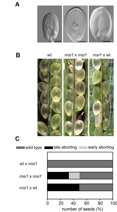
Complete loss of MSI1 is lethal, and the msi1-1allele used in this study can only be maintained in heterozygous msi1/MSI1plants (here referred to as msi1plants) (Köhler et al., 2003a; Guitton et al., 2004). Self-fertilized msi1 mutant plants form two classes of aborting seeds: an early-aborting class, which contains grossly abnormal embryos, and a late-aborting class, which contains embryos that phenotypically closely resemble fis-class mutant embryos (Köhler et al., 2003a; Guitton et al., 2004). It has been suggested that lack of MSI1 function has a gametophytic as well as a sporophytic zygotic effect, causing the formation of early- and late-aborting seeds, respectively (Guitton et al., 2004). This model predicts that 50% of the seeds inherit a maternal MSI1allele and develop normally, whereas 50% of the seeds inherit a maternal msi1-1allele and abort early if also inheriting a paternal msi1-1 allele or abort late if inheriting a paternal MSI1allele. We observed that the msi1-1 allele has 17% early-aborting and 33% late-aborting seeds, which deviates from the ratio of 25% early- to 25% late-aborting seeds predicted by the model [n=583, 2=29.72>2
0.05(2)=5.991; Fig. 1A]. One reason for this discrepancy could be a reduced transmission of the paternal msi1-1allele. We tested this hypothesis by determining the transmission of the msi1-1 allele through pollen. Indeed, we found that the transmission of the paternal msi1-1allele is reduced to 72% (n=500). Taking the reduced transmission of the paternal msi1-1allele into account, only 18% homozygous msi1mutant seeds can be expected. This number closely matches the observed number of 17% early-aborting seeds [50% wild type: 32% msi1/MSI1: 18% msi1/msi1; n=583, 2=0.536<20.05(2)=5.991]. To unequivocally test the hypothesis that early-aborting seeds require a paternally inherited msi1allele, we pollinated heterozygous msi1mutant plants with wild-type pollen. In this experiment, 51% of the seeds were phenotypically wild type and 49% of the seeds were late aborting with a fis-like phenotype [50% wild type: 50% msi1/MSI; n=487, 2=0.166<2
0.05(1)=3.841; Fig. 1B,C; Table 2]. We did not observe any early-aborting seeds, clearly proving that loss of both maternal and paternal MSI1alleles is the prerequisite for early seed abortion. This result suggests that the paternal MSI1allele is expressed and, consequently, that MSI1is not regulated by genomic imprinting, in contrast to the fis-class mutants meaand fis2.
MSI1is paternally expressed in embryo and
endosperm
[image:3.612.323.524.59.424.2]D
E
V
E
LO
P
M
E
N
T
the timing of paternal MSI1expression is comparable to that of the majority of paternal alleles (Vielle-Calzada et al., 2000). As MSI1is also expressed in sporophytic tissues (Hennig et al., 2003; Köhler et al., 2003a), transcripts of maternal MSI1 or MSI1* alleles are contributed by zygotic tissues as well as maternal sporophytic tissues. Therefore, the maternal alleles always yielded signals of higher intensity than the paternal alleles.
Imprinting of several genes has been shown to occur specifically in the endosperm, whereas the same genes are biallelically expressed in the embryo (Kinoshita et al., 1999; Kinoshita et al., 2004; Haun et al., 2007). MSI1is expressed in the embryo (Köhler et al., 2003a); therefore, we investigated whether expression of the paternal MSI1allele is confined to the embryo and is silenced in the endosperm, or whether the paternal MSI1 allele is also expressed in the endosperm. For this purpose, we performed crosses of wild-type plants with MSI1*plants and dissected F1 seeds at 6 days after pollination (DAP) into embryo and endosperm plus seed coat fractions. As shown in Fig. 2C, we could clearly detect expression of the paternal MSI1*allele in the embryo as well as in the endosperm. Thus, we conclude that MSI1 is not imprinted, but is biallelically expressed in both embryo and endosperm.
The female gametophytic defect of msi1mutants
does not affect embryo patterning
Heterozygous msi1mutant seeds abort with embryos arrested at late heart stage and displaying strongly overproliferated chalazal endosperm domains (Köhler et al., 2003a; Guitton et al., 2004). However, it remains elusive why msi1 mutant embryos arrest development and abort despite expression of the paternal MSI1 allele. It is possible that developmental defects start to accumulate early during embryogenesis when most of the paternal genome, including MSI1, is still inactive and cause gross developmental abnormalities later in embryogenesis, culminating in seed abortion. Therefore, we tested whether marker genes that define major developmental steps during early embryogenesis are correctly expressed in msi1mutant as compared with wild-type embryos. We tested markers for auxin distribution [DR5(Friml et al., 2003)], the developing suspensor [WUSCHEL-RELATED HOMEOBOX 8 (WOX8) (Haecker et al., 2004)], provascular tissue [enhancer-trap line Q0990 (Weijers et al., 2006)], the quiescent center [SCARECROW(Blilou et al., 2005)], and for cells within the region to form the shoot apical meristem [enhancer-trap lines M0221 and M0223 (Cary et al., 2002)].
The auxin-reporting DR5::GFPmarker was confined to the root pole, cotyledon tips and provascular tissue of heart stage wild-type embryos (Fig. 3). This pattern was similar in msi1 embryos, suggesting that auxin distribution is mostly normal in msi1. Expression of the WOX8reporter was confined to the suspensor in
wild-type and msi1 mutant embryos, indicating that the basal derivatives of the zygote forming the suspensor are correctly established. The enhancer-trap line Q0990 from the publicly available Haseloff collection (http://www.plantsci.cam.ac.uk/ Haseloff/construction/catalogFrame.html) is expressed in pro -vascular cells of the central region immediately adjacent to the hypophysis (Weijers et al., 2006). Because this expression pattern remained in msi1, specification of provascular cells seems to occur properly in msi1mutant embryos.
Establishment of root apical meristems was monitored using SCR::YFP, which is expressed only in the quiescent center and derivatives of the ground meristem (Wysocka-Diller et al., 2000). Expression of SCR::YFPin msi1closely resembled expression in wild type, suggesting that initiation of the root apical meristem is largely normal in msi1. To monitor formation of shoot apical meristems, enhancer-trap lines M0221 and M0223 were used. Both lines show GFP reporter activity in cells within the region forming the shoot apical meristem, and M0223 reflects expression of CUP-SHAPED COTYLEDON1[CUC1(Cary et al., 2002)]. As with the other markers used, activity of M0221 and M0223 was similar in wild type and msi1, indicating that progenitor cells for the shoot apical meristems are properly specified.
Based on these findings, we conclude that defects established in the msi1female gametophyte do not affect basic embryo pattern formation, and embryo arrest at late heart stage is caused by mechanisms that remain to be identified.
Paternal expression of MSI1immediately after
fertilization cannot rescue the msi1female gametophytic defect
[image:4.612.50.563.70.182.2]We considered two possible explanations for the female gametophytic defect of msi1mutants: (1) delayed expression of the paternal MSI1allele at only 3 DAP (Fig. 2B) is responsible for the female gametophytic defect; or (2) lack of functional MSI1 causes a defect in the female gametophyte and the consequences of this defect become obvious during later stages of seed development. We tested the first possibility by expressing paternal MSI1immediately after fertilization. We made use of the PHERES1(PHE1) promoter, which is one of the few promoters escaping early paternal silencing and is expressed immediately after fertilization (Köhler et al., 2005). We tested whether expression of MSI1under control of the PHE1 promoter (referred to as PHE1::MSI1) could be detected immediately after fertilization, by crossing wild-type plants with pollen derived from PHE1::MSI1 transgenic plants. Indeed, expression of the paternal MSI1allele was detected at 1 DAP (Fig. 4A). We compared the expression level of the PHE1::MSI1 transgene with the endogenous MSI1allele starting at 3 DAP by semi-quantitative RT-PCR. Taking into account the different amplification efficiencies of the different primer pairs, expression of Table 2. Analysis of seed development in self- and cross-fertilized msi1plants and in different transgenic backgrounds
Line Wild type (%) Early aborting (%) Late aborting (%) n 2
0.05(1)=3.841
wtwt 100 0 0 269 n.a.
msi1msi1 50 17 33 583 n.a.
wtmsi1 100 0 0 212 n.a.
msi1wt 50 17 33 487 0.166
msi1msi1; PHE1::MSI1#1 50 0 50 278 0.014
msi1msi1; PHE1::MSI1#2 50 0 50 498 0.032
msi1msi1; PHE1::MSI1#3 51.2 0 48.8 166 0.096
msi1; DD46::MSI1 #1wt 100 0 0 369 n.a.
msi1; DD46::MSI1 #2wt 100 0 0 475 n.a.
D
E
V
E
LO
P
M
E
N
T
thePHE1::MSI1transgene was about 3.5-fold higher than that of the endogenous paternal MSI1allele. In heterozygous msi1plants, 50% of the seeds carry a maternal msi1allele and thus suffer from the female gametophytic defect. If early paternal expression of MSI1 could rescue this female gametophytic msi1phenotype, then we would expect that a hemizygous PHE1::MSI1construct would rescue 25% of the seeds and lead to 75% normal seeds. However, among 11 independent transgenic PHE1::MSI1lines in an msi1 mutant background, we did not identify any plant with more than 50% normal seeds, indicating that early paternal expression is not sufficient to rescue the gametophytic msi1mutant defect. Instead, we observed a reduction in the number of early-aborting seeds by about half, suggesting that paternally expressed PHE1::MSI1is sufficient to promote development of early-aborting homozygous msi1mutant seeds up to the stage of late-aborting heterozygous msi1 seeds (data not shown). This hypothesis was tested by pollinating msi1mutant plants with pollen of three independent homozygous PHE1::MSI1transgenic lines in an msi1mutant background and
scoring subsequent seed development. In contrast to pollination with pollen from heterozygous msi1plants, which led to 17% early-aborting seeds, after pollination with pollen from PHE1::MSI1; msi1transgenic lines no early-aborting seeds were observed (Fig. 4B,D; Table 2). Thus, early paternal MSI1expression is sufficient to establish prolonged development of homozygous msi1mutant seeds. We analyzed seeds of this cross by clearing and found no significant change of seed development as compared with seeds developing on msi1 plants pollinated with wild-type pollen (Fig. 4C). To unequivocally test whether early paternal MSI1 expression can rescue the msi1 mutant phenotype, we tested transmission of the maternal msi1allele after pollination of msi1plants with pollen of PHE1::MSI1plants. We crossed msi1plants with pollen of three independent PHE1::MSItransgenic lines in a wild-type background. Using more than 100 seedlings for each line, we found no significant maternal transmission of msi1(Table 3). Thus, we conclude that early paternal MSI1expression is not sufficient to rescue the female gametophytic defect of msi1mutant seeds.
Expression of MSI1in the female gametophyte
can rescue the msi1female gametophytic defect
[image:5.612.51.282.56.361.2]Because early paternal MSI1expression could not rescue the female gametophytic defect of msi1mutants, we addressed the question of whether expression of MSI1in the female gametophyte could rescue Fig. 2. The paternal MSI1allele is expressed in the embryo and
[image:5.612.306.561.58.385.2]endosperm.(A) Schematic of the PCR assay used to amplify specifically either the MSI1or MSI1* allele. Primer combination s1-as1 amplifies only the MSI1allele (as shown beneath, left), whereas primer combination s2-as1 amplifies only the MSI1* allele (beneath, right). (B) Time-course analysis of maternal and paternal MSI1expression. Reciprocal crosses of wild-type (MSI1) and MSI1* Arabidopsisplants were performed, and expression of maternal and paternal alleles was analyzed by RT-PCR in siliques derived from these crosses. The upper panel shows results from primer combination s1-as1, the lower panel from primer combination s2-as1. (C) Seeds derived from a cross of MSI and MSI1* plants were dissected 6 days after pollination (DAP). Embryos and endosperm plus seed coat fractions were analyzed for expression of maternal (MSI1) and paternal MSI1* alleles by RT-PCR. ACTINprovided a positive control.
D
E
V
E
LO
P
M
E
N
T
the female gametophytic defect and restore wild-type seed development of msi1. The DD46promoter (At1g22015) has been shown to be active in the central cell and the synergid cells of the female gametophyte (Portereiko et al., 2006). We established transgenic plants containing the DD46promoter fused to the -GLUCURONIDASE (GUS) reporter gene (referred to as DD46::GUS) and investigated the temporal and spatial expression pattern of this reporter construct. Before fertilization, we detected GUS activity in the central cell, in the synergids and in the egg cell. After fertilization, GUS expression ceased and was almost undetectable within the seed when the embryo had reached the globular stage, at about 2 DAP (Fig. 5A). We confirmed this expression pattern using microarray data obtained from different reproductive stages of Arabidopsisdevelopment (Hennig et al., 2004). Whereas DD46was highly expressed before fertilization, reduced transcript levels were detectable after pollination (Fig. 5B). Thus, the DD46 promoter is specifically active in the female gametophyte and expression ceases after fertilization.
[image:6.612.48.562.68.144.2]We established transgenic lines containing the DD46promoter fused to the MSI1coding sequence (referred to as DD46::MSI1). Using these lines, we addressed the question of whether expression of MSI1in female gametophytes of msi1mutants could rescue the seed abortion phenotype. We obtained 11 transgenic lines in an msi1 mutant background and identified three lines with less than 50% seed abortion. Homozygous DD46::MSI1 plants from two such transgenic lines in the heterozygous msi1background were pollinated with wild-type pollen and the F1 developing seeds were analyzed. We performed at least five crosses with each line, and in all instances we found a complete rescue of seed development (Fig. 5C,D; Table 2). Microscopic analysis revealed that seed development was completed without any obvious phenotypic differences to wild-type seeds (Fig. 5E). To obtain final proof that expression of MSI1in the female gametophyte can completely restore seed development of heterozygous msi1mutant seeds, we analyzed the transmission of the msi1mutant allele through the female gametes. We pollinated two independent transgenic lines expressing DD46::MSI1with wild-type pollen and tested the F1 progeny resulting from this cross for the presence of the msi1mutant allele. Whereas the maternal msi1allele was never transmitted in non-complemented mutants, most msi1 gametophytes containing the DD46::MSI1construct were able to transmit the maternal msi1mutant allele (Table 3). Finally, we tested whether DD46::MSI1 could suppress autonomous endosperm development in msi1mutants. We emasculated 13 flowers of two independent transgenic lines showing complete rescue of the msi1 seed abortion phenotype and scored the development of the gametophytes 6 days after emasculation. Whereas the central cell of control msi1plants reproducibly underwent autonomous endosperm formation, all of the msi1; DD46::MSI1 gametophytes arrested Table 3. Transmission analysis of the msi1mutant allele through the female gametophyte in different transgenic backgrounds
Line Resistant* Sensitive Expected (%)†
msi1wt 0 465 (100%) 100
msi1/MSI1PHE1::MSI1#1 0 138 (100%) 0 msi1/MSI1PHE1::MSI1#2 1 221 (99.5%) 0 msi1/MSI1PHE1::MSI1#3 1 104 (99.0%) 0 msi1/MSI1; DD46::MSI1/+#1wt 54 133 (71.1%) 66.7‡
msi1/MSI1; DD46::MSI1/+#2wt 34 123 (78.3%) 66.7‡
*The msi1-1allele is tagged with a phosphinothricin resistance marker and msi1transmission was scored by testing resistance to phosphinothricin. †Expected percentages of sensitive seedlings.
‡In the cross
msi1/MSI1; DD46::MSI1/+ wild type, 25% of the seeds will inherit a msi1mutant allele from the female, but not a DD46::MSI1transgene. Those seeds are expected to abort. Among the surviving seeds, 33.3% will inherit the msi1mutant allele together with the DD46::MSI1transgene and are expected to transmit the msi1
mutation. Therefore, 66.7% of the seedlings are expected to be phosphinothricin sensitive.
Fig. 4. Early paternal MSI1expression does not rescue the msi1 mutant phenotype.(A) Early paternal expression of the PHE1::MSI1 transgene was tested by RT-PCR in Arabidopsisseeds derived from msi1 mutants pollinated with PHE1::MSI1pollen. The primers specifically detect only the transgene-derived MSI1transcript. Paternal MSI1 expression in seeds derived from MSI1* plants pollinated with wild-type pollen is shown as a control. ‘Standard’ refers to a dilution series of the PHE1::MSI1plasmid, with 1-4 containing 0.48, 2.4, 12 and 60 ng DNA, respectively. (B) msi1mutants pollinated with PHE1::MSI1pollen have 50% aborted seeds. (C) Cleared seeds derived from the same silique of an msi1mutant pollinated with PHE1::MSI1pollen. Wild-type (wt) seed (left), msi1mutant seed (right). (D) Quantification of seed abortion observed after crosses of msi/MSI1msi1/MSI1(n=583) and msi1/MSI1PHE1::MSI1; msi1/MSI1#1 (n=278),
msi1/MSI1PHE1::MSI1; msi1/MSI1#2 (n=498),
[image:6.612.52.287.196.566.2]D
E
V
E
LO
P
M
E
N
T
development after fusion of the polar nuclei (Fig. 5F, Table 4). Thus, we conclude that DD46::MSI1can completely rescue both aspects of the msi1mutant phenotype – seed abortion as well as autonomous endosperm development.
DISCUSSION
MSI1 has sporophytic zygotic functions
MSI1 is a subunit of the FIS PcG complex and the msi1mutant shares the parent-of-origin-dependent seed abortion phenotype with other mutants of the fis mutant class. Every seed inheriting a maternal fisallele aborts, regardless of the paternal contribution. However, in contrast to other fismutants, msi1mutant seeds form two phenotypically distinguishable classes. Here, we showed that the phenotype of early seed abortion is coupled to homozygous msi1/msi1 seeds. By contrast, seeds aborting with a fis-like
phenotype are heterozygous msi1/MSI1mutant seeds derived from an msi1mutant female gametophyte. Besides being a member of the FIS complex and related PRC2-like complexes, MSI1 is potentially part of several other chromatin-modifying complexes (Hennig et al., 2005). A central role of MSI1 in plant development is supported by the observation that reduced MSI1 levels in MSI1cosuppression plants affect many aspects of sporophytic plant development (Hennig et al., 2003). Consistent with this idea is the observation that transmission of the msi1 mutant allele through the male gametophyte is significantly reduced, suggesting that lack of MSI1 function also impairs male gametophyte development. We failed to observe a transmission defect of the msi1mutant allele through pollen in previous investigations (Köhler et al., 2003a). However, we noticed that transmission of the msi1mutant allele differs between self-pollinated and manually pollinated plants. One possible explanation for this finding is that pollen used for manual pollination is more mature than pollen of self-pollinated plants, suggesting that msi1mutant pollen development is delayed. Therefore, transmission analysis in this study was performed with freshly shed pollen.
In pollen of FIE cosuppression plants, the paternally silenced MEAallele becomes reactivated (Jullien et al., 2006b), suggesting that FIE is necessary for repression of MEAand other paternally silenced genes in pollen. It is conceivable that this repression requires a functional PRC2-like complex and that MSI1 is part of this complex. Therefore, one possible function of MSI1 during pollen development could be the repression of paternally imprinted genes like MEAand FIS2. Alternatively, MSI1 could be needed for activity of CAF-1 during pollen development. However, when testing fas1-4and fas2-4mutants (Exner et al., 2006), which lack one or other of the two CAF-1 subunits, we did not observe any transmission defect (data not shown). Future studies are needed to clarify which molecular function of MSI1 is required during pollen development. Such functions could include participation in PRC2-like complexes, in CAF-1 or in other, uncharacterized complexes.
MSI1is biallelically expressed in the embryo and
the endosperm
It has been hypothesized that the maternal effect of fismutants is caused by lack of expression of paternal FISalleles and, indeed, the paternal alleles of MEAand FIS2are imprinted (Vielle-Calzada et al., 1999; Kinoshita et al., 1999; Luo et al., 2000; Jullien et al., 2006a). However, our results demonstrate that this does not apply to all fis mutants. We show that MSI1is not paternally imprinted, but is clearly biallelically expressed in embryo and endosperm. The accumulation of transcripts of the paternal MSI1allele is delayed relative to that of the maternal MSI1allele. However, the timing of paternal MSI1 expression is comparable to that of a large number of genes investigated thus far (Vielle-Calzada et al., 2000). Thus, MSI1is not paternally imprinted. Expression of the paternal allele of the FIS-class Fig. 5. Expression of MSI1before and shortly after fertilization
can rescue the female gametophytic msi1mutant phenotype. (A)DD46::GUSexpression can be detected in the female gametophyte (left). Residual expression of DD46::GUSin seeds at 2 DAP (right). (B) Expression of DD46on microarrays. DD46is expressed before fertilization (stage I) and is reduced to baseline (dashed line) levels after fertilization (stage II) and during seed development (stage III) [data from Hennig et al. (Hennig et al., 2005)]. (C) msi1; DD46::MSI1plants pollinated with wild-type pollen do not form aborting seeds.
[image:7.612.51.290.58.364.2](D) Quantification of seed abortion observed after crosses of wild type (wt)wt (n=269), msi/MSI1wt (n=487), msi1/MSI1; DD46::MSI1 #1wt (n=369), msi1/MSI1; DD46::MSI1 #2wt (n=475). (E) Cleared seeds of msi1/MSI1; DD46::MSI1pollinated with wild-type pollen. Seeds of this cross (right) are indistinguishable from wild-type seeds (left). (F) msi1/MSI1; DD46::MSI1plants do not form endosperm without fertilization (right). Autonomous endosperm development in msi1mutants at a similar time point (left). Scale bars: 50 m in A,E,F; 200 m in C.
Table 4. Autonomous endosperm development in msi1/MSI1; DD46::MSI1/DD46::MSI1transgenic lines
Penetrance Genotype Seed-like (%)* Ovules (%) n† (%)‡
Wild type 0 100 324 0
msi1/MSI1 49 51 224 98
msi1/MSI1; DD46::MSI1#1 0 100 756 0 msi1/MSI1; DD46::MSI1#2 0 100 663 0 *Seed-like structures are defined as mainly endosperm-containing seeds developing from msi1mutant ovules without fertilization.
†Number of counted seed-like structures and ovules.
D
E
V
E
LO
P
M
E
N
T
gene FIEalso occurs around 2-3 DAP, and it has been discussed that the parent-of-origin effect on seed development in fieand meamutants is caused by different mechanisms (Yadegari et al., 2000). However, it has not been investigated whether delayed expression of the paternal FIEallele is responsible for the parent-of-origin effect of fiemutants. We tested whether delayed expression of the paternal MSI1allele is responsible for the msi1mutant phenotype by expressing MSI1under the control of a promoter that is paternally active immediately after fertilization. As early paternal MSI1expression did not rescue seed development, we conclude that MSI1 functions in the female gametophyte and establishes gene expression patterns that are required for development of the seed after fertilization. Interestingly, we also did not observe rescue of seed development when expressing the FIS2 gene under control of the PHE1promoter (data not shown). In contrast to the biallelically expressed MSI1gene, the paternal allele of FIS2is not active in the endosperm; thus, FIS2is paternally imprinted (Luo et al., 2000; Jullien et al., 2006a). Nonetheless, early paternal expression is not sufficient to rescue the fis2 mutant phenotype. Therefore, we conclude that paternal imprinting of FISgenes does not cause the parent-of-origin effect on seed development. Instead, the parent-of-origin effect of fismutants is caused by lack of expression of FISgenes in the female gametophyte.
MSI1 activity in the female gametophyte affects seed development
PRC2-like complexes have histone methyltransferase activity, and this activity of the FIS complex appears necessary for normal seed development (Gehring et al., 2006; Makarevich et al., 2006). It is likely that genes marked by histone methylation in the female gametophyte need to be kept silent after fertilization. Indeed, the FIS target gene PHE1is methylated in the female gametophyte before fertilization and lack of FIS function causes strong overexpression of PHE1after fertilization (Köhler et al., 2003b; Makarevich et al., 2006). Thus, the FIS complex establishes epigenetic modifications on its target genes that cause stable silencing during subsequent cell divisions. This function is consistent with the proposed role of PRC2 complexes in animals to stably maintain established repressive transcriptional states (Bantignies and Cavalli, 2006). A similar function has been assigned to the PRC2-like complex containing the FIS2 homolog VERNALIZATION2 (VRN2) (Gendall et al., 2001). VRN2 is required for the vernalization-dependent stable repression of the FLOWERING LOCUS C(FLC) gene. In vrn2mutants, the initial repression of FLC after vernalization is not impaired; however, FLCrepression is not stably maintained during subsequent periods of warm conditions (Gendall et al., 2001).
Function of the FIS complex after fertilization All FISgenes are also expressed after fertilization in the endosperm (Kinoshita et al., 1999; Vielle-Calzada et al., 1999; Luo et al., 2000; Köhler et al., 2003a), suggesting that the FIS complex has additional functions after fertilization, and it has been demonstrated that the FIS complex is necessary for suppression of the paternal MEAallele in the endosperm (Gehring et al., 2006; Jullien et al., 2006b). Comparing the PHE1::MSI1 and the DD46::MSI1 constructs, we found that expression of MSI1before fertilization in msi1mutant gametophytes is necessary to restore wild-type seed development. As DD46is also active after fertilization, we could not address the question of whether expression of MSI1in the female gametophyte is also sufficient to rescue the msi1maternal gametophytic defect. However, given that FISgenes are expressed after fertilization and that the FIS complex is functionally active, we consider this possibility as rather unlikely.
Seeds lacking a functional FIS complex have strongly overproliferated chalazal endosperm domains similar to those of seeds resulting from interploidy crosses of diploid maternal plants pollinated with pollen from tetraploid plants (Scott et al., 1998). Therefore, it has been hypothesized that the FIS complex regulates genomic imprinting and represses transcription of loci in the maternally derived genome that are normally expressed only when paternally contributed (Spielman et al., 2001). Consistent with this prediction is the expression of the FIS target gene PHE1, which is maternally repressed and paternally active (Köhler et al., 2005). Furthermore, pollination of fismutants mea, fieand fis2with pollen of the cdka;1mutant that only forms one generative cell causes the formation of viable seeds containing a normal zygotic embryo and homodiploid endosperm. Thus, bypassing the paternal contribution can rescue fis mutant seeds, providing strong support for this hypothesis (Nowack et al., 2007). Therefore, it is likely that FIS complex-mediated genomic imprinting of PHE1and other, as yet unidentified, genes is established in the female gametophyte and is maintained by the FIS complex after fertilization.
Embryo patterning is not affected in msi1mutant
embryos
After fertilization, the FIS complex mainly acts in the endosperm and fis mutants, including msi1, have defects in endosperm development (Grossniklaus et al., 1998; Kiyosue et al., 1999; Köhler et al., 2003a; Guitton et al., 2004). Abortion of fisseeds is preceded by an arrest in embryo development, and we hypothesized that defects established in msi1mutant gametophytes affect embryo pattern formation and cause developmental arrest of heterozygous msi1 mutant embryos. However, all markers of early embryo development and cellular differentiation tested in this study were expressed with similar patterns in wild-type and msi1 mutant embryos. We did not observe changes in expression of marker genes for auxin distribution, shoot and root apical meristem regions, provascular tissues and suspensor identity, indicating that there are no major defects in the establishment of the apical-basal axis or radial pattern formation. Therefore, we hypothesize that the female gametophytic defect caused by the msi1mutation does not directly impact on embryo pattern formation and that embryo arrest occurs by as yet undefined mechanisms. By contrast, homozygous msi1 mutant embryos arrest development after only a few cell divisions and with severe developmental aberrations (Köhler et al., 2003a), consistent with the role of MSI1 in FIS-independent complexes.
D
E
V
E
LO
P
M
E
N
T
increased proliferation of the endosperm is detrimental for embryo development (Scott et al., 1998). Conversely, bypassing the paternal contribution in fis; cdka;1double-mutant seeds restores almost wild-type-like embryo development (Nowack et al., 2007). It is conceivable that prolonged proliferation of the endosperm deprives the embryo of nutrients or, alternatively, that the endosperm does not reach the appropriate developmental stage to deliver nutrients to the developing embryo. Therefore, we suggest that lack of the FIS complex in the female gametophyte causes abnormal gene expression patterns in the central cell that persist after fertilization and produce defects in the endosperm that ultimately trigger arrest of embryo development and seed abortion.
We thank Dr B. Scheres and Dr J. Friml for generously providing SCR::YFPand
DR5rev::GFPreporter lines, respectively; Dr U. Grossniklaus for sharing green house facilities and Dr W. Gruissem for sharing laboratory facilities. This research was supported by the Swiss National Science Foundation (PP00A-106684/1 to C.K.) and by a grant from the Zürich-Basel Plant Science Center to C.K. and L.H. C.K. is supported by the EMBO Young Investigator Award.
References
Bantignies, F. and Cavalli, G.(2006). Cellular memory and dynamic regulation of Polycomb group proteins. Curr. Opin. Cell Biol. 18, 275-283.
Blilou, I., Xu, J., Wildwater, M., Willemsen, V., Paponov, I., Friml, J., Heidstra, R., Aida, M., Palme, K. and Scheres, B. (2005). The PIN auxin efflux facilitator network controls growth and patterning in Arabidopsisroots. Nature433, 39-44.
Boisnard-Lorig, C., Colon-Carmona, A., Bauch, M., Hodge, S., Doerner, P., Bancharel, E., Dumas, C., Haseloff, J. and Berger, F.(2001). Dynamic analyses of the expression of the HISTONE::YFP fusion protein in Arabidopsis
show that syncytial endosperm is divided in mitotic domains. Plant Cell13, 495-509.
Brown, R. C., Lemmon, B. E., Nguyen, H. and Olsen, O.-A.(1999). Development of the endosperm in Arabidopsis thaliana. Sex. Plant Reprod. 12, 32-42.
Cary, A. J., Che, P. and Howell, S. H.(2002). Developmental events and shoot apical meristem gene expression patterns during shoot development in
Arabidopsis thaliana. Plant J. 32, 867-877.
Chanvivattana, Y., Bishopp, A., Schubert, D., Stock, C., Moon, Y. H., Sung, Z. R. and Goodrich, J.(2004). Interaction of Polycomb-group proteins controlling flowering in Arabidopsis. Development131, 5263-5276.
Chaudhury, A. M., Ming, L., Miller, C., Craig, S., Dennis, E. S. and Peacock, W. J.(1997). Fertilization-independent seed development in Arabidopsis thaliana. Proc Natl. Acad. Sci. USA94, 4223-4228.
Drews, G. N. and Yadegari, R.(2002). Development and function of the angiosperm female gametophyte. Annu. Rev. Genet. 36, 99-124. Exner, V., Taranto, P., Schonrock, N., Gruissem, W. and Hennig, L.(2006).
Chromatin assembly factor CAF-1 is required for cellular differentiation during plant development. Development133, 4163-4172.
Friml, J., Vieten, A., Sauer, M., Weijers, D., Schwarz, H., Hamann, T., Offringa, R. and Jürgens, G.(2003). Efflux-dependent auxin gradients establish the apical-basal axis of Arabidopsis. Nature426, 147-153. Gehring, M., Huh, J. H., Hsieh, T. F., Penterman, J., Choi, Y., Harada, J. J.,
Goldberg, R. B. and Fischer, R. L.(2006). DEMETER DNA glycosylase establishes MEDEAPolycomb gene self-imprinting by allele-specific demethylation. Cell124, 495-506.
Gendall, A. R., Levy, Y. Y., Wilson, A. and Dean, C.(2001). The VERNALIZATION 2gene mediates the epigenetic regulation of vernalization in Arabidopsis. Cell 107, 525-535.
Goldberg, R. B., Paiva, G. D. and Yadegari, R.(1994). Plant embryogenesis: zygote to seed. Science266, 605-614.
Grossniklaus, U., Vielle-Calzada, J. P., Hoeppner, M. A. and Gagliano, W. B. (1998). Maternal control of embryogenesis by MEDEAa Polycomb group gene in Arabidopsis. Science280, 446-450.
Guitton, A. E. and Berger, F.(2005). Loss of function of MULTICOPY SUPPRESSOR OF IRA 1 produces nonviable parthenogenetic embryos in Arabidopsis. Curr. Biol. 15, 750-754.
Guitton, A. E., Page, D. R., Chambrier, P., Lionnet, C., Faure, J. E., Grossniklaus, U. and Berger, F.(2004). Identification of new members of FERTILIZATION INDEPENDENT SEED Polycomb group pathway involved in the control of seed development in Arabidopsis thaliana. Development131, 2971-2981.
Haecker, A., Gross-Hardt, R., Geiges, B., Sarkar, A., Breuninger, H., Herrmann, M. and Laux, T.(2004). Expression dynamics of WOXgenes mark cell fate decisions during early embryonic patterning in Arabidopsis thaliana.
Development131, 657-668.
Haun, W. J., Laoueille-Duprat, S., O’Connell, M. J., Spillane, C., Grossniklaus, U., Phillips, A. R., Kaeppler, S. M. and Springer, N. M.(2007). Genomic imprinting, methylation and molecular evolution of maize Enhancer of zeste (Mez) homologs. Plant J. 49, 325-337.
Hennig, L., Taranto, P., Walser, M., Schönrock, N. and Gruissem, W.(2003).
ArabidopsisMSI1 is required for epigenetic maintenance of reproductive development. Development130, 2555-2565.
Hennig, L., Gruissem, W., Grossniklaus, U. and Köhler, C.(2004). Transcriptional programs of early reproductive stages in Arabidopsis. Plant Physiol. 135, 1765-1775.
Hennig, L., Bouveret, R. and Gruissem, W.(2005). MSI1-like proteins: an escort service for chromatin assembly and remodeling complexes. Trends Cell Biol. 15, 295-302.
Jullien, P. E., Kinoshita, T., Ohad, N. and Berger, F.(2006a). Maintenance of DNA methylation during the Arabidopsislife cycle is essential for parental imprinting. Plant Cell18, 1360-1372.
Jullien, P. E., Katz, A., Oliva, M., Ohad, N. and Berger, F.(2006b). Polycomb group complexes self-regulate imprinting of the Polycomb group gene MEDEA
in Arabidopsis. Curr. Biol. 16, 486-492.
Kinoshita, T., Yadegari, R., Harada, J. J., Goldberg, R. B. and Fischer, R. L. (1999). Imprinting of the MEDEAPolycomb gene in the Arabidopsisendosperm.
Plant Cell11, 1945-1952.
Kinoshita, T., Miura, A., Choi, Y., Kinoshita, Y., Cao, X., Jacobsen, S. E., Fischer, R. L. and Kakutani, T.(2004). One-way control of FWAimprinting in
Arabidopsisendosperm by DNA methylation. Science303, 521-523.
Kiyosue, T., Ohad, N., Yadegari, R., Hannon, M., Dinneny, J., Wells, D., Katz, A., Margossian, L., Harada, J. J., Goldberg, R. B. et al.(1999). Control of fertilization-independent endosperm development by the MEDEAPolycomb gene in Arabidopsis. Proc. Natl. Acad. Sci. USA96, 4186-4191.
Köhler, C., Hennig, L., Bouveret, R., Gheyselinck, J., Grossniklaus, U. and Gruissem, W.(2003a). ArabidopsisMSI1 is a component of the MEA/FIE Polycomb group complex and required for seed development. EMBO J. 22, 4804-4814.
Köhler, C., Hennig, L., Spillane, C., Pien, S., Gruissem, W. and Grossniklaus, U.(2003b). The Polycomb-group protein MEDEA regulates seed development by controlling expression of the MADS-box gene PHERES1. Genes Dev. 17, 1540-1553.
Köhler, C., Page, D. R., Gagliardini, V. and Grossniklaus, U.(2005). The
Arabidopsis thalianaMEDEA Polycomb group protein controls expression of
PHERES1by parental imprinting. Nat. Genet. 37, 28-30.
Laux, T. and Jürgens, G.(1997). Embryogenesis: a new start in life. Plant Cell9, 989-1000.
Lopes, M. A. and Larkins, B. A.(1993). Endosperm origin, development and function. Plant Cell5, 1383-1399.
Luo, M., Bilodeau, P., Koltunow, A., Dennis, E. S., Peacock, W. J. and Chaudhury, A. M.(1999). Genes controlling fertilization-independent seed development in Arabidopsis thaliana. Proc. Natl. Acad. Sci. USA96, 296-301. Luo, M., Bilodeau, P., Dennis, E. S., Peacock, W. J. and Chaudhury, A.(2000).
Expression and parent-of-origin effects for FIS2, MEA, and FIE in the endosperm and embryo of developing Arabidopsis seeds. Proc. Natl. Acad. Sci. USA97, 10637-10642.
Makarevich, G., Leroy, O., Akinci, U., Schubert, D., Clarenz, O., Goodrich, J., Grossniklaus, U. and Köhler, C.(2006). Different Polycomb group complexes regulate common target genes in Arabidopsis. EMBO Rep. 7, 947-952. Nowack, M. K., Shirzadi, R., Dissmeyer, N., Dolf, A., Endl, E., Grini, P. E. and
Schnittger, A.(2007). Bypassing genomic imprinting allows seed development.
Nature447, 312-315.
Ohad, N., Margossian, L., Hsu, Y.-C., Williams, C., Repetti, P. and Fischer, R. L. (1996). A mutation that allows endosperm development without fertilization.
Proc. Natl. Acad. Sci. USA93, 5319-5324.
Ohad, N., Yadegari, R., Margossian, L., Hannon, M., Michaeli, D., Harada, J. J., Goldberg, R. B. and Fischer, R. L.(1999). Mutations in FIE, a WD Polycomb group gene, allow endosperm development without fertilization. Plant Cell11, 407-416.
Portereiko, M. F., Lloyd, A., Steffen, J. G., Punwani, J. A., Otsuga, D. and Drews, G. N.(2006). AGL80 is required for central cell and endosperm development in Arabidopsis. Plant Cell18, 1862-1872.
Schönrock, N., Bouveret, R., Leroy, O., Borghi, L., Köhler, C., Gruissem, W. and Hennig, L.(2006). Polycomb-group proteins repress the floral activator AGL19 in the FLC-independent vernalization pathway. Genes Dev. 20, 1667-1678.
Schubert, D., Clarenz, O. and Goodrich, J.(2005). Epigenetic control of plant development by Polycomb-group proteins. Curr. Opin. Plant. Biol. 8, 553-561. Schubert, D., Primavesi, L., Bishopp, A., Roberts, G., Doonan, J., Jenuwein,
T. and Goodrich, J.(2006). Silencing by plant Polycomb-group genes requires dispersed trimethylation of histone H3 at lysine 27. EMBO J. 25, 4638-4649. Schwartz, Y. B. and Pirrotta, V.(2007). Polycomb silencing mechanisms and the
management of genomic programmes. Nat. Rev. Genet. 8, 9-22.
Parent-of-D
E
V
E
LO
P
M
E
N
T
origin effects on seed development in Arabidopsis thaliana. Development125, 3329-3341.
Spielman, M., Vinkenoog, R., Dickinson, H. G. and Scott, R. J.(2001). The epigenetic basis of gender in flowering plants and mammals. Trends Genet. 17, 705-711.
Vielle-Calzada, J. P., Thomas, J., Spillane, C., Coluccio, A., Hoeppner, M. A. and Grossniklaus, U.(1999). Maintenance of genomic imprinting at the
Arabidopsis MEDEAlocus requires zygotic DDM1 activity. Genes Dev. 13, 2971-2982.
Vielle-Calzada, J. P., Baskar, R. and Grossniklaus, U.(2000). Delayed activation of the paternal genome during seed development. Nature404, 91-94.
Weijers, D., Schlereth, A., Ehrismann, J. S., Schwank, G., Kientz, M. and Jürgens, G.(2006). Auxin triggers transient local signaling for cell specification in Arabidopsisembryogenesis. Dev. Cell10, 265-270.
Wysocka-Diller, J. W., Helariutta, Y., Fukaki, H., Malamy, J. E. and Benfey, P. N.(2000). Molecular analysis of SCARECROW function reveals a radial patterning mechanism common to root and shoot. Development127, 595-603. Yadegari, R., Kinoshita, T., Lotan, O., Cohen, G., Katz, A., Choi, Y., Katz, A.,
Nakashima, K., Harada, J. J., Goldberg, R. B. et al.(2000). Mutations in the